Proteomics-Driven Analysis of Ovine Whey
Colostrum
Domenica Scumaci1‡, Francesca Trimboli2‡, Ludovica Dell’Aquila3‡, Antonio Concolino1, Giusi Pappaianni1, Laura Tammè1, Giorgio Vignola3, Alessia Luciani3, Daniela Morelli4, Giovanni Cuda1*, Andrea Boari3, Domenico Britti2
1Dpt. of Experimental and Clinical Medicine, Magna Græcia University of Catanzaro, Catanzaro, Italy, 2Dpt. of Health Science, Magna Græcia University of Catanzaro, Catanzaro, Italy,3Faculty of Veterinary Medicine, University of Teramo, Teramo, Italy,4Istituto Zooprofilattico Sperimentale dell’Abruzzo e del Molise (IZSAM)“G. Caporale”, Teramo, Italy
‡These authors contributed equally to this work.
Abstract
The aim of this study was to shed light in to the complexity of the ovine colostrum proteome, with a specific focus on the low abundance proteins. The ovine colostrum is characterized by a few dominating proteins, as the immunoglobulins, but it also contains less represented protein species, equally important for the correct development of neonates. Ovine colos-trum, collected immediately after lambing, was separated by 1D SDS-PAGE. Proteins bands were digested with trypsin and the resulting peptides were analyzed by LC-MS/MS. On the basis of the Swiss-Prot database, a total of 343 unique proteins were identified. To our knowledge, this study represents the most comprehensive analysis of ovine colostrum proteome.
INTRODUCTION
Colostrum is a complex biological fluid composed of water, proteins, carbohydrates, lipids, vitamins and minerals. Colostrum is secreted by the mammary gland immediately after partu-rition and provides nutpartu-rition, immunity and defense, and growth factors to the newborn [1]. The immunoglobulins are certainly the most important proteins of colostrum. In ruminants, the placentation types (epitheliochorial, cow; syndesmochorial, sheep) prevent the utero trans-fer of maternal immunoglobulins. For this reason, newborn ruminants rely on the ingestion
and absorption of maternal immunoglobulins from colostrum [2–4]. This process, termed
pas-sive transfer, is important for subsequent protection against neonatal infectious diseases before development of their own adaptive immunity and other post-partum environmental challenges
[4,5].
Lambs born with a negligible serum IgG concentration, so neonatal lambs depend on the passive transfer of maternal IgG in colostrum to provide humoral immunity during neonatal period. Failure of the neonatal lambs to obtain and absorb colostral IgG has been linked to in-crease risk of illness, death from bacterial septicemia, common neonatal diseases and impaired
OPEN ACCESS
Citation:Scumaci D, Trimboli F, Dell’Aquila L, Concolino A, Pappaianni G, Tammè L, et al. (2015) Proteomics-Driven Analysis of Ovine Whey Colostrum. PLoS ONE 10(2): e0117433. doi:10.1371/ journal.pone.0117433
Academic Editor:Silvia Mazzuca, Università della Calabria, ITALY
Received:July 25, 2014
Accepted:December 22, 2014
Published:February 2, 2015
Copyright:© 2015 Scumaci et al. This is an open access article distributed under the terms of the
Creative Commons Attribution License, which permits
unrestricted use, distribution, and reproduction in any medium, provided the original author and source are credited.
Data Availability Statement:All relevant data are within the paper and its Supporting Information files
Funding:This work was supported by progetto special“ricerca di eccellenza anno 2011”Fondazione cassa di risparmio della provincial di Teramo. The funders had no role in study design, data collection and analysis, decision to publish, or preparation of the manuscript.
growth performance [2,4;6–9]. On this basis, colostrum proteins can be divided into two major categories: i) high abundance proteins, mainly immunoglobulins and caseins, and, ii) a wide range of low abundant proteins. In this category are included proteins that contribute to
protection of newborns against bacterial and viral infections [10,11] and other postpartum
en-vironmental challenges such as complement factors, acute-phase proteins, anti-microbial
pro-teins and peptides, and cytokines [12,13], and proteins that contribute to the nutrition and to
the development of various parts of newborn organism, such as growth-promoting
compo-nents, important for development of gastrointestinal tract [14,15].
The biological properties of other low abundance proteins are yet to be determined, but it is interesting to report that bovine colostrum proteins have beneficial effects on some human
pa-thologies, as tumor or neurodegenerative diseases, like Alzheimer’s [16]. Many authors have
demonstrated that dietary whey proteins could prevent tumors by increasing glutathione levels in serum and tissues as well as enhancing splenic lymphocyte proliferation, T helper and
cyto-toxic T cell activity [16]. Moreover, some researchers suggest that alsoβ-lactoglobulin,α
-lactal-bumin, serum al-lactal-bumin, and lactoferrin could have anticancer potential [16]. Lactoferrin, in particular, inhibits intestinal tumors and perhaps tumors in other organs stimulating apopto-sis, inhibiting angiogenesis and modulating carcinogen metabolizing enzymes [16]. The princi-pal obstacle to the detailed study of low abundance proteins in colostrum is the high number of these proteins. In the last decade, proteomics has been established as a reliable and successful approach for the study of complex biological fluids, representing a powerful tool for the simul-taneous analysis of hundred proteins in complex mixtures.
Several proteomics studies have been performed on mammalian colostrum and milk, e.g.
human [17,18], sow [19], mare [20], and especially bovine [21–24]. In these studies,
proteo-mics has been applied to differentiate between healthy and mastitic bovine milk response of the mammary gland to various pathogens [25]. Senda et al. investigated changes in bovine whey proteome during the first ten days after calving [24], demonstrating that most of the low abundance proteins in colostrum relate to the passive immunity of neonates and some of them are important to their nutrition [22].
In an elegant study, Nissen et al. performed an extensive fractionation of colostrum prior to 2D-LC-MS/MS analysis, to gain a comprehensive picture of the bovine colostrum; this original approach brought to the identification of 403 proteins, which is, by far, the most extensive list of bovine colostrum proteins available in the literature [26].
In another study by Chiaradia et al. on ovine milk, healthy and subclinical mastitic ovine milk and MFG were analyzed to unveil a proteomic pattern that could be used as a putative sub-mastitis biomarker [27]. Currently little is known about low abundance proteins in ovine colostrum and their biological properties.
The aim of this work is to generate a map of the low abundance proteins expressed in ovine whey colostrum that will allow a better understanding of how colostrum may influence the health and the growth of lambs.
MATERIALS AND METHODS
Animals and colostrum collection
All colostrum samples were obtained from 15 Appenninic sheep [3–4 years old; 6–8 lactations)
that were provided by a commercial farm placed in a mountainous area of the Abruzzo Region, Italy.
The sheep were selected on the basis of clinical examination and following restriction: i) regu-lar vaccinations and deworming; ii) absence of infectious diseases, postpartum infections, and compromised pregnancy; iii) normal births/deaths ratio; iv) normal performance of weaning. No animals had pharmacological treatments during the previous month before samples collection.
During summer of 2012 and winter of 2013, fifteen colostrum samples, approximately 30 mL each, were collected immediately after parturition from an half of the udder by hand, brought immediately to the laboratory, and immediately stored at -80°C until use, according to proteomics good practice guidelines.
Whey preparation
To separate the cream and the skimmed fractions, colostrum samples were centrifuged at 3000 x g at 4°C for 30 min (Megafuge 1.0R; Hereus). To precipitate the casein fraction, skimmed colostrum was centrifuged at 100000 × g at 4° C for 60 min (Optima L-90K Ultracentrifuga;
Beck-man Coulter). Bright supernatant was filtered with a syringe filter 0,45μm (Minisart, Sartorius
stedim), mixed with protease inhibitor cocktail (Sigma Aldrich) and stored at−20° C until use.
Determination of protein concentration and pooling of samples
Protein determination was performed with the purpose of pooling whey samples and for 2D-LC-MS/MS sample preparation. Protein concentrations from each whey sample were quanti-fied in triplicate by the Bradford assay (Bio-Rad protein assay, Bio-Rad Laboratories GmbH) using BSA (Bovine Serum Albumin, Sigma Aldrich) as standard, according to the
manufactur-er’s instructions.
For the analysis, 15 whey samples were pooled on a proteins concentration (μg/μl) basis, in
the attempt to create a single pool with a minimal intra- sample variability.
1D SDS-PAGE analysis and protein digestion
50μg of 15 whey colostrum pooled samples were resuspended in Laemmli buffer [28], boiled,
loaded into“Any kD”precast polyacrylamide gels (Bio-Rad Laboratories, Hercules, CA),
sub-jected to electrophoresis (80 V, 2 hours), and stained with Coomassie blue [29].
Each gel lane was cut into 7 slices (Fig. 1), gel pieces were punched out manually, placed in a silicon Eppendorf tube and subjected to in-gel tryptic digestion as previously described [30].
Briefly, the gel slices were destained, washed, reduced, alkylated, and digested with trypsin;
peptides were subsequently extracted, dried, and resuspended with 40μL of 70% acetonitrile.
The resulting tryptic peptides were purified by Pierce C18 Spin Columns (Thermo Fisher
Sci-entific Inc.) according to the manufacturer’s procedure, eluted with 40μL of 70% acetonitrile
and dehydrated in a vacuum evaporator. Each purified tryptic peptide was analyzed through Nanoscale LC-MS/MS.
Nanoscale liquid chromatography tandem mass spectrometry (LC-MS/
MS) analysis
LC-MS/MS analysis was performed using an Easy LC 1000 nanoscale liquid chromatography (nanoLC) system (Thermo Fisher Scientific, Odense, Denmark) as described previously [31]. Briefly, peptide mixtures were loaded at 500 nL/min directly onto the analytical column; the
analytical nanoLC column was a pulled fused silica capillary, 75μmi.d., in-house packed to a
length of 10 cm with 3μm C18 silica particles from Dr. Maisch (Entringen, Germany). A
Fig 1. 1d-SDS-PAGE.Proteins were resolved by Any kD Mini-PROTEAN TGX precast polyacrylamide gels. Gel line was sliced up in 7 pieces; gel bands were destained, reduced, alkylated, and digested with trypsin; peptides were resuspended and analyzed by LC-MS/MS.
at 350 nL/min flow rate, and ramped from 0% B to 30% B in 15 minutes, and from 30% B to 100% B in additional 5 minutes; after 5 minutes at 100% B, the column was re-equilibrated at 0% B for 10 minutes before the following injection. MS detection was performed on a quadru-pole-orbitrap mass spectrometer Q-Exactive (Thermo Fisher Scientific, Bremen, Germany) op-erating in positive ion mode, with nanoelectrospray (nESI) potential at 1800 V applied on the column front-end via a tee piece. Data-dependent acquisition was performed by using a top-12 method with resolution (FWHM), AGC target and maximum injection time (ms) for full MS and MS/MS of, respectively, 70,000/17,500, 1e6/5e5, 50/400. Mass window for precursor ion isolation was 2.0 m/z, whereas normalized collision energy was 30. Ion threshold for trig-gering MS/MS events was 2e4. Dynamic exclusion was 15 s. Data were processed with Prote-ome Discoverer 1.3 (Thermo Fisher Scientific, Bremen, Germany), using Sequest as search engine, and the Ovis aries (Sheep)-uniprot-organism fasta as sequence database. Data were an-alyzed using the Mascot MudPIT search in Mascot Daemon. The following search parameters were used: MS tolerance 15 ppm; MS/MS tolerance 0.02 Da; fixed modifications: carbamido-methylation of cysteine; variable modification: oxidation of methionine, phosphorylation of ser-ine, threonine and tyrosine; enzyme trypsin; max. missed cleavages 2; taxonomy Human. Protein
hits based on two successful peptide identifications (Xcorr>2.0 for doubly charged peptides,
>2.5 for triply charged peptides, and>3.0 for peptides having a charge state>3) were
consid-ered valid. The false discovery rate (FDR) against reversed decoy database was below 2%.
Protein Categorization
The identified whey colostrum proteins were classified based on the PANTHER (Protein ANal-ysis THrough Evolutionary Relationships) system (http://www.pantherdb.org), a unique re-source that classifies genes and proteins by their functions. Comparisons among classes were plotted using Microsoft Excel (Microsoft Office 2010) [32].
IPA analysis
Ingenuity Pathway Analysis, version 7 (IPA; Ingenuity Systems, USA;www.analysis.ingenuity.
com) was performed to identify the molecular pathways and functional groupings based on published literature for the significant proteins.
Sheep UniProt IDs were replaced with the UniProt IDs for the closest human protein equiv-alents in order to enable the exploitation of the knowledge-based IPA software [33]
The list of protein identifications (IDs) was imported into the online software package IPA (Version 18030641, 12/07/2013) to determine their biological processes, functions, pathways, and molecular networks; analyses were performed with thresholds of 0.05 for P value; both di-rect and indidi-rect relationships were considered.
RESULTS AND DISCUSSION
The aqueous whey fraction of ovine colostrum was isolated by removing the fat layer, and ca-sein precipitate by ultracentrifugation.
1D SDS-PAGE was used for separation of the proteins prior to LC- MS/MS analysis (Fig. 1). Two distinct experiments, in which peptides extracted from a PAGE slice were analyzed sepa-rately, were performed. Mass spectrometry data were processed in MudPIT mode. As expected, in both replicates there was a high consistency in the number of peptides and proteins.
In this study, a total of 342 proteins were identified (S1 SupportingInformation). The
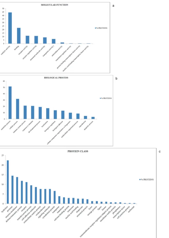
The identified proteins were classified in accordance with their molecular function and the biological process by using the PANTHER classification system.
As shown inFig. 2, the whey colostrum proteins can be classified into 10 groups with
differ-ent molecular functions (Fig. 2A), thus providing a comprehensive overview for the whey co-lostrum proteome in sheep.
About 44% of the whey colostrum proteins resulted to be involved in catalytic activity, rep-resenting the largest proportion of all the identified proteins. The second largest category refers to binding activity (22.4%), followed by receptor activity (11.2%), enzyme regulator activity (10.9%), structural molecule activity (8.9%), transporter activity (6.6%), antioxidant activity (1.6%), translation regulator activity (0.3%), protein binding transcription factor activity (0.3%) and nucleic acid binding transcription factor activity (0.3%).
Whey colostrum proteins were further categorized into 12 biological process groups (Fig. 2B). The largest group was related to metabolic process (51.6%), followed by cellular pro-cess (31.9%) immune system propro-cess (21.1%) response to stimulus (20.7%) and developmental process (19.1%).
Further analysis performed with the PANTHER software revealed that whey colostrum pro-teins could be classified into 28 distinct groups (Fig. 2C), the top ten of which are i) hydrolase (22.4%), ii) protease (14.5%), iii) enzyme modulator (13.8%), iv) defense/immunity protein (11.8%), v) receptor (11.2%), vi) transfer/carrier protein (9.5%), vii) cell adhesion molecule (8.6%), viii) extracellular matrix protein (7.6%), ix) oxidoreductase (7.6%), x) signaling mole-cule (7.6%).
Data obtained from LC-MS/MS analysis of sheep whey colostrum were uploaded into IPA software and overlaid onto a global molecular network developed from information contained in the application. Networks of these proteins were generated by IPA based on their connectivi-ty, each ranked by a score. This score is based on the hypergeometric distribution, calculated
with the right-tailed Fisher’s Exact Test, and corresponds to the negative log of this p-value.
Functional analysis in IPA identified the published biological functions that were most signifi-cantly associated with the genes in the network. Genes or gene products are represented as nodes, where shape indicates functional groups, and the biological relationship between two nodes is represented as an edge (line). All lines are supported by at least one reference in litera-ture, textbook, or from canonical information stored in the Ingenuity Pathways
knowledge database.
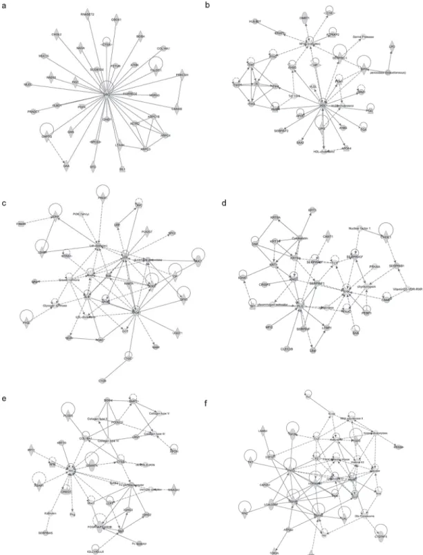
Identified proteins were mapped in 21 pathways of interacting protein clusters according to the identifiers' HomoloGene to the ortholog information in the Ingenuity Knowledge Base (IKB) (Fig. 3). In particular, 35 proteins involved in Developmental Disorder, Hereditary Dis-order and Metabolic Disease were grouped as top 1 network, [IPA score 71 (Fig. 4A)].
The associated functions of the most interesting networks are: i) Cancer, Gastrointestinal Disease, Hepatic System Disease, [IPA score 42 (Fig. 4B)]; ii) Lipid Metabolism, Small Mole-cule Biochemistry, Free Radical Scavenging [IPA score 33 (Fig. 4C)]; iii); Dermatological Dis-eases and Conditions, Neurological Disease, Organismal Injury and Abnormalitie [IPA score 33 (Fig. 4D)]; iv) Organ Morphology, Skeletal and Muscular System Development and Func-tion, Tissue Morphology [IPA score 33 (Fig. 4E)]; v) Organ Morphology, Skeletal and Muscu-lar System Development and Function, Cancer [IPA score 29 (Fig. 4F)].
Obviously, the involvement of proteins in processes such as developmental disorders, meta-bolic diseases, and cancer, occurs when they are not correctly expressed and/or regulated.
Fig 3. IPA analysis.List of signaling networks associated with identified proteins.
Fig 4. IPA analysis.In the figure are shown the top 6 signaling networks: 1) Cellular Movement, Hematological System Development and Function and Immune Cell Trafficking; 2) Cell Morphology, Cellular Movement, Digestive System Development and Function; 3) Free Radical Scavenging, Connective Tissue Disorders and Inflammatory Disease; 4) Cell-To-Cell Signaling and Interaction, Cellular Function and Maintenance, Hematological System Development and Function; 5) Organ Morphology, Skeletal and Muscular System Development and Function, Tissue Morphology; 6) Organ Morphology, Skeletal and Muscular System Development and Function, Cancer.
Function, Organismal Development and Cellular Growth and Proliferation, as shown inS2 SupportingInformation. Lastly, IPA allowed us to deduce the upstream regulator of identified proteins. Intriguingly, the molecules that regulate the major number of proteins are lipopoly-saccharide, TGFB1, beta-estradiol, dexamethasone and TNF. The putative target proteins are
listed inS3 SupportingInformation.
Going into details on the characterization of the identified proteins in ovine whey colos-trum, we found that several of them belong to the casein family (mainly alpha casein), most likely representing unassembled casein micelles. It is demonstrated that caseins are responsible for important biological functions such as ion carriers (calcium, phosphate, iron, zinc, copper), bioactive peptide precursors and immunomodulators [34]. It was also demonstrated that the casein proteolytic fragments have an antimicrobial activity, suggesting that proteases may also play a role in the host defence [35]. In addition, peptides derived from caseins are receiving much attention as possible sources of natural bioactivity with health benefits for humans, prob-ably because they stimulate the innate immune system within the mammary gland and prevent udder infections during the dry phase [36]. As expected, proteins involved in metabolic and immune system processes account for 51.6% and 21.1% of the total proteins, respectively [36]. In our study we also found many proteins involved in lipid metabolism (fatty acid synthase, apolipoproteins A-I, butyrophilin, xanthine dehydrogenase, and endothelial lipase) suggesting that colostrum contains the enzymes necessary to balance the equilibrium between lipid syn-thesis and breakdown. While lipases contribute to effective digestion and retention of colos-trum fat, the apolipoproteins acts as carriers of fatty acids in the circulation. Fatty acid synthase is an indispensible component of lipogenesis and energy production [37]. Xanthine dehydrogenase and butyrophilin, appears to be essential for milk fat globule production [38, 39]. Xanthine dehydrogenase and butyrophilin with other four milk fat globule membrane pro-teins (Milk Fat Globule-EGF factor 8 or lactadherin, ATP-binding cassette sub-family G mem-ber 2, protein disulfide isomerase family A memmem-ber 3, and ras-related protein RAB11A) probably represent a small fraction dissociated from the membranes.
Particularly, butyrophilin is concentrated in the apical membranes of mammary epithelial cells and is expressed only during lactation [40]. Regarding the other four milk fat globule membrane proteins, lactadherin may play a role in the membrane vesicle secretion, such as budding or shedding of plasma membrane (micro-vesicles) and exocytosis of endocytic multi-vesicular bodies (exosomes) [41]. ATP-binding cassette sub-family G member 2 is included in the superfamily of ATP-binding cassette (ABC) transporters. Significant expression of this pro-tein has been observed in the placenta, which may suggest a potential role for this molecule in this tissue. It likely serves as a cellular defense mechanism in response to xenobiotic exposure [42].
Ras-related protein RAB11A is a low molecular-weight GTP-binding proteins that coordi-nate stages of transport in the secretory pathway, and annexins, which include a group of calci-um-dependent membrane aggregating proteins that can initiate contacts between secretory vesicle membranes, which subsequently fuse [43].
Finally, protein disulfide isomerase family A member 3 is a protein of the endoplasmic retic-ulum that interacts with lectin chaperones calreticulin and calnexin to modulate folding of newly synthesized glycoproteins [44].
Interestingly, we found a group of proteins involved in cellular growth and proliferation,
such as transforming growth factor beta (TGF-β), and Bone morphogenetic protein 1 (BMP1).
The TGF-βbelongs to a large family of proteins involved in regulating the proliferation and
differentiation of many cell types, i.e. mammary gland epithelial cells or human colon
adhesions that plays a crucial roles in controlling the differentiation of epithelial cells and in maintaining the integrity of the epithelium [45].
BMP1 is a metallopepsidase involved in extra-cellular matrix (ECM) formation by
activa-tion a subset of the TGFβsuperfamily of proteins [46].
Moreover, we identified some proteins involved in carbohydrate metabolism including gli-ceraldeide 3 fosfate deydrogenase, aldolase, phosphoglycerate kinase or beta 1,4-galattosyl-transferase I, most of which are involved in glycolysis. We hypothesize that these proteins derive from somatic cells disrupted during whey preparation.
As stated earlier, 21.1% of proteins was identified as immune or inflammation modulators: among them, lactoferrin, bactinecin, and gelsolin isoform b were detected. Interestingly, gelso-lin isoform b is described only in bovine colostrum [22] and in human milk [47]. Gelsogelso-lin, a
Ca2+-dependent actin-regulatory protein, mapped in IPA network 4, is necessary for rapid
mo-tile responses in cell types involved in stress response and inflammation [22,48]. Thus, the
presence of gelsolin in colostrum could be related to newborns protection from noxious agents. Other proteins of interest that play an essential role in the innate immune system are serum
amyloid A (SAA) andα1-antitrypsin. SAA take part in the acute phase of inflammation: it is
demonstrated that SAA concentration increases by 1000-fold within 24–48 h following
infec-tion/inflammation [36]. The role ofα1-antitrypsin is to reduces the biologically active trypsin,
decreasing the proteolysis. This protein has been described only in bovine colostrum where the
concentration is 100 times higher than in milk. The presumed role ofα1-antitrypsin is to
pro-tect immune components against the proteolytic cleavage, allowing the absorption of the intact proteins by the newborn ruminant [36].
Another group of proteins comprises proteins with anti-oxidant function, such as transfer-rin, lactoperoxidase, peroxiredoxin 2, superoxide dismutase (SOD2) and Glutathione peroxi-dase (GPx).
To the best of our knowledge, this is the most exhaustive and comprehensive report in ovine colostrum of an interesting set of proteins related with IPA network 6 and 14 (Fig. 3), and implicated in protection from stress stimuli: heat shock protein 90 (HSP90) and heat
shock protein 70 (HSP70) [49–51]. These two proteins are involved in the organization of
cyto-skeleton and in microtubule dynamics. Moreover, Hsp70 is important in the protection and re-generation of bowel [52] and its expression is reported in sheep lung epithelial cells [53], myocardium [54], brain [55], and uterine tissue [56].
Our study, which takes advantage of a Gel-LC-MS/MS integrated approach, provides the first systematic classification of the ovine whey colostrum low abundance proteome. Data ob-tained from this analysis enhance our understanding of the biochemical complexity of the nutritional components of colostrum, helping to gain new insights into proteins involved in colostrogenesis and, possibly, into proteins having bioactivity in the recipient lamb with impor-tant effects on immune system and the development of gastrointestinal tract. The normal de-velopment of gastrointestinal tract will be important for the food nutrients absorption and for the growth and weight gain of the newborn.
Further studies will be necessary in order to understand how this primary source of food passes through the gastrointestinal tract of the newborn lamb how the above-mentioned regu-latory proteins and peptides influence the lamb development and growth, but we are confident that this work represent a milestone in this way.
Supporting Information
of peptides, GO Terms, molecular weight (kDa) and isoelectric point. (XLSX)
S2 Supporting Information. IPA analysis.In the figure are shown the proteins biofunction classification. Chart were customized for molecular and cellular function and physiological sys-tem development and function.
(TIF)
S3 Supporting Information. IPA analysis.In the table are listed the putative upstream regula-tors and their relative target.
(XLS)
Author Contributions
Conceived and designed the experiments: DS FT GC DB. Performed the experiments: LD AC LT GP. Analyzed the data: DS FT. Contributed reagents/materials/analysis tools: AB GC GV. Wrote the paper: DS FT GC DB AB. Collected samples: AL DM.
REFERENCES
1. Bauman DE, Mather IH, Wall RJ, Lock AL (2006) Major advances associated with the biosynthesis of milk. J Dairy Sci 89: 1235–1243. PMID:16537956
2. Halliday R (1978) Immunity and health in young lambs. Vet Res 103: 489–492.
3. Levieux D (1982) Transmission de l’immunité passive colostrale: le point des connaissances. In: Jar-rige R (Ed), Physiologie et Pathologie Périnatales chez les Animaux de Ferme, Paris, INRA. pp 345– 369. doi:10.1016/j.ejvs.2014.10.022PMID:25510183
4. Campbell SG, Siegel MJ, Knowlton BJ (1977) Sheep immunoglobulins and their transmission to the neonatal lamb. N Z Vet 25: 361–365. PMID:353601
5. Britti D, Massimini G, Peli A, Luciani A, Boari A (2005) Evaluation of serum enzyme activities as predic-tors of passive transfer status in lambs. JAVMA 226: 951–955. PMID:15786999
6. Hunter AG, Reneau JK, Williams JB (1997) Factors affecting IgG concentration in day-old lambs. J Anim Sci 45: 1146–1151.
7. Sawyer M, Willadsen CH, Osburn BI, McGuire TC (1977) Passive transfer of colostral immunoglobulins from ewe to lamb and its influence on neonatal lamb mortality. J Am Vet Med Assoc 171: 1255–1259. PMID:604324
8. Halliday R (1974) Variation in immunoglobulin concentrations in merino and Scottish Blackface lambs. Anim Prod 19: 301–308.
9. Massimini G, Mastellone V, Britti D, Lombardi P, Avallone L (2007) Effect of passive transfer status on preweaning growth performance in dairy goat kids. JAVMA 231: 1873–1877. PMID:18081529
10. Sozmen M, Beytut E (2012) An investigation of growth factors and lactoferrin in naturally occurring ovine pulmonary adenomatosis. J Comp Pathol 147: 441–451. doi:10.1016/j.jcpa.2012.04.004PMID: 22721818
11. Yekta MA, Cox E, Goddeeris BM, Vanrompay D (2011) Reduction of Escherichia coli O157: H7 excre-tion in sheep by oral lactoferrin administraexcre-tion. Vet Microbiol 150: 373–378. doi:10.1016/j.vetmic.2011. 02.052PMID:21511407
12. Korhonen H, Marnila P, Gill HS (2000) Milk immunoglobulins and complement factors. Br J Nutr 84: S75–S80. PMID:11242450
13. Stelwagen K, Carpenter E, Haigh B, Hodgkinson A, et al. (2008) Immune components of bovine colos-trum and milk. J Anim Sci 87: 3–9. doi:10.2527/jas.2008-1377PMID:18952725
14. Patureau-Mirand P, Mosoni L, Levieux D, Attaix D, Bayle G, et al. (1990) Effect of colostrum feeding on protein metabolism in the small intestine of newborn lambs. Biology of the Neonate 57: 30–36. PMID: 2302435
15. Kelly DD, Coutts AGP (2000) Development of digestive and immunological function in neonates: role of early nutrition. Lives Prod Sci 66: 161–167.
17. Palmer DJ, Kelly VC, Smit AM, Kuy S, Knight CG, et al. (2006) Human colostrum: identification of minor proteins in the aqueous phase by proteomics. Proteomics 6: 2208–2216. PMID:16502470
18. Picariello G, Ferranti P, Mamone G, Roepstorff P, Addeo F (2008) Identification of N-linked glycopro-teins in human milk by hydrophilic interaction liquid chromatography and mass spectrometry. Proteo-mics 8: 3833–3847. doi:10.1002/pmic.200701057PMID:18780401
19. Ogawa S, Tsukahara T, Nishibayashi R, Nakatani M, Okutani M, et al. (2014) Shotgun proteomic analy-sis of porcine colostrum and mature milk. Anim Sci J 85:440–448. doi:10.1111/asj.12165PMID: 24450292
20. Pecka E, Dobrzanski Z, Zachwieja A, Szulc T, Czyz K (2012) Studies of composition and major protein level in milk and colostrum of mares. Animal Sci J 83: 162–168. doi:10.1111/j.1740-0929.2011.00930. xPMID:22339698
21. Galvani M, Hamdan M, Righetti PG (2001) Two-dimensional gel electrophoresis/matrix-assisted laser desorption/ionisation mass spectrometry of commercial bovine milk. Rapid Communication in Mass Spectrometry 15: 258–264. PMID:11223956
22. Yamada M, Murakami K, Wallingford JC, Yuki Y (2002) Identification of low-abundance proteins of bo-vine colostral and mature milk using two-dimensional electrophoresis followed by microsequencing and mass spectrometry. Electrophoresis 23: 1153–1160. PMID:11981865
23. Natale M, Bisson C, Monti G, Peltran A, Garoffo LP, et al. (2004) Cow’s milk allergens identification by two-dimensional immunoblotting and mass spectrometry. Molecular Nutrition & Food Research 48: 363–369. PMID:15672476
24. Senda A, Fukuda K, Ishii T, Urashima T (2011) Changes in the bovine whey proteome during the early lactation period. Animal Sci J 82: 698–706. doi:10.1111/j.1740-0929.2011.00886.xPMID:21951907
25. Hogarth CJ, Fitzpatrick JL, Nolan AM, Young FJ, Pitt A, et al. (2004) Differential protein composition of bovine whey: a comparison of whey from healthy animals and from those with clinical mastitis. Proteo-mics 4: 2094–2100. PMID:15221770
26. Nissen A Bendixen E, Ingvartsen KL, Røntved CM (2012) In-depth analysis of low abundant proteins in bovine colostrum using different fractionation techniques. Proteomics 12: 2866–2878. doi:10.1002/ pmic.201200231PMID:22848049
27. Chiaradia E, Valiani A, Tartaglia M, Scoppetta F, Renzone G, et al. (2013): Ovine subclinical mastitis: Proteomic analysis of whey and milk fat globules unveils putative diagnostic biomarkers in milk. J Prote-omics 83: 144–159. doi:10.1016/j.jprot.2013.03.017PMID:23563085
28. Laemmli UK (1970) Cleavage of structural proteins during the assembly of the head of bacteriophage T4. Nature 227: 680–685. PMID:5432063
29. Candiano G, Bruschi M, Musante L, Santucci L, Ghiggeri GM, et al. (2004) Blue silver: a very sensitive colloidal Coomassie G-250 staining for proteome analysis. Electrophoresis 25: 1327–1333. PMID: 15174055
30. Pujia A, De Angelis F, Scumaci D, Gaspari M, Liberale C, et al. (2010) Highly efficient human serum fil-tration with water-soluble nanoporous nanoparticles. Int J Nanomedicine 5: 1005–1015. doi:10.2147/ IJN.S12865PMID:21187942
31. Morelli M, Scumaci D, Di Cello A, Venturella R, Donato G, et al. (2014) DJ-1 in Endometrial Cancer: A Possible Biomarker to Improve Differential Diagnosis Between Subtypes. Int J Gynecol Cancer 103: 550–557.
32. Mi H, Muruganujan A, Thomas PD (2013) PANTHER in 2013: modeling the evolution of gene function, and other gene attributes, in the context of phylogenetic trees. Nucleic Acids Res. 41 (Database issue): D377–D386. doi:10.1093/nar/gks1118PMID:23193289
33. Addis MF, Pisanu S, Ghisaura S, Pagnozzi D, Marogna G, et al. (2011) Proteomics and pathway analy-ses of the milk fat globule in sheep naturally infected by Mycoplasma agalactiae provide indications of the in vivo response of the mammary epithelium to bacterial infection. Infect Immun. 79: 3833–3845. doi:10.1128/IAI.00040-11PMID:21690237
34. Korhonen H, Pihlanto A (2007) Technological options for the production of health-promoting proteins and peptides derived from milk and colostrum. Curr Pharm Des 13: 829–843. PMID:17430184
35. Lahov E, Regelson W (1996) Antibacterial and immunostimulating casein-derived substances from milk: Casecidin, isracidin peptides. Food Chem Toxicol 34: 131–145. PMID:8603791
36. Hernández-Castellano LE, Almeida AM, Castro N, Argüello A (2014) The colostrum proteome, rumi-nant nutrition and immunity: a review. Curr Protein Pept Sci 15: 64–74. PMID:24555887
38. McManaman L, Palmer CA, Wright RM, Neville MC (2002) Functional regulation of xanthine oxidore-ductase expression and localization in the mouse mammary gland: evidence of a role in lipid secretion. J Physiol 545: 567–579. PMID:12456835
39. Ogg SL, Weldon AK, Dobbie L, Smith AJH, Mather IH (2004) Expression of butyrophilin (Btn1a1) in lac-tating mammary gland is essential for the regulated secretion of milk-lipid droplets. Proc. Natl. Acad. Sci. USA 101: 10084–10089. PMID:15226505
40. Robenek H, Hofnagel O, Buers I, Lorkowski S, Schnoor M, et al. (2006) Butyrophilin controls milk fat globule secretion. Proc Natl Acad Sci USA 103: 10385–10390. PMID:16801554
41. Oshima K, Aoki N, Kato T, Kitajima K, Matsuda T (2002) Secretion of a peripheral membrane protein, MFG-E8, as a complex with membrane vesicles. Eur J Biochem 269: 1209–1218. PMID:11856354
42. Gu HM, Li G, Gao X, Berthiaume LG, Zhang DW (2013) Characterization of palmitoylation of ATP bind-ing cassette transporter G1: effect on protein traffickbind-ing and function. Biochim Biophys Acta 1831: 1067–1078. doi:10.1016/j.bbalip.2013.01.019PMID:23388354
43. Wu CC, Yates JR, Neville MC, Howell KE (2000) Proteomic analysis of two functional states of the Golgi complex in mammary epithelial cells. Traffic 1: 769–782. PMID:11208067
44. Chen J, Lobachev KS, Grindel BJ, Farach-Carson MC, Hyzy SL, et al. (2013) Chaperone properties of pdia3 participate in rapid membrane actions of 1α,25-dihydroxyvitamin d3. Mol Endocrinol 27: 1065–
1077. doi:10.1210/me.2012-1277PMID:23660595
45. Moses HL, Yang EY, Pietenpol JA (1990) TGF-beta stimulation and inhibition of cell proliferation: new mechanistic insights. Cell 63: 245–247. PMID:2208284
46. Delana R, Hopkins SK, Greenspan DS (2007) The bone morphogenetic protein 1/Tolloid-like metallo-proteinases. Matrix Biology 26: 508–523. PMID:17560775
47. Liao Y, Alvarado R, Phinney B, Lönnerdal B (2011) Proteomic characterization of human milk whey pro-teins during a twelve-month lactation period. J Proteome Res 10: 1746–1754. doi:10.1021/pr101028k PMID:21361340
48. Napolitano F, Annicchiarico G, Catillo G, CrisàA, Grandoni F, et al. (2014) Identification of Ovis aries
Gelsolin isoform b, a candidate gene for milk quality. Small Ruminant Research 116: 21–27. 49. Craig EA, Lindquist S (1988) The heat shock proteins. Annu Rev Genet 22: 631–677. PMID:2853609 50. Welch WJ (1990) The mammalian stress response: cell physiology and biochemistry of stress proteins.
In: Morimoto RI, Tissieres A, Georgopoulos C (eds) Stress proteins in biology and medicine. Cold Spring Harbor Laboratory, New York. pp 223–278.
51. Kregel KC, Kevin C (2002) Heat shock proteins: modifying factors in physiological stress responses and acquired thermotolerance. J Appl Physiol 92: 2177–2186. PMID:11960972
52. Petrof EO, Ciancio MJ, Chang EB (2004) Role and regulation of intestinal epithelial heat shock proteins in health and disease. Chin J Dig Dis 5: 45–50. PMID:15612656
53. Kramer BW, Kramer S, Ikegami M, Jobe AH (2002) Injury, inflammation, and remodeling in fetal sheep lung after intra-amniotic endotoxin Am J Physiol Lung Cell Mol Physiol 283: L452–L459. PMID: 12114208
54. Scharte M, Baba HA, Van Aken H, Schulzki C, Mayer J, et al. (2001) Alanyl-glutamine dipeptide does not affect hemodynamics despite a greater increase in myocardial heat shock protein 72 immunoreac-tivity in endotoxemic sheep. J Nutr 131: 1433–1437. PMID:11340095
55. Andrews MH, Matthews SG (2000) Regulation of glucocorticoid receptor mRNA and heat shock protein 70 mRNA in the developing sheep brain. Brain Res 878: 174–182. PMID:10996148