Factor Essential for the Development of Plague
Avital Tidhar, Yehuda Flashner, Sara Cohen, Yinon Levi, Ayelet Zauberman, David Gur, Moshe Aftalion, Eytan Elhanany, Anat Zvi, Avigdor Shafferman, Emanuelle Mamroud*
Department of Biochemistry and Molecular Genetics, Israel Institute for Biological Research, Ness-Ziona, Israel
Abstract
Yersinia pestisis the causative agent of plague. Previously we have isolated an attenuatedY. pestistransposon insertion mutant in which thepcmgene was disrupted. In the present study, we investigated the expression and the role ofpcm locus genes in Y. pestis pathogenesis using a set of isogenic surE, pcm, nlpD and rpoS mutants of the fully virulent Kimberley53 strain. We show that inY. pestis, nlpDexpression is controlled from elements residing within the upstream genessurEand pcm. The NlpD lipoprotein is the only factor encoded from thepcmlocus that is essential for Y. pestis virulence. A chromosomal deletion of thenlpDgene sequence resulted in a drastic reduction in virulence to an LD50of at
least 107cfu for subcutaneous and airway routes of infection. The mutant was unable to colonize mouse organs following
infection. The filamented morphology of the nlpD mutant indicates that NlpD is involved in cell separation; however, deletion ofnlpD did not affect in vitro growth rate. Trans-complementation experiments with theY. pestis nlpD gene restored virulence and all other phenotypic defects. Finally, we demonstrated that subcutaneous administration of thenlpD mutant could protect animals against bubonic and primary pneumonic plague. Taken together, these results demonstrate thatY. pestisNlpD is a novel virulence factor essential for the development of bubonic and pneumonic plague. Further, the nlpDmutant is superior to the EV76 prototype live vaccine strain in immunogenicity and in conferring effective protective immunity. Thus it could serve as a basis for a very potent live vaccine against bubonic and pneumonic plague.
Citation:Tidhar A, Flashner Y, Cohen S, Levi Y, Zauberman A, et al. (2009) The NlpD Lipoprotein Is a NovelYersinia pestisVirulence Factor Essential for the Development of Plague. PLoS ONE 4(9): e7023. doi:10.1371/journal.pone.0007023
Editor:Ludovic Tailleux, Institut Pasteur, France
ReceivedJune 3, 2009;AcceptedAugust 13, 2009;PublishedSeptember 14, 2009
Copyright:ß2009 Tidhar et al. This is an open-access article distributed under the terms of the Creative Commons Attribution License, which permits unrestricted use, distribution, and reproduction in any medium, provided the original author and source are credited.
Funding:Israel Institute for Biological Research funds, grant number SB/5112-28. The funders had no role in study design, data collection and analysis, decision to publish, or preparation of the manuscript.
Competing Interests:The authors have declared that no competing interests exist.
* E-mail: [email protected]
Introduction
Yersinia pestisis the etiological agent of plague, which has caused millions of deaths in three world pandemics and is still a public health issue in some regions of the world. The most prevalent form of the disease is the bubonic plague, which develops following transmission of the pathogen from rodent reservoirs to humans via infected fleas [1]. Primary pneumonic plague is less abundant in nature and results from inhalation ofY. pestisdroplets or aerosols. It is a rapidly progressing disease leading to high mortality rates in untreated patients and can spread from person to person [1]. These characteristics led to the recognition ofY. pestisas a potential threat agent [2].
The ability ofY. pestisto respond to the host environment and to overcome immune systems is attributed to the combined activity of multiple virulence mechanisms. Among these mechanisms, only few have been found to be absolutely required for virulence in animal model systems. The type III secretion system (TTSS) is essential for survival of the pathogen within the mammalian host environment. This was demonstrated by the inability ofY. pestis strains devoid of the plasmid carrying the TTSS (pCD12) genes to colonize host tissues and to produce systemic disease following infection via both subcutaneous (s.c.) and airway routes [3–6]. The TTSS, shared by the closely related enteropathogensY. enterocolitica and Y. pseudotuberculosis, comprises a secretion apparatus,
chaper-ones and several effectors (Yops) and leads to the modulation of cell signaling networks necessary for an effective immune response [7,8]. Other virulence factors were found to be indispensable forY. pestispathogenesis via the s.c. route of infection. These include the plasminogen activator factor, which is encoded on the pPCP1 plasmid [9–11]; the Yersiniabactin (Ybt) iron acquisition system, which is encoded within the high pathogenicity island [12,13]; the chromosomally encoded PurH involved in the synthesis of purines [4,14]; adenylate kinase, which is involved in nucleotide metabolism [15]; and the recently characterized YadBC [16].
InE. coli, these two genes share a bicistronic operon, butpcmcan be transcribed independently from its own promoter [19]. The nlpDgene is located downstream ofpcm and codes for an outer membrane lipoprotein that is assumed to be involved in cell wall formation and maintenance [20,21]. The last gene in the locus is rpoS, which encodes an alternative RNA polymerase Sigma factor (RpoS). The rpoS and the nlpD genes constitute an operon; however, the majorrpoSpromoter is located within thenlpDgene [22–25]. The expression ofrpoSis induced during stress conditions, such as starvation and extreme pH, and during stationary growth phase [26,27]. This factor has been found to be involved in S. typhimuriumvirulence in a mouse infection model [28].
Genes within thepcm‘‘stress locus’’ were extensively studied in many species of the Enterobacteriaceae family, including Yersinia enterocolitica[29,30]. However, their importance for the pathogen-esis of plague has not been evaluated so far. In the present study, we have characterized the pcm genomic locus in Y. pestis. The expression pattern of the genes was evaluated along with their respective contributions toY. pestisvirulence using mouse models of bubonic and pneumonic plague. Our findings indicate that within thepcm locus,nlpD is the only essential gene forY. pestis virulence. In addition, a highly attenuated Y. pestis nlpD-null
mutant has been shown to induce an efficient protective immunity against bubonic and pneumonic plague.
Results
Thepcmlocus of Y. pestis
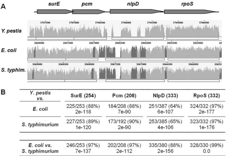
In a previous screen designed to isolate attenuated Y. pestis Kimberley53 mutants, a transposon insertion was identified at the 39 end of the pcm gene of the Kim53-K9 mutant [4]. When administered subcutaneously to mice, the virulence of Kim53-K9 was severely attenuated [4]. Inspection of the genome sequence of Y. pestis CO92 [31] revealed that the pcm locus shows high conservation of the gene order with respect to the related enteropathogensE. coliandSalmonella(Fig. 1A). However, whereas the Y. pestisSurE, Pcm and RpoS proteins share high levels of identity with their relatedE. coli/Salmonellaproteins (88–97%), the NlpD lipoproteins are more divergent (64–65%, Fig. 1B). The most noticeable disparity between the NlpDs ofY. pestis and E. coli/Salmonellais a protein sequence containing a unique proline and glutamine-rich repeat region at the N-terminus that is present in the proteins of the enteric pathogens but is completely absent from NlpD ofY. pestisand all otherYersiniaspecies. Examination of
Figure 1.In silicoanalysis of thepcmgenomic locus ofY. pestis.(A) A similarity plot representing the alignment of theY. pestisCO92surE,pcm,
nlpDandrpoSgenomic locus (gray arrows) with the corresponding orthologous regions ofE. coliK12 andS. typhimuriumLT2. Peaks represent regions of sequence conservation; regions that are conserved among all three genomes are in light gray, while regions that are conserved only betweenE. coliandS. typhimuriumare in dark gray. (B) Similarity comparison of the amino acid sequences of theY. pestisCO92pcmlocus-encoded proteins to the corresponding proteins inE. coliandS. typhimurium. Y. pestisprotein length is indicated in parenthesis and the similarity and E-values with respect to theE. coliorS. typhimuriumproteins are provided. In the bottom row, the similarity between theE. coliandS. typhimuriumproteins is presented.
the NlpD sequences within the Yersinia genus revealed that the nlpDgene products inY. pseudotuberculosisand Y. enterocoliticahave relatively high levels of sequence similarity to the corresponding gene product ofY. pestis(98% and 94%, respectively).
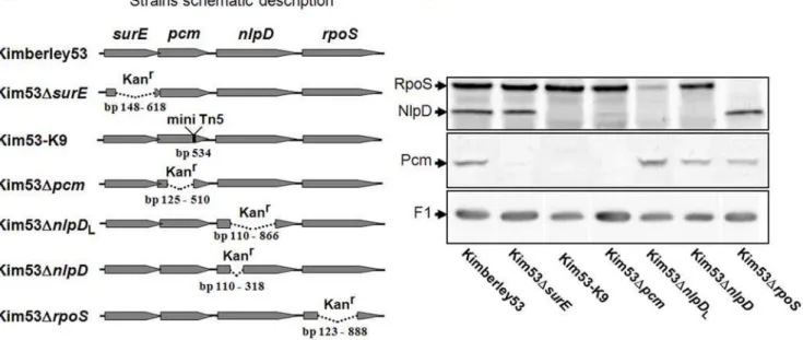
To characterize the factor/s involved in the attenuated phenotype of Kim53-K9, we used the virulentY. pestis Kimber-ley53 strain to generate a series of isogenic deletion mutants within thepcmlocus genes (Table 1). First, the DNA sequence of thepcm locus of Kimberley53 was determined (GenBank acc. no. FJ666123) and was found to be identical to CO92. In each of the newly constructed mutants, a defined region of a single gene was deleted and replaced with a kanamycin resistance cassette (see details in ‘‘Materials and Methods’’), resulting in Y. pestis Kimberley53 derivatives that were designated Kim53DsurE, Kim53Dpcm, Kim53DnlpD and Kim53DrpoS (Fig. 2A). PCR analysis verified that all of the Kimberley53-derived strains carry the pMT1, pCD1, pPCP1 plasmids and the chromosomal pgm locus. To evaluate the expression of Pcm, NlpD and RpoS in these strains, bacterial cultures were grown to stationary phase (24 hours) at 37uC and 28uC in heart infusion broth (HIB) and then subjected to Western blot analysis with highly specific antibodies. The patterns of expression were independent of the growth temperature (data not shown). In the Kim53DsurEmutant, Pcm was not detected, whereas the levels of NlpD and RpoS were comparable to the wild type strain (Fig. 2B). This result suggests that a control element influencing pcm expression resides within the surE gene, as has been reported for E. coli pcm[19]. In the Kim53-K9 strain, Pcm and NlpD were not detected, whereas the level of RpoS was comparable to the wild type strain (Fig. 2B). This finding suggests that expression ofnlpD is regulated by an element within the pcm gene. This assumption was further supported by the observation that the level of NlpD was reduced in Kim53Dpcm, whereas that of RpoS was not altered. Of note, in E. coli and Salmonella, expression of nlpDis driven by promoters located within the intergenic region between pcm and nlpD [21,22,25,27,32]. The genomic sequence in this region differs considerably betweenY. pestisand the enteric pathogens (Fig. 1A). The rpoS gene resides downstream of nlpD. In bacterial enteropathogens, the major rpoS promoter has been identified within the nlpD gene [33 and references therein,34,35]. We attempted to locate these known enterobacterial rpoS promoter
sequences in the correspondingY. pestisKimberley53 and CO92 genomic regions. PutativerpoSpromoter elements within thenlpD gene (547 bp upstream of the ATG start codon of rpoS) were identified according to tandem presence of 235 and 210 consensus sequences (Fig. S1). We therefore constructed two nlpD-null mutants: KimDnlpDL, in which the putative rpoS promoter elements were deleted, and KimDnlpD, in which these sequences were preserved (Fig. 2A). As expected, the level of RpoS was decreased in KimDnlpDLbut not in KimDnlpD(Fig. 2B). Of note is that the Kanr phenotype does not appear to derive expression of downstream ORFs, see for example the expression levels of Pcm in Kim53DsurE, NlpD in Kim53Dpcm, and RpoS in Kim53DnlpDL.
The last gene in thepcmlocus that was analyzed wasrpoS, which is predicted to encode an alternative sigma factor expressed during stress conditions [36]. The Pcm and NlpD expression levels in Kim53DrpoSwere comparable to the levels found in the wild type strain (Fig. 2B). Likewise, inE. coli, expression of thepcmandnlpD genes is RpoS-independent [19,37].
Identification of the sites of transcription initiation of the
pcm, nlpDandrpoSgenes ofY. pestisand their relation to the observed expression profiles
To further characterize the elements regulating the expression of the pcm, nlpD and rpoSgenes, total RNA was prepared from cultures ofY. pestisbacteria grown to stationary phase (24 hours) at 37uC and then was used for primer extension analysis and RT-PCR. Using a primer complementary to the 59region of thepcm gene, a single transcription initiation site designated Txn2 was identified within thesurEgene, 176 bp upstream of the ATG start codon of the pcm gene (Fig. 3A left panel). Similar analysis performed with a primer complementary to the 59region ofnlpD allowed identification of two transcriptional initiation sites within thesurEandpcmgenes located 988 bp (Txn1) and 300 bp (Txn3) upstream of the ATG start codon ofnlpD, respectively (Fig. 3A middle panel). To corroborate these findings, RT-PCR analysis was performed using primers complementary to regions within the nlpD and surE genes. An 1123-bp fragment was observed extending from thenlpDgene to thesurEgene at a site between Txn1 and Txn2 (Fig. 3B right panel, R2+F3). This result indicates
Table 1.Y. pestisstrains and plasmids used in this study.
Strain or plasmid Relevant characteristic(s) Reference or source
Y. pestisstrains
Kimberley53 Virulent strain [57]
EV76 pgm- (Girard’s strain) [58]
Kim53-K9 Kimberley53 strain in which the mini-Tn5 transposon was inserted into thepcmgene (nt no. 532 in the coding sequence).
[4]
Kim53DsurE Kimberley53 strain in which bp 148 to 618 (out of 765) of thesurEgene were deleted;kanR This study
Kim53Dpcm Kimberley53 strain in which bp 125 to 510 (out of 624) of thepcmgene were deleted;kanR This study
Kim53DnlpDL Kimberley53 strain in which bp 112 to 866 (out of 999) of thenlpDgene deleted;kanR This study
Kim53DnlpD Kimberley53 strain in which bp 112 to 318 (out of 999) of thenlpDgene were deleted;kanR This study
Kim53DrpoS Kimberley53 strain in which bp 123 to 888 (out of 996) of therpoSgene were deleted;kanR This study
Kim53DyopJ Kimberley53 deleted inyopJ [59]
Plasmid
pnlpD The completenlpDcoding sequence and the 442 bp region directly upstream were cloned into the pBR322 plasmid (Promega);ampR
This study
that a bicistronic pcm-nlpD message is indeed transcribed from Txn1. No message could be detected using primers located upstream of Txn1 (Fig. 3B right panel, R2+F4). RT-PCR analysis performed using primers complementary to regions within therpoS andpcmgenes indicated that Txn3 drives transcription of an nlpD-rpoS bicistronic message (Fig. 3B left panel, R1+F1). No RNA message could be detected using primers located upstream of Txn3 (Fig. 3B left panel, R1+F2). The majorrpoS transcription initiation start site (Txn4) was identified within thenlpDgene at the exact location predicted by thein silico-analysis (Fig. S1 and Fig. 3A right panel).
Integration of the data obtained from primer extension, RT-PCR and Western blot analyses (Fig. 2 and Fig. 3) helped to unravel the complex regulation controlling the expression of Y. pestis pcm, nlpDandrpoSgenes (Fig. S2). Transcription of thepcm gene is driven by control elements within the surE gene, and a major transcription start site was identified at Txn2 (Fig. 3C). Consistent with this result, Pcm was not detected in the Kim53DsurEmutant (see Fig. 2). Transcription of the nlpDgene can start in principle from three different positions: Txn1, Txn2 and Txn3. However, in our analysis we failed to see nlpD transcripts from Txn2. Transcription starting at position Txn1 leads to the synthesis of a pcm-nlpDbicistronic message, whereas transcription starting from position Txn3 drives the synthesis of an nlpD-rpoSbicistronic message (Fig. 3C). The finding that the NlpD level was severely reduced in the Kim53Dpcmmutant in which the genomic region containing Txn3 was deleted (Fig. 2) indicates that this region includes a majornlpDcontrol element/s. In agreement with this result, deletion of the Txn1 genomic region hardly interfered with NlpD expression (see Fig. 2, Kim53DsurE). Putative Y. pestis rpoS promoter sequences were identified within thenlpD gene byin-silicoanalysis (Fig. S1), and therpoStranscription start point was indeed identified at that region (Txn4, Fig. 3C). A significant reduction in the RpoS level was observed upon deletion
of the Txn4 region, indicating that this region indeed contains majorrpoScontrol elements (see Fig. 2, Kim53DnlpDL). The low RpoS level detected in Kim53DnlpDL(Fig. 2) probably represents the basal expression level of the nlpD-rpoS bicistronic message transcribed from Txn3 (see Fig. 3C). In accordance with these results, a deletion generated within the nlpD gene that did not affect Txn4 did not cause a significant reduction in the RpoS level (see Fig. 2 Kim53DnlpD).
To characterize the expression patterns of theY. pestis pcm, nlpD andrpoSgenes during logarithmic and stationary growth phases, wild type Kimberley53 and the Kim53Dpcm and Kim53DnlpD mutants were grown in HIB for 48 hours, and equal amounts of total protein were loaded on sodium dodecyl sulfate-polyacryl-amide gel electrophoresis (SDS-PAGE). Western blot analysis of Kimberley53 cells indicated that Pcm and NlpD levels remained essentially constant during the transition from logarithmic to stationary growth phase, whereas the RpoS expression pattern was growth phase-dependent (Fig. 4). One should note that during the stationary growth phase an NlpD precursor starts to accumulate, appearing as a slightly slower migrating band than NlpD. Similar findings were reported forE. coli [21,36,38]. The Western blot analyses with the various mutants (Fig. 4), together with the RT-PCR results (Fig. 3B), indicate that the low expression levels of NlpD and RpoS were governed by distal control elements (Txn1 and Txn3, respectively). We therefore evaluated the contribution of each of the distal elements to NlpD (Txn1) and RpoS (Txn3) expressionin vitroduring growth. In Kim53Dpcm, the NlpD level was constant but was lower than that of the wild type strain (Fig. 4). Consequently, the region in proximity to Txn3 contains the major nlpDcontrol element. In Kim53DnlpDL, in which the Txn4 region was deleted, the level of RpoS was low and growth phase-dependent (Fig.4). This finding indicates that the major control elements regulating RpoS expression are located in close proximity to the Txn4 genomic region.
Figure 2. Expression ofpcm,nlpDandrpoSinY. pestisderivatives.(A) Schematic description ofY. pestismutants. A kanamycin resistance cassette (Kanr) was inserted in place of the deleted region. In all mutants the Kanrcassette is oriented in the same direction as thepcmlocus genes and the runthrough transcription is minimal as evident from the expression analyses of Kim53DsurE, Kim53Dpcmand Kim53DnlpDL. (B) Assessment of bacterial Pcm, NlpD and RpoS expression. Cultures of theY. pestisstrains were inoculated (initial OD660= 0.01) and incubated for an additional 24 hours at 37uC. Western blot analysis was performed with anti-Pcm, anti-NlpD and anti-RpoS antibodies. F1 expression levels in the indicated strains were detected with anti-F1 antibodies and are shown to indicate that comparable amounts of bacterial extracts were blotted on the membrane.
NlpD is an essential factor for the development of bubonic and pneumonic plague
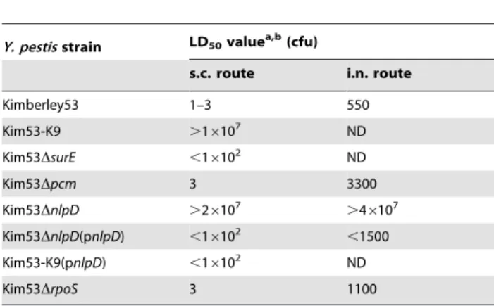
To test the contribution of SurE, Pcm, NlpD and RpoS toY. pestis pathogenesis, we evaluated the virulence of Kimberley53-deletion mutants in mouse models of bubonic plague (s.c. infection) and pneumonic plague (intranasal infection). In the bubonic plague mouse model, Kim53DsurE, Kim53Dpcm and Kim53DrpoSwere found to be highly virulent as reflected by the mortality rate, the mean time to death and the LD50value (Fig. 5A, Table 2). In contrast, The Kim53-K9 and Kim53DnlpDstrains were avirulent (Fig. 5A, Table 2).
The inability of Kim53-K9 and Kim53DnlpDto overcome host defense systems was further emphasized by the finding that infecting mice with higher doses of these mutants, up to 107cfu of each strain, was non-lethal (Table 2) and that infected mice did not show any visible disease symptoms, such as ruffled hair or a hunched back.
Intranasal (i.n.) infection of mice was demonstrated recently to serve as a model for studying the development of primary pneumonic plague [3]. We therefore used this model system to study the involvement of our genes of interest in development of pneumonic plague. All mice infected intranasally with 26104cfu of wild type Kimberley53 succumbed to the infection by day 4 (data not shown), and the LD50was found to be 550 cfu (Table 2). Similar LD50values were reported recently for other virulentY. pestis strains [39,40]. The Kim53Dpcmand Kim53DrpoSmutants were able to produce disease upon i.n. infection of mice, and the LD50 values of these mutants were 6 and 2 fold higher (respectively), than the LD50of the wild type strain (Table 2). In
contrast, i.n. infection of mice with up to 46107 cfu of Kim53DnlpDwas non-lethal (Table 2), and infected mice did not show disease symptoms.
To substantiate our observation that the attenuated phenotype of the NlpD-null mutants (Kim53DnlpDand Kim53-K9) resulted from the loss of NlpD expression, we examined the ability of a trans-complemented Y. pestis nlpD gene to restore the mutant’s virulence. Due to the toxicity associated with over-expression of NlpD ([21] and our unpublished data), we expressed the gene using its native control element. The complete coding sequence of the Y. pestis Kimberley53 nlpD gene preceded by the 442 bp upstream of the nlpD ATG, a region that includes the Txn3 transcription initiation region, was cloned into pBR322. The plasmid was introduced into Kim53DnlpDand Kim53-K9 to give the complemented strains Kim53DnlpD(pnlpD) and Kim53-K9(pnlpD). Western blot analysis performed using an overnight culture of the complemented strains grown at 37uC or 28uC (not shown) confirmed that in both strains the NlpD expression level was comparable to expression in the wild type strain (Fig. 5B). All mice infected subcutaneously with 100 cfu of the NlpD-complemented strains (the bubonic infection model) succumbed to the infection by day 8 (Fig. 5C). Moreover, Kim53DnlpD(pnlpD) also regained virulence following i.n. infection (the pneumonic infection model) (Table 2). These results confirmed that NlpD is a novel virulence factor ofY. pestisand is essential for development of both bubonic and pneumonic plague. Finally, NlpD expression on the background of the Kim53-K9 mutant also restored virulence, confirming that the attenuation of Kim53-K9 was not due to the Figure 3. Identification ofY. pestis pcm, nlpDandrpoStranscription start sites.(A) Primer extension (PE) electropherogram used to analyze the PE product generated using primers R1 [rpoS-rev (137)], R2 [nlpD-rev (151)] or R3 [pcm-rev (126)] (Table S1). PE peaks (black) corresponding to the MapMarkerH1000 internal size standards [gray, see actual size (bp) at the bottom of the electropherogram] are marked with the related nucleotide from the Kimberley53 genomic sequence. The size of the PE product is indicated at the top of the electropherogram. (B) RT-PCR products generated with the reverse primers specified above and the forward primers: F1 [rpoS-for (21355)], F2 [rpoS-for (21493)], F3 [nlpD-for (2972)] and F4 [nlpD-for (21389]. (C) Positions of R1-R3 primers used for RT-PCR and PE analyses is indicated by dashed, solid and double lines, respectively. The position of transcription start sites determined according to the PE products length (Txn 1–4), and the position of the F1-F4 primers used for RT-PCR are indicated.
absence of thepcmgene product but resulted from the elimination ofnlpDexpression.
Deletion ofnlpDimpairsY. pestiscolonization of internal organs in mice
In an attempt to evaluate the level of attenuation of Kim53DnlpD, we used mice to monitor the colonization of internal organs by Kim53DnlpD and the wild type strain Kimberley53. A dramatic disparity between the two strains was observed following infection via both the s.c. and the airway routes. In contrast to the wild type strain, which reached high bacterial loads in all organs within 48 hours, Kim53DnlpD could not be detected in the draining inguinal lymph nodes (I-LN), the spleen, the lungs or the blood of the subcutaneously infected mice at 24 hours post infection (data not shown) and up to 10 days post infection (Fig. 6A). No visible lesions were observed at the site of the injection.
When mice were infected intranasally with Kim53DnlpD, the bacterial load in the lungs decreased rapidly by two orders of magnitude within 48 hours to an average of 110 cfu (Fig. 6B), and bacteria were cleared within 3 days post infection (data not shown). In contrast, the bacterial load increased rapidly when the wild type strain was used. Moreover, Kim53DnlpDwas unable to colonize the mediastinal lymph node (M-LN), the spleen or the blood, whereas the number of colony forming units in all organs increased rapidly to an average of 104–107cfu within 48 hours for the wild type strain (Fig. 6B), causing the death of all mice within 4 days.
These observations indicate that NlpD is required forY. pestis propagation and dissemination to target organs, and they corroborate the non-virulent phenotype of Kim53DnlpDin both pneumonic and bubonic infection models.
In vitrocharacteristics of thenlpD-null mutant
Bacterial lipoproteins are components of the cell envelope of Gram-negative bacteria and are usually localized at the periplas-mic space anchored to either the outer or the inner membrane [41].Y. pestisNlpD has a threonine residue immediately after the fatty-acylated cysteine and is therefore predicted to reside in the outer membrane similarly to theE. coliNlpD [41,42]. Microscope analyses indicated thatin vitroculturing of Kim53DnlpDat 28uC leads to formation of unsegmented chains containing an average of 7.265.6 cells/chain as opposed to the more common aggregative morphology of Kimberley53 (Figures 7B and 7A, respectively). Elevation of the culturing temperature to 37uC (Fig. 7D), reduced significantly the number of Kim53DnlpDcells/chain to 462.5 (T-test P,1E-14), while the wild type morphology is temperature independent (Fig. 7C). In spite of this change, NlpD is not essential for Y. pestiscell growth, at least under laboratory conditions, as Figure 4. Growth phase-dependent expression of Pcm, NlpD
and RpoS in Y. pestis. Western blot analysis of bacterial extracts prepared from culture samples taken at the indicated time points was performed with anti-NlpD, anti-RpoS and anti-Pcm antibodies. doi:10.1371/journal.pone.0007023.g004
Figure 5. Virulence ofY. pestis pcmlocus mutants in mice.(A) Virulence of theY. pestis pcmlocus mutants in the mouse model of bubonic plague. Groups of five mice were infected subcutaneously with 100 cfu/mouse of the indicatedY. pestisstrains; Kimberley53 (closed circles), Kim53-K9 (open squares), Kim53Dpcm (closed triangles), Kim53DnlpD (stars) and Kim53DrpoS (open diamonds). (B) Episomal expression ofnlpDin attenuatedY. pestismutants. Western blot analysis of the indicated cultures was performed as described in the legend to Figure 2B (C) Virulence of thenlpD-complementedY. pestismutants in the mouse model of bubonic plague. Groups of five mice were infected subcutaneously with 100 cfu/mouse of the indicatedY. pestisstrains; Kim53DnlpD (stars), Kim53-K9 (open squares), Kim53DnlpD(pnlpD) (closed squares) and Kim53-K9(pnlpD) (closed circles).
shown by the normal growth rate of thenlpDmutant (Fig. 7E). Growth rate of Kim53DnlpDwas found to be similar to that of the wild type strain underin vitroculture conditions at 37uC (Fig. 7E) and at 28uC (data not shown) as determined by ANOVA test of regression (P.0.15). Of note is that there is a significant difference
between the intercept of thenlpDmutant and the wild type strain (P,0.05) due to the filamentous phenotype of the mutant (Fig. 7B and 7D). Complementation of nlpD expression eliminated completely the abnormal cell morphology and restored the wild type morphology (data not shown).
It can be assumed that deletion ofnlpDinY. pestismay influence membrane-related functions, such as TTSS activity, which is known to be essential forY. pestisvirulence, and thus could lead to the attenuated phenotype. However, preservation of TTSS functionality, at least in vitro, was demonstrated by retention of calcium-dependent growth at 37uC and expression and secretion of Yop effectors such as YopE (Fig. 8A and 8B) and YopJ (data not shown), into the culture medium following exposure to inducing conditions. Moreover, thenlpD-null mutant preserves the ability to translocate Yop effectors into host cells, like the wild type strain, as evidenced by suppression of TNF-a secretion from infected RAW264.7 macrophages (Fig. 8C). The genes flankingnlpDwere found in many enteropathogens to be involved in survival during stationary phase and under other environmental stress conditions [36]. In search of a possible explanation for the attenuation of Kim53DnlpD, we evaluated the involvement of Y. pestis NlpD in resistance to prolonged growth in rich (HIB) and minimal (M9) broth, and in acidic and oxidative conditions that simulate the intra-phagosomal milieu. Exposure of Kim53DnlpDand Kimber-ley53 to prolongedin vitrogrowth (up to 96 h), and to oxidative stress (10–50 mM H2O2) indicated that NlpD is not essential for resistance to these conditions (data not shown). In contrast, Kim53DnlpD was slightly more susceptible than wild type Table 2.Virulence ofY. pestisstrains in mouse models of
bubonic and pneumonic plague.
Y. pestisstrain LD50valuea,b(cfu)
s.c. route i.n. route
Kimberley53 1–3 550
Kim53-K9 .16107 ND
Kim53DsurE ,16102 ND
Kim53Dpcm 3 3300
Kim53DnlpD .26107 .4
6107
Kim53DnlpD(pnlpD) ,16102 ,1500
Kim53-K9(pnlpD) ,16102 ND
Kim53DrpoS 3 1100
ND = not determined.
aThe ‘‘,’’ symbol indicates that the calculated LD
50value is the minimal
infection dose tested, under which more than 50 percent of the animals died.
bThe ‘‘.’’ symbol indicates that the calculated LD
50value is the maximal
infection dose tested under which less than 50 percent of the animals died. doi:10.1371/journal.pone.0007023.t002
Figure 6. Colonization of mouse organs byY. pestisstrains.(A) Bacterial colonization of mouse organs after s.c. infection. Mice were injected subcutaneously with either 16104cfu of Kimberley53 (closed symbols) or 16105cfu of Kim53DnlpD(open symbols). Animals were sacrificed at 48 h post infection. Blood was collected and the draining inguinal lymph nodes (I-LN), the spleen and the lungs were harvested from each mouse, homogenized in 1 ml PBS and cultured on BHIA plates at 28uC for 48 hours. Values represent total bacteria in the organs (cfu/organ), or the bacterial concentration in blood (cfu/ml). The dashed line indicates the limit of detection. Horizontal bars represent the average value of the bacterial load in each case. (B) Bacterial colonization of mouse organs after i.n. infection. Dissemination ofY. pestisstrains into the blood and to the internal organs following i.n. inoculation with 16105cfu of either Kimberley53 (closed symbols) or Kim53DnlpD(closed symbols). Mice were sacrificed at the indicated time points, and the bacterial load in the indicated organs was determined as described above.
Kimberley53 to acidic pH (Fig. 8D). Moreover, the Kim53DnlpD(pnlpD) strain, in which NlpD expression was restored, regained resistance to these conditions (Fig. 8D). The possible linkage between acidic susceptibility and attenuation of Kim53DnlpDawaits further study.
Evaluation of the immunization potential of the Kim53DnlpDstrain
The high level of attenuation of Kim53DnlpDmotivated us to evaluate the potential of this defined mutant to serve as a vaccine. In a model of bubonic plague, mice were injected subcutaneously with 16105 cfu of either Kim53DnlpD or the Y. pestis EV76 prototype vaccine strain. Fifty days later, mice were challenged subcutaneously with 16105LD50of the fully virulent Kimberley53 strain. As shown in Table 3, the geometric mean ELISA titers of aF1 andaV antibodies measured in sera from the mice at 30 days
post immunization with Kim53DnlpD were 17,100 and 26,
respectively. SimilaraF1 andaV titers were measured following immunization with Kim53-K9. In contrast, mice vaccinated with the EV76 strain demonstrated anti-F1 geometric mean titers of less than 30 and anti-V antibody ELISA titers below the limit of detection (Table 3). These results suggest that although Kim53DnlpDwas highly impaired in colonization of host internal organs, its persistence within the host lasted long enough for the host to mount an effective immune response. Consistent with these results, all mice vaccinated with Kim53DnlpD survived s.c. challenge with 16105 LD50 of Kimberley53 without showing signs of illness whereas a similar dose of EV76 failed to elicit significant protective immunity (Table 3). The EV76 strain appears to be effective only when applied at a high vaccination dose (107cfu, Flashneret al2004).
In the mouse model of pneumonic plague, a single s.c. immunization with 16105–16107 cfu of Kim53DnlpDor EV76 was followed 50 days later by i.n. challenge of virulentY. pestis Kimberley53 (Table 4). The challenge dose was 5,500 cfu, which is equivalent to 10 LD50. While all control mice died within 4 days, a protection level of 33% was obtained following immunization with 105cfu of Kim53DnlpD. Higher protection levels of 66% and 82% were obtained by immunization with increasing doses of Kim53DnlpD (106
cfu and 107 cfu, respectively). In contrast, EV76 was unable to elicit protection following immunization with 105cfu, and a protection level of 33% was attained only after the immunization dose was increased to 107cfu (Table 4). Comparing total survival in the nlpD-vaccinated mice to EV76-vaccinated mice by Chi-square analysis revealed highly significant difference (P = 0.0013). These findings strongly accentuate the potency of Kim53DnlpD in establishing effective protective immunity and suggest that it may be a suitable platform for a live vaccine.
Discussion
Studies performed in the last decade with several Gram negative bacteria have demonstrated that the genomic region including the surE, pcm, nlpD and rpoS genes is important for survival under environmental stress conditions [18,36,43,44]. In the present study, we have further characterized theY. pestis pcm locus genes and analyzed their expression and involvement in the pathogenesis ofY. pestisusing mouse models of bubonic and pneumonic plague. Systematic deletion mutagenesis of the surE, pcm, nlpDand rpoS genes and complementation studies allowed us to identify the NlpD lipoprotein as the only essential factor for Y. pestis pathogenesis in this locus.
Subcutaneous and intranasal administration of aY. pestis nlpD -null strain to mice demonstrated that this strain is severely attenuated (LD50.107cfu, Table 2), and is impaired in its ability to colonize internal organs (Fig. 6). In addition, thenlpD-null mutant was unable to produce a systemic disease following intravenous inoculation with 106 cfu and infected mice lacked any visible disease symptoms (data not shown).
Trans-complementation experiments verified that NlpD is an essentialY. pestisvirulence factor (Fig. 5C, Table 2). Using a similar strategy, virulence was also restored to the original transposon insertion mutant (Kim53-K9), confirming that its attenuation resulted from elimination of nlpD expression and not from disruption ofpcmexpression (Fig. 5C, Table 2; note also that the Kim53Dpcmis as virulent as the wild type strain). To the best of our knowledge, this is the first demonstration of a single chromosomalY. pestisfactor that is essential for development of both bubonic and pneumonic plague.
Yersinia enterocoliticaNlpD was also suggested to be involved in pathogenesis [45]. In the latter study, transposon insertion within Figure 7. Growth curves and microscope analyses ofY. pestis
strains followingin vitrogrowth.Y. pestisstrains were grown for 24 hours at 28uC (A and B) or at 37uC (C and D) in HIB. Gram staining of Kimberley53 (A and C) and Kim53DnlpD(B and D) was performed and bacilli were observed by light microscopy at a magnification of61000. Scale bar = 10mm. (E) Growth curves of Kimberley53 (closed squares) and Kim53DnlpD (open squares). Bacteria were inoculated (initial OD660= 0.05) and grown at 37uC in HIB for 48 hours. Cultures were sampled at the indicated time points after inoculation and cfu values were determined by plating on BHI agar.
theY. enterocolitica nlpD gene led to attenuation of virulence in a mouse infection model. Interestingly, as opposed to the dramatic attenuation of theY. pestis nlpDmutant shown in the present study, theY. enterocolitica nlpDmutant displayed only a limited impairment of virulence [45]. This finding might imply that Y. pestis nlpDis required for an activity specific to the development of plague disease.
Recently, an additionalY. pestislipoprotein, Braun’s lipoprotein (Lpp), was found to be important for virulence [40]. It should be noted, however, that in contrast to NlpD, Lpp contributes only to the production of bubonic plague but not to that of the pneumonic plague [40]. Lipoproteins have been implicated in the pathogen-esis of other bacterial pathogens as well. For example, disruption of the lsp and lgt genes, which are involved in lipoproteins Figure 8. Analysis of TTSS activity and resistance to acidic pH ofY. pestisstrains.To assay the functionality of the TTSS, wild type Kimberley53 and avirulent Kim53DnlpDstrains were grown for 4 hours at inducing and non–inducing conditions (37uC6calcium). Western blot analyses of bacterial extracts (A) and culture supernatants (B) were performed with anti-YopE antibodies. (C) Suppression of TNF-asecretion from infected macrophages. RAW264.7 cells were infected by impaction with 100 MOI of Kimberley53, 50 MOI of Kim53DnlpDor 100 MOI of Kim53DyopJ. Secretion of TNF-ainto the culture medium was monitored 2 h after initiation of infection by ELISA. (D) Resistance to acidic pH. Overnight cultures of Kimberley53 (black columns), Kim53DnlpD(dark gray columns) and Kim53DnlpD(pnlpD) (light gray columns) were used to inoculate HIB (initial OD660= 0.1). These cultures were incubated at 37uC for 4 hours, washed with phosphate buffered saline and then exposed to acidic stress (pH 4.2) for the indicated time. Viable cell counts were determined by plating dilutions on BHI agar and incubating at 28uC for 48 hours.
doi:10.1371/journal.pone.0007023.g008
Table 3.Vaccine potential of theY. pestisKim53DnlpDstrain against bubonic plague.
Immunizationa Antibody responsebGMT (GeoStDv)
Percent survivalc(alive/total)
aF1 aV
Kim53-K9 9,250 (2.0) 93 (7.7) 90 (9/10)
Kim53DnlpD 17,100 (3.4) 26 (29.3) 100 (11/11)
EV76 29 (4.2) ,10 - 10 (1/10)
Control ,10 - ,10 - 0 (0/10)
aMice were immunized subcutaneously with 105cfu of the indicatedY. pestisstrains. bAnti-F1 and anti-V titers were determined by ELISA.
cFifty days post immunization, mice were challenged subcutaneously with 105LD
50of the virulentY. pestisKimberley53 strain (1 LD50= 1–3 cfu).
metabolism, caused attenuation of virulence inM. tuberculosis, S. aureusandS. pneumoniae[46–48].
The dramatic loss of virulence of theY. pestis nlpD-null strain is reminiscent of the non-virulent phenotype of Y. pestis strains lacking the pCD1 plasmid that carries the genes encoding the type III secretion system. However, our results indicate that thenlpD mutant is able to properly express and secrete Yop effectors during in vitro inducing conditions (Fig. 8A and 8B) and is able to translocate effectors into host cells, as reflected by its ability to suppress TNFa secretion from infected macrophages (Fig. 8C). Moreover, the V-antigen multifunctional virulence factor impor-tant for type III secretion system activity is expressed by thenlpD mutant during infection as reflected by development of aV antibodies following immunization of mice (Table 3 and 4). Taken together, these observations suggest that the type III secretion system is functional in thenlpDmutant.
The finding that theY. pestis nlpDgene is expressed from control elements shared with eitherpcm orrpoSraised the possibility that NlpD might also be important for survival under environmental stress conditions. However, the nlpD-null mutant did not differ significantly from the wild type strain in the ability to survive during prolonged growth in rich and minimal broth and during exposure to oxidative conditions. Yet, we found a slight increase in the mutant’s sensitivity toin vitroacidic conditions (Fig. 8), similar to the conditions present in the intra-phagosomal milieu. While the latter does not seem to account for the dramatic loss of virulence, further studies are needed to understand the importance of the sensitivity of the mutant to acidic conditions.
The aberrant shape of theY. pestisDnlpDbacilli (Fig. 7B and 7D) suggests that NlpD is important for cell separation. Consistent with this assumption is the structure of NlpD (Fig. 9), which contains an N-terminal LysM domain found in a variety of enzymes involved in bacterial cell wall degradation [49–51]. NlpD also contains a C-terminal M23 metallopeptidase region (Fig. 9). The M23 family of endopeptidases includes proteins that are involved in bacterial cell separation [52], however, no proteolytic activity has been demonstrated for bacterial lipoproteins of this family (see the MEROPS peptidase database, http://merops.sanger.ac.uk [53]). Of note is that in vitro cell growth of the nlpD mutant was not affected (Fig. 7E). The observed phenotype of thenlpD-null mutant (Fig. 7B and 7D) suggests a linkage between impairment of cell separation and attenuation of virulence. Interestingly, similar links have already been described for other bacterial pathogens [54,55]. Furthermore Kajimura and colleagues [54] hypothesized that
cluster formation of the attenuatedS. aureusSle1 mutant, which lacks the lysM-containing N-acetylmuramyl-L-alanine amidase, inhibits the dissemination of daughter cells and thus could affect the spread of the bacteria during infection. Indeed, the Y. pestis nlpDmutant is totally impaired in its ability to disseminate from the site of infection to the internal organs (Fig. 6).
Interestingly, the highly attenuated phenotype of the nlpD mutant and its inability to colonize host organs did not seem to prevent the development of immunity against plague following s.c. infection. Rather, this strain seemed to effectively stimulate a long-term adaptive immune response as demonstrated by the generation of high F1 antibody titers. Accordingly, immunization of mice with low doses of Kim53DnlpDresulted in remarkably higher humoral response and protection levels against bubonic and pneumonic plague than did immunization with theY. pestis EV76 vaccine strain (Tables 3 and 4). The differential behavior of the two vaccine strains may result from the complete absence of the chromosomal pgm locus (102 kb including the pathogenicity island), from EV76. The contribution of thepgmlocus to Y. pestis survival in host cells is well documented [56]. The observed development of protective immunity could have practical implications in the design of futureY. pestisvaccines or therapies against both bubonic and pneumonic plague.
Materials and Methods
Bacterial strains, plasmids, mutant construction and routine growth conditions
TheY. pestisstrains and the plasmids used in this study are listed in Table 1. The primers used for construction of the Y. pestis derivatives are listed in Table S1. Construction of the Kimber-ley53 deletion mutants was performed by replacing part of the gene coding sequence with a linear fragment containing the Kanr GeneBlockTMresistance cassette (pUC4K plasmid, Pharmacia) by homologous recombination, using previously established method-ologies [60]. In all constructs the Kanr resistance cassette is oriented in the same direction as thepcmlocus genes (Fig. 2A). To preserve expression of downstream genes, the resistance cassette does not include transcription terminator sequences.
Genotype verification of all obtained phenotypes was done by PCR and Western blot analysis. All of the Kimberley53-derived strains carry the pMT1, pCD1, pPCP1 plasmids and thepgmlocus. Bacteria were isolated from stocks on selective BIN medium [61]. Routine propagation was performed on brain heart infusion Table 4.Vaccine potential of theY. pestisKim53DnlpDstrain against pneumonic plague.
Immunizationa Antibody responsebGMT (GeoStDv)
Percent survival (alive/total)c
Y. pestisstrain Dose (cfu)d aF1 aV
105 4,500 (10.5) 10 (3.6) 33 (2/6)
Kim53DnlpD 106 31,800 (4.8) 32 (36) 66 (4/6)
107 7,200 (4.6) 12 (28) 82 (9/11)
105 20 (4.9)
,10 - 0 (0/6)
EV76 106 100 (15.3)
,10 - 17 (1/6)
107 635 (4.2) 18 (21) 33 (2/6)
Control - ,10 - ,10 - 0 (0/6)
aMice were immunized subcutaneously with the indicated dose of each strain. bAnti-F1 and anti-V titers were determined by ELISA.
cFifty days post immunization, mice were challenged intranasally with 10 LD
50of the virulentY. pestisKimberley53 strain (1 LD50= 550 cfu). dUnder the specific experimental conditions, one colony forming unit of Kim53DnlpDis estimated to be 1-6 bacilli.
agar (BHIA) (Difco) or HIB (Difco). For generation of nlpD -complemented strains, a 1444 kb fragment extending from the 39 end of the nlpD gene up to 442 nt upstream of the ATG start codon ofnlpDwas amplified from the DNA of the virulentY. pestis Kimberley53 strain. The resulting PCR product was cloned into pBR322 between the NheI and PstI sites and its sequence was verified. Plasmids were introduced into the Kim53DnlpD and Kim53-K9 strains by electroporation to give the Kim53DnlpD(pnlpD) and Kim53-K9(pnlpD) strains. These strains were routinely grown in media supplemented with 100mg/ml ampicillin (Sigma, Israel).
DNA sequence determination of theY. pestis
Kimberley53pcm locus andin-silicoanalysis
TheY. pestisCO92 sequence corresponding to thepcmlocus (nt 3744222 to 3747841) was compared to the Kimberley53 sequence at the equivalent region and was found to be identical) GenBank acc. no. FJ666123). All sequencing reactions were performed with an ABI310 genetic analyzer (Applied Biosystems) using the ABI PRISM BigDye terminator reaction kit. The genome alignment of the orthologous regions comprising thesurE, pcm, nlpD and rpoS genes was generated by the multiple genome alignment software Mauve [62]. The sequences were extracted from the following GenBank (NCBI) entries: NC_003143 (Y. pestis CO92), NC_003197 (S. typhimuriumLT2), NC_000913 (E. coliK12).
Growth curves
Y. pestisstrains were grown by shaking (100 rpm) at 28uC in HIB supplemented with 0.2% (+)xylose (Sigma) and 2.5 mM CaCl2 (Sigma). The resulting cultures were diluted in fresh broth to an OD660of 0.01 and allowed to grow for 48 hours at either 28uC or 37uC while shaking at 180 rpm. At the indicated time points, aliquots were removed for determination of the OD660 and the number of colony forming units (cfu) after plating on BHI agar plates and incubation at 28uC for 48 hours. Experiments were repeated for three times. The growth of Y. pestis strains under nutrient-limiting conditions was assessed in M9 medium at pH 8.0 [0.05% NaCl (Merck), 0.1% NH4Cl (Merck), 0.3% KH2PO4 (Merck), 0.7% Na2HPO4(Merck) 1024M CaCl2(Merck), 1023M MgSO4 (Merck), 0.4% D-(+)-glucose (Sigma), 5mg/ml thiamine (Sigma), 50mg/ml L-arginine (Sigma), 50mg/ml L-cysteine (Sigma), 50mg/ml glycine (Sigma), 50mg/ml L-methionine (Sigma), 50mg/ml L-phenylalanine (Sigma)]. Aliquots of the liquid cultures were taken at different time points for cfu enumeration and Western blot analysis. Comparison of theY. pestitstrain’s growth rates was performed by log-log transformation. The slop of the curves was compared using an ANOVA test of regression.
Western blot analysis
bicinchoninic acid (BCA Protein Assay Reagent, Pierce) according to the manufacturer’s instructions. Equal amounts of protein were loaded and separated by 12% SDS-PAGE. After transfer to nitrocellulose membranes, duplicate membranes were developed with rabbit polyclonal anti-peptide Pcm and NlpD antibodies, with rabbit polyclonalS. typhimuriumRpoS antibodies (a generous gift from Franc¸ois Norel, Institute Pasteur), or with goat anti-YopE antibodies (bL-20, Santa Cruz Biotechnology) and with rabbit polyclonal anti-peptide YopJ [60]. Probing with the primary antibody was followed by incubation of the membranes with HRP-conjugated second antibody, and then the reactive protein bands were visualized by ECL. The Pcm and NlpD anti-peptide antibodies were raised by immunizing rabbits with maleimide-activated KLH (Pierce) conjugated to the synthetic peptides CERLLQAIEAVPRER (amino acids 21–34 of Pcm) and CVGGDRSGTMLSKANT (amino acids 39–53 of NlpD). Antibodies were affinity purified using a Sulfolink-peptide coupling gel column (Pierce) according to manufacturer’s instructions.
Total RNA isolation, primer extension and RT-PCR analyses
HIB supplemented with 0.2% (+)xylose and 2.5 mM CaCl2was inoculated with Y. pestis bacteria (initial OD660= 0.01), and incubated for 24 hours at 28uC or 37uC with shaking at 100 rpm. Total RNA was isolated using a RiboPureTM-bacteria Kit (Ambion) and was then treated with DNase I (Ambion) according to the manufacturer’s instructions.
Primer extension (PE) reactions were carried out according to Lloyd and colleagues [63]. Briefly, for each reaction, FAM-labeled primer (Table S1, final concentration 5–10 nM) was added to 20mg of total RNA, and first strand cDNA synthesis was performed using the AMV RT enzyme (Promega). FAM-labeled cDNAs were cleaned using a PreformaH DTR Gel Filtration Cartridge (Edge BioSystems) according to the manufacturer’s instructions and then air-dried with a heated SpeedVac centrifuge and resuspended in 25ml of UltraPureTMFormamide (Invitrogen) with 1.5ml MapMarkerH1000 (BioVentures, Inc.). The mixture was heated to 90uC for 2 minutes, chilled on ice for 5 minutes and then used for electrophoresis, using an ABI310 genetic analyzer (Applied Biosystems). The DNA fragments were sized using the GeneScanR Analysis Software version 3.1 (Applied Biosystems). For reverse transcriptase-PCR analyses (RT-PCR), primers were added to 1mg of total RNA and synthesis was carried out with AMV RT (Promega) at 42uC for 1 hour according to the manufacturer’s instructions. After first strand synthesis the cDNA was diluted 1:2 and an aliquot (3ml) was taken for PCR amplification. Positive and negative control reactions were performed with 200 ng of DNA or with total RNA, respectively.
Infection of mice
All experiments were performed in accordance with the Israeli law and were approved by the Ethics Committee for animal experiments at the Israel Institute for Biological Research. Female OF1 mice, 5–6 weeks old (IFFA CREDO S.A., France), were used in all infection studies. For s.c. infections of mice, the Y. pestis strains were grown on BHIA for 48 hours at 28uC. Bacterial colonies were harvested from the BHIA plates at the end of the incubation period, suspended in 5 ml of saline solution (0.9% NaCl) and diluted in saline solution to the required infection dose. Bacteria were quantified by counting colony forming units after plating and incubating on BHI agar plates. In s.c. infections, samples of 100ml were administered into the lower back of the mice. Under these conditions, the LD50of the Kimberley53 strain is 1–3 cfu [4]. For i.n. infections, theY. pestisstrains were grown on
BHIA for 48 hours at 28uC. Bacterial colonies were suspended in HIB supplemented with 0.2% (+)xylose and 2.5 mM CaCl2to give an initial OD660 of 0.01, and the cultures were incubated for additional 22 hours at 28uC or 37uC with shaking at 100 rpm. At the end of the incubation period, the cultures were washed, diluted in saline solution to the required infection dose and quantified by cfu counting as described above. Prior to infection, mice were anaesthetized with a mixture of 0.5% ketamine HCl and 0.1% xylazine injected intraperitoneally and were then infected intranasally with 35ml of the bacterial suspension. The LD50 experiments were performed with groups of 8 mice (i.n.) or 5 mice (s.c.). The LD50 values were calculated according to the method described by Reed and Muench [64], protection analyses were performed by Chi-square test.
Monitoring disease progression
Mortality rates were expressed in accordance with the required information by either the proportion of dead animals at the end of the monitoring period (14 days unless otherwise indicated), by MTTD, or by the 50% lethal dose (LD50). Monitoring of disease progression was performed by tracking bacterial dissemination to the internal organs and blood. In s.c. infection experiments, organs were aseptically removed 48 hours after infection with 16104cfu of the wild type Kimberley53 strain (a terminal stage of disease) or with 16105 of Kim53DnlpD. Groups of four mice were anaesthetized, tail vein blood was collected and spleens, lungs and draining inguinal lymph nodes (I-LN) were harvested. Tissue homogenates were prepared in 1 ml PBS/organ. Bacterial enumeration in tissue homogenates or in blood samples was done by plating serial dilutions in PBS on BHI agar and calculating the cfu/organ or cfu/1 ml blood. In i.n. infection experiments, groups of four mice were anesthetized, and tail vein blood was collected one hour and 48 hours post infection with 16105cfu. The lungs, spleens and the mediastinal lymph nodes (M-LN) were harvested and prepared as described above for bacterial enumeration.
Morphology analysis
Y. pestisbacilli were observed by light microscopy using a Nikon Eclipse E200 microscope at a magnification of61000. Gram staining was performed using a HT90A kit (Sigma-Aldrich) according to the manufacturer’s instructions. Y. pestiscells in at least 20 random non-overlapping microscopic fields were counted.
TTSS functionality analyses and stress survival assays.
plating stationary-phase bacteria on BHI agar after exposure to hydrogen peroxide (10–50 mM in 16PBS, pH = 7) for up to 60 minutes.
Antisera and serological tests
Mouse anti-F1 and anti-V antigen IgG antibody titer determi-nations were performed by ELISA as previously described by Flashner and colleagues. [4]. Briefly, microtiter plates were coated with purified recombinant F1 antigen [65] or with 500 ng of purified Glutathione-S-Transferase -V- antigen fusion protein prepared according to Leary et al. [66]. The tested sera were serially diluted by 2-fold dilutions in a final volume of 50ml and were incubated in the wells for 1 hour at 37uC. Alkaline phosphatase–labeled rabbit anti-mouse IgG (1/2500 dilution, Sigma) was used as the secondary antibody. Titers were defined as the reciprocal values of the endpoint serum dilutions, which had OD405values two fold higher than the normal serum controls.
Supporting Information
Figure S1 PutativeY. pestis rpoSpromoter sequences identifiedin silico. (A) Sequences of rpoS promoters in different bacteria; the consensus235 sequences is indicated by ambiguity code. (B) The DNA region upstream of the Kimberley53 and CO92rpoScoding sequences, containing the putative 210 and/or 235 promoter sequences.
Found at: doi:10.1371/journal.pone.0007023.s001 (0.73 MB TIF)
Figure S2 Organization and expression of Y. pestis pcm locus genes in wild-type and mutant strains. Transcription start sites (TS) within Y. pestis pcm locus are depicted. Deleted region are represented by dashed line and replaced by a kanamycin
resistance cassette. The expression level of each gene is indicated by the intensity of its color: dark gray - comparable to the wild type strain, light gray - lower than the wild-type strain, colorless - no expression. The mini-Tn5 transposon inserted within Kim53-K9 is indicated by a black arrow (the arrow points to the direction of transcription of the kanamycin resistance cassette). The (*) symbol indicates a putative transcription start site (see also in Results). Found at: doi:10.1371/journal.pone.0007023.s002 (0.60 MB TIF)
Table S1 Sequences of primers used in this study. a. Numbers in parenthesis indicate the position of the primer relative to the first nucleotide in the ORF. b. Underlined bases indicate restriction sites for cloning of the amplified sequence. c. FAM (6-carboxy-fluorescein) labeled primers for primer extension. These primers where also used for first strand synthesis in the RT-PCRs. Found at: doi:10.1371/journal.pone.0007023.s003 (0.05 MB DOC)
Acknowledgments
We thank Mrs. Lazar S., Mrs. Brut P. and Mrs. Stein D. for excellent technical assistance. We thank Dr. Halperin G. for his assistance in statistical analyses of the data. We thank Dr. Velan B. and Dr. Ariel N. for critical reading of the manuscript. We also thank Dr. Franc¸ois Norel from the Institute Pasteur Paris, France for the generous gift of RpoS antibodies.
Author Contributions
Conceived and designed the experiments: AT YF SC AZ DG AS EM. Performed the experiments: AT YL AZ DG MA EM. Analyzed the data: AT YF SC AZ DG AZ AS EM. Contributed reagents/materials/analysis tools: EE AZ. Wrote the paper: AT AS EM.
References
1. Perry RD, Fetherston JD (1997) Yersinia pestis–etiologic agent of plague. Clin Microbiol Rev 10: 35–66.
2. Inglesby TV, Dennis DT, Henderson DA, Bartlett JG, Ascher MS, et al. (2000) Plague as a biological weapon: medical and public health management. Working Group on Civilian Biodefense. Jama 283: 2281–2290.
3. Lathem WW, Crosby SD, Miller VL, Goldman WE (2005) Progression of primary pneumonic plague: a mouse model of infection, pathology, and bacterial transcriptional activity. Proc Natl Acad Sci U S A 102: 17786–17791. 4. Flashner Y, Mamroud E, Tidhar A, Ber R, Aftalion M, et al. (2004) Generation of Yersinia pestis attenuated strains by signature-tagged mutagenesis in search of novel vaccine candidates. Infect Immun 72: 908–915.
5. Bleves S, Cornelis GR (2000) How to survive in the host: the Yersinia lesson. Microbes Infect 2: 1451–1460.
6. Viboud GI, Bliska JB (2005) Yersinia outer proteins: role in modulation of host cell signaling responses and pathogenesis. Annu Rev Microbiol 59: 69–89. 7. Cornelis GR (2002) Yersinia type III secretion: send in the effectors. J Cell Biol
158: 401–408.
8. Marketon MM, DePaolo RW, DeBord KL, Jabri B, Schneewind O (2005) Plague bacteria target immune cells during infection. Science 309: 1739–1741. 9. Lathem WW, Price PA, Miller VL, Goldman WE (2007) A plasminogen-activating protease specifically controls the development of primary pneumonic plague. Science 315: 509–513.
10. Sodeinde OA, Subrahmanyam YV, Stark K, Quan T, Bao Y, et al. (1992) A surface protease and the invasive character of plague. Science 258: 1004–1007. 11. Welkos SL, Friedlander AM, Davis KJ (1997) Studies on the role of plasminogen activator in systemic infection by virulent Yersinia pestis strain C092. Microb Pathog 23: 211–223.
12. Bearden SW, Fetherston JD, Perry RD (1997) Genetic organization of the yersiniabactin biosynthetic region and construction of avirulent mutants in Yersinia pestis. Infect Immun 65: 1659–1668.
13. Fetherston JD, Bertolino VJ, Perry RD (1999) YbtP and YbtQ: two ABC transporters required for iron uptake in Yersinia pestis. Mol Microbiol 32: 289–299.
14. Brubaker RR (1970) Interconversion of Purine Mononucleotides in Pasteurella pestis. Infect Immun 1: 446–454.
15. Munier-Lehmann H, Chenal-Francisque V, Ionescu M, Chrisova P, Foulon J, et al. (2003) Relationship between bacterial virulence and nucleotide metabolism: a mutation in the adenylate kinase gene renders Yersinia pestis avirulent. Biochem J 373: 515–522.
16. Forman S, Wulff CR, Myers-Morales T, Cowan C, Perry RD, et al. (2008) yadBC of Yersinia pestis, a new virulence determinant for bubonic plague. Infect Immun 76: 578–587.
17. Fu JC, Ding L, Clarke S (1991) Purification, gene cloning, and sequence analysis of an L-isoaspartyl protein carboxyl methyltransferase from Escherichia coli. J Biol Chem 266: 14562–14572.
18. Visick JE, Cai H, Clarke S (1998) The L-isoaspartyl protein repair methyltransferase enhances survival of aging Escherichia coli subjected to secondary environmental stresses. J Bacteriol 180: 2623–2629.
19. Li C, Wu PY, Hsieh M (1997) Growth-phase-dependent transcriptional regulation of the pcm and surE genes required for stationary-phase survival of Escherichia coli. Microbiology 143 (Pt 11): 3513–3520.
20. Ichikawa JK, Li C, Fu J, Clarke S (1994) A gene at 59 minutes on the Escherichia coli chromosome encodes a lipoprotein with unusual amino acid repeat sequences. J Bacteriol 176: 1630–1638.
21. Lange R, Hengge-Aronis R (1994) The nlpD gene is located in an operon with rpoS on the Escherichia coli chromosome and encodes a novel lipoprotein with a potential function in cell wall formation. Mol Microbiol 13: 733–743. 22. Cunning C, Brown L, Elliott T (1998) Promoter substitution and deletion
analysis of upstream region required for rpoS translational regulation. J Bacteriol 180: 4564–4570.
23. Hirsch M, Elliott T (2002) Role of ppGpp in rpoS stationary-phase regulation in Escherichia coli. J Bacteriol 184: 5077–5087.
24. Lange R, Fischer D, Hengge-Aronis R (1995) Identification of transcriptional start sites and the role of ppGpp in the expression of rpoS, the structural gene for the sigma S subunit of RNA polymerase in Escherichia coli. J Bacteriol 177: 4676–4680.
25. Paesold G, Krause M (1999) Analysis of rpoS mRNA in Salmonella dublin: identification of multiple transcripts with growth-phase-dependent variation in transcript stability. J Bacteriol 181: 1264–1268.
26. Hengge-Aronis R (2002) Signal transduction and regulatory mechanisms involved in control of the sigma(S) (RpoS) subunit of RNA polymerase. Microbiol Mol Biol Rev 66: 373–395.
27. Lange R, Hengge-Aronis R (1994) The cellular concentration of the sigma S subunit of RNA polymerase in Escherichia coli is controlled at the levels of transcription, translation, and protein stability. Genes Dev 8: 1600–1612. 28. Fang FC, Libby SJ, Buchmeier NA, Loewen PC, Switala J, et al. (1992) The
29. Badger JL, Miller VL (1995) Role of RpoS in survival of Yersinia enterocolitica to a variety of environmental stresses. J Bacteriol 177: 5370–5373.
30. Iriarte M, Stainier I, Cornelis GR (1995) The rpoS gene from Yersinia enterocolitica and its influence on expression of virulence factors. Infect Immun 63: 1840–1847.
31. Parkhill J, Wren BW, Thomson NR, Titball RW, Holden MT, et al. (2001) Genome sequence of Yersinia pestis, the causative agent of plague. Nature 413: 523–527.
32. Li C, Ichikawa JK, Ravetto JJ, Kuo HC, Fu JC, et al. (1994) A new gene involved in stationary-phase survival located at 59 minutes on the Escherichia coli chromosome. J Bacteriol 176: 6015–6022.
33. Navarro-Llorens JM, Martinez-Garcia E, Tormo A (2002) Enterobacter cloacae rpoS promoter and gene organization. Arch Microbiol 179: 33–41. 34. Hirsch M, Elliott T (2005) Stationary-phase regulation of RpoS translation in
Escherichia coli. J Bacteriol 187: 7204–7213.
35. Hirsch M, Elliott T (2005) Fis regulates transcriptional induction of RpoS in Salmonella enterica. J Bacteriol 187: 1568–1580.
36. Hengge-Aronis R (2002) Recent insights into the general stress response regulatory network in Escherichia coli. J Mol Microbiol Biotechnol 4: 341–346. 37. Weber H, Polen T, Heuveling J, Wendisch VF, Hengge R (2005) Genome-wide analysis of the general stress response network in Escherichia coli: sigmaS-dependent genes, promoters, and sigma factor selectivity. J Bacteriol 187: 1591–1603.
38. Lange R, Hengge-Aronis R (1991) Identification of a central regulator of stationary-phase gene expression in Escherichia coli. Mol Microbiol 5: 49–59. 39. Honko AN, Sriranganathan N, Lees CJ, Mizel SB (2006) Flagellin is an effective
adjuvant for immunization against lethal respiratory challenge with Yersinia pestis. Infect Immun 74: 1113–1120.
40. Sha J, Agar SL, Baze WB, Olano JP, Fadl AA, et al. (2008) Braun lipoprotein (Lpp) contributes to virulence of yersiniae: potential role of Lpp in inducing bubonic and pneumonic plague. Infect Immun 76: 1390–1409.
41. Seydel A, Gounon P, Pugsley AP (1999) Testing the ‘+2 rule’ for lipoprotein sorting in the Escherichia coli cell envelope with a new genetic selection. Mol Microbiol 34: 810–821.
42. Robichon C, Vidal-Ingigliardi D, Pugsley AP (2005) Depletion of apolipoprotein N-acyltransferase causes mislocalization of outer membrane lipoproteins in Escherichia coli. J Biol Chem 280: 974–983.
43. Visick JE, Ichikawa JK, Clarke S (1998) Mutations in the Escherichia coli surE gene increase isoaspartyl accumulation in a strain lacking the pcm repair methyltransferase but suppress stress-survival phenotypes. FEMS Microbiol Lett 167: 19–25.
44. Zhang RG, Skarina T, Katz JE, Beasley S, Khachatryan A, et al. (2001) Structure of Thermotoga maritima stationary phase survival protein SurE: a novel acid phosphatase. Structure 9: 1095–1106.
45. Darwin AJ, Miller VL (1999) Identification of Yersinia enterocolitica genes affecting survival in an animal host using signature-tagged transposon mutagenesis. Mol Microbiol 32: 51–62.
46. Mei JM, Nourbakhsh F, Ford CW, Holden DW (1997) Identification of Staphylococcus aureus virulence genes in a murine model of bacteraemia using signature-tagged mutagenesis. Mol Microbiol 26: 399–407.
47. Petit CM, Brown JR, Ingraham K, Bryant AP, Holmes DJ (2001) Lipid modification of prelipoproteins is dispensable for growth in vitro but essential for virulence in Streptococcus pneumoniae. FEMS Microbiol Lett 200: 229–233. 48. Sander P, Rezwan M, Walker B, Rampini SK, Kroppenstedt RM, et al. (2004)
Lipoprotein processing is required for virulence of Mycobacterium tuberculosis. Mol Microbiol 52: 1543–1552.
49. Bateman A, Bycroft M (2000) The structure of a LysM domain from E. coli membrane-bound lytic murein transglycosylase D (MltD). J Mol Biol 299: 1113–1119.
50. Buist G, Steen A, Kok J, Kuipers OP (2008) LysM, a widely distributed protein motif for binding to (peptido)glycans. Mol Microbiol 68: 838–847.
51. Lai X, Weng J, Zhang X, Shi W, Zhao J, et al. (2006) MSTF: a domain involved in bacterial metallopeptidases and surface proteins, mycobacteriophage tape-measure proteins and fungal proteins. FEMS Microbiol Lett 258: 78–82. 52. Bernhardt TG, de Boer PA (2004) Screening for synthetic lethal mutants in
Escherichia coli and identification of EnvC (YibP) as a periplasmic septal ring factor with murein hydrolase activity. Mol Microbiol 52: 1255–1269. 53. Rawlings ND, Morton FR, Kok CY, Kong J, Barrett AJ (2008) MEROPS: the
peptidase database. Nucleic Acids Res 36: D320–325.
54. Kajimura J, Fujiwara T, Yamada S, Suzawa Y, Nishida T, et al. (2005) Identification and molecular characterization of an N-acetylmuramyl-L-alanine amidase Sle1 involved in cell separation of Staphylococcus aureus. Mol Microbiol 58: 1087–1101.
55. Pilgrim S, Kolb-Maurer A, Gentschev I, Goebel W, Kuhn M (2003) Deletion of the gene encoding p60 in Listeria monocytogenes leads to abnormal cell division and loss of actin-based motility. Infect Immun 71: 3473–3484.
56. Pujol C, Grabenstein JP, Perry RD, Bliska JB (2005) Replication of Yersinia pestis in interferon gamma-activated macrophages requires ripA, a gene encoded in the pigmentation locus. Proc Natl Acad Sci U S A 102: 12909–12914.
57. Grosfeld H, Bino T, Flashner Y, Ber R, Mamroud E, et al. (2003) Vaccination with plasmid DNA expressing the Yersinia pestis capsular protein F1 protects mice against plague. Adv Exp Med Biol 529: 423–424.
58. Ben-Gurion R, Shafferman A (1981) Essential virulence determinants of different Yersinia species are carried on a common plasmid. Plasmid 5: 183–187. 59. Zauberman A, Velan B, Mamroud E, Flashner Y, Shafferman A, et al. (2007) Disparity between Yersinia pestis and Yersinia enterocolitica O:8 in YopJ/ YopP-dependent functions. Adv Exp Med Biol 603: 312–320.
60. Zauberman A, Cohen S, Mamroud E, Flashner Y, Tidhar A, et al. (2006) Interaction of Yersinia pestis with macrophages: limitations in YopJ-dependent apoptosis. Infect Immun 74: 3239–3250.
61. Ber R, Mamroud E, Aftalion M, Tidhar A, Gur D, et al. (2003) Development of an improved selective agar medium for isolation of Yersinia pestis. Appl Environ Microbiol 69: 5787–5792.
62. Darling AC, Mau B, Blattner FR, Perna NT (2004) Mauve: multiple alignment of conserved genomic sequence with rearrangements. Genome Res 14: 1394–1403.
63. Lloyd AL, Marshall BJ, Mee BJ (2005) Identifying cloned Helicobacter pylori promoters by primer extension using a FAM-labelled primer and GeneScan analysis. J Microbiol Methods 60: 291–298.
64. Reed LJ, Muench H (1938) A simple method of estimating fifty percent endpoints. The American Journal of Hygiene 27: 493–497.