PAULO FERNANDO RIBEIRO ORTEGA
NOVAS NANOESTRUTURAS DE POLIDIACETILENO: FUNDAMENTOS E APLICAÇÕES
Dissertação apresentada à Universidade Federal de Viçosa, como parte das exigências do Programa de Pós-Graduação em Agroquímica, para obtenção do título de Magister Scientiae.
VIÇOSA
Ficha catalogháfica phepahada pela Seção de Catalogação e Classificação da Biblioteca Centhal da UFV
T
Ortega, Paulo Fernando Ribeiro,
1989-O77n Novas nanoestruturas de polidiacetileno : fundamentos e 2013 aplicações / Paulo Fernando Ribeiro Ortega. – Viçosa, MG,
2013.
xi, 45 f. : il. (algumas color.) ; 29 cm.
Texto em potuguês e inglês.
Orientador: Luis Henrique Mendes da Silva.
Dissertação (mestrado) - Universidade Federal de Viçosa. Inclui bibliografia.
1. Polidiacetileno. 2. Materiais nanoestruturados. 3. Sensores. 4. Calorimetria. 5. Nanotecnologia. I. Universidade Federal de Viçosa. Departamento de Química. Programa de
PAULO FERNANDO RIBEIRO ORTEGA
NOVAS NANOESTRUTURAS DE POLIDIACETILENO: FUNDAMENTOS E APLICAÇÕES
Dissertação apresentada à Universidade Federal de Viçosa, como parte das exigências do Programa de Pós-Graduação em Agroquímica, para obtenção do título de Magister Scientiae.
APROVADA: 2 de julho de 2013.
Ana Clarissa dos Santos Pires Luciano de Moura Guimarães
ii
“
Aos meus queridos pais,
iii
AGRADECIMENTOS
À Deus pela saúde e dons que me possibilitaram chegar até aqui;
À Universidade Federal de Viçosa e ao Programa de Pós-graduação em Agroquímica por proporcionarem a realização deste trabalho;
À Coordenação de Aperfeiçoamento de Pessoal de Nível Superior (CAPES) pela bolsa de estudos;
Ao Instituto Nacional de Ciências e Tecnologias Analíticas Avançadas (INCTAA), à Fundação de Amparo a Pesquisa do Estado de Minas Gerais (FAPEMIG) e ao Conselho Nacional de Desenvolvimento Científico e Tecnológico (CNPq) pelo apoio financeiro ao projeto;
Aos professores e meus orientadores Luis Henrique e Maria do Carmo que contribuíram muito para minha formação profissional;
Aos amigos que fiz no grupo Quivecom e, em especial, ao Jardel que foi um grande parceiro neste trabalho e em todos os outros nestes últimos anos (grande Dong);
iv
SUMÁRIO
LISTA DE SÍMBOLOS E ABREVIAÇÕES ... v
LISTA DE FIGURAS ... vii
LISTA DE TABELAS ... ix
RESUMO... x
ABSTRACT ... xi
CAPÍTULO 1: Revisão de literatura ... 1
1. Introdução ... 1
2. Polidiacetilenos: Estrutura e Transição Eletrônica ... 2
3. Aplicações dos Polidiacetilenos como sensores colorimétricos ... 9
4. Referências bibliográficas ... 15
CAPÍTULO 2: Synthesis and optical properties of polydiacetylenes in triblock copolymers aqueous solution ... 18
1. Introduction ... 18
2. Material and methods ... 19
2.1. Materials ... 19
2.2. Vesicle preparation ... 20
2.3. Nanoblends preparation ... 20
2.4. Quantifying the colorimetric response (CR) ... 20
2.5. Isothermal Titration Calorimetry experiments ... 21
3. Results and discussion ... 22
3.1. Synthesis and evaluation of the thermochromism of vesicles and nanoblends ... 22
3.2. Effect of NaOH addition on the PDA nanostructured systems ... 30
3.3. Effect of Na2CO3 addition on the PDA nanostructured systems ... 36
4. Conclusion ... 41
v
LISTA DE SÍMBOLOS E ABREVIAÇÕES
CAPÍTULO 1: Revisão de literatura
B: razão entre as intensidades de absorção em 640 nm em relação a soma das
intensidades em 640 e 540 nm; DA: diacetileno;
FAT: fenilacetamida;
I: Absorbância;
ITC: Titulação Microcalorimétrica Isotérmica; LB: técnica de Langmuir-Blodgett;
Li-PCDA: sal de lítio de PCDA;
PCDA: ácido 10,12-pentacosadiinóico; PDA: polidiacetileno;
PEO: poli (óxido de etileno); PMMA: polimetacrilato de metila; PVA: Polivinilacetato;
RC: resposta colorimétrica;
RMN: ressonância magnética nuclear; UV: radiação ultravioleta;
Vis: radiação visível; λ: comprimento de onda.
CAPÍTULO 2: Synthesis and optical properties of polydiacetylenes in triblock copolymers aqueous solution
A: absorbance;
AEPCDA: N-(2-aminoethyl)pentacosa-10,12-diynamide; cmc: critic micelar concentration;
CR: colorimetric response; DA: diacetylene;
EO: ethylene oxide;
vi
Mn: molar mass;
NEO: number of ethylene oxide segments; NPO: number of propylene oxide segments;
PB: ratio between the absorption intensities at 640 nm and sum of the intensities at 640 and 540 nm;
PCDA: 10,12-pentacosadiynoic acid; PDA: polydiacetylene;
PO: propylene oxide;
PVDF: Polyvinylidene difluoride; TCDA: 10,12-tricosadiynoic acid; Ttr: chromatic transition temperature; UV: ultraviolet radiation;
Vis: visible radiation; ε: molar absorptivity;
∆H: enthalpy change;
∆Hap-int: apparent enthalpy change of interaction;
vii
LISTA DE FIGURAS
CAPÍTULO 1: Revisão de literatura ... 1
Figura 1. Polimerização dos monômeros de Diacetileno por reação de adição 1,4. ... 2 Figura 2. Espectros de absorção UV-Vis típicos dos PDAs. ... 3 Figura 3. Reestruturação da cadeia conjugana en-ino para a forma de butatrieno. ... 4 Figura 4. Representação esquemática da rotação em torno das ligações simples na estrutura conjugada dos PDAs. ... 4 Figura 5. Fotografias da solução de Li-PCDA (a) e lipossomas de PCDA (b) embebidas em filme de PVA em diferentes temperaturas. ... 6 Figura 6. Representação esquemática da formação de nanofibras de PDA-polímero através de eletrofiação. ... 7 Figura 7. Esquema representando a auto-organização dos monômeros de PCDA e a formação de vesículas de PDA após a irradiação UV. ... 9 Figura 8. Espectros de absorção UV-Vis das vesículas de PDA na presença de diferentes concentrações de NaOH. ... 10 Figura 9. RC das vesículas de PDA em função da [NaOH].. ... 11 Figura 10. Fotografia das soluções de PDA funcionalizado com grupos azida e alquino na presença de diversos cátions. ... 12 Figura 11. Representação esquemática do mecanismo proposto transição colorimétrica induzida por Pb2+. ... 13
CAPÍTULO 2: Synthesis and optical properties of polydiacetylenes in triblock copolymers aqueous solution ... 18
viii
Figure 3. Absorption spectra of PCDA vesicles (a) and PCDA/L64 nanoblends solution (b) at different temperatures. CR curves as a function of the temperature of PCDA vesicles solution (c) and PCDA/L64 nanoblends solution (d). ... 24 Figure 4. CR curves as a function of the temperature of nanoblends formed in different concentrations of L64 (a) and Chromatic Transition Temperature as a function L64 concentration (b). ... 26 Figure 5. Chromatic Transition Temperature as a function copolymer concentration using PCDA (a) and TCDA (b) monomers. ... 28 Figure 6. CR as a function NaOH concentration of the PCDA vesicle (■), PCDA/L64 nanoblend (▲), TCDA vesicle (□) and TCDA/L64 nanoblend (∆), at 25 ºC. ... 31 Figure 7. CR as a function NaOH concentration of the PCDA/L64 (a) and TCDA/L64 (b) nanoblends made in different concentrations of L64, at 25 ºC. . 32 Figure 8. Apparent molar enthalpy change of interaction between (■) PCDA and (●) TCDA vesicles versus NaOH concentration, at 298.15 K. ... 33 Figure 9. Apparent molar enthalpy change of interaction between (■) PCDA/L64 and (●) TCDA/L64 nanoblends versus NaOH concentration, at 298.15 K. ... 35 Figure 10. Reactions equilibrium involved in aqueous solution of Na2CO3. ... 37
Figure 11. CR as a function NaOH concentration of the PCDA vesicle or PCDA/L64 nanoblends (a) and TCDA vesicle or TCDA/L64 (b) nanoblends made in different concentrations of L64, at 25 ºC. ... 37 Figure 12. ∆ 𝑎𝑝 − 𝑖𝑛𝑡 between (■) PCDA and (●) TCDA vesicles versus [Na2CO3], at 298.15 K (a). ∆ 𝑎𝑝 − 𝑖𝑛𝑡 between (■) PCDA/L64 and (●)
ix
LISTA DE TABELAS
CAPÍTULO 2: Synthesis and optical properties of polydiacetylenes in triblock copolymers aqueous solution
x
RESUMO
ORTEGA, Paulo Fernando Ribeiro, M.Sc., Universidade Federal de Viçosa, julho de 2013. Novas Nanoestruturas de Polidiacetileno: Fundamentos e Aplicações. Orientador: Luis Henrique Mendes da Silva. Coorientadora: Maria do Carmo Hespanhol da Silva.
Este trabalho apresenta a proposta de síntese e avaliação de novas nanoblendas de polidiacetilenos constituídos por monômeros de ácido 10,12-pentacosadiinóico (PCDA) e ácido 10,12-tricosadiinóico (TCDA) auto-organizados em meio anfifílico formado por soluções aquosas de copolímeros tribloco. As transições eletrônicas das nanoblendas foram estudadas em diferentes temperaturas e na presença de diferentes concentrações de NaOH e Na2CO3, examinando os seguintes parâmetros: resposta colorimétrica (RC) e
xi
ABSTRACT
ORTEGA, Paulo Fernando Ribeiro, M.Sc., Universidade Federal de Viçosa, July, 2013. New Polydiacetylene Nanostructures: Fundamentals and Applications. Adviser: Luis Henrique Mendes da Silva. Co-Adviser: Maria do Carmo Hespanhol da Silva.
This work presents the proposal of synthesis and evaluation of new nanoblends of polidiacetilenos made of pentacosadiynoic acid (PCDA) and 10,12-tricosadiynoic acid (TCDA) monomers self-assembled in a amphiphilic mid formed by aqueous solutions of triblock copolymers . The electronic transitions of nanoblends were studied at different temperatures and in the presence of different concentrations of NaOH and Na2CO3, examining the following
1
CAPÍTULO 1
Revisão de literatura
1. Introdução
O desenvolvimento de novos materiais que objetivem aplicações tecnológicas e que sejam de baixo custo e de simples construção tem sido alvo de grande pesquisa pela comunidade científica de forma geral. Materiais, como os polidiacetilenos, que apresentam sensível mudança em suas propriedades físico-químicas, na presença de baixas concentrações de substâncias estratégicas, têm sido extensivamente estudados devido a sua natureza complexa e ao seu potencial para diferentes aplicações1.
Os PDAs são polímeros que possuem características estruturais, eletrônicas e óticas únicas, e sofrem uma mudança de cor e uma indução de fluorescência de forma abrupta, sob a ação térmica, mecânica, química e biológica2. Apesar do PDA ter sido preparado pela primeira vez por Wegner3
em 1969, apenas em 1993 que Charych4 e colaboradores exploraram seu
potencial como sensor utilizando um filme de PDA em monocamada ligada a ácido siálico para o reconhecimento específico do vírus Influenza. Desde então, as pesquisas acadêmicas no desenvolvimento desses materiais inteligentes contendo PDAs aumentaram exponencialmente na tentativa de criar novos sensores químicos, biológicos e materiais específicos para aplicação em eletrônica1,5.
2
Portanto, neste trabalho uma nova metodologia de auto-estruturação de moléculas de PDA em meio anfifílico formado por solução aquosa de copolímeros tribloco é mostrada. Esse novo ambiente, no qual os diacetilenos se agregam, facilita não só o processo de auto-organização e polimerização dos monômeros, mas também modifica a barreira energética rotacional da estrutura conjugada dos PDAs, permitindo o controle de sua faixa de transição cromática.
2. Polidiacetilenos: Estrutura e Transição Eletrônica
Os PDAs são polímeros, com ligações duplas e triplas alternadas, formados a partir de monômeros de diacetileno (DA) em uma reação de adição 1,4 induzida por radiação ultravioleta6, como mostrado na Figura 1.
Figura 1. Polimerização dos monômeros de Diacetileno por reação de adição 1,4.
Os grupos R na Figura 1 conferem as características anfifílicas destas moléculas, onde R1 geralmente contém um grupamento polar e R2 uma
cadeia carbônica saturada. Além disso, os PDAs são formados em estruturas auto-organizadas como filmes monomoleculares7 ou
multicamadas8, micelas9, partículas nanoestruturadas10 ou vesículas11. Em
qualquer uma destas estruturas o sistema eletrônico π-conjugado confere a estes materiais características especiais como a transição cromática brusca sobre determinados estímulos físico-químicos.
A absorção de radiação por estes materiais ocorre via transição π-π*
3
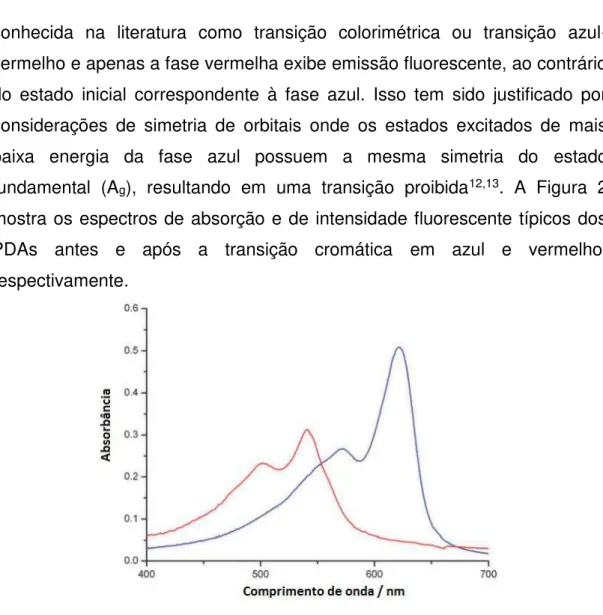
conhecida na literatura como transição colorimétrica ou transição azul-vermelho e apenas a fase vermelha exibe emissão fluorescente, ao contrário do estado inicial correspondente à fase azul. Isso tem sido justificado por considerações de simetria de orbitais onde os estados excitados de mais baixa energia da fase azul possuem a mesma simetria do estado fundamental (Ag), resultando em uma transição proibida12,13. A Figura 2
mostra os espectros de absorção e de intensidade fluorescente típicos dos PDAs antes e após a transição cromática em azul e vermelho, respectivamente.
Figura 2. Espectros de absorção UV-Vis típicos dos PDAs.
Os espectros eletrônicos dos PDAs antes do estímulo apresentam uma banda de absorção típica em 640 nm e um ombro vibrônico em 590 nm de tal forma que o material apresenta uma coloração azul (fase azul). Após o estímulo estes sistemas apresentam bandas resultantes de transições eletrônicas em 540 e 490 nm e o material apresenta-se com coloração vermelha (fase vermelha).
4
(Figura 3).
Figura 3. Reestruturação da cadeia conjugana en-ino para a forma de butatrieno.
Entretanto, estudos espectroscópicos e cálculos quanto-mecânicos mostram que a forma en-ino alternada está presente em ambas as fases15,16.
A segunda hipótese sugere que a transição colorimétrica é resultado de mudanças conformacionais na estrutura conjugada do polímero que geram uma redução do comprimento da caixa dos elétrons π-conjugados. A fase azul e a fase vermelha teriam suas cadeias principais em uma conformação planar (mais conjugada) e não-planar, respectivamente17. Alguns estudos
utilizando ressonância magnética nuclear (RMN) e espectroscopia no infra-vermelho suportam este modelo, sendo, portanto, o mais aceito pela comunidade científica atualmente18,19,20,21. Além disso, cálculos teóricos
afirmam que apenas uma pequena rotação em torno da ligação simples C─C reduz a sobreposição dos orbitais π, alterando o espectro eletrônico22.
A Figura 4 traz uma representação esquemática da rotação em torno das ligações simples na estrutura central do polímero.
Figura 4. Representação esquemática da rotação em torno das ligações simples na estrutura conjugada dos PDAs.
5
diferentes aplicações para cada caso. Os PDAs podem formar arranjos supramoleculares em uma, duas ou até mesmo em três dimensões, devido a facilidade com que os diacetilenos são estruturados e modificados. É possível também, através de reações químicas simples, modificar a cadeia hidrocarbônica e inserir grupos funcionais específicos nos DAs, alterando as interações intermoleculares e, consequentemente, modificando a barreira energética rotacional do polímero que é uma função da diferença de energia dos estados conformacionais das fases azul e vermelha1.
Na maioria dos artigos e revisões da literatura, os PDAs sofrem uma transição cromática irreversível que é um fator limitante no desenvolvimento de alguns tipos de sensores colorimétricos. Nesse contexto, Chen e Yoon23
(2011) modificaram monômeros do ácido 10,12-pentacosadiinóico (PCDA) incorporando um grupo fenilacetamida (FAT) na cabeça polar do ácido carboxílico. A partir dos monômeros de FAT-PCDA, vesículas de polidiacetileno foram preparadas apresentando termocromismo reversível na faixa de temperatura entre 30 e 70 ºC. Os autores atribuíram a causa dessa reversibilidade às ligações de hidrogênio e às interações de empacotamento
π entre as cabeças adjacentes dos FAT-PCDA.
Balakrishnan e colaboradores24 (2010) também investigaram o
6
Figura 5. Fotografias da solução de Li-PCDA (a) e lipossomas de PCDA (b)
embebidas em filme de PVA em diferentes temperaturas24.
As diferentes arquiteturas formadas pelos PDAs quando estes são preparados ainda é de difícil controle e elas são formadas na maioria dos casos aleatoriamente1. A formação de estruturas unidimensionais é
comumente favorecida de tal forma que Russel e colaboradores25 (2004),
estudando a atividade antibacteriana desses nanomateriais, verificaram que sais de monômeros de diacetileno anfifílicos contendo um grupamento amina em sua cabeça formam tubos finos bem definidos em solventes orgânicos.
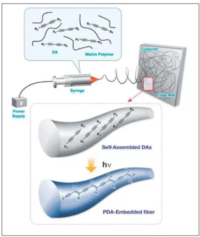
Outro método utilizado para a formação de PDAs unidimensionais foi feito através do processo de eletrofiação26. Nesta técnica uma solução de
7
formando, por fim, PDAs inseridos em fibras poliméricas e arranjados em uma única dimensão. Essas fibras apresentam diferentes respostas colorimétricas quando expostas em solventes orgânicos e foram aplicadas com um sensor para a identificação da qualidade da gasolina27.
A Figura 6 ilustra o método de obtenção das fibras de polímero contendo os PDAs com diâmetro de alguns nanômetros até micrômetros.
Figura 6. Representação esquemática da formação de nanofibras de PDA-polímero
através de eletrofiação26,27.
A formação de estruturas bidimensionais também tem sido explorada na literatura devido a sua possibilidade de revestir superfícies para tecnologia de imagem28. A deposição de monocamadas através da técnica
8
Já os PDAs estruturados em três dimensões incluem os cristais de PDA30, micelas9 e vesículas6,31, e possuem a vantagem de ter uma maior
área superficial e maior número de sítios de interação, quando comparados com estruturas uni e bidimensionais. Essas características fazem com que as estruturas tridimensionais sejam as mais indicadas na aplicação de sensoriamento de moléculas ou íons com relevância tecnológica e ambiental.
As vesículas são as formas tridimensionais dos PDAs mais encontradas na literatura e se assemelham, até certo grau, à organização de uma membrana celular. Assim como as membranas biológicas, as vesículas podem reconhecer seletivamente algumas moléculas estratégicas em sua interface, se os monômeros apresentarem sítios de ligação específicos, assim como as “proteínas antena” na superfície da célula.
Essas estruturas geralmente são encontradas na forma de membranas esféricas com características essencialmente fluídicas que englobam em seu interior uma fase aquosa ou orgânica. No caso das vesículas de PDA, a sua construção é feita em meio aquoso e os monômeros diacetilênicos devem ser organizados e orientados favoravelmente para que se consiga a posterior reação de adição 1,4.
Na formação das vesículas de PDA, os DAs são hidratados e individualizados em uma etapa de sonicação. Em seguida, este sistema é acondicionado a 4 ºC por um tempo superior a oito horas, necessário para a auto-organização dos monômeros na forma de bicamada. A temperatura mais baixa também é necessária para que os monômeros alinhados estejam preferencialmente na conformação trans, de forma que após a fotopolimerização o PDA esteja na conformação planar.
9
Figura 7. Esquema representando a auto-organização dos monômeros de PCDA e
a formação de vesículas de PDA após a irradiação UV.
3. Aplicações dos Polidiacetilenos como sensores colorimétricos As mudanças em suas propriedades óticas e eletrônica têm motivado os pesquisadores a utilizarem os PDAs como sensores de natureza diversa. Vários estudos na literatura avaliaram a transição cromática dos PDAs induzida por bactérias32, biomoléculas33,34, solventes orgânicos8,35,
temperatura36, pH37, surfactantes38, íons39, dentre outros. A resposta
colorimétrica (RC) é um parâmetro proposto por Charych (1993) capaz de informar de forma quantitativa a extensão da transição de cor após um determinado estímulo, além de ser útil como propriedade analítica do sensor4.
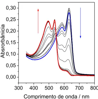
A Figura 8 ilustra os espectros de absorção de vesículas formadas a partir do PCDA antes e após a adição de quantidades crescentes de solução de NaOH. Nota-se que o máximo de absorção em ~ 640 nm diminui sua
absorbância, enquanto a fraca absorção em ~ 540 nm aumenta sua
10
300 400 500 600 700 800
0,00 0,05 0,10 0,15 0,20 0,25 0,30
Ab
s
ror
bâni
c
ia
Comprimento de onda / nm
Figura 8. Espectros de absorção UV-Vis das vesículas de PDA na presença de diferentes concentrações de NaOH.
Para quantificar a resposta colorimétrica dos PDAs, os espectros UV-Vis são analisados antes e após o estímulo. Define-se então, a razão entre a intensidade de absorção no comprimento de onda ( ) de 640 nm em relação a soma das intensidades em 640 e 540 nm que correspondem às fases azul e vermelha, respectivamente.
= / + (1)
onde B0 corresponde a razão das intensidades antes do estímulo. I640 e I540
são as absorbâncias em 640 e 540 nm, respectivamente. Após a adição da solução de NaOH ou de agente perturbador dos PDAs, define-se:
= / + (2)
onde B1 corresponde a razão das intensidades após o estímulo.
11
A RC é expressa em porcentagem de acordo com a equação 3.
𝑅 = − × % (3)
Como a RC é uma função da concentração do agente perturbador ou da intensidade do estresse aplicado no sistema, tal como o aumento da temperatura do sistema, as curvas colorimétricas permitem uma melhor visualização e compreensão do fenômeno. A Figura 9 ilustra a RC em função da concentração de NaOH calculada a partir dos espectros da Figura 8.
0,0 0,5 1,0 1,5 2,0 2,5 3,0
0 20 40 60 80 100
R
C
/
%
[NaOH] / mmol L-1
Figura 9. RC das vesículas de PDA em função da [NaOH].
12
interações fracas com os receptores, uma perturbação no esqueleto conjugado dos PDAs irá resultar em uma transição colorimétrica total ou parcial.
Yoon e colaboradores40 (2011) prepararam vesículas de PDA a partir
de monômeros contendo um grupo imidazólio ligados a grupos azida e alquino. Essas vesículas funcionalizadas foram utilizadas para a detecção de Cu2+ em sua superfície através de uma reação click. Os íons Cu2+ foram
adicionados à suspensão de vesículas na presença de ácido ascórbico sendo reduzidos a Cu+. Essa redução, promovida pelo ácido ascórbico, é
catalisada pelos grupos azida e alquino implicando na modificação conformacional da estrutura do polímero conjugado. Os autores obtiveram um sensor específico para íons cobre visto que nenhum outro cátion foi capaz de promover uma resposta colorimétrica superior a 60% em concentrações 50 vezes maior que a da espécie de interesse (Figura 10).
Figura 10. Fotografia das soluções de PDA funcionalizado com grupos azida e alquino na presença de diversos cátions.
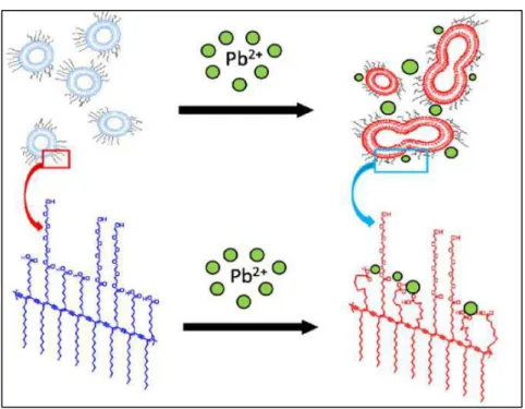
Sukwattanasinitt e colaboradores41 (2011) sintetizaram diferentes
ésteres de etileno glicol a partir de monômeros de PCDA e construíram vesículas com misturas destes novos monômeros e PCDA na razão molar de 1:9 para a detecção seletiva de íons Pb2+. O sensor foi capaz de detectar
o metal em níveis de partes por milhão e sem RC significativa na presença de vários íons tais como Cd2+, Co2+, Cu2+, Fe2+, Hg+, Ni2+ e Zn2+. A transição
colorimétrica foi atribuída à perturbação das cadeias provocada pela fusão das vesículas induzida pela ligação do Pb2+ aos grupos carboxilatos. Essa
13
A Figura 11 mostra a representação esquemática do mecanismo proposto pelos autores para a detecção seletiva de íons Pb2+.
Figura 11. Representação esquemática do mecanismo proposto transição
colorimétrica induzida por Pb2+ 41.
Outro exemplo importante da aplicação direta dos PDAs foi descrita por Thongmalai e colaboradores42 (2011). Estes autores utilizaram os PDAs
14
interações intermoleculares envolvidas nesses processos de reconhecimento molecular seletivo.
Pires e colaboradores43 (2010) mediram pela primeira vez a energia
associada às transições colorimétricas induzidas por diferentes solventes orgânicos em vesículas de PCDA e PCDA + colesterol + esfingomielina dispersas em água. Utilizando a Microcalorimetria de Titulação Isotérmica (ITC), os autores investigaram a resposta colorimétrica induzida por diferentes solventes clorados e encontraram um mecanismo de transição diferente para cada um. Tetracloreto de carbono (CCl4), clorofórmio (CHCl3)
e diclorometano (CH2Cl2) promoveram uma transição colorimétrica nas
vesículas de PCDA de aproximadamente 48, 98 e 52 %, respectivamente. Já nas vesículas contendo colesterol e esfingomielina, as RCs para CCl4,
CHCl3 e CH2Cl2 foram de aproximadamente 32, 62 e 42 %, respectivamente.
Entretanto, os resultados obtidos por ITC indicaram que apenas o CHCl3
induz uma transição entalpicamente dirigida enquanto os solventes CCl4 e
15
4. Referências bibliográficas
[1] Yarigama, O.; Jaworski, J.; Yoon, B.; Kim, J. Chem. Commun. 48, 2012, 2469-2485.
[2] Carpick, R. W.; Sasaki, D. Y.; Marcus, M. S.; Eriksson, M. A.; Burns, A. R. J. Phys.: Condens. Matter 16, 2004, R679-R697.
[3] Wegner, G. Z. Naturforsch Teil B 24, 1969, 824-832.
[4] Charych, D. H.; Nagy, J. O.; Spevak, W.; Bednarski, M. D. Science 261, 1993, 585-588.
[5] Chen, X.; Zhou, G.; Peng, X.; Yoon, J. Chem. Soc. Rev. 41, 2012, 4610-4630.
[6] Okada, S.; Peng, S.; Spevak, W.; Charych, D. Acc. Chem. Res. 31, 1998, 229-239.
[7] Endo, O.; Ootsubo, H.; Toda, N.; Suhara, M.; Ozaki, H.; Mazki, Y. J. Am. Chem. Soc. 126, 2004, 9894-9895.
[8] Champaiboon, T.; Tumcharern, G.; Potisatityuenyong, A.; Wacharasindhu, S.; Sukwttanasinitt, M. Sens. Actuators B 139, 2009, 532-537.
[9] Perino, A.; Klymchenko, A.; Morere, A.; Contal, E.; Rameau, A.; Guenet, J.; Mély, Y.; Wagner, A. Macromol. Chem. Phys. 212, 2011, 111-117.
[10] Nagy, J. O.; Zhang, Y.; Liu, E. Y. X.; Motari, E.; Song, J. C.; Lejeune, J. T.; Wang, P. G. Bioorg. Med. Chem. Lett. 18, 2008, 700-703.
[11] Jose, D. A.; König, B. Org. Biomol. Chem. 8, 2010, 655-662.
[12] Soos, Z. G.; Galvao, D. G.; Etemad, S. Adv. Mater. 6, 1994, 280-287. [13] Carpick, R. W.; Sasaki, D. Y.; Burns, A. R. Langmuir 16, 2000, 1270-1278.
[14] Chance, R. R.; Baughman, R. H.; Muller, H.; Eckhardt, C. J. J. Chem. Phys. 67, 1997, 3616-3618.
16
[16] Bässler, H.; Sixl, H.; Enkelmann, V. Advances in Polymer Science ed H-J Cantow., 1984, Springer.
[17] Sandman, D. J. Trends Polym. Sci. 2, 1994, 44-55.
[18] Lio, A.; Reichert, A.; Ahn, D. J.; Nagy, J. O.; Salmeron, M.; Charych, D. H.Langmuir 13, 1997, 6524-6532.
[19] Lee, D. C.; Sahoo, S. K.; Cholli, A. L.; Sandman D. J. Macromolecules 35, 2002, 4347-4355.
[20] Tanaka, H.; Gomez, M. A.; Tonelli A. E.; Thakur, M. Macromolecules 22, 1989, 1208-1215.
[21] Rubner, M. F.; Sandman, D. J.; Velazquez, C. Macromolecules 20, 1987, 1296-1300.
[22] Hankin, S. H. W.; Downey, M. J.; Sandman, D. J. Polymer 33, 1992, 5098-5101.
[23] Chen, X.; Yoon, J. Dyes and Pigments 89, 2011, 194-198.
[24] Balakrishnan, S.; Lee, S.; Kim, J. J. Mater. Chem. 20, 2010, 2302-2304. [25] Lee, S. B.; Koepsel, R.; Stolz, D. B.; Warriner, H. E.; Russel, A. J. J. Am. Chem. Soc. 126, 2004, 13400-13405.
[26] Yoon, J.; Kim, J. M. Macromol. Chem. Phys. 209, 2008, 2194-2203. [27] Lee. J.; Balakrishnan, S.; Cho, J.; Jeon, S. H.; Kim, J. M. J. Mater. Chem. 21, 2011, 2648-2655.
[28] Siegel, A. C.; Philips, S. T.; Wiley, B. J.; Whitesides, G. M. Lab Chip 9, 2009, 2775-2781.
[29] Coe, E.; Kane, J. J.; Nguyen, T. L.; Toledo, L. M.; Wininger, E.; Fowler, F. W.; Lauher, J. W. J. Am. Chem. Soc. 119, 1997, 86-93.
[30] Luo, L.; Wilhelm, C.; Sun, A.; Grey, C. P.; Lauher, J. W. Goroff, N. S. J. Am. Chem. Soc. 130, 2008, 7702-7709.
17
[32] Scindia, Y.; Silbert, L.; Volinsky, R.; Kolusheva, S.; Jelinek, R. Langmuir 23, 2007, 4682-4687.
[33] Biesalski, M.; Tu, R.; Tirrell, M. V. Langmuir 21, 2005, 5663-5666.
[34] Lee, S. W.; Kang, C. D.; Yang, D. H.; Lee, J. S.; Kim, J. M.; Ahn, D. J. Macromol. Res. 14, 2006, 483-485.
[35] Eaidkong, T.; Mungkarndee, R.; Phollookin, C.; Tumcharern, G.; Sukwattanasinitt, M.; Wacharasindhu, S. J. Mater. Chem. 22, 2012, 5970-5977.
[36] Charoenthai, N.; Pattanatornchai, T.; Wacharasindhu, S.; Sukwattanasinitt, M.; Traiphol, R. J. Coll. Interf. Sci. 360, 2011, 565-573. [37] Kew, S. J.; Hall, E. A. H. Anal. Chem. 78, 2006, 2231-2238.
[38] Lee, K. M.; Moon, J. H.; Jeon, H.; Chen, X.; Kim, H. J.; Kim, S.; Kim, S. J.; Lee, J. Y.; Yoon, J. J. Mater. Chem. 21, 2011, 17160-17166.
[39] Upcher, A.; Lifshitz, Y.; Zeiri, L.; Golan, Y.; Berman, A. Langmuir 28, 2012, 4248-4258.
[40] Xu, Q.; Lee, K. M.; Wang, F.; Yoon, J. J. Mater. Chem. 21, 2011, 15214-15217.
[41] Narkwiboonwong, P.; Tumcharern, G.; Potisatityuenyong, A.; Wacharasindhu, S.; Sukwattanasinitt, M. Talanta 83, 2011, 872-878.
[42] Thongmalai, W.; Eaidkong, T.; Ampornpun, S.; Mungkarndee, R.; Tumcharern, G.; Sukwattanasinitt, M.; Wacharasindhu, S. J. Mater. Chem. 21, 2011, 16391-16397.
18
CAPÍTULO 2
Synthesis and optical properties of polydiacetylenes in
triblock copolymers aqueous solution
1. Introduction
Polydiacetylenes (PDAs) are en-yne conjugated polymers formed by the 1,4-addition of diacetylene monomers1. For this reaction of
polymerization to occur, the monomers must be properly oriented and excited by UV radiation with the occurrence of the polymerization in several self-assembly forms such as monolayer2, multilayer3, nanostructured particles4,
micelles5 and vesicles6.
The PDAs exhibit interesting optical properties as evidenced in their ability to color change from blue to red, under various stimuli such as temperature7, pH8 and in the presence of various species as surfactants9-11,
ions10,11, organic solvents12,13, biomolecules14,15, including virus16 and
bacteria17. However, the mechanism involved in this process are not yet fully
understood, due to different behaviors observed of the materials when exposed to various environmental perturbations18. Nevertheless, a large
number of articles have explored the chromatic properties of PDAs to the development of smart sensors19. However, there is a particular difficulty for
developing and implementing this material as sensors due to the limited range of response for chromatic transition of the PDA. For a complete blue-red transition, the stimulus applied is restricted at a specific and very small range intensity. For example, the vesicles prepared with 10,12-pentacosadiynoic acid monomers (PCDA) undergo an gradual colorimetric transition with increasing of the temperature. However, only at 55 ºC the absorption spectrum and the appearance of the material is abruptly changed, and the PDA molecules are completely converted from blue to red only at ~68 ºC20. The original packing state of the monomers is indeed the main
19
diacetylene structure, is of great scientific and technological importance. One possible approach to modulate the PDA chromatic range is making the auto-organization process to occur into geometric controlled template.
The (EO)n-(P)m-(EO)n (EO = ethylene oxide, PO = propylene oxide)
block copolymers, commercially known as Pluronics, or Synperonics, are amphiphilic molecules capable of self-aggregating in aqueous solution, forming micelles with a hydrophobic core composed mainly by PO segments and hydrophilic shell composed by EO units21. The size of aggregates as well
as the hydrophobic core depends mainly of molecular weight and the EO/PO ratio of the macromolecule22. Thus, the copolymers micelles provide a
suitable place for insertion and orientation of diacetylene monomers in the hydrophobic core of the aggregates, which act as special reactors for the polymerization process. Comparing with the PDA vesicles synthesized in water, in the PDA/copolymer blends, the packing state of the monomers is modified.
Therefore, we synthesize the PDAs composed by 10,12-pentacosadiynoic acid or 10,12-tricosadiynoic acid in amphiphilic environments formed by copolymers of different hydrophobicity/hydrophilicity balance, molecular weight and concentration. The chromatic transition of nanoblends was evaluated by the temperature changes and by addition of sodium hydroxide and sodium carbonate in the system. The optic properties of the nanoblends were ever compared with blue-red transition of the classic vesicle. Apparent molar enthalpy of interaction was determined by microcalorimetric technique in order to contribute to elucidating the molecular mechanism associated with the chromatic transition.
2. Material and methods 2.1. Materials
10,12-pentacosadiynoic acid (PCDA) and 10,12-tricosadiynoic acid (TCDA) were purchased from Sigma-Aldrich (U.S.A.). Sodium hydroxide (NaOH) and sodium carbonate (Na2CO3) were obtained from Vetec (Brazil).
20
Milli-QII water (Millipore, USA) water was used for the preparation of all solutions.
2.2. Vesicle preparation
PCDA or TCDA monomers were dissolved in CHCl3, which was then
removed by a stream of N2 gas. Deionized water was added to make the
total lipid concentration 1 mM. The resulting systems was sonicated for 10 minutes to obtain a clear solution, wich was immediately filtered using a 0.45 m PVDF filter (Milipore). The suspensions were then stored at 4 ºC overnight to induce crystallization of lipid membranes. Polymerization was carried out under irradiation at 254 nm wavelength for 5 min and resulted in blue vesicles.
2.3. Nanoblends preparation
PCDA or TCDA monomers were dissolved in copolymer aqueous solution made with different structures and concentrations without any previous step, such that the total lipid concentration is 1 mM. The resulting systems were sonicated for 10 minutes to obtain a clear solution, wich was
immediately filtered using a 0.45 μm PVDF filter (Milipore). The suspensions
were then stored at 4 ºC overnight and the polymerization was carried out under irradiation at 254 nm wavelength for 5 min, resulting in blue nanoblends.
2.4. Quantifying the colorimetric response (CR)
The temperature effect on PDA vesicles and nanoblends was analyzed by addition of 0.7 L of sample, diluted by factor of 1:10, into quartz cell spectrophotometer. The heating was promoted by Peltier system (Shimadzu TCC-240A) coupled to the UV-Vis spectrophotometer (Shimadzu UV-2550), with temperature control of 15.0 to 70.0 ºC. The spectra were obtained between 350 and 900 nm after thermal equilibrium. The chromogenic effect promoted by Na2CO3 and NaOH solutions was evaluated
21
quantify the extent of blue-to-red color transitions, the CR (%) was calculated using the following equation:
𝑅 % = [𝑃 − 𝑃𝑃 ] × (1)
where PB = Ablue/(Ablue+Ared). A is the blue (650 nm) and red (540 nm)
absorbance obtained by UV-vis spectroscopy. The terms blue and red are related to material appearance. PB0 is the control, blue-ratio of PDA pure at
15 ºC, while PB1 is the value of sample exposed to different temperature. PB0
and PB1 also represent the absorbances before and after the salt solution
exposition, respectively.
2.5. Isothermal Titration Calorimetry experiments
The energetic analyses of systems containing PCDA or TCDA vesicles and nanoblends with NaOH or Na2CO3 were performed on an isothermal
titration microcalorimeter model CSC-4200 (Calorimeter Science Corporation), controlled by ITCRun software. The whole calorimetric procedure was chemically and electrically calibrated to the heat of protonation of (tris(hydroxymethyl)aminomethane) and the joule effect, as recommended.
The titrations were carried out by step-by-step injections (5 L) of each one of concentrated salt solutions into a 1.75 mL reaction cell containing PCDA or TCDA suspension in the same concentration of the CR experiments. The injections of electrolytes solution were performed by a gastight Hamilton syringe (250 L) and controlled by an instrument, with time interval of 20 minutes between each injection. The solutions were titrated in the sample cell with stirring at 300 rpm using a helix stirrer, and measurements were conducted at a constant temperature of 25.000 ± 0.001 °C. The reference cell was filled with water, initially placed in the reaction cell. Raw data were obtained as a plot of power ( W) against time (minutes). These raw data were then integrated to obtain ∆H values associated with each injection. ∆Hobs parameters were calculated by dividing ∆H per
22
3. Results and discussion
3.1. Synthesis and evaluation of the thermochromism of vesicles and nanoblends
Figure 1 shows the UV-Vis electronic spectra of the PCDA vesicles (a) and the PCDA/L64 nanoblends (b) for both red and blue forms. The PCDA/L64 nanoblends were synthesized with the same concentration of diacetylene monomer, present in the vesicles, and the concentration of L64 used was 1.0 % (w/w).
400 500 600 700 800 900 0.00 0.05 0.10 0.15 A b s o rb a n c e
/ nm
25.0 ºC 60.0 ºC (a)
400 500 600 700 800 0.00
0.15 0.30 0.45
(b)
/ nm
A b s o rb a n c e
25.0 ºC 60.0 ºC
Figure 1. Absorption spectra of PCDA vesicles solution at 25 and 60 ºC (a) and
absorption spectra of PCDA/L64 nanoblends solution at 25 and 60 ºC (b).
The spectra of the vesicles and PCDA nanoblends are very similar. The absorption of radiation occurs in the same range of wavelengths and there is no significant change in the spectral form including maximum, minimum, and isosbestic point. At 25.0 ºC, both spectra show an absorption band at 640 nm and vibronic shoulder at 590 nm in the blue phase. After the stimulus, the solutions revealed electronic bands at 540 and 490 nm, respectively. The only difference between PCDAs formed into vesicles and nanoblends is shown in the absorption intensities. The molar absorptivity of the nanoblends PCDA/L64 and PCDA vesicles, both in blue phases, correspond to
ε
= (4.0999 ± 0.0454) and (1.3554 ± 0.07537) cm-1 mmol-1 L,23
copolymers, favors the formation of a larger number of resonant species shaped by its hydrophobic core.
The effect of the concentration of L64 in the synthesis of polymeric PCDA nanoblends was also evaluated. Figure 2 shows the spectra of the PCDA nanostructures formed in different concentrations of L64 (0.10, 1.00, 2.00 and 8.00 % (w/w)) and at the same PCDA monomeric concentration.
400 500 600 700 800 0.0
0.1 0.2 0.3 0.4 0.5 0.6
A
b
s
o
rb
a
n
c
e
nm L64 0.10 % (m/m) L64 1.00 % (m/m) L64 2.00 % (m/m) L64 8.00 % (m/m)
Figure 2. Absorption spectra of PCDA/L64 nanoblends solution made at different copolymer concentrations, at 15.0 ºC.
These spectra refer to blue form of the solutions containing the nanoblends after polymerization. Increasing the copolymer concentration, the absorption intensity increases. This result corroborates with the last ones, showing that copolymers optimize the polymerization of the PCDA monomers, creating an environment more suitable for the process of PCDA self-organization. The higher concentrations of L64 increases the number of copolymer aggregates facilitating the polymerization of PDAs. A possible mechanism for that process is: below the critical micelle concentration (cmc) of the copolymer, the PCDA structures are formed containing traped L64 monomers. Above the cmc of L64 equal to 2.55 % (m/m)21, the copolymers
24
in different tamplates, getting far enough for the non-occurrence of the 1,4-addition.
The amphiphilic environment, formed by copolymer in an aqueous solution, affect not only the synthesis of PCDA nanoblends but the chromatic transition range is also changed. Figure 3 shows the electronic absorption spectra of PCDA vesicles (a) and PCDA/L64 nanoblends (b) at different temperatures. In these experiments, the concentration of L64 for the formation of the nanoblend was 1,0 % (w/w). The PCDA structures were diluted in a solvent containing equal concentrations of copolymer used in the synthesis and the temperature difference defined between each spectrum corresponds to 5 ºC. The CR was calculated from these spectra for both PCDA vesicle (c) and PCDA nanoblend (d). The CR represents quantitatively the percentage of molecules that underwent blue-to-red transition after the stimulus.
400 500 600 700 800 0.00 0.05 0.10 0.15 A b s o rb a n c e
/ nm
Increasing Temperature (a)
400 500 600 700 800 0.00
0.15 0.30 0.45
(b)
/ nm
A b s o rb a n c e Increasing Temperature
20 30 40 50 60 70 0 20 40 60 80 100 C R / %
Temperature / ºC
(c)
20 30 40 50 60 70 0 20 40 60 80 100 C R / %
Temperature / ºC
(d)
Figure 3. Absorption spectra of PCDA vesicles (a) and PCDA/L64 nanoblends
solution (b) at different temperatures. CR curves as a function of the temperature of
25
The temperature increase induces a color transition in both the PCDA vesicles and PCDA/copolymer nanoblends, but at different temperature ranges. The PCDA vesicles are predominantly in the blue form up to approximately 45 ºC, with a gradual increase in red species not exceeding 20% (Figure 3 (a) and 3 (c)). Above 55 ºC, an abrupt change in the spectra occurs indicating the existence of a cooperative process. The temperature change causes a complete conversion (100%) of PCDAs vesicles to red form at 70 ºC. However the PCDA nanoblends are predominantly blue to a temperature below 30 ºC and, from this, these species also suffer an abrupt transition, with full electronic conversion at 50 º C (Figure 3 (b) and 3 (d)).
On the basis of our results, the PCDA/L64 nanoblends has a maximum temperature of transition reduced by 20 ºC, when compared to PCDA vesicles. This indicates that the polydiacetylene macromolecules arranged in amphiphilic reactors make different intermolecular interactions in comparison with vesicular form.
The most accepted model for scientific community suggests that the chromatic transition is a result of rotation about the C-C bond in the conjugated structure of the polymer. This reduces the planarity of the backbone, causing dramatic changes in the π-orbital overlap. Therefore, to occur this process is necessary to disturb the system with energy greater than rotational barrier of the C-C bond. Accordingly, these conformational changes lead to alternations in electronic states and the characteristic optical absorption that can be easily monitored by UV-Vis spectrophotometry25-29.
PCDA polymer arranged in the nanoblends can form PCDA-PCDA and PCDA-H2O interactions, as well as in vesicles, but also PCDA-L64
26
The CRs were also obtained for nanoblends formed in different concentrations of L64. As mentioned above, the nanoblends synthesized in higher concentrations of L64 form resonant structures that generate identical spectra and differ only in intensity of absorption. Interestingly, the chromatic range induced by temperature change is affect by copolymer concentrations. Figure 4 (a) shows the RCs obtained for the PCDA/L64 nanoblends, at different copolymer concentration, versus temperature. Each of these graphs were conveniently adjusted using the Origin software and the temperature where the transition corresponds to 50% of the maximum value of CR was defined as the chromatic transition temperature (Ttr).Figure 4 (b) shows the
Ttr values as a function of the copolymer concentration in the nanoblends.
This parameter is useful because it promotes a better view of the chromatic transition interval in terms of a midpoint. The results indicated as 0.00 % (m/m) of L64 refer to PCDA vesicle.
20 30 40 50 60 70 80 0 20 40 60 80 100 0.00% 0.01% 0.05% 0.10% 0.30% 1.00% 2.00% 5.00% 8.00% 12.0% C R / %
Temperature / ºC
(a)
0 2 4 6 8 10 12
20 25 30 35 40 45 50 55 60 65 (b) Ttr / º C
Concentration of L64 / % (m/m)
Figure 4. CR curves as a function of the temperature of nanoblends formed in
different concentrations of L64 (a) and Chromatic Transition Temperature as a
function L64 concentration (b).
27
relation to vesicles around 8 ºC. A more abrupt decrease in the Ttr values is
observed up to a concentration of 1.00 % (w/w) of L64 with ∆Ttr ≈ 24 ºC.
However at a concentration of 12.00 % (w/w) of L64, the Ttr values decrease
in ∆Ttr ≈ 34 ºC.
These results show that the synthesis of PCDAs in copolymer solutions generate species with chromatic transition bands highly dependent on the concentration of the macromolecule. The temperature range effect on the blue-red transition was highly extended without changing the nature of the diacetylene monomers.
To better understand the cause of these effects of amphiphilic reactors for the synthesis of PCDAs, different triblock copolymers were also used for the construction of polymeric nanoblends at different concentrations. These copolymers are different in terms of the number of ethylene oxide segments (NEO), number of propylene oxide segments (NPO) and the average molar mass (Mm) of the macromolecule. These parameters define their
physicochemical properties such as hydrophobicity, hydrophilicity, and interfacial tension. Furthermore, depending on the nominal composition of these copolymers, the critical micelle concentration also changes, as well as the size and shape of the aggregate and hydrophobic core. Thus, various copolymers with different structure permits the construction of PDAs in different environments that will result in chromatic ranges also different, depending on the stimulus applied. The Table 1 shows the nominal composition of the polymers used in this work.
Table 1. Nominal composition of the studied copolymers22.
Copolymer NEO NPO Mm / (g mol-1)
L35 2 X 11 17 1900
L64 2 X 13 31 2900
P123 2 X 19 69 5800
F68 2 X 76 30 8400
28
We evaluate the thermochromism associated the various PCDA/copolymer nanoblends at various concentrations of copolymer. In the synthesis of different nanoblends and obtaining of these spectra was used the same conditions applied for the PCDA/L64 nanoblends at the copolymer concentration of 1.00 % (w/w).
Was also synthesized and evaluated the thermochromism of TCDA/L64 nanoblends copolymer at various concentrations. The PCDA monomers have carbon chain greater than TCDA monomers in two carbon atoms, but both have the same carboxylic acid functional group.
From these spectra were obtained Ttr values as a function of
copolymer concentration. Figure 5 shows the Ttr curves of nanoblends as a
function of the concentration of macromolecule.
0 10 20 30 40 20
30 40 50
60 L64 F68
L35 P123 F127 T tr / º C
[Copolymer] / mmol L-1
(a)
PCDA
0 10 20 30 40 20 30 40 50 60 TCDA (b)
[Copolymer] / mmol L-1
Ttr / º C L64 F68 P123 F127
Figure 5. Chromatic Transition Temperature as a function copolymer concentration
using PCDA (a) and TCDA (b) monomers.
All copolymers used at the study, have caused a reduction in the Ttr of
29
occurs causing the weakening of the interaction between diacetylene monomers. In addition, larger hydrophobic cores tend to orient monomers diacetylenes in larger distances, contributing to the decrease of the monomer-monomer interactions. Both processes together, decrease the rotational energy barrier about the C-C bond justifying the reduction in the values of Ttr nanoblends.
Comparing the structures formed with different DA monomers, we expected that PCDA/copolymer nanoblends have transition temperatures always higher than TCDA/copolymer nanoblends for the same copolymer. This could be justified by the greater number of van der Waals interactions between the PCDA tail and the hydrophobic region of the copolymer and due to PCDA-PCDA interactions, compared to TCDA, that has a lower carbon chain. However, this trend has been not observed. Therefore, the hydrophobicity is not the only driving force able to influence the chromatic transition of the nanostructures, but also, other secondary parameters such as hydrophilicity and molecular weight of polymers.
Thus, the nanoblends containing copolymers with PDAs embedded have their chromatic transition temperature lowered mainly due to weakening of the interactions DA-DA, what is promoted by the highest number of interactions DA-copolymer formed in the hydrophobic environment of the aggregates.
30
3.2. Effect of NaOH addition on the PDA nanostructured systems The PDA vesicles show an irreversible transition from blue-to-red when the pH of the system is increased. This effect has been studied by Hall et al. (2006) by addition of the NaOH solutions and other metal hydroxides in suspensions of TCDA vesicles, with the objective to understand how the acid-base equilibrium affects the chromogenic responses of liposomes8.
According to these authors, the chromatic transition could be the result of a increased osmotic pressure across the membrane, the electrostatic effect associated with changes to the diffuse layer of counterions and the Coulomb repulsion between ionized groups on the surface of the bilayer. In this case, the Coulomb repulsion between charged heads is the main factor responsible for the disturbance in the PDA backbone, which reduced conjugation lenght and therefore induced blue-red transformations.
Traiphol et al. (2011) evaluated the effect of the head group on the pH-chromism of the PDA vesicles using the 10,12-pentacosadiynoic acid (PCDA) and N-(2-aminoethyl)pentacosa-10,12-diynamide (AEPCDA) diacetylene monomers20. The addition of hydroxide ions (OH-) in a
suspension of PCDA vesicles leads to breaking of the hydrogen bonds on the surface by transforming the carboxylic acid group to the carboxylate one. With that, the ionic repulsion generated on the surface causes the rearrangement of PDA segments. The behavior of AEPCDA vesicles is very different and opposite to the one observed in the PCDA vesicles. The addition of OH- does not affect the structure of the lipid membranes, but the
addition of the H+ protonates the surface of the structures causing a full color
31
In all previous studies, the pH-chromism range was modified using only monomers with different structures. The polymer nanoblends formed by diacetylene monomers and copolymers respond to different pH ranges depending on the packaging of the diacetylene monomers. Figure 6 shows the colorimetric response of PCDA and TCDA vesicles and PCDA/L64 and TCDA/L64 nanoblends, made at a copolymer concentration of 0.05 % (w/w), as a function of the concentration of NaOH in the system at 25 ºC.
0.0 0.5 1.0 1.5 2.0 2.5 3.0 0
20 40 60 80 100
C
R
/
%
[NaOH] / mmol L-1 PCDA vesicle PCDA/L64 blend TCDA vesicle TCDA/L64 blend
Figure 6. CR as a function NaOH concentration of the PCDA vesicle (■), PCDA/L64
nanoblend (▲), TCDA vesicle (□) and TCDA/L64 nanoblend (∆), at 25 ºC.
The TCDA vesicles, as already described, are more sensitive to hydroxyl ions than PCDA vesicles, due to differences in the length of the carbon chain of monomers. In the NaOH concentration of 1.0 mM, the RC of PCDA vesicles is smaller than the vesicles TCDA in approximately 23 units. Furthermore, the full electronic transition occurs at concentrations above 2.5 and 2.2 mM to PCDA and TCDA, respectively. Though, the polymer nanoblends are much more sensitive to the basic environment and show a colorimetric transition above 95% at approximately 1.8 and 0.5 mM to PCDA and TCDA, respectively.
32
action disturbs the skeleton of PCDA, being the polymer nanoblends the most sensitive.
The chromatic transition of the nanoblends synthesized at different concentrations of copolymer promoted by OH- ions were also evaluated.
Figure 7 shows RC curves as a function of the concentration of NaOH, for different PCDA/L64 (a) and TCDA/L64 (b) nanoblends.
0.0 0.5 1.0 1.5 2.0 2.5 3.0 3.5 0 20 40 60 80 100 (a) R C / %
[NaOH] / mmol L-1 PCDA vesicle
PCDA/L64 (0.05 % (w/w)) PCDA/L64 (0.10 % (w/w)) PCDA/L64 (1.00 % (w/w)) PCDA/L64 (2.00 % (w/w)) PCDA/L64 (8.00 % (w/w))
0.0 0.5 1.0 1.5 2.0 2.5 3.0 0 20 40 60 80 100 (b) RC / %
[NaOH] / mmol L-1 TCDA vesicle
TCDA/L64 (0.05 % (w/w)) TCDA/L64 (0.10 % (w/w)) TCDA/L64 (1.00 % (w/w)) TCDA/L64 (2.00 % (w/w)) TCDA/L64 (8.00 % (w/w))
Figure 7. CR as a function NaOH concentration of the PCDA/L64 (a) and TCDA/L64
(b) nanoblends made in different concentrations of L64, at 25 ºC.
The sensitivity of the nanostructures in relation to the OH- ion depends
33
concentrations of L64, the nanoblends have abrupt transition range and maximum transition colorimetric very similar to the nanoblends made in high concentrations of copolymer. The full colorimetric transition occurs at 0.50 and 0.37 mM of NaOH to 0.05 % (w/w) and 8.00 % (w/w) of L64, respectively.
Isothermal titration calorimetry (ITC) is a sensitive and reproductive technique that gives us valuable information about the processes of interaction between PCDA or TCDA structures and the species that promote the chromatic transition30. Thus, the characteristics of the apparent molar
enthalpy of interaction curves between PCDA or TCDA vesicles and nanoblends with NaOH (∆ 𝑎𝑝−𝑖𝑛 ) versus [NaOH], provide quantitative energetics informations for verifying the proposed model for the chromatic transition induced by this electrolyte. To determine values of ∆ 𝑎𝑝−𝑖𝑛 , we subtract the dilution curve in solvent from the titration curve of electrolyte in solution containing the nanostructures for each electrolyte concentration. As the amount of ions interacting with the structures of polydiacetylene is unknown, so we cannot calculate the exact molar enthalpy change of this interaction, but only an apparent molar enthalpy change.
Figure 8 shows the enthalpic titration curves obtained from addition of NaOH to PCDA or TCDA solutions, where the apparent molar enthalpy change of interaction for each injection of electrolyte solution is plotted against total electrolyte concentration.
0.0 0.2 0.4 0.6 0.8 1.0 1.2 -60 -50 -40 -30 -20 -10 0 10 PCDA vesicle TCDA vesicle ap
-i n t H / k J m o l -1
[NaOH] / mmol L-1
34
The curves for both polydiacetylenes shows that up 0.17 mM of the NaOH, the enthalpy change is exothermic, with initial values of -49.8 kJ mol -1. The energy released has become less negative with the increase of NaOH
concentration in the system. Above 0.17 mM of NaOH, the process became almost athermal. Based on this behavior, the interaction between the vesicles with ions in solution is enthalpically favorable and the ∆ 𝑎𝑝−𝑖𝑛 values were
mainly associated with the deprotonation of the carboxylic groups on the vesicle surface with subsequent neutralization reaction between protons released and hydroxide ions added on the system. This can be justified by the values of the heats of dissociation of carboxylic acid groups (∆ 𝑖 =
-0,544 kJ mol-1) and neutralization reaction (∆
𝑛 = -57,35 kJ mol-1), both
exothermic, which are values of the order of ∆ 𝑎𝑝−𝑖𝑛 obtained by ITC31.
Furthermore, for both vesicles, the abrupt increase in the values of ∆ 𝑎𝑝−𝑖𝑛 shows the occurrence of a cooperative process caused by
interactions between the hydroxide ions and the carboxylic groups on the vesicle surface, implying in the possible aggregation of nanostructures caused by formation of hydrogen bonds between the functional groups of adjacent vesicles with partially ionized surface. Therefore, the result of all these molecular processes originates in the disruption of the coplanarity of the en-yne conjugated polymer that leads to the chromatic transition, being the distance amidst adjacent monomers the main factor, caused by the electrostatic repulsion of the charged heads. However, despite the colorimetric transition of the vesicles occur at concentrations above 0.5 mM, the ITC curves over this concentration, reveal that the energy required to overcome the rotational barrier of the backbone conjugated is negligible in comparison with the processes described above. It is also important to emphasize that there is no significant difference between the processes occurring in PCDA and TCDA vesicles. The coincidence in the ∆ 𝑎𝑝−𝑖𝑛 curves shows that the size of the tail of the diacetylene monomers, arranged in vesicles, does not influence in the interaction process of these nanostructures with hydroxide ions, corroborating with the surface model suggested.
35
titration of NaOH solutions on PCDA or TCDA nanoblends synthesized in concentration of 1.0 % (w/w) of L64.
0.0 0.3 0.6 0.9 1.2 -10 0 10 20 30 40 50 60 70 80 90 100 110 120
130 PCDA/L64 nanoblends TCDA/L64 nanoblends
ap
-i n t H / k J m o l -1
[NaOH] / mmol L-1
Figure 9. Apparent molar enthalpy change of interaction between (■) PCDA/L64
and (●) TCDA/L64 nanoblends versus NaOH concentration, at 298.15 K.
The ∆ 𝑎𝑝−𝑖𝑛 curves for nanoblends are completely different from the curves obtained for the vesicles. For PCDA/L64 nanoblends, the enthalpy values are endothermic until concentration of ~0.30 mM of NaOH. In the first injections of alkaline solution, until 0.128 mM, the system absorbs an energy of around 122 kJ mol-1. From this concentration, an abrupt decrease in the
values of ∆ 𝑎𝑝−𝑖𝑛 shows the occurrence of a cooperative process. In the curves obtained for the TCDA/L64 nanoblends, the profile is similar in relation the nanostructures synthesized with PCDA. Although, different from the behavior observed for the vesicles when they interact with NaOH, the PCDA and TCDA nanoblends differ in terms of magnitude and the concentration range in which thermodynamic processes occur. The first injections of NaOH, the energies are endothermic with values of ~117 kJ mol -1 to the concentration of 0.076 mM. After, occurs an abrupt drop in the
∆ 𝑎𝑝−𝑖𝑛 values until ~0.14 mM of NaOH, where the process became almost
athermal.
36
monomers and the electrostatic repulsion generated on the surface is sufficient to cause conformational changes in the copolymer structure containing chains of polymerized PCDA or TCDA monomers trapped. The conformational changes in the L64 structures are processes entropically driven, with endothermic curves, despite the ionization and neutralization reaction on the surface that results in exothermic enthalpy changes. Thus, these thermodynamic processes prevail at the start of titration for both PCDA and TCDA nanoblends until concentrations of 0.128 and 0.076 mM, respectively.
The abrupt decrease in the values of apparent enthalpy of interaction for the nanoblends is the result of possible aggregation processes of the copolymer chains containing diacetylene monomers adsorbed. All these molecular processes seek to achieve a more stable conformation, however, occurs the polydiacetylene backbone disturbance causing the colorimetric transition. In the case of nanoblends, the decrease in the coplanarity of the en-yne structure occurs by the same effects that cause the chromatic change in the vesicles, but these structures are formed on copolymer templates and the synergistic effect between the conformational changes of macromolecule and trapped chains of diacetylenes becomes very important. Thus, the difference between PCDA and TCDA nanoblends curves is justified by the greater number of hydrophobic interactions between PCDA monomers and PO units of L64. The increase of these interactions implies in a lower rotational freedom of copolymers molecules into the nanoblend structure, so that the energy required to overcome the rotational energy barrier in PCDA nanoblends is greater, and the concentration of NaOH needed to promote self-aggregation processes in nanoblends of TCDA is lower. Moreover, as in the microcalorimetric experiments for vesicles, the energy involved in the transition chromatic process of polydiacetylenes in nanoblends is negligible compared to other thermodynamic processes observed by ITC.
3.3. Effect of Na2CO3 addition on the PDA nanostructured systems Aqueous solutions of Na2CO3 present a series of equilibria reactions
37 Na2CO3→ 2 Na+ + CO3
2-CO32- + H2O ↔ HCO3- + OH- K1 = 4.5 x 10⁻⁷ HCO3- + H2O ↔ H2CO3 + OH- K2 = 4.5 x 10⁻¹¹
H2CO3 → CO2 + H2O
Figure 10. Reactions equilibrium involved in aqueous solution of Na2CO3.
Na2CO3 solutions are considered basic salts due to the ability to
originate hydroxide ions in solution, increasing the pH. Thus, an aqueous solution of this salt also promote the colorimetric transition in PDA structures. However, there is a greater number of ionic species with different sizes and charge distribution involved in these equilibria, which interacts differently with the blue nanostructures.
Figure 11 shows the colorimetric response of PCDA or TCDA vesicles and nanoblends as a function of the Na2CO3 concentration in the system, at
25 ºC. In the same way as done in the experiments with NaOH, the nanoblends used in this study were synthesized in the copolymer solution at various L64 concentrations.
0.0 0.5 1.0 1.5 0 20 40 60 80 100 (a) PCDA vesicle
PCDA/L64 (0.05 % (w/w)) PCDA/L64 (0.10 % (w/w)) PCDA/L64 (1.00 % (w/w)) PCDA/L64 (2.00 % (w/w)) PCDA/L64 (8.00 % (w/w))
R
C
/
%
[Na2CO3] / mmol L-1
0.0 0.5 1.0 1.5
0 20 40 60 80 100 (b) RC / %
[Na2CO3] / mmol L-1
TCDA vesicle
TCDA/L64 (0.05 % (w/w)) TCDA/L64 (0.10 % (w/w)) TCDA/L64 (1.00 % (w/w)) TCDA/L64 (2.00 % (w/w)) TCDA/L64 (8.00 % (w/w))
Figure 11. CR as a function NaOH concentration of the PCDA vesicle or PCDA/L64
nanoblends (a) and TCDA vesicle or TCDA/L64 (b) nanoblends made in different
concentrations of L64, at 25 ºC.
At intervals from 0.0 to 1.5 mM, the aqueous solutions of Na2CO3


![Figura 9. RC das vesículas de PDA em função da [NaOH].](https://thumb-eu.123doks.com/thumbv2/123dok_br/15367770.62992/24.892.304.620.453.781/figura-rc-das-vesículas-de-pda-função-naoh.webp)