Volume 2013, Article ID 395672,8pages http://dx.doi.org/10.1155/2013/395672
Research Article
Exercise and Caloric Restriction Alter the Immune System of
Mice Submitted to a High-Fat Diet
Frederick Wasinski,
1Reury F. P. Bacurau,
2Milton R. Moraes,
1Anderson S. Haro,
1Pedro M. M. Moraes-Vieira,
3Gabriel R. Estrela,
1Edgar J. Paredes-Gamero,
1Carlos C. Barros,
4Sandro S. Almeida,
1Niels O. S. Câmara,
3and Ronaldo C. Araujo
11Department of Biophysics, Federal University of S˜ao Paulo, 04023-062 S˜ao Paulo, SP, Brazil
2School of Arts, Sciences and Humanities, University of Sao Paulo, Avenue Arlindo Bettio 1000, 03828-000 S˜ao Paulo, SP, Brazil 3Department of Immunology, Laboratory of Transplantation Immunobiology, Institute of Biomedical Sciences, University of S˜ao Paulo,
05508-900 S˜ao Paulo, SP, Brazil
4Department of Nutrition, School of Nutrition, Federal University of Pelotas, 96010-610 Pelotas, RS, Brazil
Correspondence should be addressed to Ronaldo C. Araujo; [email protected]
Received 5 December 2012; Revised 5 February 2013; Accepted 6 February 2013
Academic Editor: Franc¸ois Mach
Copyright © 2013 Frederick Wasinski et al. his is an open access article distributed under the Creative Commons Attribution License, which permits unrestricted use, distribution, and reproduction in any medium, provided the original work is properly cited.
As the size of adipocytes increases during obesity, the establishment of resident immune cells in adipose tissue becomes an important source of proinlammatory mediators. Exercise and caloric restriction are two important, nonpharmacological tools against body mass increase. To date, their efects on the immune cells of adipose tissue in obese organisms, speciically when a high-fat diet is consumed, have been poorly investigated. hus, ater consuming a high-fat diet, mice were submitted to chronic swimming training or a 30% caloric restriction in order to investigate the efects of both interventions on resident immune cells in adipose tissue. hese strategies were able to reduce body mass and resulted in changes in the number of resident immune cells in the adipose tissue and levels of cytokines/chemokines in serum. While exercise increased the number of NK cells in adipose tissue and serum levels of IL-6 and RANTES, caloric restriction increased the CD4+/CD8+ cell ratio and MCP-1 levels. Together, these data demonstrated that exercise and caloric restriction modulate resident immune cells in adipose tissues diferently in spite of an equivalent body weight reduction. Additionally, the results also reinforce the idea that a combination of both strategies is better than either individually for combating obesity.
1. Introduction
Chronic, low-grade inlammation is associated with insulin resistance, type 2 diabetes, and several types of cancer [1,2]. hese detrimental conditions are also associated with obesity [3]. As visceral and subcutaneous adipocytes increase in size, monocytes and CD4+ and CD8+ T cells migrate to adipose tissue (AT) [4] initiating the release of proinlammatory mediators (e.g., IL-1�, IL-6, RANTES, MCP-1, and IL-18) inducing local insulin resistance [5]. hus, the expanded ATs and their populations of resident immune cells consti-tute the main microenvironment in which proinlammatory cytokines are produced and released in the organism [6].
Physical activity and caloric restriction (CR) are both nonpharmacological strategies recommended to reduce obe-sity [7]. Although the beneicial efects of exercise are well described in skeletal muscle and the liver, the same is not true for AT [7–9]. Additionally, there is little information concerning the efects of both weight reduction strategies on immune cell populations that reside in AT. In relation to CR, though its importance in reducing body weight is unquestionable, consumption of a healthy diet requires such marked lifestyle changes that many individuals are unable to comply with one of them.
maintaining a high-fat (HF) diet. he aim of this study was investigate the efect of physical exercise or CR on AT immune cells in diet-induced obese mice.
2. Materials and Methods
2.1. Animals. Male C57BL/6 (� = 20, 5 per group) mice (aged 8–12 weeks; 23–26 g) were obtained from the Animal Care Facility at the Federal University of S˜ao Paulo (UNIFESP). All animals were housed in standard, individual cages and had access to water and food. To examine the changes in stromal vascular cell populations in adipose tissue under conditions of diet-induced obesity, we divided the C57BL/6 mice into four groups and fed them either a standard chow diet (6% fat, Nuvilab mod. CR-1) or a high-fat diet (D12451, 45% Kcal fat, Research Diets). At 16 weeks, the mice were further subdivided into the following groups: (1) a control group fed a normal, low-fat (LF) chow; (2) a control group fed a high-fat (HF) diet; (3) a dietary restriction group fed a 30% high-fat diet (HFREST); and (4) an exercise group fed a high-fat diet that participated in 60 minutes of swimming (HFEX). Food consumption was controlled every day. Based on the quantity of high-fat diet chow consumed and using of the macronutrient composition as reference, we calculated the energy intake. he 30% caloric restriction was designed taking HF consumption as a reference.
All procedures were previously reviewed and approved by the internal ethical committee of the institution.
2.2. Exercise Protocol. he HFEX animals were subject to swimming sessions in a swimming system adapted for mice with water heated to 30∘C. he 300 liter tank had 10 lanes and was itted with air pumps that maintained the mice in constant motion. Swimming sessions began with 15 minutes in the irst week and gradually increased in length until the mice were able to swim for 60 minutes a day. At this point, the HFEX mice were subjected to swimming sessions 5 times per week for 6 weeks. Both the exercise and the dietary restriction groups were subject to their respective intervention for 6 weeks.
he mice were anesthetized with ketamine/xylazine for blood collection via retroorbital venous plexus and then killed by cervical dislocation. he blood was centrifuged at 1000 g for 10 minutes. he serum was removed and stored at −80∘C for future analysis. We collected a 1 g sample of adipose inguinal from each group and subjected the sample to enzymatic degradation. All animals were weighed weekly until the end of the experiments.
2.3. Isolation of the Stromal Vascular Fraction (Sfv) and Flow Cytometry. Ater sacriicing the mouse, inguinal adipose tis-sue (IAT) was extracted, weighed, and subjected to enzymatic degradation as previously described [10]. Ater the isolation of the IAT SFV cells, 200�L of FCS washing bufer (1x PBS, 2% SFC) was added, and the solution was centrifuged for 5 minutes. Ater the supernatant was discarded, the pellet was resuspended in FCS and centrifuged for 5 minutes at
600 g. he cells were stained with anti-CD8 (Caltag-FITC-Medsystems, Buckingham, UK), anti-CD4 (blue-Paciic-BioLegend), anti-F4/80 (PerCP-Bioscience), and anti-NK.1 (PE-Bioscience) antibodies. he stromal cells were acquired via FACS in a Canto II low cytometer (BD, Becton Dickin-son, NJ, USA). he data analyses were completed using the program FlowJo 8.7.4. (Tree Star Inc., Ashland, OR, USA).
2.4. Analysis of Cytokines in Serum. Serum samples were stored at −80∘C. he panel used for the Milliplex Mouse cytokine/chemokine immunoassay included the following cytokines: MCP-1, RANTES, TNF-alpha (tumor necrosis factor), IL-6, and IL1-�. Testing was conducted in accordance with the procedures previously described by the manufac-turer (Milliplex Mouse cytokine/chemokine panel).
2.5. Glucose Tolerance Test. he glucose tolerance test (GTT) was carried out in animals fasted for 12 hours. To avoid stress, there was an interval of 7 days between tests. Glycemia was measured using a glucometer (Accu-Chek Advantage) measuring blood drops obtained from the tail vein. For GTT 1 g glucose per kg of body weight (BW) was injected intraperitoneally. Glucose levels were determined at baseline, 0, 15, 30, 60, and 120 min ater the injection of glucose.
2.6. Statistical Analysis. he data were presented as the mean±standard error in the descriptive text and graphics. All experiments were compared using One Way ANOVA followed by post-hoc Tukey test. Signiicant diferences were determined when the�value was less than 0.05 (� < 0.05). he graphics were developed in Prism 5.0.
3. Results
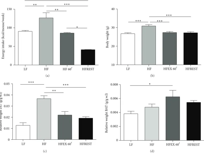
Animals subjected to the HF diet consumed more calories compared with mice from the LF diet group (Figure 1(a)). Higher caloric consumption was accompanied by an increase in mouse total body mass (Figure 1(b)). Swimming combined with the HF diet was able to reduce mouse body weight similar to that observed in animals administered caloric restriction (Figure 1(b)). here was no diference in inguinal adipose tissue (IAT) and brown adipose tissue (BAT) between trained animals and those submitted to caloric restriction (Figures1(c)and1(d)); however, trained animals presented more BAT than animals from LF group (Figure 1(d)).
It is known that obesity is associated with systemic, low-grade inlammation. herefore, we investigated the efects of a HF diet, exercise, and diet restriction in our study groups by evaluating the serum levels of several proinlammatory cytokines. We observed increased levels of IL-1�in the HF diet group and a reduction of this cytokine in both interven-tion groups (exercise and caloric restricinterven-tion) (Figure 2(a)). TNF-�serum levels were not afected by the changes in diet and exercise investigated in this study (Figure 2(b)).
0 50 100 150
Ener
g
y in
ta
k
e
(k
cal/mo
u
se/w
eek)
LF HF HFREST
∗ ∗∗
∗∗∗ ∗∗
∗∗
HF60�
(a)
10 20 30 40
B
o
d
y w
eig
h
t (g)
∗∗∗ ∗∗∗ ∗∗∗
LF HF HFEX60� HFREST
(b)
0 0.01 0.02 0.03 0.04 0.05
Rela
ti
ve
w
eig
h
t
IA
T (g/g
w
.l)
LF HF HFREST
∗∗
∗∗∗ ∗∗∗
HFEX60�
(c)
0 0.002 0.004 0.006 0.008
Rela
ti
ve
w
eig
h
t
B
A
T (g/g
w
.l)
∗
LF HF HFEX60� HFREST
(d)
Figure 1: (a) Weekly caloric intake of animals subjected to the control diet (LF), high-fat diet (HF), high-fat diet with exercise (HFEX 60�), and high-fat diet with 30% food restriction (HFREST). (b) Mouse body weights (g). (c) Relative weight of IAT (inguinal adipose tissue). (d) Relative weight of BAT (brown adipose tissue). Mean±SEM,� = 5mice per group.∗∗∗� < 0.001,∗∗� < 0.01,∗� < 0.05.
Reduction of body weight was unable to induce changes in IL-6 levels as no diferences were observed in animals from the caloric restriction group (Figure 2(c)).
Animals subjected to swimming had signiicantly increased RANTES serum levels (regulated upon activation, normal T cells expressed and secreted). he increases in body mass and IAT observed in animals submitted to the HF diet did not lead to changes in RANTES levels, albeit AT lymphocyte iniltration during obesity is expected. Nevertheless, physical exercise increased RANTES serum levels, while caloric restriction did not change its levels (Figure 2(d)). Moreover, MCP-1 (monocyte chemoattractant protein-1) was reduced by exercise and was not afected by caloric restriction (Figure 2(e)).
Because immune cells that reside in obese AT actively secrete proinlammatory cytokines and chemokines, we eval-uated the efect of the HF diet and both interventions on AT leukocyte populations. We observed a reduction in CD4+ and CD8+ T lymphocytes in AT in animals submitted to both interventions in comparison with animals consuming the HF diet (Figures3(a)and3(b)). Because CD8+ cells were more
reduced than CD4+ cells, an increased CD4+/CD8+ ratio was observed (Figure 3(e)). he natural killer cell marker (NK1.1) was afected only by caloric restriction (Figure 3(c)). Also, we observed that both exercise and caloric restriction were able to reverse the increased macrophage iniltration observed in the HF diet group (Figure 3(d)).
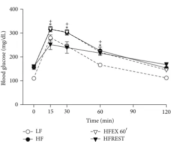
Concerning glucose tolerance test, it was observed that only caloric restriction was able to improve this parameter in comparison to HF group (Figure 4).
4. Discussion
0 500 1000 1500
IL
-1
�
(pg/mL)
∗∗
LF HF HF60� HFREST
(a)
0 10 20 30 40
∗
LF HF HF60� HFREST
TNF
-𝛼
(pg/mL)
(b)
0 20 40 60 80 100
IL
-6 (pg/mL)
∗
∗ ∗
LF HF HF60� HFREST
(c)
0 50 100 150
RANTES (pg/mL)
∗∗
∗ ∗
LF HF HF60� HFREST
(d)
0 50 100 150 200 250
M
CP
-1 (pg/mL)
∗ ∗
∗ ∗
LF HF HF60� HFREST
(e)
Figure 2: Cytokine concentrations (pg/mL) in mouse serum subjected to control diet (LF,� = 12), high-fat diet (HF,� = 12), high-fat diet with exercise 60�(HFEX 60�,� = 8), and high-fat diet with 30% food restriction (HFREST,� = 8). (a) IL-1�, (b) TNF-�, (c) IL-6, (d) RANTES, and (e) MCP-1. Mean±SEM.∗∗∗� < 0.001,∗∗� < 0.01,∗� < 0.05.
mass and counterbalances several deleterious consequences due to its anti-inlammatory efects [12]. Normally, exercise intervention with dietary modiications is suggested to com-bat obesity; the combination of both interventions works better than exercise alone [13–15]. However, little is known about the beneits of exercise when dietary modiications are not prescribed.
0 100 200 300
LF HF HFREST
∗∗∗ ∗∗∗
∗∗∗
HFEX60�
CD
4
(
×1
0
3cells)
(a)
0 50 100 150 200
LF HF HFREST
∗
∗∗∗
∗∗
∗ ∗∗∗
HFEX60�
CD
8
(
×1
0
3cells)
(b)
6.97
0 20 40 60 80 100
NK
1.1
(
×1
0
3 cells)
LF HF HFREST
∗ ∗∗∗
NK1.1
FSC
HFEX60�
(c)
0 200 400 600
F
4/
80
(
×1
0
3 cells)
LF HF HFREST
∗∗∗ ∗∗∗ ∗∗
24.8
F3/80
FSC
HFEX60�
(d)
0 2 4 6 8
CD4/CD8
LF HF HFREST
∗
∗∗
∗∗∗ ∗∗∗
15.8
19
HF60�
CD4
CD8
(e)
Figure 3: Quantiication of cells expressing CD4, CD8, NK1.1, and F4/80 (macrophage) in inguinal adipose tissue of mice subjected to control diet (LF), high-fat diet (HF), high-fat diet with exercise (HFEX 60�), and high-fat diet with 30% food restriction (HFREST). (a) CD4 cells, (b) CD8 cells, (c) NK1.1 cells, (d) F4/80 cells, and (e) ratio of CD4/CD8. Mean±SEM,� = 5mice per group.∗∗∗� < 0.001,∗∗� < 0.01,∗� < 0.05.
an inlammatory state in AT. Studies have also shown that reducing the presence of inlammatory cells in AT improves glucose tolerance in IFN-�-deicient mice [16]. IFN-�is also able to inhibit the Hedgehog signaling pathway involved
∗ +
0 30 60 90 120
0 100 200 300 400
15
Time (min)
B
lo
o
d g
lucos
e (m
g/dL)
∗+
∗ +
LF
HF HFREST
HFEX60�
Figure 4: Glucose tolerance test in mouse subjected to control diet (LF), high-fat diet (HF), high-fat diet with exercise 60�(HFEX 60�), and high-fat diet with 30% food restriction (HFREST). he glucose levels of LF were diferent from the other groups at time 0, 30, and 60 minutes (� < .05). Mean±SEM,� = 5mice per group (+� < 0.05, HFEX 60�versus HFREST;∗� < 0.05, HF versus HFREST).
in the number of NK cells corroborates with two previous studies which reported that loss of NK cells has little or no efect on metabolic parameters ater a 45% HF diet for 26 weeks or 60% of the same diet for 12 weeks [18,19].
In concordance with previous results [20, 21] we observed an increased frequency of macrophages in obese AT. Macrophages are responsive to TLR stimuli producing marked amounts of proinlammatory cytokines (e.g., IL-12, TNF, IL-1�, and IL-6) increasing AT inlammatory response [22]. Additionally, these immune mediators are involved in insulin resistance and type 2 diabetes in obese organisms [23]. hus, our data regarding macrophages and adipose tissue from the HF diet animals were in accordance with previous studies that showed that an increased iniltration of macrophages in AT was observed. Although we did not evaluate the macrophage proile (e.g., M1 and M2) in AT, we veriied that the increase in macrophage number and MCP-1, an important molecule in the recruitment of these cells [24], promoted by HF, was reversed by exercise.
In this study, increased macrophage iniltration due to HF was followed by an increased number of CD8+ T cells in the AT. Conversely, macrophage reduction promoted by exercise and caloric restriction was followed by a decreased number of CD8+ T cells. Adaptive T cells are also related to macrophage iniltration in AT [25]. CD8+ T cells increase 3 to 4 times in the AT of humans and animals subjected to high-fat diets and produce large amounts of cytokines and chemokines. Nishimura and et al. [25] reported that CD8+ T cell neutralization reduced macrophage iniltration and insulin resistance in mice fed with a high-fat diet. he adoptive transfer of CD8+ T cells to mice deicient in this cell population aggravates inlammation in the AT. Together, these data suggest that CD8+ T cells are activated in the AT
of obese mice and that these lymphocytes induce macrophage activation and migration to AT.
CD4+ T cells also play a pivotal role in the progression of obesity and are associated with inlammation via cytokine secretion. In Rag-1 KO mice, reconstitution of CD4+ T cells reduced the increment in body weight, adipocytes size, glucose tolerance, and insulin signaling [26,27]. In this sense, it is tempting to speculate that the increase of CD4+ T cells due to a HF diet could compensate for the increased inlammation in AT. he reduction of these cells induced by exercise and caloric restriction suggests that inlammation improvement in AT induces a reduction in CD4+ T cells.
In the blood as well as in most tissues, the CD4+/CD8+ T-cell ratio is generally greater than 2 to 1. HF consumption induced a reduction in this ratio in AT. It is important to note that only caloric restriction was able to restore the CD4+ and CD8+ numbers to the levels observed in LF group, and this could be related to the diferent responses observed in the glucose tolerance test.
Since immune cells that reside in adipose tissue are an important source of proinlammatory cytokines and chemokines in obesity, we decided to investigate whether an HF diet and interventions afected cytokines. he HF diet reduced MCP-1 and increased RANTES in serum compared with the control. RANTES is a potent chemoattractant for several cell types [28], including NK cells [29]; its increase in the serum of control animals could be due to the higher number of NK cells in these animals.
Interleukin-6 is a widely studied cytokine in exercise. It has been demonstrated to be increased by up to 100 times in exhaustive acute exercise promoted by skeletal muscle glycogen depletion [12]. Investigations on the efects of chronic exercise on IL-6 revealed that physical training reduces its levels. Our data, in opposition, demonstrated that chronic swimming increased 6. Higher increases in IL-6 levels, in response to exercise, have been associated with the reduction of glycogen stores [12]. In our study, mice were subjected to an HF diet during initial swimming training, and this diet was maintained throughout the study. his diference in study design could explain the discrepancy among our data and those from other studies that investigated the efect of chronic exercise on IL-6 levels. Our results show that the anti-inlammatory efects of exercise are present regardless of whether the exercise promotes body mass reduction [12]. In our study, all the changes promoted by exercise were accompanied by a reduction in body mass, AT, and caloric intake. Interesting, it seems that these reductions were not the only factor in determining the changes in AT cell populations or serum cytokines. Although exercise and caloric restriction have both induced a reduction in body mass and AT mass, the biological repercussions of both interventions were diferent. For example, fewer NK and CD4+ cells were observed in response to changes in diet as compared to exercise.
intake promoted by exercise. In accordance with previous studies [31, 32], such efect could be related to the efect of exercise on leptin sensitivity on central nervous system. Trained animals also presented an enhancement of BAT in comparison to LF group and it is tempting to speculate that a change in thermogenesis could inluence body weight reduction in trained animals. herefore, our results suggest that the efect of chronic exercise was not restricted to caloric expenditure due exercise practice.
A few limitations must also be discussed. Because the changes we observed in the levels of circulating cytokines were not strictly related to the local number of resident immune cells, it is important to note that the absence of detection experiments of adipose tissue cytokines constitute a limitation of our study. Also, note that exercise intensity is an important factor for adaptations to occur. hus, other dif-ferent intensities from that which we evaluated could induce diferent immune changes from those observed herein, and this must be considered as a limitation of our design.
5. Conclusions
Our data demonstrate that both exercise and caloric restric-tion were able to counterbalance the deleterious efects induced by an HF diet. he interventions induced a reduction in body mass and body fat. However, these reductions could not explain all the results because the efects of dietary restriction and exercise were not the same. he exercise appears to afect innate immunity (i.e., NK1.1), while dietary restriction inluenced adaptive immunity (i.e., CD4+/CD8+ ratio). Both interventions afected cytokine and chemokine levels in diferent manners.
Conflict of Interests
he authors declare that they have no competing interests.
Acknowledgment
his study was supported by Grants from FAPESP (2011/ 03528-0).
References
[1] A. H. Mokdad, B. A. Bowman, E. S. Ford, F. Vinicor, J. S. Marks, and J. P. Koplan, “he continuing epidemics of obesity and diabetes in the United States,”Journal of the American Medical Association, vol. 286, no. 10, pp. 1195–1200, 2001.
[2] P. M. Moraes-Vieira, E. J. Bassi, R. C. Araujo, and N. O. Camara, “Leptin as a link between the immune system and kidney-related diseases: leading actor or just a coadjuvant?”Obesity Reviews, vol. 13, no. 8, pp. 733–743, 2012.
[3] S. Hafner and H. Taegtmeyer, “Epidemic obesity and the metabolic syndrome,”Circulation, vol. 108, no. 13, pp. 1541–1545, 2003.
[4] H. Yang, Y. H. Youm, B. Vandanmagsar et al., “Obesity increases the production of proinlammatory mediators from adipose tissue T cells and compromises TCR repertoire diversity:
implications for systemic inlammation and insulin resistance,” Journal of Immunology, vol. 185, no. 3, pp. 1836–1845, 2010. [5] A. S. Greenberg and M. S. Obin, “Obesity and the role of adipose
tissue in inlammation and metabolism,”American Journal of Clinical Nutrition, vol. 83, no. 2, pp. 461S–465S, 2006.
[6] V. Z. Rocha and P. Libby, “he multiple facets of the fat tissue,” hyroid, vol. 18, no. 2, pp. 175–183, 2008.
[7] R. H. Coker, R. H. Williams, S. E. Yeo et al., “he impact of exercise training compared to caloric restriction on hepatic and peripheral insulin resistance in obesity,”Journal of Clinical Endocrinology and Metabolism, vol. 94, no. 11, pp. 4258–4266, 2009.
[8] N. Kawanishi, H. Yano, T. Mizokami et al., “Exercise training attenuates hepatic inlammation, ibrosis and macrophage inil-tration during diet induced-obesity in mice,”Brain, Behavior, and Immunity, vol. 26, no. 6, pp. 931–941, 2012.
[9] A. D. Krisan, D. E. Collins, A. M. Crain et al., “Resistance training enhances components of the insulin signaling cascade in normal and high-fat-fed rodent skeletal muscle,”Journal of Applied Physiology, vol. 96, no. 5, pp. 1691–1700, 2004. [10] S. Gesta, M. Bl¨uhet, Y. Yamamoto et al., “Evidence for a role
of developmental genes in the origin of obesity and body fat distribution,”Proceedings of the National Academy of Sciences of the United States of America, vol. 103, no. 17, pp. 6676–6681, 2006.
[11] S. Sun, Y. Ji, S. Kersten, and L. Qi, “Mechanisms of inlammatory responses in obese adipose tissue,”Annual Review of Nutrition, vol. 32, pp. 261–286, 2012.
[12] A. M. W. Petersen and B. K. Pedersen, “he anti-inlammatory efect of exercise,”Journal of Applied Physiology, vol. 98, no. 4, pp. 1154–1162, 2005.
[13] L. H. Colbert, M. Visser, E. M. Simonsick et al., “Physical activity, exercise, and inlammatory markers in older adults: indings from the health, aging and body composition study,” Journal of the American Geriatrics Society, vol. 52, no. 7, pp. 1098–1104, 2004.
[14] M. L. Kohut, D. A. McCann, D. W. Russell et al., “Aerobic exercise, but not lexibility/resistance exercise, reduces serum IL-18, CRP, and IL-6 independent of �-blockers, BMI, and psychosocial factors in older adults,” Brain, Behavior, and Immunity, vol. 20, no. 3, pp. 201–209, 2006.
[15] E. Goldhammer, A. Tanchilevitch, I. Maor, Y. Beniamini, U. Rosenschein, and M. Sagiv, “Exercise training modulates cytokines activity in coronary heart disease patients,” Interna-tional Journal of Cardiology, vol. 100, no. 1, pp. 93–99, 2005. [16] V. Z. Rocha, E. J. Folco, G. Sukhova et al., “Interferon-�, a
h1 cytokine, regulates fat inlammation: a role for adaptive immunity in obesity,”Circulation Research, vol. 103, no. 5, pp. 467–476, 2008.
[17] J. Todoric, B. Strobl, A. Jais et al., “Cross-talk between interferon-�and hedgehog signaling regulates adipogenesis,” Diabetes, vol. 60, no. 6, pp. 1668–1676, 2011.
[18] M. E. Kotas, H. Y. Lee, M. P. Gillum et al., “Impact of CD1d deiciency on metabolism,”PLoS One, vol. 6, no. 9, Article ID e25478.
[20] H. Kanda, S. Tateya, Y. Tamori et al., “MCP-1 contributes to macrophage iniltration into adipose tissue, insulin resistance, and hepatic steatosis in obesity,”Journal of Clinical Investigation, vol. 116, no. 6, pp. 1494–1505, 2006.
[21] C. N. Lumeng, J. L. Bodzin, and A. R. Saltiel, “Obesity induces a phenotypic switch in adipose tissue macrophage polarization,” Journal of Clinical Investigation, vol. 117, no. 1, pp. 175–184, 2007. [22] S. Gordon, “Alternative activation of macrophages,” Nature
Reviews Immunology, vol. 3, no. 1, pp. 23–35, 2003.
[23] A. C. Konner and J. C. Bruning, “Toll-like receptors: link-ing inlammation to metabolism,”Trends in Endocrinology & Metabolism, vol. 22, no. 1, pp. 16–23, 2011.
[24] A. Ito, T. Suganami, A. Yamauchi et al., “Role of CC chemokine receptor 2 in bone marrow cells in the recruitment of macrophages into obese adipose tissue,”Journal of Biological Chemistry, vol. 283, no. 51, pp. 35715–35723, 2008.
[25] S. Nishimura, I. Manabe, M. Nagasaki et al., “CD8+ efector T cells contribute to macrophage recruitment and adipose tissue inlammation in obesity,”Nature Medicine, vol. 15, no. 8, pp. 914–920, 2009.
[26] P. Mombaerts, J. Iacomini, R. S. Johnson, K. Herrup, S. Tone-gawa, and V. E. Papaioannou, “RAG-1-deicient mice have no mature B and T lymphocytes,”Cell, vol. 68, no. 5, pp. 869–877, 1992.
[27] Y. Shinkai, G. Rathbun, K. P. Lam et al., “RAG-2-deicient mice lack mature lymphocytes owing to inability to initiate V(D)J rearrangement,”Cell, vol. 68, no. 5, pp. 855–867, 1992.
[28] T. J. Schall, K. Bacon, R. D. R. Camp, J. W. Kaspari, and D. V. Goeddel, “Human macrophage inlammatory protein� (MIP-1�) and MIP-1� chemokines attract distinct populations of lymphocytes,”Journal of Experimental Medicine, vol. 177, no. 6, pp. 1821–1825, 1993.
[29] P. Loetscher, M. Seitz, I. Clark-Lewis, M. Baggiolini, and B. Moser, “Activation of NK cells by CC chemokines: chemotaxis, Ca2+ mobilization, and enzyme release,”Journal of Immunol-ogy, vol. 156, no. 1, pp. 322–327, 1996.
[30] Q. Wang, X. D. Perrard, J. L. Perrard et al., “Diferential efect of weight loss with low-fat diet or high-fat diet restriction on inlammation in the liver and adipose tissue of mice with diet-induced obesity,”Atherosclerosis, vol. 219, no. 1, pp. 100–108, 2011.
[31] M. A. Cornier, E. L. Melanson, A. K. Salzberg et al., “he efects of exercise on the neuronal response to food cues,”Physiology & Behavior, vol. 105, no. 4, pp. 1028–1034, 2012.
Submit your manuscripts at
http://www.hindawi.com
Stem Cells
International
Hindawi Publishing Corporation
http://www.hindawi.com Volume 2014
Hindawi Publishing Corporation
http://www.hindawi.com Volume 2014
INFLAMMATION
Hindawi Publishing Corporation
http://www.hindawi.com Volume 2014
Behavioural
Neurology
Endocrinology
International Journal ofHindawi Publishing Corporation
http://www.hindawi.com Volume 2014
Hindawi Publishing Corporation
http://www.hindawi.com Volume 2014
Disease Markers
Hindawi Publishing Corporation
http://www.hindawi.com Volume 2014
BioMed
Research International
Oncology
Journal of Hindawi Publishing Corporationhttp://www.hindawi.com Volume 2014
Hindawi Publishing Corporation
http://www.hindawi.com Volume 2014
Oxidative Medicine and Cellular Longevity
Hindawi Publishing Corporation
http://www.hindawi.com Volume 2014
PPAR Research
The Scientiic
World Journal
Hindawi Publishing Corporation
http://www.hindawi.com Volume 2014
Immunology Research
Hindawi Publishing Corporation
http://www.hindawi.com Volume 2014
Journal of
Obesity
Journal ofHindawi Publishing Corporation
http://www.hindawi.com Volume 2014
Hindawi Publishing Corporation
http://www.hindawi.com Volume 2014 Computational and Mathematical Methods in Medicine
Ophthalmology
Journal ofHindawi Publishing Corporation
http://www.hindawi.com Volume 2014
Diabetes Research
Journal ofHindawi Publishing Corporation
http://www.hindawi.com Volume 2014
Hindawi Publishing Corporation
http://www.hindawi.com Volume 2014
Research and Treatment
AIDS
Hindawi Publishing Corporation
http://www.hindawi.com Volume 2014
Gastroenterology Research and Practice
Hindawi Publishing Corporation
http://www.hindawi.com Volume 2014
Parkinson’s
Disease
Evidence-Based Complementary and Alternative Medicine
Volume 2014