Reduction of Adipose Tissue Mass by the
Angiogenesis Inhibitor ALS-L1023 from
Melissa officinalis
Byung Young Park1,2, Hyunghee Lee3, Sangee Woo3, Miso Yoon3, Jeongjun Kim3, Yeonhee Hong3, Hee Suk Lee2, Eun Kyu Park2, Jong Cheon Hahm2, Jin Woo Kim1, Soon Shik Shin4, Min-Young Kim2*, Michung Yoon3*
1Department of Biological Sciences, Korea Advanced Institute of Science and Technology (KAIST), Daejeon, Korea,2AngioLab, Inc., Daejeon, Korea,3Department of Biomedical Engineering, Mokwon University, Daejeon, Korea,4Department of Formula Sciences, College of Oriental Medicine, Dongeui University, Busan, Korea
*yoon60@mokwon.ac.kr(Michung Yoon);mykim@angiolab.co.kr(MYK)
Abstract
It has been suggested that angiogenesis modulates adipogenesis and obesity. This study was undertaken to determine whether ALS-L1023 (ALS) prepared by a two-step organic
solvent fractionation fromMelissaleaves, which exhibits antiangiogenic activity, can
regu-late adipose tissue growth. The effects of ALS on angiogenesis and extracellular matrix
remodeling were measured usingin vitroassays. The effects of ALS on adipose tissue
growth were investigated in high fat diet-induced obese mice. ALS inhibited VEGF- and bFGF-induced endothelial cell proliferation and suppressed matrix metalloproteinase
(MMP) activityin vitro. Compared to obese control mice, administration of ALS to obese
mice reduced body weight gain, adipose tissue mass and adipocyte size without affecting appetite. ALS treatment decreased blood vessel density and MMP activity in adipose tis-sues. ALS reduced the mRNA levels of angiogenic factors (VEGF-A and FGF-2) and MMPs (MMP-2 and MMP-9), whereas ALS increased the mRNA levels of angiogenic inhibitors (TSP-1, TIMP-1, and TIMP-2) in adipose tissues. The protein levels of VEGF, MMP-2 and MMP-9 were also decreased by ALS in adipose tissue. Metabolic changes in plasma lipids, liver triglycerides, and hepatic expression of fatty acid oxidation genes occurred during ALS-induced weight loss. These results suggest that ALS, which has antiangiogenic and MMP inhibitory activities, reduces adipose tissue mass in nutritionally obese mice, demon-strating that adipose tissue growth can be regulated by angiogenesis inhibitors.
Introduction
The development of obesity is associated with extensive modifications of adipose tissue
involv-ing adipogenesis, angiogenesis and extracellular matrix (ECM) remodelinvolv-ing [1].
Similar to tumor growth, angiogenesis (the formation of new blood vessels from pre-exist-ing vessels) occurs in growpre-exist-ing adipose tissue of adults. Most tissues normally do not grow throughout adulthood and the supporting vasculature is quiescent, whereas adipose tissue can OPEN ACCESS
Citation:Park BY, Lee H, Woo S, Yoon M, Kim J, Hong Y, et al. (2015) Reduction of Adipose Tissue Mass by the Angiogenesis Inhibitor ALS-L1023 from
Melissa officinalis. PLoS ONE 10(11): e0141612. doi:10.1371/journal.pone.0141612
Editor:Utpal Sen, University of Louisville, UNITED STATES
Received:July 6, 2015
Accepted:October 9, 2015
Published:November 23, 2015
Copyright:© 2015 Park et al. This is an open access article distributed under the terms of the
Creative Commons Attribution License, which permits unrestricted use, distribution, and reproduction in any medium, provided the original author and source are credited.
Data Availability Statement:All relevant data are within the paper.
grow and regress throughout life. Adipose tissue is highly vascularized and each adipocyte is
nourished by an extensive capillary network [1–3]. Extensive changes in ECM remodeling have
also been observed during adipose tissue growth [4]. The matrix metalloproteinase (MMP)
sys-tem plays an important role in the development of adipose tissue and microvessel maturation
via ECM modulation [4–6]. Therefore, it has been suggested that adipose tissue growth is
dependent upon angiogenesis and may be inhibited by angiogenesis inhibitors.
Actively growing adipocytes produce an array of vascular growth factors, including vascular endothelial growth factor (VEGF), fibroblast growth factor (FGF), soluble VEGF receptor-2
(VEGFR2), hepatocyte growth factor, angiopoietin-2 and angiogenin [7]. The serum
concen-trations of these growth factors are higher in overweight or obese individuals compared to
nor-mal-weight individuals [8]. In addition to secreting vascular growth factors, adipocytes release
several MMPs that modulate the ECM and allow matrix-bound vascular growth factors to induce angiogenesis. Thus, angiogenesis and MMP inhibitors are in development as targeted
antiobesity therapeutics [9].
We examined the antiangiogenic and MMP inhibitory activities of medicinal herbs and
found thatMelissa officinalisL. (Labiatae; lemon balm) exhibited antiangiogenic and MMP
inhibitory activities [10].Melissahas been used as a medicinal plant to treat nervousness,
insomnia, gastrointestinal disorders, herpes virus infection and Alzheimer's disease [11,12]. An
active fraction denominated ALS-L1023 (ALS) extracted fromMelissaleaves by organic
sol-vents exhibited antiangiogenic and MMP inhibitory activities. In this study, we investigated the effect of ALS on angiogenesis and MMP activities and whether ALS can regulate adipose tissue growth in high fat diet-induced obese mice. When high fat diet-induced obese mice were treated with ALS for 8 weeks, adipose tissue mass and adipocyte size were significantly reduced in treated mice compared to control mice. The mRNA expression of angiogenic factors (VEGF and bFGF), MMPs (MMP-2 and -9), and their inhibitors (TIMP-1, TIMP-2, and TSP-1) were also modulated by ALS in obese mice. Metabolic changes in circulating lipids, liver lipid accu-mulation, and hepatic expression of fatty acid oxidation-related genes were found during ALS-induced weight reduction. These studies suggest that ALS can inhibit the growth of adipose tis-sue by inhibiting angiogenesis and MMPs.
Materials and Methods
Preparation of ALS
Melissa officinalisL. leaves were purchased from Alfred Galke GmbH, (Harz, Germany) and
ALS was manufactured by activity-guided fractionation. The driedMelissaleaves were
extracted with aqueous ethanol and the extract was filtered and concentrated. The concen-trated ethanol extract was further fractionated with ethyl acetate, concenconcen-trated and dried to obtain ALS in a dried powder form. ALS was standardized with two reference compounds of rosmarinic acid and caffeic acid by high-performance liquid chromatography (HPLC). ALS
was dissolved in 100% DMSO and used forin vitrotests.
In vitro
Cytotoxicity Test
Human umbilical vein endothelial cells (HUVECs) were purchased from Lonza (Basel, Swit-zerland) and cultured in EBM-2 supplemented with SingleQuots (Lonza, Basel, SwitSwit-zerland) in a 37°C incubator with a humidified atmosphere containing 5% CO2. HUVECs were plated on
96 well plate at a density of 1 × 10⁴cells/well and incubated for 24 h at 37°C with culture
medium in the absence or presence of 10, 25, 50, 75, 100 or 150μg/ml ALS. Cell viability was
detected by 2,3-bis[2-methoxy-4-nitro-5-sulfophenyl]-2H-tetrazolium-5-carboxanilide diso-dium salt (XTT) assay using a Cell Proliferation Kit II (Roche, Basel, Switzerland).
study design, data collection and analysis, decision to publish, or preparation of the manuscript.
HUVEC Proliferation Assay
To perform VEGF-induced or bFGF-induced HUVEC proliferation assay, HUVECs were cul-tured in EBM-2 supplemented with SingleQuots in a 37°C incubator with a humidified
atmo-sphere containing 5% CO2. HUVECs were plated on 96-well plates at a density of 3 × 10⁴cells/
well with EBM-2 medium containing 2% fetal bovine serum. After 24 hours, the cells were washed twice with phosphate-buffered saline (PBS) and then EBM-2 medium with or without
10 ng/ml of VEGF or bFGF was added to these cells in the absence or presence of 25 or 50μg/
ml of ALS. After 48 h, the proliferation of HUVECs was measured by the XTT test.
MMP Assay
MMP activity was measured using an LS50B spectrofluorometer (Perkin-Elmer, Waltham, MA, USA) using the substrate 2,4-dinitrophenyl-Pro-Leu-Gly-Met-Trp-Ser-Arg (Calbiochem,
San Diego, CA, USA), as previously described [13]. Recombinant human MMP-2 and MMP-9
were purchased from R&D Systems (Minneapolis, MN, USA) and used after activation with 1 mM APMA (amino-phenyl mercuric acetate) before the assay. MMP (10 nM) and substrate
(1μM) were mixed in 2 ml of reaction buffer (50 mM Tricine, pH 7.5, 10 mM CaCl2, 200 mM
NaCl) in the presence or absence of ALS. Fluorescence intensity was measured at room temper-ature using a 280-nm excitation wavelength and a 360-nm emission wavelength.
Animal Studies
Eight-week-old male wild-type C57BL/6J mice (n = 8/group) were housed and bred at the Mokwon University under pathogen-free conditions with a standard 12-h light/dark cycle.
Prior to the administration of special diets, mice were fed standard rodent chow and waterad
libitum. Mice were randomly divided into four groups, which were fed for 8 weeks with a stan-dard chow diet (10% kcal fat, Research Diets, New Brunswick, NJ, USA), a high fat diet (45% kcal fat, Research Diets), or the same high fat diet supplemented with 0.4 or 0.8% (w/w) ALS. 4 or 8 g ALS-L1023 powder was mixed with 1 kg high fat diet. The body weight of each animal was measured daily by a person blinded to the treatments. Food intake was determined by measuring the amount of food consumed by the mice throughout the treatment period. A known amount of a diet was given to the animal. The food was reweighed every week and the amount consumed was calculated by difference. After a 12 h fast on the last day of the study,
the animals were sacrificed by cervical dislocation [14]. Visceral (VSC) and subcutaneous (SC)
fat pads were removed, weighed, snap-frozen in liquid nitrogen and stored at−80°C until use.
Portions of the VSC and SC fat pads were prepared for histology. Plasma levels of triglycerides and free fatty acids were measured using an automatic blood chemical analyzer (CIBA Corn-ing, Oberlin, OH) and SICDIA NEFAZYME (Shinyang Chemical, Seoul, Korea), respectively. All animal experiments were approved by the Institutional Animal Care and Use Committees of Mokwon University (permit number: NVRGS AEC-5) and conducted according to National Research Council Guidelines.
Histological Analysis
For hematoxylin and eosin (H&E) staining, organs were fixed in 10% phosphate-buffered
for-malin for 1 day and processed in a routine manner for paraffin sections. Tissue sections (5μm)
were cut and stained with H&E for microscopic examination. To quantify the size of epididy-mal and inguinal adipocytes, the H&E-stained sections were analyzed using the Image-Pro Plus analysis system (Media cybernetics, Bethesda, MD). Blood vessel staining was performed
thick) of adipose tissues were incubated with a rabbit von Willebrand Factor (vWF) anti-body as a primary antianti-body, goat anti-rabbit antianti-body as a biotinylated secondary antianti-body and streptavidin-alkaline phosphatase solution. A freshly prepared chromogen reagent was added to sections for the visualization of blood vessel. Blood vessel density was determined by Image-Pro Plus analysis system and blood vessel density was normalized with the number of
adipocytes.
Zymography
MMP-2 and MMP-9 activities were monitored by zymography with extracts of epididymal and
inguinal adipose tissues [15]. Adipose tissues were weighed and extracted with 10 mM sodium
phosphate buffer (pH 7.2) containing 150 mM NaCl, 1% Triton X-100, 0.1% sodium dodecyl
sulfate (SDS), 0.5% sodium deoxycholate and 0.2% NaN3at 4°C. Adipose tissue extracts were
mixed with zymography sample buffer (63 mM Tris HCl, 10% glycerol, 2% SDS, 0.0025% bro-mophenol blue, pH 6.8) without heat denaturation. The HT1080 cell culture medium was used for the molecular weight markers for MMP. Electrophoresis was performed at 125 V on 10% SDS-polyacrylamide gels containing 0.1% gelatin. After electrophoresis, the gels were incu-bated in renaturing buffer containing 0.25% Triton X-100 for 30 min at room temperature and equilibrated in developing buffer (50 mM Tris base, 40 mM HCl, 200 mM NaCl, 5 mM CaCl2, 0.2% Brij-35) for 30 min at room temperature. The gels were then incubated in developing buffer overnight at 37°C. The gels were stained with 0.1% Coomassie Blue R-250 and destained with 10% acetic acid in 40% methanol.
Reverse Transcription-Polymerase Chain Reaction (RT-PCR)
Total cellular RNA from retroperitoneal and inguinal adipose tissues was prepared using Trizol
reagent (Gibco-BRL, Grand Island, NY, USA). After 2μg total RNA was reverse-transcribed
using Moloney murine leukemia virus reverse transcriptase and an antisense primer, cDNA was generated. Synthesized cDNA fragments were amplified by PCR in an MJ Research Ther-mocycler (Waltham, MA, USA). The PCR primers used for gene expression analysis are shown inS1 Table. The cDNA was mixed with PCR primers,TaqDNA polymerase (Nanohelix, Dae-jeon, Korea), and a deoxyribonucleotide triphosphate mixture. The reaction consisted of 30 cycles of denaturation for 1 min at 94°C, annealing for 1 min at 58°C and elongation for 1 min at 72°C. PCR products were quantified from agarose gels using GeneGenius (Syngene, Cam-bridge, UK).
Western Blot Analysis
Epididymal and inguinal adipose tissues were lysed in ice-cold lysis buffer (50 mM Tris-Cl (pH 8.0), 150 mM NaCl, 0.02% Sodium azide and 1% Triton X-100) containing protease inhibitors (phenylmethylsulfonyl fluoride and aprotinin). Lysates were centrifuged at 12,000 rpm for 20
min at 4°C and the resulting supernatants (10μg) were subjected to electrophoresis on 10%
Statistics
The data distribution was analyzed for normality and the group means and standard deviation (SD) were measured. All values were expressed as mean ± SD. Statistical analysis was
per-formed by ANOVA followed by the post hoc Tukey’s multiple comparison test to analyze
which group means differ and how many group means differ from each other. Statistical
signif-icance was defined as a value ofp<0.05.
Results
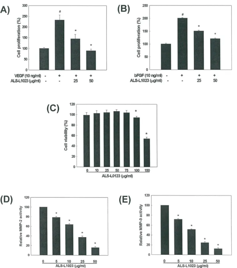
Inhibition of Angiogenesis and MMP Activity by ALS
The effect of ALS on angiogenesis was examined using HUVEC proliferation assays. VEGF and bFGF are angiogenic stimulators that play important roles in pathogenic angiogenesis by inducing endothelial cell proliferation. ALS added to the culture medium at a concentration of
25μg/ml or 50μg/ml inhibited VEGF-induced endothelial cell proliferation by 68% and 100%,
respectively, compared to control (p<0.05;Fig 1A). ALS also inhibited bFGF-induced HUVEC
proliferation by 48% and 86% at a concentration of 25μg/ml and 50μg/ml, respectively,
com-pared to control (p<0.05;Fig 1B). Inhibition of endothelial cell proliferation by ALS was not
due to cytotoxic effect since the viability of HUVECs was not affected by ALS at concentrations
between 10μg/ml and 75μg/ml determined by XTT assays (Fig 1C). Thus, the antiangiogenic
effects of ALS were mediated by inhibition of VEGF-induced or bFGF-induced endothelial cell proliferation.
MMP-2 and MMP-9 contribute to tissue remodeling by degrading the ECM in angiogenic
processes as well as participate in adipocyte differentiation [6]. The activities of MMP-2 and
MMP-9 were significantly inhibited by ALS treatment in a concentration-dependent manner
(p<0.05;Fig 1D and 1E). The IC50values were 17.7±1.0μg/ml for MMP-2 and 12.3±1.4μg/ml
for MMP-9. Thus, ALS possesses inhibitory effects on both angiogenesis and MMP activity.
Effects of ALS on Body Weight and Adipose Tissue Mass
To determine whether ALS reduces adipose tissue mass and prevents body weight gain in obese mice, mice were fed a low fat diet, a high fat diet or the same high fat diet supplemented with 0.4% or 0.8% ALS for 8 weeks. After 8 weeks of treatment, the high fat diet-fed mice had 115% greater body weight gains compared to that of standard chow diet-fed mice (14.56±1.89
g vs. 6.78±1.45 g, respectively, p<0.05). In contrast, mice fed a high fat diet supplemented with
0.4% and 0.8% ALS had 34% (9.56±1.69 g) and 43% (8.32±3.13 g) lower body weights,
respec-tively, compared to obese mice fed a high fat diet alone (p<0.05;Fig 2A). Adipose tissue mass
was significantly decreased by ALS treatment in high fat diet-fed obese mice. As shown inFig
2B and 2C, the mass of both VSC and SC fat pads in the ALS-treated mice was reduced in com-parison to that of high fat diet-fed control mice. Both VSC and SC adipose tissue weights were
decreased by 31% (p<0.05) and 36% (p<0.05) after 0.4% ALS and 48% (p<0.05) and 44%
(p<0.05) after 0.8% ALS administration, respectively.
Mice fed a high fat diet had higher food consumption compared to mice fed a low fat diet, but there was not a significant difference in food consumption between the high fat diet control
group and ALS-treated group (Fig 2D). In pair feeding experiments, 0.8% ALS had no
signifi-cant appetite effect (Fig 2E). ALS treatment had no effect on organ weight of the kidney and
Fig 1. Inhibitory effects of ALS on angiogenesis and MMP activity.(A) Inhibition of VEGF-induced HUVEC proliferation by ALS. (B) Inhibition of bFGF-induced HUVEC proliferation by ALS. #p<0.05 compared to vehicle control,*p<0.05 compared to VEGF- or bFGF-treated control group. (C) The effect of
ALS on HUVEC cell viability by MTT assay.*p<0.05 compared to the vehicle control. ALS-mediated inhibition of (D) MMP-2 and (E) MMP-9 activity measured by spectrofluorometry and the IC50values were determined.*p<0.05 compared to the vehicle control.
Fig 2. Regulation of body weight gain, adipose tissue mass and food consumption in high fat diet-fed obese mice.Adult male mice were fed a standard chow diet, a high fat diet, or high fat diet supplemented with 0.4 or 0.8% ALS for 8 weeks. (A) Modulation of body weight gain by ALS. Body weight gains at the end of the treatment period are significantly different between the chow diet group and the high fat diet group (#p<0.05) and between the high fat diet group and the groups fed a high fat diet supplemented with 0.4 or 0.8% ALS (*p<0.05). Regulation of VSC (B) and SC (C) fat mass by ALS. (D) Effects of ALS on food intake. (E) Appetite of ALS-treated mice. We fed the amount consumed per day by each treated mouse to a paired mouse and measured body weights daily. (F) Organ weights for liver, kidney, heart and pancreas. All values are expressed as the mean±SD. #p<0.05 compared to the chow group,
*p<0.05 compared to the high fat group.
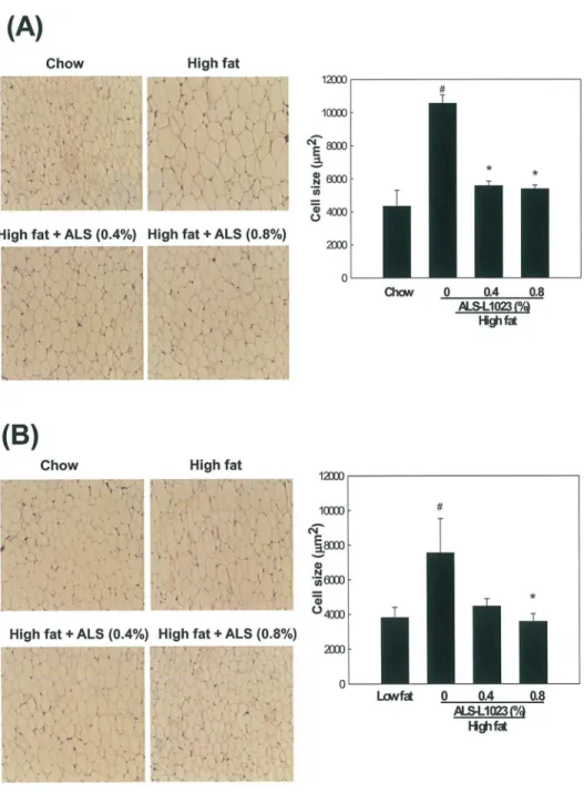
Effects of ALS on Adipocyte Size
Analysis of H&E-stained adipose tissue sections showed that ALS treatment significantly decreased the size of adipocytes. ALS at concentrations of 0.4% and 0.8% decreased the average
size of VSC adipocytes by 47% and 48%, respectively (p<0.05;Fig 3A). The average size of
Fig 3. Light microscopy analysis of adipocyte size in adipose tissue.Adult male mice were fed a standard chow diet, a high fat diet, or the same high fat diet supplemented with 0.4 or 0.8% ALS for 8 weeks. Representative H&E-stained sections (5-μm thick) of (A) epididymal VSC and (B) inguinal SC adipose tissues are shown (original magnification ×100). Adipocyte size in the high fat diet plus ALS groups was smaller compared to the high fat diet groups. The size of adipocytes in a fixed area (1,000,000μm2) was
measured. All values are expressed as the mean±SD. #p<0.05 compared to the chow group,*p<0.05
compared to the high fat group.
adipocytes was 5571±271μm2in 0.4% ALS-treated mice and 5394±216μm2in 0.8%
ALS-treated mice, which were smaller compared to the high fat diet-fed obese mice (10565
±495μm2). ALS at concentrations of 0.4% and 0.8% also decreased the average size of SC
adi-pocytes by 40% and 52%, respectively (p<0.05;Fig 3B). The average size of adipocytes was
4475±420μm2in 0.4% ALS-treated mice and 3585±446μm2in 0.8% ALS-treated mice, which
were smaller compared to the high fat diet-fed mice (7544±1978μm2).
Effects of ALS on Vascularization in Adipose Tissue
To determine whether the decrease of adipose tissue mass by ALS treatment resulted from the inhibition of angiogenesis, we studied the effects of ALS on blood vessel density in both VSC and SC adipose tissue. Staining of adipose tissue sections with an antibody against vWF, an endothelial cell marker showed that the blood vessel density of both VSC and SC adipose tissue from the ALS-treated mice was significantly lower compared to the high fat diet-fed control
mice. ALS treatment decreased blood vessel density by 78% in VSC adipose tissue (p<0.05;Fig
4A) and by 65% in SC adipose tissue (p<0.05;Fig 4B).
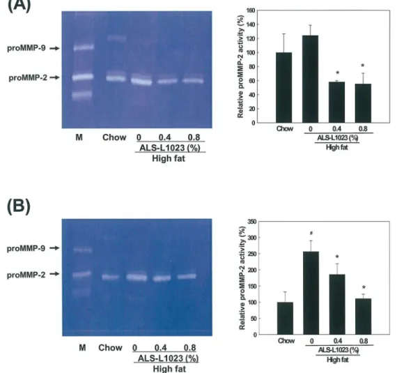
Effects of ALS on MMP Activity in Adipose Tissue
MMP activity in adipose tissue extracts was examined using zymography on gelatin-containing gels. Gelatin zymography showed that proMMP-2 activity was significantly reduced in the adi-pose tissue from the ALS-treated mice compared to the control group, whereas proMMP-9 lev-els were not detectable. The proMMP-2 activity in VSC adipose tissue was significantly
reduced by 48% and 49% after the administration of 0.4% and 0.8% ALS, respectively (p<0.05;
Fig 5A). Similarly, proMMP-2 activity in SC adipose tissue was also decreased by 23% and 36%
after treatment with 0.4% and 0.8% ALS, respectively (p<0.05;Fig 5B). Thus ALS reduced
proMMP-2 activity more in VSC adipose tissue than in SC adipose tissue.
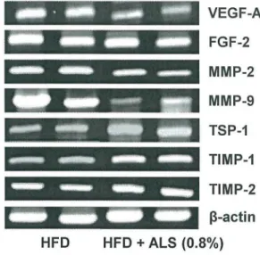
Effects of ALS on mRNA and Protein Expression of Angiogenic Factors,
MMPs, and Their Inhibitors in Adipose Tissue
The expression patterns of genes involved in angiogenesis were investigated in VSC and SC adipose tissues from C57BL/6J mice fed a high fat diet. The mRNA expression of angiogenic and antiangiogenic factors was downregulated and upregulated, respectively, in ALS-treated mice compared to high fat diet-fed control mice. In VSC adipose tissue, the mRNA expression
of the VEGF-A angiogenic factor was decreased by 45% (p<0.05) compared to control mice,
whereas the mRNA level of the TSP-1 antiangiogenic factor was increased by 41% in
ALS-treated mice compared to control mice (p<0.05;Fig 6A). In SC adipose tissue, the mRNA
lev-els of VEGF-A and FGF-2 were significantly reduced by 83% (p<0.05) and 29% (p<0.05),
respectively, whereas TSP-1 mRNA level was increased by 11% in ALS-treated mice compared
to control mice (Fig 6B).
MMP mRNA expression was significantly inhibited by ALS treatment. In VSC adipose
tis-sue, MMP-2 and MMP-9 mRNA levels were significantly decreased by 19% (p<0.05) and 81%
(p<0.05), respectively (Fig 6A), and in SC adipose tissue significantly decreased by 20% and
41% (p<0.05), respectively (Fig 6B). In contrast, TIMP-1 and TIMP-2 mRNA levels were
sig-nificantly increased by 69% (p<0.05) and 31% (p<0.05), respectively, in VSC adipose tissue
(Fig 6A) and significantly increased by 122% (p<0.05) and 43% (p<0.05), respectively, in SC
adipose tissue (Fig 6B) from ALS-treated mice compared to control mice.
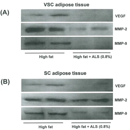
decreased in ALS-treated mice compared to high fat diet-fed control mice in both VSC and SC
adipose tissue (Fig 7).
Effects of ALS on Plasma Lipid Levels, Hepatic Lipid Accumulation, and
mRNA Expression of Hepatic Fatty Acid Oxidation-Related Genes
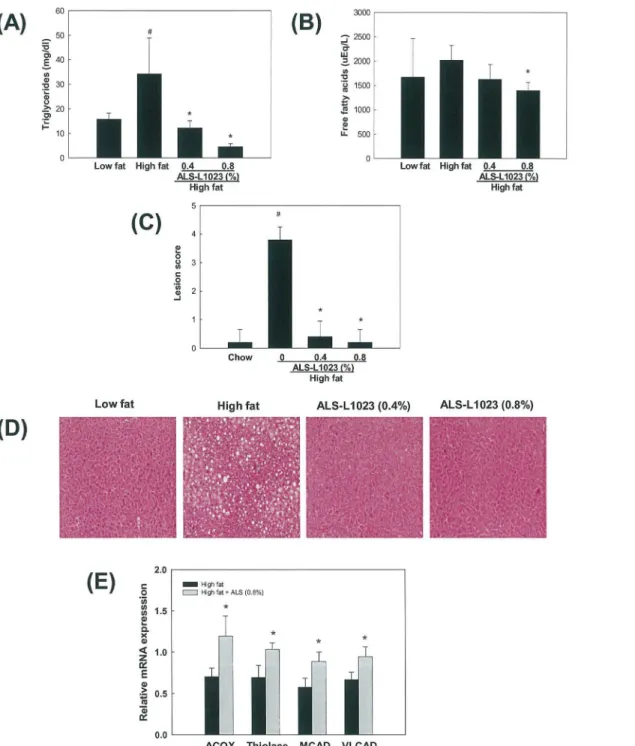
Circulating levels of triglycerides and free fatty acids were higher by 117% and 21%,
respec-tively, in high fat diet-fed mice compared with chow diet-fed mice (Fig 8A and 8B). However,
triglyceride and free fatty acid levels were decreased by 87% and 31%, respectively, in 0.8% Fig 4. Histological analysis of blood vessels in adipose tissue stained with an antibody against vWF.
The blood vessels of (A) epididymal VSC and (B) inguinal SC adipose tissues derived from mice fed a high fat diet or a high fat diet supplemented with 0.8% ALS for 8 weeks were stained and analyzed (original
magnification ×100). Higher magnitude of bracket area is shown (original magnification ×200). All values are expressed as the mean±SD.*p<0.05 compared to the high fat group.
ALS-L1023 treated mice compared with those in high fat diet-fed mice. Significant hepatic lipid accumulation was induced by high fat, as indicated by the increased hepatic lesion score (Fig 8C and 8D). In contrast, ALS-L1023 almost completely abolished hepatic triglycerides. We then determined the levels of mRNA encoding peroxisome proliferator-activated receptor
α(PPARα) target enzymes responsible for fatty acidβ-oxidation in the livers. Compared with
high fat group, the mRNA levels of two peroxisomal PPARαtargets, namely acyl-CoA oxidase
(ACOX) and thiolase, were increased in 0.8% ALS-treated group by 54% and 49%, respectively (Fig 8E). mRNA encoding mitochondrial PPARαtargets, medium chain acyl-CoA dehydroge-nase (MCAD) and very long chain acyl-CoA dehydrogedehydroge-nase (VLCAD), were also elevated by 54% and 40%, respectively.
Discussion
Angiogenesis, the formation of new capillary blood vessels, is a tightly regulated process. Under normal physiological conditions, angiogenesis only takes place during embryonic
devel-opment, wound healing and female menstruation [16]. Failure in the regulation of angiogenesis
Fig 5. Zymographic analysis of adipose tissue.Extracts from (A) epididymal VSC and (B) inguinal SC adipose tissues obtained from mice fed a high fat diet or a high fat diet supplemented with 0.4 and 0.8% ALS for 8 weeks were applied to a gelatin-containing gel. Gelatinolytic activity was measured by zymography. The HT1080 cell culture medium was used for the molecular weight markers for MMP-2 and MMP-9. All values are expressed as the mean±SD. #p<0.05 compared to the chow group,*p<0.05 compared to the high fat group. M, molecular weight marker.
is correlated with many diseases such as cancer, rheumatoid arthritis, psoriasis and
prolifer-ative retinopathy [17,18]. Similarly, the growth and expansion of adipose tissue requires the
formation of new blood vessels to provide oxygen and nutrients to adipocytes. Recently, we Fig 6. Effects of ALS on the expression of angiogenic factors, MMPs and their inhibitors in epididymal VSC and inguinal SC adipose tissues of diet-induced obese mice.Adult male mice were fed a high fat diet or a high fat diet supplemented with 0.8% ALS for 8 weeks. Analysis of VEGF-A, FGF-2, MMP-2, MMP-9, TIMP-1, TIMP-2 and TSP-1 mRNA levels by RT-PCR in epididymal VSC (A) and inguinal SC (B) adipose tissues. Representative PCR bands from one of three independent experiments are shown. All values are expressed as the mean±SD.*p<0.05 compared to the high fat group.
demonstrated that the antiangiogenic dietary supplement, Ob-X which is composed of a
stan-dardized mixture ofMelissa officinalisL.,Morus albaL. andArtemisia capillarisThunb.
aque-ous extract reduces adipose tissue mass and suppresses obesity by inhibiting angiogenesis in
nutritionally and genetically obese mice [14,19].
Since one of the Ob-X ingredients, aqueous extract ofMelissaleaves showed anti-angiogenic
effects we extractedMelissaleaves with organic solvents in an attempt to increase
anti-angio-genic activity. In the present study we prepared ALS by a two-step organic solvent fractionation
fromMelissaleaves which was standardized with reference compounds by HPLC. The
antian-giogenic and MMP inhibitory activities of ALS were enhanced compared to the water extract
or aqueous ethanol extract ofMelissaleaves suggesting that hydrophobic active components in
ALS extracted with organic solvents may have more potent antiangiogenic effects compared to the hyrophilic active components extracted with water or aqueous ethanol (data not shown).
We examined the effects of ALS on angiogenesis and MMP activityin vitro. ALS reduced
VEGF- and bFGF-induced HUVEC proliferation, demonstrating that ALS exerts
Fig 7. Effects of ALS on the protein expression of VEGF, MMP-2 and MMP-9 in epididymal VSC and inguinal SC adipose tissues of diet-induced obese mice.The protein levels of epididymal VSC and inguinal SC adipose tissues derived from mice fed a high fat diet or a high fat diet supplemented with 0.8% ALS for 8 weeks were analyzed by Western blotting. Representative bands from one of three independent experiments are shown.
antiangiogenic effects by inhibiting the proliferation of endothelial cells. ALS also inhibited the
activities of two major MMPs (MMP-2 and MMP-9) in a concentration-dependent mannerin
vitro. Since it was suggested that inhibition of angiogenesis and MMP activity impairs adipose Fig 8. Effects of ALS on plasma lipid levels, hepatic lipid accumulation and liver PPARαtarget gene expression in diet-induced obese mice.Adult male mice were fed a high fat diet or a high fat diet supplemented with 0.8% ALS for 8 weeks. Plasma levels of (A) triglycerides and (B) free fatty acids. (C) Histological analyses of hepatic lipid accumulation. Pathological scores of hepatic lipid accumulation are as follows: 0, no lesion; 1, mild; 2, moderate; 4, very severe. (D) Representative H&E-stained liver sections are shown (original magnification ×100). (E) Modulation of liver PPARαtarget gene expression. All values are expressed as the mean±SD.*p<0.05 compared to the high fat group.
tissue development [4,20], it is likely that ALS may suppress adipose tissue growth by inhibit-ing angiogenesis and MMPs.
We then treated high fat diet-induced obese mice with ALS. Consistent with our hypothesis that ALS can inhibit adipose tissue growth, ALS substantially reduced adipose tissue mass and prevented body weight gain. We previously had performed animal experiments using ALS at doses of 0.1, 0.25, 0.5% in high fat diet-induced obese rats and found that 0.5% ALS (100mg/ kg/day) significantly reduced adipose tissue mass (results not published yet). Considering dos-age conversion from rat to mouse, we selected ALS at doses of 0.4% (200mg/kg/day) and 0.8% (400mg/kg/day) to supplement the HFD in an obese mouse model to increase the effects for a mechanism study. Treatment with 0.4% and 0.8% ALS for 8 weeks decreased body weight gain by 70% and 84%, respectively. Both VSC and SC adipose tissue weights were decreased by 65%
(p<0.05) and 68% (p<0.05) after 0.4% ALS and 74% (p<0.05) and 72% (p<0.05) after 0.8%
ALS administration, respectively. The ALS-mediated decreases in VSC and SC adipose tissue mass were greater compared to reductions in VSC (59%) and SC (65%) adipose tissue mass obtained after a 12-week administration of galardin, a broad spectrum inhibitor of MMPs and
angiogenesis [4]. Our data are also supported by other results which indicate that several types
of angiogenesis inhibitors, such as angiostatin, endostatin and TNP-470 and its analog
CKD-732 inhibit fat mass expansion in mice [20–22]. Similar to the effects of ALS on adipose tissue
mass, the average size of adipocytes in both VSC and SC adipose tissues was substantially smaller in ALS-treated mice than in untreated obese mice. These results indicate that ALS can reduce adipose tissue mass and inhibit adipocyte hypertrophy. VSC obesity is known to be
closely associated with metabolic syndromes including insulin resistance [23,24]. Given that
adipogenesis and obesity are closely associated with angiogenesis, modulating angiogenesis
may be a novel therapeutic approach to obesity and obesity-related diseases [25].
During ALS-induced adipose tissue loss, food intake was not changed, showing that ALS did not affect appetite. Furthermore, ALS-treated mice weighed less than pair-fed mice having similar calorie intake. Similarly, it was reported that the administration of angiogenesis
inhibi-tors, such as endostatin, toob/obmice resulted in weight loss without changes in appetite [20]
and tolylsam, an MMP inhibitor with specificity for gelatinases, reduced body weight and fat
pad weight without an effect on food intake [26]. ALS may target adipose tissue and cause
weight reduction. The weight loss induced by antiangiogenic ALS arose specifically from the loss of adipose tissue mass, as the weights of other organs such as the heart and kidneys were not reduced in ALS-treated obese mice. However, the liver weight increased by high fat diet was decreased to weight of chow-fed mice after ALS treatment. Thus, ALS can reduce adipose tissue mass by targeting only the growing adipose tissue without any side effects on other organs.
Blood vessel staining showed that the blood vessel density of both VSC and SC adipose tis-sues was reduced in ALS-treated mice compared to untreated obese mice. These data were sup-ported by our previous results which showed that the antiangiogenic herbal composition Ob-X
decreased blood vessel density of VSC adipose tissue in nutritionally obese mice [14]. Our
pres-ent results suggest that ALS is a potpres-ent angiogenesis inhibitor and regulates adipose tissue mass by inhibiting angiogenesis.
As angiogenesis may represent a target for treating obesity, it is important to determine the expression profiles of genes involved in angiogenesis. Growing adipose tissue contains a diverse population of cell, which determines the expression of several angiogenic modulators. Angio-genic factors, such as VEGFs and FGF-2, promote the proliferation, differentiation and
migra-tion of endothelial cells within fat tissue [27,28] Moreover, VEGF-A and FGF-2 synergistically
induce angiogenesis [29]. Blockage of the VEGFR2 signaling system by a neutralizing antibody
endothelial cells to regulate preadipocyte differentiation [30]. In contrast, TSP-1 inhibits
angio-genesisin vivoand impairs the migration and proliferation of cultured microvascular
endothe-lial cells [31]. Our RT-PCR analysis showed that ALS administration to obese mice decreased
VEGF-A and FGF-2 mRNA expression, whereas the antiangiogenic TSP-1 mRNA expression was increased in both VSC and SC adipose tissues. Western blot analysis confirmed that ALS treatment decreased VEGF protein level in VSC and SC adipose tissues.
In obesity, MMP expression is modulated in adipose tissue and MMPs (e.g., MMP-2 and MMP-9) potentially affect adipocyte differentiation. It has been reported that the different
MMPs do not present the same behavior in all the obesity models. Maquoi et al [32] showed
that the expression of MMP-3, -11, -12, -13 and -14 and TIMP-1 mRNA was upregulated, whereas that of MMP-7, -9, -16, and -24 and TIMP-4 was downregulated in high fat diet-induced obese mice. Similar results were reported in two genetic models of obesity (ob/ob and
db/db mice) and in a diet-induced model of obesity [33]. In contrast to the data of Maquoi
et al, Chavey et al found that mRNA levels for MMP-2, -3, -12, -14, -19 and TIMP-1 were strongly induced in obese adipose tissue, but MMP-7 and TIMP-3 mRNAs are markedly decreased in obesity. Other study demonstrated that MMP-2 and MMP-9 activity in VSC
adi-pose tissue was decreased in an animal model of early insulin resistance [34]. However, this is
not animal model of obesity and there was no difference in body weight compared with control mice. High MMP-2 expression was observed in the adipose tissue of mice with nutritionally
induced obesity as well as in genetically obese mice [35]. Treatment with MMP inhibitors
impairs adipose tissue development in mice fed a high fat diet [4,36]. Furthermore, the
secre-tion of MMP-2 and MMP-9 increases during adipocyte differentiasecre-tion in both human
adipo-cytes and mouse preadipocyte cell lines [6,33], suggesting that MMP-2 and MMP-9 are
important for adipocyte development. In this study, treating obese mice with ALS decreased MMP-2 and MMP-9 protein levels in adipose tissues. ALS also reduced MMP-2 and MMP-9 mRNA expression and increased TIMP-1 and TIMP-2 mRNA expression in both VSC and SC adipose tissues, indicating that ALS exerts a specific regulatory effect on genes and conse-quently proteins involved in angiogenesis and the MMP system in adipose tissues. Our obser-vations further indicate that the inhibition of adipose tissue growth by angiogenesis inhibitors may alter the expression of genes responsible for angiogenesis and the MMP system.
Gelatin zymography of VSC and SC adipose tissue extracts showed that ALS reduced proMMP-2 activity compared to the control, although proMMP-9 activity was not detectable. It has been reported that, in galardin-treated animals, zymography on gelatin-containing gels showed reduced MMP-2 activity in SC and VSC adipose tissues, whereas MMP-9 level were
not observed [4].
MMPs and TIMP expression profiles were profoundly altered in adipose tissue of high fat diet-induced obese mice. Most of these modulations were specific to the gonadal VSC fat, not
to the SC fat [16]. In our study, gelatin zymography showed that MMP activity in VSC adipose
tissue was more significantly reduced by ALS than in SC adipose tissue. However, there is no difference in reduction of adipose tissue mass between VSC and SC fat. The effect of ALS on reduction of adipose tissue mass might arise from anti-angiogenic activity as well as MMPs inhibitory activities.
Treatment with MMP inhibitors impairs adipose tissue development in mice fed a high fat
diet [36]. Indeed, MMP-2−/−mice had reduced fat mass and smaller adipocyte size in both
VSC and SC adipose tissue compared to MMP-2+/+mice, suggesting a functional role of
MMP-2 in adipose tissue growth [37]. Thus, these data demonstrate that the inhibition of
MMP activity by ALS may lead to reduced adipose tissue mass in obese mice.
endogenous, inhibit angiogenic responses bothin vivoandin vitro[38,39]. Moreover, MMP-deficient mice exhibit delayed or diminished angiogenic responses during development or in
response to tumor xenograft [40]. On the other hand, it was also reported that MMP-based
proteolysis of ECM proteins releases anti-angiogenic cryptic fragments such as angiostatin and
endostatin [41,42], showing that inhibiting MMP activity may decrease endogenous
angio-genic inhibitors. However, studies demonstrated that MMPs could increase the bioavailability
of potent angiogenic factors by releasing matrix-bound VEGF and basic FGF (bFGF) [43,44].
Thus, suppressing MMP activity may decrease endogenous anti-angiogenic substances, whereas it decreases potent angiogenic factors such as VEGF and bFGF. In addition, MMPs have novel function of modulating adipocyte differentiation, which is independent of
angio-genesis. Therefore, MMP inhibitors can block the adipocyte differentiation process [6,33,45].
Collectively, it seems that MMPs and their inhibitors play a pivotal role in controlling adipo-genesis and adipose tissue growth.
Metabolic changes including plasma lipids, liver triglycerides, and hepatic expression of fatty acid oxidation-related genes occurred during ALS-induced weight loss. ALS treatment not only decreased circulating triglycerides and free fatty acids, but also inhibited liver lipid accumulation. Consistently, ALS-treated mice had significantly higher mRNA levels of hepatic
PPARαtarget enzymes involved in fatty acidβ-oxidation, indicating that the elevated fatty acid
oxidation in the livers may be paralleled by large reductions in plasma lipids and weight gain. It
is well known that PPARαincreases the fatty acid catabolism by stimulating the transcription
of rate-limiting enzymes in mitochondrial and peroxisomalβ-oxidation and microsomalω
-oxidation [46]. It is likely that ALS stimulates the transcriptional expression of PPARαtarget
enzymes in the livers, which reduces the intracellular levels of fatty acids available for triglycer-ide synthesis. This in turn curbs circulating triglycertriglycer-ide levels and fat accumulation. Thus, ALS may play a critical role in the regulation of obesity-associated disorders, such as hypertriglycer-idemia, hepatic steatosis, insulin resistance and type 2 diabetes.
In conclusion, our present results demonstrate that ALS, which inhibits angiogenesis and MMP activity, suppresses the growth and development of adipose tissue in obese mice. These events may be mediated by changes in the expression of genes involved in angiogenesis and the MMP system. Thus, antiangiogenic ALS provides a potential therapeutic approach for prevent-ing and treatprevent-ing human obesity and its related disorders.
Supporting Information
S1 Table. Sequences of primers used for RT-PCR assays.
(DOC)
Author Contributions
Conceived and designed the experiments: BP JWK SSS MYK Michung Yoon. Performed the experiments: HL YH SW Miso Yoon JK HSL EKP JCH. Analyzed the data: BP JWK SSS MYK Michung Yoon HL YH SW Miso Yoon JK HSL EKP JCH. Contributed reagents/materials/ analysis tools: BP JWK SSS MYK Michung Yoon HL YH SW Miso Yoon JK HSL EKP JCH. Wrote the paper: BP MYK Michung Yoon. Obtained funding: SSS Michung Yoon.
References
1. Crandall DL, Hausman GJ, Kral JG (1997) A review of the microcirculation of adipose tissue: anatomic, metabolic, and angiogenic perspectives. Microcirculation 4: 211–232. PMID:9219215
2. Bouloumié A, Lolmède K, Sengenès C, Galitzky J, Lafontan M (2002) Angiogenesis in adipose tissue.
3. Silverman KJ, Lund DP, Zetter BR, Lainey LL, Shahood JA, Freiman DG, et al. (1998) Angiogenic activ-ity of adipose tissue. Biochemical and Biophysical Research Communications 153: 347–352. 4. Lijnen HR, Maquoi E, Hansen LB, Van Hoef B, Frederix L, Collen D (2002) Matrix metalloproteinase
inhibition impairs adipose tissue development in mice. Arteriosclerosis, Thrombosis, and Vascular Biol-ogy 22: 374–379. PMID:11884277
5. Galardy RE, Grobelny D, Foellmer HG, Fernandez LA (1994) Inhibition of angiogenesis by the matrix metalloprotease inhibitor N-[2R-2-(hydroxamidocarbonymethyl)-4-methylpentanoyl)]-L-tryptophan methylamide. Cancer Research 54: 4715–4718. PMID:7520359
6. Bouloumié A, Sengenès C, Portolan G, Galitzky J, Lafontan M (2001) Adipocyte produces matrix metal-loproteinases 2 and 9: involvement in adipose differentiation. Diabetes 50: 2080–2086. PMID:
11522674
7. Li J, Yu X, Pan W, Unger RH (2002) Gene expression profile of rat adipose tissue at the onset of high fat-diet obesity. Americal Journal of Physiology, Endocrinology and Metabolism 282: E1334–E1341 8. Silha JV, Krsek M, Sucharda P, Murphy LJ (2005) Angiogenic factors are elevated in overweight and
obese individuals. International Journal of Obesity 29: 1308–1314. PMID:15953938
9. Valentino MA, Lin JE, Waldman SA (2010) Central and peripheral molecular targets for antiobesity pharmacotherapy. Clinical Pharmacology and Therapeutics 87: 652–662. doi:10.1038/clpt.2010.57
PMID:20445536
10. Kim JS, Park BY, Park EK Lee HS, Hahm JC, Bae KH, et al. (2006) Screening of Anti-angiogenic Activ-ity from Plant Extract. Korean Journal of Pharmacognosy 37: 253–257.
11. Blumenthal M, Goldberg A, Brinckmann J (2000) Herbal Medicine-Expanded Commission E Mon-graphs. Newton, MA: Integrative Medicine Communications 123: 230–232.
12. Akhondzadeh S, Noroozian M, Mohammadi M, Ohadinia S, Jamshidi AH, Khani M, et al. (2003) Melissa officinalis extract in the treatment of patients with mild to moderate Alzheimer's disease: a dou-ble blind, randomized, placebo controlled trial. Journal of Neurology, Neurosurgery, and Psychiatry 6: 625–632.
13. Kim MY, Park BY, Lee HS, Park EK, Hahm JC, Lee J, et al. (2010) The anti-angiogenic herbal composi-tion Ob-X inhibits adipose tissue growth in obese mice. Internacomposi-tional Journal of Obesity 34: 820–830.
doi:10.1038/ijo.2010.13PMID:20179671
14. Lee H, Park D, Yoon M (2013) Korean red ginseng (Panax ginseng) prevents obesity by inhibiting angiogenesis in high fat diet-induced obese C57BL/6J mice. Food and Chemical Toxicology 53: 402–
408 doi:10.1016/j.fct.2012.11.052PMID:23232078
15. Hong Y, Kim MY, Yoon M (2011) The anti-angiogenic herbal extracts Ob-X from Morus alba, Melissa officinalis, and Artemisia capillaris suppresses adipogenesis in 3T3-L1 adipocytes. Pharmaceutical Biology 49: 775–783. doi:10.3109/13880209.2010.547208PMID:21449830
16. Folkman J (1985) Tumor angiogenesis. Advances in Cancer Research. 43: 175–203. PMID:2581424 17. Celletti FL, Waugh JM, Amabile PG, Brendolan A, Hilfiker PR, Dake MD (2001) Vascular endothelial
growth factor enhances atherosclerotic plaque progression. Nature Medicine 7: 425–429. PMID:
11283668
18. Ferrara N, Davis-Smyth T (1997) The biology of vascular endothelial growth factor. Endocrine Reviews 18: 4–25. PMID:9034784
19. Yoon M, Kim MY (2011) The anti-angiogenic herbal composition Ob-X from Morus alba, Melissa offici-nalis, and Artemisia capillaris regulates obesity in genetically obese ob/ob mice. Pharmaceutical Biol-ogy 49: 614–619. doi:10.3109/13880209.2010.539617PMID:21554004
20. Rupnick MA, Panigrahy D, Zhang C, llabrida SM, Lowell BB, Langer R, et al. (2002) Adipose tissue mass can be regulated through the vasculature. Proceedings of the National Academy of Sciences of the United States of America 99: 10730–10735. PMID:12149466
21. Bråkenhielm E, Cao R, Gao B, Gelin B, Cannon B, Parini P, et al. (2004) Angiogenesis inhibitor,
TNP-470, prevents diet-induced and genetic obesity in mice. Circulation Research 94: 1579–1588. PMID:
15155527
22. Kim YM, An JJ, Jin YJ, Rhee Y, Cha BS, Lee HC, et al. (2007) Assessment of the anti-obesity effects of the TNP-470 analog, CKD-732. Journal of Molecular Endocrinology 38: 455–465. PMID:17446235 23. Després JP, Lemieux I (2006) Abdominal obesity and metabolic syndrome. Nature 444: 881–887.
PMID:17167477
24. Jeong S, Yoon M (2009) Fenofibrate inhibits adipocyte hypertrophy and insulin resistance by activating adipose PPARαin high fat diet-induced obese mice. Experimental and Molecular Medicine 41: 397–
25. Cao Y (2007) Angiogenesis modulates adipogenesis and obesity. The Journal of Clinical Investigation 117: 2362–2368. PMID:17786229
26. Van Hul M, Lijnen HR (2011) Matrix metalloproteinase inhibition impairs murine adipose tissue devel-opment independently of leptin. Endocrine Journal 58: 101–107. PMID:21242647
27. Carmeliet P, Ferreira V, Breier G, Pollefeyt S, Kieckens L, Gertsenstein M, et al. (1996) Abnormal blood vessel development and lethality in embryos lacking a single VEGF allele. Nature 380: 435–439.
PMID:8602241
28. Bikfalvi A, Klein S, Pintucci G, Rifkin DB (1997) Biological roles of fibroblast growth factor-2. Endocrine Reviews 18: 26–45. PMID:9034785
29. Cao R, Brakenhielm E, Wahlestedt C, Thyberg J, Cao Y (2001) Leptin induces vascular permeability and synergistically stimulates angiogenesis with FGF-2 and VEGF. Proceedings of the National Acad-emy of Sciences of the United States of America 98: 6390–6395. PMID:11344271
30. Fukumura D, Ushiyama A, Duda DG, Xu L, Tam J, Krishna V, et al. (2003) Paracrine regulation of angiogenesis and adipocyte differentiation during in vivo adipogenesis. Circulation Research 93: e88–
e97. PMID:14525808
31. Armstrong LC, Bornstein P (2003) Thrombospondins 1 and 2 function as inhibitors of angiogenesis. Matrix Biology 22: 63–71. PMID:12714043
32. Maquoi E, Munaut C, Colige A, Collen D, Lijnen HR (2002) Modulation of adipose tissue expression of murine matrix metalloproteinases and their tissue inhibitors with obesity. Diabetes 51: 1093–1101.
PMID:11916931
33. Chavey C, Mari B, Monthouel MN, Bonnafous S, Anglard P, Van Obberghen E, et al. (2003) Matrix metalloproteinases are differentially expressed in adipose tissue during obesity and modulate adipo-cyte differentiation. The Journal of Biological Chemistry 278: 11888–11896. PMID:12529376 34. Miksztowicz V, Morales C, Zago V, Friedman S, Schreier L, Berg G (2014) Effect of insulin-resistance
on circulating and adipose tissue MMP-2 and MMP-9 activity in rats fed a sucrose-rich diet. Nutrition, Metabolism, and Cardiovascular Disease: NMCD 24: 294–300.
35. Lijnen HR, Maquoi E, Holvoet P, Mertens A, Lupu F, Morange P, et al. (2001) Adipose tissue expres-sion of gelatinases in mouse models of obesity. Thrombosis and Haemostasis 85: 1111–1116. PMID:
11434693
36. Demeulemeester D, Collen D, Lijnen HR (2005) Effect of matrix metalloproteinase inhibition on adipose tissue development. Biochemical and Biophysical Research Communications 329: 105–110. PMID:
15721280
37. Van Hul M, Lijnen HR (2008) A functional role of gelatinase A in the development of nutritionally induced obesity in mice. Journal of Thrombosis and Haemostasis 6: 1198–1206. doi:
10.1111/j.1538-7836.2008.02988.xPMID:18433461
38. Bergers G, Brekken R, McMahon G, Vu TH, Itoh T, Tamaki K, et al. (2000) Matrix metalloproteinase-9 triggers the angiogenic switch during carcinogenesis. Nature Cell Biology 2: 737–744. PMID:
11025665
39. Stetler-Stevenson WG (1999) Matrix metalloproteinases in angiogenesis: a moving target for therapeu-tic intervention. The Journal of Clinical Investigation 103: 1237–1241. PMID:10225966
40. Itoh T, Tanioka M, Yoshida H, Yoshioka T, Nishimoto H, Itohara S (1998) Reduced angiogenesis and tumor progression in gelatinase A-deficient mice. Cancer Research 58: 1048–1051. PMID:9500469 41. O'Reilly MS, Holmgren L, Shing Y, Chen C, Rosenthal RA, et al. (1994) Angiostatin: a novel
angiogene-sis inhibitor that mediates the suppression of metastases by a Lewis lung carcinoma. Cell 79: 315–
328. PMID:7525077
42. O'Reilly MS, Boehm T, Shing Y, Fukai N, Vasios G, Moses M, et al. (1997) Endostatin: an endogenous inhibitor of angiogenesis and tumor growth. Cell 88: 277–285. PMID:9008168
43. Rundhaug JE (2003) Matrix metalloproteinases, angiogenesis, and cancer: commentary re: A. C. Lock-hart et al., Reduction of wound angiogenesis in patients treated with BMS-275291, a broad spectrum matrix metalloproteinase inhibitor. Clinical Cancer Research 9: 551–554. PMID:12576417 44. Hawinkels LJ, Zuidwijk K, Verspaget HW, de Jonge-Muller ES, van Duijn W, Ferreira V, et al. (2008)
VEGF release by MMP-9 mediated heparan sulphate cleavage induces colorectal cancer angiogene-sis. European Journal Cancer 44: 1904–1913.
45. Croissandeau G, Chrétien M, Mbikay M (2002) Involvement of matrix metalloproteinases in the adipose conversion of 3T3-L1 preadipocytes. The Biochemical Journal 364: 739–746. PMID:12049638 46. Yoon M (2009) The role of PPARαin lipid metabolism and obesity: focusing on the effects of estrogen
on PPARαactions. Pharmacological Research 60: 151–159. doi:10.1016/j.phrs.2009.02.004PMID: