UNIVERSIDADE ESTADUAL PAULISTA “JULIO DE MESQUITA FILHO” FACULDADE DE MEDICINA
CAMPUS DE BOTUCATU
AVALIAÇÃO DO NÍVEL SÉRICO DE IL-6, IL-10 E IL-13 ANTES
E APÓS O TRATAMENTO DO LINFOMA DE HODGKIN
CLÁSSICO
RAFAEL DEZEN GAIOLLA
ORIENTADOR: PROF. DR. DEILSON ELGUI DE OLIVEIRA
CO-ORIENTADORA: PROF
A. DR
A. LÍGIA NIÉRO-MELO
Dissertação apresentada ao Programa de Pós-Graduação em Patologia da Faculdade de Medicina de Botucatu, Universidade Estadual Paulista - UNESP, para obtenção do título de Mestre.
FICHA CATALOGRÁFICA ELABORADA PELA
SEÇÃO TÉCNICA DE AQUISIÇÃO E TRATAMENTO DA INFORMAÇÃO
DIVISÃO TÉCNICA DE BIBLIOTECA E DOCUMENTAÇÃO - CAMPUS DE BOTUCATU - UNESP BIBLIOTECÁRIA RESPONSÁVEL: Sulamita Selma Clemente Colnago – CRB 8/4716 Gaiolla, Rafael Dezen.
Avaliação do nível sérico de IL-6, IL-10 e IL-13 antes e após o tratamento do Linfoma de Hodgkin clássico / Rafael Dezen Gaiolla. – 2008.
Dissertação (mestrado) – Faculdade de Medicina de Botucatu, Universidade Estadual Paulista, 2008.
1. Hodgkin, Doença de.
CDD 616.99446
À P
Paula, que faz a minha vida
simplesmente completa.
Aos meus filhos SSofia e Guilherme,
minha dose-dupla diária de amor e
felicidade.
Aos meus Pais, A
Antonio e
Ao P
Paulo, Paula e Raquel, pela
amizade, cumplicidade e, acima de
tudo, por sempre estarem por perto,
mesmo que de longe.
À minha querida tia D
Dica, por 32
anos de amor e dedicação
incansáveis.
Ao CCóto, Ângela, Fábio, Denise,
Otávio e Marcela, pelo amor e
carinho a mim dedicados.
Ao P
Prof. Dr. Deilson Elgui de Oliveira, pela disponibilidade em
me orientar e pela oportunidade de vivenciar a pesquisa científica
realizada com grande competência e dedicação.
À P
Prof
a. Dr
a. Lígia Niéro-Melo, por quem tenho grande
admiração. Agradeço por todos os ensinamentos, oportunidades
e por estar sempre presente nas minhas conquistas pessoais e
profissionais.
Ao D
Dr. Vergílio Antonio Rensi Colturato, por não medir esforços
em viabilizar a realização desse trabalho e por sempre, sem
exceções, apoiar minhas decisões profissionais. Tenho como
exemplo a perseverança e amor com os quais realiza seu
trabalho.
Ao D
Dr. Mair Pedro de Souza, pela amizade e por seu exemplo de
dedicação e competência profissional.
Ao P
Prof. Adj. Sérgio Alberto Rupp de Paiva, ao D
Dr. Marcos
Ferreira Minicucci e à D
Dra. Suzana Erico Tanni Minamoto, pelo
Aos membros do Laboratório de Patologia Molecular da FMB –
UNESP:
Alice Silva Gonçalves
Ana Paula Ferraz da Silva
Suzane Ramos da Silva
Leila Giron
Ana Rachel Oliveira Leda
Danielle Amorim
Celene Maria C. Gandin
Marcos Roberto Franchi
Luis Fernando Franchi
Aos meus queridos amigos,
Andréa Miranda Pedro
Ricardo Augusto Monteiro de Barros Almeida
David Nicolleti Gumieiro e Lisa Milaré Gumieiro
Marcos Ferreira Minicucci e Fernanda Chiuso Minicucci
Fernando Seara Cordaro e Larissa Chambô Cordaro
Aos médicos do Departamento de Oncologia Clínica e
Hematologia do Hospital Amaral Carvalho de Jaú:
Dra. Ana Lúcia Coradazzi
Dra. Andiara Vasconcellos Cantarelli Boneto Pires
Dra. Andréa Miranda Pedro
Dr. Carlos Augusto de Mendonça Beato
Dra. Cláudia Teresa de Oliveira
Dr. Éderson Roberto de Mattos
Dr. Fabrício Pascolat
Dr. Gustavo Fernando Veraldi Ismael
Dr. José Getúlio Martins Segalla
Dr. Marcos Augusto Mauad
Dr. Mair Pedro de Souza
Dra. Maura Rosane Valério Ikoma
Dr. Vergílio Antonio Rensi Colturato
É uma honra poder trabalhar com vocês.
Aos colegas
Prof
a. Dr
a. Lucilene Silva Ruiz e Resende, por todo apoio e
incentivo à realização deste trabalho e pelo grande auxílio
durante a prova de qualificação.
Prof
aDr
a. Maria Aparecida Custódio Domingues pela revisão
histopatológica dos casos desse estudo.
Prof
a. Dr
a. Alexandrina Sartori, por contribuir para a melhoria
desse trabalho durante a prova de qualificação.
Aos médicos residentes da Disciplina de Hematologia da
Faculdade de Medicina de Botucatu e do Hospital Amaral
Carvalho de Jaú, pela assistência prestada aos pacientes e
auxílio nas coletas de dados e materiais.
Aos funcionários do Serviço de Hematologia do Hospital Amaral
Carvalho de Jaú, pela disponibilidade, dedicação e atenção
dispensadas em todas as etapas da realização desse estudo.
Ao Hemocentro da Faculdade de Medicina de Botucatu, na
pessoa do diretor Dr. José Mauro Zanini, que viabilizou a
realização do grupo-controle deste estudo.
Aos funcionários do Laboratório de Análises Clínicas do
Hospital das Clínicas da FMB – UNESP, em especial a SSra.
Zezé, pela colaboração na coleta das amostras de sangue dos
pacientes.
Ao Departamento de Patologia do Hospital Amaral Carvalho de
Jaú, na pessoa do D
Dr. Francisco Carlos Quevedo, pela
disponibilização do material necessário para a realização deste
estudo.
Ao P
Prof. Dr. Carlos Eduardo Bacchi, pela disponibilização do
material necessário para a complementação desse estudo.
A todos os funcionários do Departamento de Patologia da
Faculdade de Medicina de Botucatu que colaboraram para a
realização desse trabalho.
Aos funcionários da Pós-Graduação e da Biblioteca: Janete
Aparecida Herculano Nunes Silva, Andrea Paula Longo
Devidé, Lílian Cristina Nadal Bianchi Nunes, Regina Célia
Spadin, Nathanael Pinheiro Salles, Sulamita Selma Colnago e
Tânia Alice de Andrade, pela competência, prontidão e gentileza
com as quais realizam seu trabalho.
Às funcionárias do Laboratório de Citometria de Fluxo do
Hospital Amaral Carvalho de Jaú: M
Marcimara, Marina e
Fátima, pelo auxílio no processamento e estocagem das amostras
de soro.
Aos pacientes, coadjuvantes dessa conquista, que mesmo no
momento de insegurança e fragilidade de seu diagnóstico, foram
grandiosos em aceitar participar do estudo. A todos vocês, minha
eterna gratidão e admiração.
1. Revisão da Literatura... 18
1.1. Aspectos gerais do Linfoma de Hodgkin... 19
1.2. Vírus de Epstein-Barr no Linfoma de Hodgkin... 23
1.3. Aspectos clínicos do Linfoma de Hodgkin... 26
1.4. Citocinas e Linfoma de Hodgkin... 32
Referências Bibliográficas... 37
Artigo Científico... 49
Resumo... 51
Abstract... 53
1. Introduction... 55
2. Design and Methods... 58
2.1. Patients... 58
2.2. Disease staging and laboratory evaluation... 58
2.3. Immunohistochemistry and EBV status... 59
2.4. Determination of interleukin serum levels... 60
2.5. HL treatment... 60
2.6. Statistical analysis... 61
3. Results... 62
3.1. Association of serum interleukin and disease features……… 63
4. Discussion... 67
References... 72
1.1 Aspectos gerais do Linfoma de Hodgkin
O Linfoma de Hodgkin (LH) foi inicialmente descrito em 1832 por Thomas Hodgkin, em seu trabalho intitulado On Some Morbid Appearances of the Absorbent Glands and Spleen, no qual descreveu a autópsia de sete
pacientes que apresentavam um processo expansivo primário de nódulos linfáticos e baço1. Com base nessas observações, e acrescentando a descrição de novos casos, Samuel Wilks definiu o linfoma de Hodgkin como entidade nosológica em 1856, diferenciando-o de outras doenças associadas ao aumento de nódulos linfáticos2. Carl Sternberg (1898)3 e Dorothy Reed (1902)4, independentemente, descreveram a célula diagnóstica da doença, que foi denominada célula de Reed-Sternberg. Tipicamente, essa é uma célula gigante e multinucleada, mas formas mononucleares também foram descritas, denominadas células de Hodgkin.
FIGURA 1 - Aspecto histopatológico do Linfoma de Hodgkin clássico, subtipo celularidade mista. Notar a presença de duas células Hodgkin/Reed-Sternberg (setas) em meio à população heterogênea de células inflamatórias (hematoxilina & eosina, 400x)
linfóide na maioria dos casos de LH 8,9. Por outro lado, em cerca de 1 a 2% dos casos elas apresentam algumas características de linfócitos T10.
Em mais de 98% dos casos de LH é observada a expressão de CD30, glicoproteína pertencente à superfamília dos receptores para o fator de necrose tumoral (Tumoral Necrosis Factor – TNF), geralmente com padrão de
imunocoloração positiva na membrana e/ou perinuclear. Ainda que inespecífico, outro marcador freqüente é o CD15, expresso em aproximadamente 80% dos casos de LH. O antígeno leucocitário comum CD45, bem como marcadores característicos de linhagem linfóide B geralmente não são expressos. Entretanto, aproximadamente 25% dos casos de LH exibem expressão de CD20. Também são expressas pelas células tumorais outras proteínas comumente detectadas em diferentes tipos celulares, incluindo células dendríticas (fascina, TARC), granulócitos e monócitos (CD15), plasmócitos (MUM-1, CD138), linfócitos B (Pax-5) e linfócitos ativados (CD30), caracterizando um imunofenótipo bastante complexo e peculiar7,11 Para o diagnóstico de rotina do LH, é recomendada a pesquisa de pelo menos 5 marcadores linfóides nas células neoplásicas: CD45, CD30, CD15, CD20 e CD3.
Estudos que avaliam a expressão gênica em linhagens de células H-RS revelam supressão ou expressão reduzida de moléculas importantes da via de sinalização deflagrada pelo receptor de células B (B Cell Receptor - BCR)12, incluindo proteínas com atividade tirosina-quinase e fatores
de transcrição. Por esse motivo, assume-se que essas células têm comprometido o programa de expressão característico de células B13. Nesse
transcrição Notch-1, que geralmente se associa com a diferenciação de células T, em detrimento da expressão de fatores associados à diferenciação de células B8.
Alguns mecanismos responsáveis pela transformação celular no LH já estão bem estabelecidos. A instabilidade genômica da célula H-RS é documentada pela presença de alterações citogenéticas em virtualmente todos os casos da doença, notadamente alterações cromossômicas numéricas. Adicionalmente, é verificada ativação constitutiva de vias importantes de sinalização intracelular, particularmente as que convergem para a ação de fatores de transcrição da família NFNB, cuja expressão proporciona menor
susceptibilidade à apoptose, marcante nas células H-RS8.
O LH representa aproximadamente 1% de todas as neoplasias malignas nos EUA e cerca de 10% do total dos linfomas. Segundo informações do Instituto Nacional do Câncer dos Estados Unidos (National Cancer Institute – NCI) estima-se que em 2008 sejam diagnosticados naquele país
aproximadamente 8.220 novos casos da doença (4.400 homens e 3.820 mulheres), que deve responder por 1.350 óbitos no período14. O LH acomete homens numa freqüência um pouco maior que mulheres (razão 1,3:1 a 1,4:1) apresentando padrão bimodal de distribuição etária nos países desenvolvidos, com o primeiro pico observado na terceira década de vida e o segundo após a quinta década15. Nos países em desenvolvimento, por outro lado, predomina a incidência na infância, com pico entre 7 e 12 anos7,16. Infelizmente dados epidemiológicos recentes sobre o LH no Brasil são escassos.
REAL), e recentemente adotado na classificação de neoplasias hematopoéticas da Organização Mundial da Saúde (World Health Organization
- WHO), o LH é dividido em dois grupos principais: o LH clássico e LH predomínio linfocítico nodular (LH-PLN)7,17. Casos de LH clássico são ainda subdivididos em LH Esclerose Nodular EN), LH Celularidade Mista (LH-CM), LH Rico em Linfócitos (LH-RL) e LH Depleção Linfocítica (LH-DL). O LH clássico e LH-PLN diferem quanto ao comportamento clínico, histopatologia, imunofenótipo, mutações nos genes que codificam para cadeias de imunoglobulinas e na freqüência de infecção pelo vírus de Epstein-Barr (Epstein-Barr virus - EBV)18,19. De fato, há evidências suficientes para se
considerar o LH-PLN uma entidade distinta do LH clássico20,21. No presente estudo, serão discutidos apenas os aspectos etiopatogenéticos e clínicos do LH clássico, doravante denominado simplesmente LH.
1.2 Vírus de Epstein-Barr no Linfoma de Hodgkin
Vários estudos sugerem participação do EBV na etiopatogenia de parcela variável dos casos de LH. Trata-se de um vírus da família
Herpesviridae, subfamília Gamaherpesvirinae, que infecta mais de 90% da
A ligação do EBV à superfície de linfócitos B faz-se pela interação entre a principal glicoproteína do envelope viral, a gp350, com a molécula CD21, receptor linfóide para o fragmento C3d do sistema complemento. Outras glicoproteínas são requeridas para a entrada do vírus na célula, incluindo moléculas da classe II do complexo principal de histocompatibilidade (Major Histocompatibility Complex – MHC). Após a
entrada no linfócito, o genoma viral é transportado para o núcleo e se mantém em conformação circular, denominada epissomo22.
Durante os programas de infecção latente pelo EBV, entretanto, apenas um pequeno número dos genes virais é expresso. A expressão de proteínas virais é restrita ao mínimo requerido para manutenção da infecção, dificultando o reconhecimento das células infectadas pelo sistema imunológico22. Métodos moleculares permitem detectar o genoma do EBV ou proteínas virais nas células H-RS em cerca de 40 a 50% dos casos de LH, notadamente LH-CM23, 24, 25,26. As células neoplásicas do LH, latentemente infectadas pelo vírus, comumente expressam dois pequenos RNAs não-codificadores denominados EBERs 1 e 2 (EBV-encoded RNAs), cinco
proteínas nucleares, os antígenos nucleares 1, 2, 3A, 3B e 3C (Epstein-Barr virus Nuclear Antigens - EBNAs), e duas proteínas de superfície, as proteínas
latentes de membrana 1 e 2a (Latent Membrane Proteins - LMPs).
viral, além de ser necessário para a replicação do vírus nas células infectadas. O EBNA-2 regula a expressão gênica de diferentes produtos do EBV, aumentando a expressão de LMPs, bem como de proteínas celulares que participam da proliferação e eventual transformação de células B. Membros da família EBNA-3 (EBNA-3A, 3B e 3C) também regulam a expressão de genes celulares22. A LMP-1, por outro lado, mimetiza moléculas da superfamília de receptores do fator de necrose tumoral (Tumor Necrosis Factor Receptor –
TNFR). Esta é considerada a mais importante oncoproteína do EBV e sua expressão é capaz de ocasionar a transformação de linfócitos in vitro 28. LMP-1
1.3 Aspectos clínicos do Linfoma de Hodgkin
O diagnóstico e a classificação do LH são baseados no estudo anatomopatológico, complementado pela imunoistoquímica. A forma mais comum de apresentação clínica da doença é o surgimento de linfonodomegalia indolor, geralmente em região cervical ou supraclavicular (em 60 a 80% dos casos), axilar (em 30 a 35% dos casos), ou massa mediastinal identificada por exames de imagem (em 60% dos casos). A região inguinal é acometida em menor freqüência (5 a 15% dos casos) 31. O acometimento mediastinal pode estar associado a massas volumosas com poucos sintomas clínicos, sendo incomum o paciente apresentar manifestações indicativas de síndrome da veia cava superior.
O LH-EN e o LH-CM tendem a apresentar um padrão centrípeto de acometimento dos nódulos linfáticos: regiões cervical, mediastinal e para-aórtica. Por outro lado, enquanto o LH-EN costuma se apresentar como doença supradiafragmática, o LH-CM geralmente se revela como doença subdiafragmática disseminada, sendo comum o acometimento de órgãos abdominais. No LH-RL e no LH-PLN, o padrão inicial de acometimento é de nódulos linfáticos periféricos, principalmente cadeias cervical, submandibular, axilar e inguinal. O LH-DL apresenta-se geralmente com acometimento de linfonodos retroperitoneais e órgãos abdominais, sendo mais comum, nesse subtipo, a infiltração de medula óssea.
noturna (sintomas B) 32. A febre tende a aparecer no período vespertino e, embora seja rara, a febre do padrão Pel-Ebstein, quando identificada, favorece o diagnóstico de LH. Outros sintomas inespecíficos podem ser encontrados, incluindo prurido cutâneo (em 10 a 15% dos casos), fadiga e, embora incomum, pode ocorrer dor nas áreas acometidas pelo linfoma, poucos minutos após a ingestão de bebidas alcoólicas. Manifestações pára-neoplásicas cutâneas, neurológicas, renais e metabólicas são raras, mas podem preceder o diagnóstico da doença 33.
A primeira avaliação do paciente com LH deve ser criteriosa, pois dela depende a definição da melhor estratégia terapêutica a ser empregada. Além de história clínica e exame físico detalhados, é imprescindível o estadiamento da doença, baseado em tomografia computadorizada contrastada das regiões cervical, torácica, abdominal e pélvica, análise de aspirado e biópsia bilateral de medula óssea, hemograma e velocidade de hemossedimentação (VHS) e perfil bioquímico sérico, incluindo função renal, função hepática e desidrogenase lática (DHL). Também contribuem para o estadiamento, a cintilografia de corpo inteiro com Gálio 33 e a tomografia por emissão de pósitrons acoplada à tomografia computadorizada (Positron Emission Tomography/Computadorized Tomography - PET-CT). O
pós-tratamento 36. Seu alto custo, entretanto, inviabiliza seu emprego de forma rotineira.
O curso clínico do LH está diretamente relacionado à sua classificação histopatológica, ao grau de extensão da neoplasia, à classificação histopatológica da doença e a alguns fatores prognósticos adversos bem estabelecidos. As taxas de cura são elevadas nos estadios precoces, com sobrevida livre de doença (SLD) e sobrevida global (SG) acima de 95% 37, 38. Esses índices são particularmente relevantes tendo em vista que o LH acomete, predominantemente, população jovem e economicamente ativa 39. Por outro lado, embora as taxas de resposta inicial possam alcançar 80% nos estadios avançados, de maneira geral, é observada redução significativa da SG (aproximadamente e 65% em 5 anos) 37, 40. Entre 10 e 15% dos pacientes com doença localizada e 25 a 30% dos pacientes com doença avançada apresentam falha terapêutica ou recaída após tratamento inicial. Nesses casos, a SLD em 5 anos varia de 18% a 45%, de acordo com o número de fatores adversos identificados 41, 42, 43.
mostarda nitrogenada, possibilitando a cura de parcela importante dos pacientes com LH.
Entre 1965 e meados de 1980, avanços importantes no tratamento do LH consolidaram princípios fundamentais para o tratamento das doenças oncológicas de maneira geral. A radioterapia exclusiva era a principal modalidade para o tratamento do LH na década de 70. Na década de 80, a aplicabilidade da poliquimioterapia tornou-se evidente com o estabelecimento de esquemas eficazes, como MOPP (mostarda nitrogenada, vincristina, procarbazina e prednisona) e ABVD (doxorrubicina, bleomicina, vimblastina e dacarbazina)44, e foi impulsionada pela necessidade de desenvolvimento de esquemas sem os efeitos tóxicos tardios da radioterapia. Desde então, a evolução do tratamento do LH tem sido caracterizada pela substituição da radioterapia em campo extendido associada à quimioterapia com agentes alquilantes, por esquemas quimioterápicos baseados em antraciclinas, associados ou não à radioterapia restrita ao campo envolvido e com doses reduzidas. Adicionalmente, foi consolidado o benefício do transplante autólogo de células progenitoras hematopoiéticas em casos de doença recidivada e esquemas quimioterápicos mais agressivos têm sido propostos para o tratamento em primeira linha do LH avançado 42,45,46,52.
antineoplásico ideal: atingir o máximo de eficácia terapêutica com a menor toxicidade possível.
Estudos recentes têm buscado a identificação de marcadores clínicos, histopatológicos e moleculares capazes de estratificar, mais adequadamente, os indivíduos que apresentem LH de evolução clínica semelhante. Uma vez definidos, esses marcadores podem propiciar o delineamento da estratégia terapêutica mais apropriada para pacientes nos estadios precoces, com diminuição nos efeitos adversos a médio e longo prazos, além de indicar, com maior segurança, o emprego de esquemas mais efetivos para pacientes em estadios avançados 40, 48, 49.
compõem a Pontuação Prognóstica Internacional (International Prognostic Score - IPS), são: sexo masculino, idade superior a 45 anos, nível de
hemoglobina, leucocitose, linfocitopenia, albumina sérica e estadio IV 52.
Variações morfológicas e imunofenotípicas do LH, expressão de proteínas associadas a apoptose e a natureza e quantidade de linfócitos T no microambiente tumoral recentemente têm sido estudados como potenciais fatores prognósticos. Em 2007, Horning e colaboradores analisaram criticamente os atuais marcadores e índices de estratificação de risco para o LH. Os autores referem que todos foram capazes de predizer quais pacientes apresentam risco elevado para falha terapêutica e recaída, notadamente a idade do paciente. No entanto, em todos os grupos de risco gerados foram identificados pacientes que se beneficiariam de terapêutica mais agressiva. Adicionalmente, pacientes com LH avançado com quatro ou mais fatores de risco representam um desafio em termos de tratamento, dadas as altas taxas de recaída 53. Considerando as informações disponíveis até o momento, nenhum marcador já descrito possibilita redirecionar a conduta terapêutica no LH com benefício para o paciente.
produção dessas moléculas é modulada pelas células neoplásicas do LH. Alguns trabalhos têm sugerido que alterações da resposta imunológica do paciente com LH podem se associar a características clínicas e histopatológicas da doença 54, 55, 56. Nesse sentido, merecem atenção os estudos que avaliam a influência dos níveis de diferentes citocinas (produzidas tanto pela célula de H-RS como pelo infiltrado inflamatório de permeio) na caracterização do LH e na evolução clínica dos pacientes acometidos pela doença.
1.4 Citocinas e Linfoma de Hodgkin
Citocinas são proteínas de baixo peso molecular secretadas por diferentes tipos celulares com atividade no sistema imunológico. Essas moléculas atuam na regulação das imunidades inata e adquirida, além de contribuírem na hematopoiese e outros processos biológicos 57, 58. Citocinas são ativas em concentrações muito baixas e podem atuar de maneira autócrina, parácrina ou mesmo endócrina.
não-neoplásicas no tumor também produzem citocinas, que devem contribuir para a proliferação e sobrevivência das células H-RS 59.
Diversas citocinas e quimiocinas têm sido estudadas no LH, e os resultados apontam para uma produção desbalanceada de moléculas do padrão de resposta humoral (perfil Th2) 55, 57. Dentre as principais citocinas estudadas, merecem destaque a interleucina-6 60, 61, interleucina-10 62 e interleucina-13 63.
de modo que a citocina também pode atuar como fator de crescimento autócrino no LH 60.
A IL-10 atua como inibidor de macrófagos e células dendríticas ativadas, com conseqüente diminuição dos níveis de IL-12, da expressão de moléculas co-estimuladoras e de moléculas de classe II do MHC. Essa citocina está envolvida no controle das reações da imunidade inata e da imunidade mediada por células, sendo produzida pelo próprio macrófago ativado e por linfócitos T. No LH, a IL-10 e seu receptor (IL-10R) são detectados nas células H-RS. Uma vez que a IL-10 aumenta a expressão de bcl-2 e protege células normais da apoptose induzida por fármacos, acredita-se que níveis elevados de IL-10 atuem de modo a proteger as células H-RS da apoptose deflagrada pelo tratamento quimioterápico, além da morte celular programada decorrente de mutações não-funcionais adquiridas e da perda do fenótipo típico de células B 62, 66. Níveis séricos elevados de IL-10 foram relacionados a estadio mais avançado do LH, presença de sintomas constitucionais e menor sobrevida livre de progressão 62, 67. São comumente detectados nos casos de LH associados à infecção pelo EBV 68, 69. Em 2007, da Silva e colaboradores relataram que a presença de certos polimorfismos de nucleotídeos simples (Single Nucleotide Polymorphisms – SNPs) na região promotora do gene da IL-10 está
relacionada a maior susceptibilidade ao desenvolvimento de LH associado à infecção pelo EBV 70.
imunomodulatórias de células monocíticas/macrofágicas. Embora não se atribua à IL-13 participação direta na diferenciação de células de perfil Th2, ela atua estimulando outras células a produzirem quimiocinas que recrutam células indutoras de resposta imunitária humoral 63. Um possível papel da IL-13 como fator autócrino de crescimento de células H-RS foi originalmente demonstrado
in vitro 63, 71. Posteriormente, estudos in vivo revelaram elevada expressão de
IL-13 citoplasmática em células de H-RS provenientes de LH, especialmente nos subtipos LH-EN e LH-CM. A expressão de receptores para a IL13 (IL-13R) foi observada em células H-RS, linfócitos, macrófagos, fibroblastos e células endoteliais, fato que subsidia a hipótese de uma possível participação da citocina como fator de crescimento autócrino para a célula neoplásica e indutora de fibrose, particularmente no LH-EM 72, 73. Níveis séricos elevados de IL-13 foram detectados em pacientes recém-diagnosticados para LH quando comparados a indivíduos saudáveis, sugerindo relevância clínica da IL-13 na doença 74.
Acerca das citocinas com atividade na resposta imunitária celular (Perfil Th1), foi observada produção variável de IL-2 e INF-J em
linhagens derivadas de LH e tumores primários 57. Em estudo recente (2008),
Cozen e colaboradores mostraram níveis diminuídos de IL-12 em indivíduos jovens com LH, sugerindo sua possível participação na etiopatogenia da doença nesse grupo de pacientes 75.
histopatológicas dos tumores 60-62, 76, 77. As informações sobre os níveis séricos de citocinas após o tratamento do LH e seu potencial impacto na sobrevida do paciente são igualmente insuficientes 65. Os dados atualmente disponíveis sugerem que algumas citocinas, de fato, têm papel na história natural do LH. Entretanto, não foi demonstrada a eventual contribuição do monitoramento de seus níveis séricos durante a evolução da doença, bem como sua potencial aplicabilidade clínica.
Caracteristicamente o LH exibe disfunção do sistema imunológico, com deficiência sistêmica da resposta imunitária celular. Tendo em vista que algumas citocinas têm se revelado importantes na fisiopatogenia da doença, a determinação dos níveis séricos de algumas dessas moléculas ao diagnóstico e após o tratamento do LH pode fornecer informações significativas sobre o comportamento biológico da doença e sua evolução. Cotejado com dados clínicos, laboratoriais e anatomopatológicos, esse parâmetro pode vir a ser fator prognóstico per se, permitindo a individualização do tratamento de
REFERÊNCIAS BIBLIOGRÁFICAS
1. Hodgkin T. On some morbid appearances of the absorbent glands and spleen. Med Chir Trans 1832;17: 69-114.
2. Wilks, S. Enlargement of the lymphatic glands and spleen (or, Hodgkin’s disease), with remarks. Guy’s Hosp Rep 1865;11:56-7.
3. Sternberg C. Uber einer eigenartige unter dem Blide der Pseudoleukamie velufende Tubercolose dês lymphathishem Apparates. Ztschr Heilk 1898;19:21-90.
4. Reed D. On the pathological changes in Hodgkin’s disease, with special reference to its relation to tuberculosis. Johns Hopkins Hosp Rep 1902;10:133-96.
5. Papadaki T, Stamatopoulos K. Hodgkin disease immunopathogenesis: long standing questions, recent answers, further directions. Immunology 2003;117:508-11.
6. Thomas RK, Re D, Zander T, Wolf J, Diehl V. Part 1: Hodgkin’s lymphoma – molecular biology of Hodgkin and Reed-Sternberg cells. Lancet 2004 5:11-18.
8. Re D, Kuppers R, Diehl V. Molecular Pathogenesis of Hodgkin’s Lymphoma. J Clin Oncol 2005;23: 6379-86.
9. Marafioti T, Hummel M, Foss HD, Laumen H, Korbjuhn P, Anagnostopoulos I et al. Hodgkin and Reed-Sternberg cells represent an expansion of a single clone originating from a germinal center B-cell with functional immunoglobulin gene rearrangements but defective immunoglobulin transcription. Blood 2000; 95:1443-50
10. Kuppers R. Molecular biology of Hodgkin’s lymphoma. Adv Cancer Res. 2002;84:277-312.
11. Weiss LM, Warnke RA, Hansmann ML, Chan JKC, Mueller-Hermelink HK, Harris NL et al. Pathology of Hodgkin Lymphoma. In: Hoppe RT, Mauch PT, Armitage JO, Diehl V, Weiss LM, editors. Hodgkin Lymphoma. 2nd ed. Philadelphia: Lippincott Williams and Wilkins; 2007.p. 43-71.
12. Pileri SA, Ascani S, Leoncini L, Sabattini E, Zinzane PL, Piccaluga PP et al. Hodgkin’s lymphoma: the pathologist’s viewpoint. J Clin Pathol 2002;55:162-176.
14. Surveillance, Epidemiology, and End Results (SEER) Program (www.seer.cancer.gov) SEER Stat Database. National Cancer Institute, DCCPS, Surveillance Research Program, Cancer Statistics Branch, released April 2006, based on the November 2005 SEER data submission.
15. Thomas RK, Re D, Zander T, Wolf J, Diehl V. Epidemiology and etiology of Hodgkin’s lymphoma. Ann Oncol 2002;13:147-52.
16. Macmahon B. Epidemiology of Hodgkin’s Disease. Cancer Res 1966;26:1189-200.
17. NL Harris, ES Jaffe, H Stein, Banks PM, Chan JK, Cleary ML et al. A revised European-American classification of lymphoid neoplasms: a proposal from the International Lymphoma Study Group. Blood 1994; 84:1361-92.
18. Diehl V, Sextro M, Franklin, Hansmann ML, Harris N, Jaffe E et al. Clinical Presentation, Course, and Prognostic Factors in Lymphocyte-Predominant Hodgkin’s Disease and Lymphocyte-Rich Classical Hodgkin’s Disease: Report From the European Task Force on Lymphoma Project on Lymphocyte-Predominant Hodgkin’s Disease. J Clin Oncol 1999;17:776-83.
20. S Bodis, MD Kraus, G Pinkus, Silver B, Canellos GPS, Schulman LN et al. Clinical presentation and outcome in lymphocyte-predominant Hodgkin's disease. J Clin Oncol. 1997;15:3060-66.
21. Aster JC. Lymphocyte-Predominant Hodgkin's Disease: How Little Therapy Is Enough? J Clin Oncol 1999;17:744-6.
22. Cohen JI. Epstein-Barr virus infection. N Engl J Med 2000;343:481-92.
23. Dolcetti R, Boiocchi M. Epstein-Barr virus in the pathogenesis of Hodgkin’s disease. Biomed & Pharmacother 1998;52:13-25.
24. Kuppers R, Schmitz R, Distler V, Renné C, Bräuninguer A, Hansmann ML. Pathogenesis of Hodgkin’s lymphoma. Eur J Haematol 2005;75(Suppl. 66):26-33.
25. Herling M, Rassidakis GZ, Medeiros LJ, Vassilakopoulos TP, Kliche KO, Nadali G et al. Expression of Epstein-Barr vírus latent membrane protein-1 in Hodgkin and Reed-Sternberg cells of Classical Hodgkin’s Lymphoma: associations with presenting features, serum interleukin 10 levels, and clinical outcome. Clinical Cancer Research 2003;9:2114-20.
26. Ambinder RF. Epstein-Barr Virus and Hodgkin Lymphoma. Hematology 2007:204-9.
28. Dirmeier U, Hoffmann R, Kilger E, Schultheiss U, Briseño C, Gires O et al. Latent membrane protein 1 of Epstein-Barr virus coordinately regulates proliferation with control of apoptosis. Oncogene 2005;24:1711-7.
29. Herbst H, Foss HD, Samol J, Araujo I, Klotzbach H, Krause H et al.
Frequent Expression of Interleukin-10 by Epstein-Barr Virus-Harboring Tumor Cells of Hodgkin’s Disease. Blood 1996;87:2918-29.
30. Kapatai G, Murray P. Contribution of the Epstein-Barr virus to the molecular pathogenesis of Hodgkin’s lymphoma. J Clin Pathol 2007;60:1342-9.
31. Stain RS, Morgan DS. Hodgkin Disease. In: Greer JP, Foerstner J, Lukens JN, Rodgers GM, Paraskevas F, Glader B, editors. Wintrobe’s Clinical Hematology. 2nd ed. Philadelphia: Lippincott Williams and Wilkins, 2004 p. 2521-54.
32. Diehl V, Thomas RK, Re D. Part II: Hodgkin’s lymphoma – diagnosis and treatment. Lancet 2004;5:19-26.
33. Hodgson DC, Gospodarowicz MK. Clinical Evaluation and Staging of Hodgkin Lymphoma. In: Hoppe RT, Mauch PT, Armitage JO, Diehl V, Weiss LM, editors. Hodgkin Lymphoma. 2nd ed. Philadelphia: Lippincott Williams and Wilkins; 2007.p. 123-32.
35. Kwee TC, Kwee RM, Nievelstein RAJ. Imaging in staging of malignant lymphomas: a systematic review. Blood 2008;111:504-16.
36. Schaefer NG, Taverna C, Strobel K, Wastl C, Kurrer M, Hany TF. Hodgkin disease: diagnostic value of FDG PET/CT after first-line therapy – is biopsy of FDG-avid lesions still needed? Radiology 2007;244:257-62.
37. Connors, JM. Evolving Approaches to Primary Treatment of Hodgkin Lymphoma. Hematology 2005; 239-51.
38. Straus DJ. Treatment of early-stage nonbulky Hodgkin lymphoma. Curr Opin Oncol 2006;18:432-6.
39. Diehl V. Chemotherapy or Combined Modality Treatment: the optimal treatment for Hodgkin’s Disease. J Clin Oncol 2004;22:15-8.
40. Laskar S, Gupta T, Vimal S, Muckaden MA, Siakia TK, Pai SK et al. Consolidation Radiation After Complete Remission in Hodgkin's Disease Following Six Cycles of Doxorubicin, Bleomycin, Vinblastine, and Dacarbazine Chemotherapy: Is There a Need? J Clin Oncol 2004;22:62-8.
42. Barlett, NL. Therapies for Relapsed Hodgkin Lymphoma: Transplant and Non -Transplant Approaches Including Immunotherapy. Hematology (Am Soc Hematol Educ Program); 245-51.
43. Brice P. Managing relapsed and refractory Hodgkin lymphoma. Br J Haematol 2008;141:3-13.
44. Bonadonna G, Zucali R, Monfardini S, De Lena M, Uslenghi C. Combination chemotherapy of Hodgkin's disease with adriamycin, bleomycin, vinblastine, and imidazole carboxamide versus MOPP. Cancer 1975;36:252-9.
45. Sureda A. Autologous and Allogeneic Stem Cell Transplantation in Hodgkin’s Lymphoma. Hematol Oncol Clin North Am 2007;21:943-60.
46. Diehl V, Behringer K. Could BEACOPP be the new standard for the treatament of advanced Hodgkin’s lymphoma ? Cancer Investigation 2006;24:461-5.
47. Meyer RM, Ambinder RF, Stroobants S. Hodgkin’s Lymphoma: evolving concepts with implications for pratice. Hematology 2004;184-201.
48. Connors JM. Hodgkin’s Lymphoma: The Hazards of Success. J Clin Oncol 2003;21:3388-90.
50. Diehl V, Stein H, Hummel M, Zollinger R, Connors JM. Hodgkin’s Lymphoma: Biology and Treatment Strategies for Primary, Refractory and Relapsed Disease. Hematology (Am Soc Hematol Educ Program) 2003;225-472.
51. Klimm B, Diehl V, Pfistner B, Engert A. Current treatment strategies of the German Hodgkin Study Group (GHSG). Eur J Haematol 2005;75:125-34.
52. Hasenclever D, Diehl V. A Prognostic Score for Advanced Hodgkin’s Disease. N Engl J Med 1998; 339:1506-14.
53. Horning SJ. Risk, Cure and Complications in Advanced Hodgkin Disease. Hematology (Am Soc Hematol Educ Program) 2007:197-203.
54. Poppema S. Immunobiology and Pathophysiology of Hodgkin Lymphomas. Hematology (Am Soc Hematol Educ Program) 2005;231-8.
55. Gruss HJ, Pinto A, Duyster J, Poppema S, Herrmann F. Hodgkin’s disease: a tumor with disturbed immunological pathways. Immunology Today 1997;18:156-63.
56. Marshall NA, Christie LE, Munro LR, Culligan DJ, Johnston PW, Barker RN et al. Immunosuppressive regulatory T cells are abundant in thee reactive lymphocytes of Hodgkin lymphoma. Blood 2004;103:1755-62.
58. Abbas AK, Lichtman AH. In: Cellular and Molecular Immunology. 5th ed. Rio de Janeiro: Elsevier; 2005. p. 251-82..
59. Khan G. Epstein-Barr virus, cytokines, and inflammation: A cocktail for the pathogenesis of Hodgkin’s lymphoma. Experimental Hematology 2006;34:399-406.
60. Cozen W, Gill PS, Ingles SA, Masood R, Martinez-Maza O, Cockburn MG et al. IL-6 levels and genotype are associated with risk of young adult Hodgkin lymphoma. Blood 2004;103:3216-21.
61. Reynolds GM, Billingham LJ, Gray LJ, Flavell JR, Najafipour S, Crocker J et al. Interleukin 6 expression by Hodgkin/Reed-Sternberg cells is associated with the presence of 'B' symptoms and failure to achieve complete remission in patients with advanced Hodgkin's disease. Br J Haematol. 2002;118:195-201.
62. Vassilakopoulos TP, Nadali G, Angelopoulou MK, Siakantaris MP, Dimopoulou MN, Kontopidou FN et al. Serum interleukin-10 levels are an independent prognostic factor for patients with Hodgkin’s lymphoma. Haematologica 2001;86:274-81.
64. Kurzrock R, Redman J, Cabanillas F, Jones D, Rothberg J, Talpaz M. Serum interleukin 6 levels are elevated in lymphoma patients and correlate with survival in advanced Hodgkin’s disease and with B symptoms. Can Res 1993;53:2118-22.
65. Seymor JF, Talpaz M, Hagemeister FB, Cabanillas F, Kurzrock R. Clinical Correlates of Elevated Levels of Interleukin-6 in Patients with Untreated Hodgkin’s Disease. Am J Med 1997;102:21-8.
66. Sarris AH, Kliche KO, Pethambaram P, Preti A, Tucker S, Jackow C et al. Interleukin-10 levels are often elevated in serum of adults with Hodgkin’s disease and are associated with inferior failure-free survival. Ann Oncol 1999;10:433-40.
67. Herling M, Rassidakis GZ, Medeiros JL, Vassilakopoulos TP, Kliche KO, Nadali G et al. Expression of Epstein-Barr Virus Latent Membrane Protein-1 in Hodgkin and Reed-Sternberg Cells of Classical Hodgkin’s Lymphoma: Associations with Presenting Features, Serum Interleukin 10 Levels, and Clinical Outcome. Clin Can Res 2003;9:2114-20.
69. Duckers DF, Jaspars LH, Voss W, Oudejans JJ, Hayes D, Cillessen S et al. Quantitative immunohistochemical analysis of cytokine profiles in Epstein-Barr virus-positive and negative cases of Hodgkin’s disease. J Pathol 2000;190:143-9.
70. Da Silva GN, Bacchi MM, Rainho CA, de Oliveira DE. Epstein-Barr virus infection and single nucleotide polymorphisms in the promoter region of interleukin 10 gene in patients with Hodgkin lymphoma. Arch Pathol Lab Med 2007;131:1691-6.
71. Kapp U, Yeh WC, Patterson B, Elia AJ, Käji D, Ho A et al. Interleukin 13 is secreted by and stimulates the growth of Reed-Sternberg cells. J Exp Med 1999;189:1939-46.
72. Oshima K, Akaiwa M, Umeshita R, Suzumiya J, Izuhara K, Kikuchi M et al. Interleukin-13 and Interleukin-13 receptor in Hodgkin’s disease: possible autocrine mecanism and involvement in fibrosis. Histopathology 2001;38:368-75.
73. Skinnider BF, Elia AJ, Gascoyne RD, Trümper LH, von Bonin F, Kapp U et al. Interleukin 13 and interleukin 13 receptor are frequently expressed by Hodgkin and Reed-Sternberg cells of Hodgkin lymphoma. Blood 2001;97:250-5.
75. Cozen W, Gill OS, Salam MT, Nieters A, Masood R, Cockburn MG et al. Interleukin-2, interleukin-12, and interferon- levels and risk of young adult Hodgkin lymphoma. Blood 2008;111:3377-82.
76. Fischer M, Bijman M, Molin D, Cormont F, Uyttenhove C, van Snick J et al. Increased serum levels of interleukin-9 correlate to negative prognostic factors in Hodgkin’s lymphoma. Leukemia 2003;17(12):2513-6.
Serum levels of Interleukins 6, 10 and 13 before and after the treatment of classical Hodgkin’s lymphoma
Authors:
Rafael Dezen Gaiolla, MD Lígia Niéro-Melo, MD, PhD
Maria Aparecida Custódio Domingues, MD, PhD Deilson Elgui de Oliveira, PhD
Institutions:
All authors from Botucatu Medical School, São Paulo State University (UNESP) - Botucatu, SP, Brazil
Mail Adress
Rafael Dezen Gaiolla, MD
Botucatu School of Medicine – UNESP
Pathology Department - Molecular Pathology Laboratory Rubião Junior, s/n - CEP 18618-970,
Resumo
Abstract
Background and objectives. Hodgkin’s lymphoma (HL) is a malignant
revealed that lymphocytopenia and B-symptoms were good predictors of IL-6 levels in serum before treatment of patients with HL. Interpretation and
Conclusions. Pre-treatment levels of IL-6 and IL-10 in serum are frequently elevated in patients with HL and decrease substancially after treatment. In addition, this elevated levels are associated with some clinical and laboratory features of the disease. The potential association verified between post-treatment levels of IL-6 and post-treatment failure remains to be confirmed in a larger group of patients.
1. Introduction
Although multiagent chemotherapy is effective for treatment of Hodgkin’s lymphomas (HL), with a high proportion of cure, in some cases, particularly patients presenting with advanced disease, the tumor may progress during treatment or relapse after achieving complete remission1-6. Therefore, identification of patients with HL that would benefit from more or less aggressive therapeutic approaches is of pivotal importance. In this regard, several investigators have attempted to identify clinical and laboratory markers to predict unfavorable outcome. Prognostic models described so far are useful in identifying patients with poor response to standard therapy. However, none of them has been enough to stratify patients properly to allow changes in the standard treatment7, 8, 9, 10. In addition, most of the proposed models do not take into consideration many important biologic features of HL, notably changes in the immune system during disease.
known to play an important role in the pathogenesis of cHL11, 14, and high levels of some interleukins have been correlated with disease activity and prognosis. Noteworthy, the majority of cHL cases are characterized by an unbalanced production of Th2 cytokines13,15-23 .
2. Design and Methods
2.1. Patients
Patients selected for this study were all HIV-negative, had diagnosis of cHL confirmed by histopathology and immunophenotyping, and were not subjected previously to cytotoxic or radiation treatment. Patients were treated between 2005 and 2008 at Botucatu Medical School, São Paulo State University (UNESP Botucatu, SP), and Amaral Carvalho Hospital (Jaú, SP), both in Sao Paulo State, Brazil. Serum samples were collected at the initial medical visit upon diagnosis of HL as well as one month after the end of chemotherapy. Additionally, serum samples of 26 healthy blood-donnors volunteers were collected to compose the control group. All samples were stored at -86°C until use. The present study was approved by the Ethical Comitee on Research of both institutions where subjects came from, and informed consent was obtained from all participants, in acordance with institutional guidelines.
2.2. Disease staging and laboratory evaluation
aspiration and biopsy. The presence of B symptoms was defined by one or more of the following symptons: unexplained fever of 38oC and higher, drenching night sweats or unexplained weight loss of more than 10% of body weight in the preceding 6 months. Bulky disease was defined as the presence of nodal mass greater than 10cm in diameter. Hemoglobin, white blood cell counts, serum albumin, erythrocyte sedimentation rate (ESR) and serum lactate dehydrogenase (LDH) were measured by standard assays set up at each participant institution. Anemia was defined as the presence of hemoglobin levels less than 12 g/dL. In both institutions, the lower limit of albumin was 3,5 g/dL. Serum -2 microglobulin was measured by radioimmunoassay and its upper normal limit was 3,5mg/dL.
2.3. Immunohistochemistry and EBV status
Immunohistochemical stainings were performed in sections of paraffin-embedded tumor biopsies. Histological sections were stained with CD43 (DF-T1 clone, 1:150), CD45 (2B11 + PD7/26 clone, 1:200), anti-CD20 (L26 clone, 1:200), anti-CD30 (Ber-H2 clone, 1:40), anti-CD15 (C3D-1 clone, 1:150) and polyclonal anti-CD3 (1:150), all from Dako Cytomation
(Carpinteria, CA, USA). In situ hybridization (ISH) for assessment of EBV
2.4. Determination of interleukin serum levels
Levels of IL-6, IL-10 and IL-13 were determined for controls and patients in serum samples. Samples were stored frozen in small aliquots (500μL) and thawed only once. Quantification of cytokines was performed by the following commercially-avalilable sandwich enzyme-linked immunoassays, accordingly to manufacturer instructions: human IL-13 Quantikine™ (R&D Systems, Inc., Minneapolis, MN, USA), human IL-6 ELISA MAX™ and human IL-10 ELISA MAX™ (Biolegend, Inc., San Diego, CA, USA),. Each serum sample was assayed in duplicate and cytokine levels were determined in the Bio-Rad microplate reader model 680 (Bio-Rad Laboratories Inc., Hercules, CA, USA) set at wavelength 450nm, with (IL-13) or without (IL-6 and IL-10) corrections at 570nm. According to information provided by the manufacturer of the ELISA kits used, the lower limit of interleukin detection for 6, 10 and IL-13 were 4 pg/mL, 2 pg/mL and 32 pg/mL, respectively.
2.5. HL treatment
Progressive disease (PD) was defined as enlargement (higher than 25%) of a preexisting site of disease, or development of disease in a previously uninvolved site. Primary treatment failure was defined as PD during initial treatment, as well as failure to achieve CR or PR after initial therapy, or PD within five months after CR.
2.6. Statistical analysis
Data were presented as medians, lower and upper quartiles. Categorical variables were compared by the chi-square test and continuous variables by the Mann-Whitney test or the one-way ANOVA test. Wilcoxon test was used to compare serum levels of both IL-6 and IL-10 pre and post-treatment. Spearman’s test was conducted to study correlations between serum levels of the studied interleukins and the storage time of samples, to investigate any detrimental effect on the detected levels of cytokines by ELISA.
Clinical and laboratorial data were used in two models of multiple linear regressions for prediction of serum levels of IL-6 and IL-10. Before performing the statistical tests, values of interleukins levels were transformed to their neperian logarithms, in order to normalize their distribution. Collinearity among variables was tested using the chi-square test. The models were checked for linearity, independence and normality.
3. Results
Serum levels of IL-6 in the blood donors (control group) ranged from less than 4 pg/mL to 20,2 pg/mL (median of less than 4 pg/mL), and serum levels of IL-10 from less than 2 pg/mL to 50,1 pg/mL (median less than 2 pg/mL). Serum levels of IL-13 were always below the limit of detection of the assay (less than 32 pg/mL). Eighteen out of 26 controls (69%) had IL-6 serum levels below the limit of detection of the assay (less than 4pg/mL), while 19 controls (73%) had serum levels below the limit of detection of the assay for IL-10 (less than 2 pg/mL).
Twenty-eight patients with HL were studied. The distribution of age and sex was similar among the HL patients and healthy controls. The median value of serum levels of IL-6 at diagnosis was 22,5 pg/mL (ranged less than 4 - 434,2 pg/mL). Only 4 (14,3%) patients had serum IL-6 levels below the detection limit of the assay. The serum levels of IL-6 were significantly higher in HL patients before treatment than in the controls (p<0,001). In the case of IL-10, the median value of pre-treatment serum levels was 14,8 pg/mL (ranged less than 2 - 250 pg/mL), and only 4 (14,3%) patients had serum levels below de limit of detection of the assay. As observed for IL-6, the levels of IL-10 in serum were significantly higher in HL patients before treatment compared to controls (p<0,001).
pre-treatment levels (p<0,001). The same was observed for IL-10 (median of less than 2 pg/mL after treatment; p<0,001). Before and after treatment, all HL patients had IL-13 serum levels below the limit of detection of the assay. The median time of sample storage before analysis by ELISA was 18,6 months for IL-6 (range 4,4 – 40,7 months), 18,8 months for IL-10 (range 13,3 – 30,5 months) and 18,6 months for IL-13 (range 13,2 – 30,3 months). The storage time had no influence in the interleukins levels detected (for IL-6 and IL-10, R=-0,14 / p=0,47 and R=-R=-0,14 / p=0,46, respectively).
3.1 Association of serum interleukins and disease features
hepa tomeg
aly
-hepa tome
galy + abdom en -abdom en + B-sym ptoms -B-sy mptom s + anemi a -anem ia+ IL -6 co nc en tra tio n ( pg /mL ) 0 100 200 300 400 500
Figure 1 – Association between pre-treatment IL-6 serum levels and clinical features of 28 patients with Hodgkin’s lymphoma. Bars represent medians (low quartile, upper quartile) and circles represent individual values of serum IL-6.
In respect to IL-10, serum levels were higher in patients with low serum albumin. There was also a trend considering higher IL-10 levels and advanced Ann-Arbor stage, abdominal involvement, hepatomegaly, B-symptoms and anemia (Figure 2). There was no association between IL-10 levels and the remaining variables previously mentioned.
norm al albu
min
low a
lbuminstage I/ II
stag e III/I
V
abdo men
-abdome n +
hepatome galy
-hep atomeg
aly +
B-sym ptoms -B-sy mptoms +an emia -ane mia + IL-10 c oncentrat ion (pg/mL) 0 50 100 150 200 250 300
Figure 2 – Association between pre-treatment IL-10 serum levels and clinical and laboratory features of 28 patients with Hodgkin’s lymphoma. The bars represent medians (low quartile, upper quartile) and the circles represent individual values of serum IL-10.
Multiple linear regression was performed with Ann-Arbor stage, mediastinal involvement, B-symptoms, leucocytosis and lymphocytopenia, known to be clinical relevant in cHL. Gender and age were included independently of association with IL-6 or IL-10 levels because they are well-known prognostic variables for HL. Mediastinal involvement, splenomegaly, extranodal disease and anemia were excluded from the model due to collinearity. Serum -2 microglobulin, LDH, serum albumin and EBER-1 were not included because informations on these parameters were missing for some patients.
*
P=0,1p=0,09
P=0,08
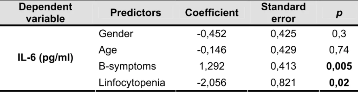
B-symptoms and lymphocytopenia were identified as potent predictors of the serum levels of IL-6 before treatment. These results are shown in Table 1. Although hepatomegaly was associated with high IL-10 levels (p=0,01), none of the studied variables were able to predict the cytokine levels in serum.
Table 1 – Multiple linear regression analysis of predictors for IL-6 serum levels in 28 patients with Hodgkin lymphoma.
Dependent
variable Predictors Coefficient
Standard
error p
Gender -0,452 0,425 0,3
Age -0,146 0,429 0,74
B-symptoms 1,292 0,413 0,005 IL-6 (pg/ml)
4. Discussion
Classical HL is characterized by well-defined histological features, notably H-RS cells surrounded by a heterogeneous and abundant inflammatory infiltrate, which provides a propicious microenvironment for their proliferation and survival. A complex network of interactions mediated by cytokines, chemokines and cell-cell interactactions does exist between different cell types in HL, and it seems crucial for the development and progression of the tumor38, 39. Several aspects about different cytokines that contribute to the pathogenesis of HL have been studied, providing a better understanding of the immune disfunction and symptomatology associated with this disease11, 12, 14. However, the exact role of cytokines in the cause-effect relationship between H-RS cells and microenvironment remains obscure.
In the present study, serum levels of IL-6 and IL-10 were frequently elevated in cHL patients at diagnosis compared to normal volunteers, and they decreased substancially after treatment. On the other hand, IL-13 was aways indetectable in serum by ELISA method used. It was observed that IL-6 and IL-10 serum levels were associated with some clinical and laboratory features of HL. Remarkable, in the HL patients evaluated the IL-6 levels in serum could be predicted by the presence of B-symptoms (high) and lymphocytopenia (low levels).
immune response have been documented40, 41. IL-6 plays a central role in inflammation as a regulator of the transition between humoral and cellular responses, which could explain, at least in part, some features on the pathobiology of HL and its clinical manifestations.
Clinical and laboratory correlates of IL-6 levels in HL have been previously studied in both untreated and relapsed patients, with some discordant results16, 19, 20, 42. In agreement with previously reported data, in the
present study it was observed an association between higher IL-6 serum levels before treatment and presence of B-symptoms19, 20. Additionally, patients with higher levels of IL-6 presented more often hepatomegaly, abdominal disease and anemia. On the other hand, no association with gender, Ann-Arbor stage and bulky disease were observed. A possible explanation for this might be the small number of patients studied in comparison to other series19, 20.
advanced HL9. Interestingly, in the present study it predicted lower levels of IL-6. One possible explanation for this phenomenon is that the low lymphocyte count might reflect a histological change in HL towards a pattern with more proliferation of H-RS cells, rather than stimulation of reactive inflammatory cells. Taken together, these data suggest that a subset of HL may exihibts a more discrete inflammatory component along with a higher number of H-RS cells and worse evolution as consequence.
For IL-10, higher levels of this cytokine in serum before treatment of HL patients were related to low serum albumin and hepatomegaly. There was also a trend of association with advanced Ann-Arbor stages, abdominal involvement, B-symptoms and anemia, which are in agreement with previous studies17, 21. However, the previously reported association between high IL-10 levels and EBV infection was not observed. Herling (2003) et al. reported that higher serum levels of IL-10 before treatment were associated with the immunostaining for EBV LMP-1, mainly in mixed cellularity subtype22. Unfortunatelly, in the present study the EBV status could not be retrieved for all patients, and the lack of association between IL-10 levels and EBV infection might be due to the limited number of EBV-positive HL patients in the present casuistic, or even a low number of MCHL cases.
expressed in the tumors, almost exclusively by H-RS cells. The IL-13 receptor transcripts are expressed not only in H-RS cells, but also by a large proprotion of other cells within the reactive infiltrate, including fibroblasts. This data suggests that IL-13 may also support the maintenance of the reactive infiltrate in HL. In addition, treatment of HL-derived cell line with a neutralizing antibody to IL-13 resulted in a dose-dependent inhibition of H-RS proliferation33 – 35. The accumulating evidence indicates that co-expression of IL-13 and its receptor is a common feature of H-RS cells, strengthening the IL-13 activity as an autocrine growth factor in HL. The findings of indetectable serum levels of IL-13 do not refute this hypothesis, but it may suggest that IL-13 acts predominantly at the level of the tumor microenvironment, possibly in very low concentrations and with short range of action.
necessary to state conclusively whether the reduction of IL-6 with treatment is a clear-cut predictor of good response.
In summary, it was observed that pre-treatment serum levels of IL-6 and IL-10 were associated with some clinical and laboratory parameters in HL patients, and that these cytokines decrease significantly after treatment. Higher IL-6 serum levels after chemotherapy seem to be associated with treatment failure, but this data requires confirmation in large series.
Acknowledges
The authors would wish to thank Ms. Celene Maria Gandin, Mr. Marcos Roberto Franchi, and Mr. Luis Fernando Franchi for their technical assistance in histological and immunohistochemical techniques; Carlos Eduardo Bacchi, MD, PhD and Francisco Carlos Quevedo, MD, for providing us access to HL biopsies for analysis; Sergio Alberto Rupp de Paiva,MD, PhD, Marcos Ferreira Minicucci, MD and Suzana Erico Tani Minamoto, MD for their support with the statistical analysis; and Alice de Oliveira Gonçalves, BSc and the current staff of the Molecular Pathology Laboratory at Botucatu School of Medicne (UNESP) for their help with samples and experiments. This work was supported by FAPESP grant# 2006/00591-5.
Disclosures
References
1. Straus DJ. Treatment of early-stage nonbulky Hodgkin lymphoma. Curr Opin Oncol 2006;18:432-36.
2. Diehl V. Chemotherapy or Combined Modality Treatment: the optimal treatment for Hodgkin’s Disease. J Clin Oncol 2004;22:15-18.
3. Laskar S, Gupta T, Vimal S, Muckaden MA, Siakia TK, Pai SK et al. Consolidation Radiation After Complete Remission in Hodgkin's Disease Following Six Cycles of Doxorubicin, Bleomycin, Vinblastine, and Dacarbazine Chemotherapy: Is There a Need? J Clin Oncol 2004;22:62-68.
4. Lazarus HM, Rowlings PA, Zhang MJ, Vose JM, Armitage JO, Bierman PJ et al. Autotransplants for Hodgkin's Disease in Patients Never Achieving Remission: A Report From the Autologous Blood and Marrow Transplant Registry. J Clin Oncol 1999;17:534.
5. Barlett, NL. Therapies for Relapsed Hodgkin Lymphoma: Transplant and Non -Transplant Approaches Including Immunotherapy. Hematology (Am Soc Hematol Educ Program); 245-51.
7. Diehl V, Stein H, Hummel M, Zollinger R, Connors JM. Hodgkin’s Lymphoma: Biology and Treatment Strategies for Primary, Refractory and Relapsed Disease. Hematology (Am Soc Hematol Educ Program) 2003;225-472.
8. Klimm B, Diehl V, Pfistner B, Engert A. Current treatment strategies of the German Hodgkin Study Group (GHSG). Eur J Haematol 2005;75:125-34.
9. Hasenclever D, Diehl V. A Prognostic Score for Advanced Hodgkin’s Disease. N Engl J Med 1998; 339:1506-14.
10. Diehl V, Thomas RK, Re D. Part II: Hodgkin’s lymphoma – diagnosis and treatment. Lancet 2004;5:19-26.
11. Skinnider BF, Mak TW. The role of cytokines in classical Hogdkin lymphoma. Blood 2002;99:4283-97.
12. Khan G. Epstein-Barr virus, cytokines, and inflammation: A cocktail for the pathogenesis of Hodgkin’s lymphoma. Experimental Hematology 2006;34:399-406.
14. Gruss HJ, Pinto A, Duyster J, Poppema S, Herrmann F. Hodgkin’s disease: a tumor with disturbed immunological pathways. Immunology Today 1997;18:156-63.
15. Cozen W, Gill PS, Ingles SA, Masood R, Martinez-Maza O, Cockburn MG et al. IL-6 levels and genotype are associated with risk of young adult Hodgkin lymphoma. Blood 2004;103:3216-21.
16. Reynolds GM, Billingham LJ, Gray LJ, Flavell JR, Najafipour S, Crocker J et al. Interleukin 6 expression by Hodgkin/Reed-Sternberg cells is associated with the presence of 'B' symptoms and failure to achieve complete remission in patients with advanced Hodgkin's disease. Br J Haematol. 2002;118:195-201.
17. Vassilakopoulos TP, Nadali G, Angelopoulou MK, Siakantaris MP, Dimopoulou MN, Kontopidou FN et al. Serum interleukin-10 levels are an independent prognostic factor for patients with Hodgkin’s lymphoma. Haematologica 2001;86:274-81.
18. Skinnider BF, Kapp U, Mak TW. The role of interleukin 13 in classical Hodgkin lymphoma. Leuk Lymphoma 2002;43:1203-10.
20. Seymor JF, Talpaz M, Hagemeister FB, Cabanillas F, Kurzrock R. Clinical Correlates of Elevated Levels of Interleukin-6 in Patients with Untreated Hodgkin’s Disease. Am J Med 1997;102:21-28.
21. Sarris AH, Kliche KO, Pethambaram P, Preti A, Tucker S, Jackow C et al. Interleukin-10 levels are often elevated in serum of adults with Hodgkin’s disease and are associated with inferior failure-free survival. Ann Oncol 1999;10:433-40.
22. Herling M, Rassidakis GZ, Medeiros JL, Vassilakopoulos TP, Kliche KO, Nadali G et al. Expression of Epstein-Barr Virus Latent Membrane Protein-1 in Hodgkin and Reed-Sternberg Cells of Classical Hodgkin’s Lymphoma: Associations with Presenting Features, Serum Interleukin 10 Levels, and Clinical Outcome. Clin Can Res 2003;9:2114-20.
23. Herbst H, Foss HD, Samol J, Araujo I, Klotzbach H, Krause H et al. Frequent expression of interleukin-10 by Epstein-Barr Virus-harboring tumor cells of Hodgkin’s disease. Blood 1996;87:2918-29.
24. Akira S, Taga T, Kishimoto T. Interleukin-6 in biology and medicine. Adv Immunol 1993;54:1-78
26. Herbst H, Samol J, Foss HD, Raff T, Niedobitek G. Modulation of interleukin-6 expression in Hodgkin and Reed-Sternberg cells by Epstein-Barr virud. J Pathol 1997;182:299-306.
27. Jucker, M, Abts H, Li W, Schindler R, Merzs H, Gunther A et al. Expression of Interleukin-6 and Interleukin-6 Receptor in Hodgkin’s Disease. Blood 1991;11:2413-18.
28. Foss HD, Herbst H, Oelman E, Samol J, Grebe M, Blankenstein T, et al. Lymhpotoxin, tumor necrosis factro and interleukin-6 gene transcripts are presetn in Hodgkin and Reed-Sternberg cells of most Hodgkin’ disease cases. Br J Haematol 1993;84627-35.
29. Levy Y, Brouet JC. Interleukin-10 prevents spontaneous death of germinal center B cells by induction of the bcl-2 protein. J Clin Investig 1994;93:424-28.
30. Duckers DF, Jaspars LH, Voss W, Oudejans JJ, Hayes D, Cillessen S et al. Quantitative immunohistochemical analysis of cytokine profiles in Epstein-Barr virus-positive and negative cases of Hodgkin’s disease. J Pathol 2000;190:143-49.
32. Viviani S, Notti P, Bonfante V, Verderio P, Valagussa P, Bonddonna G. Elevated pretreatmant serum of IL-10 are associated with poor prognosis in Hodgkin’s disease, the Milan Cancer Institute experience. Med Oncol 2000;17:59-63.
33. Kapp U, Yeh WC, Patterson B, Elia AJ, Käji D, Ho A et al. Interleukin 13 is secreted by and stimulates the growth of Reed-Sternberg cells. J Exp Med 1999;189:1939-46.
34. Oshima K, Akaiwa M, Umeshita R, Suzumiya J, Izuhara K, Kikuchi M et al. Interleukin-13 and Interleukin-13 receptor in Hodgkin’s disease: possible autocrine mecanism and involvement in fibrosis. Histopathology 2001;38:368-75.
35. Skinnider BF, Elia AJ, Gascoyne RD, Trümper LH, von Bonin F, Kapp U et al. Interleukin 13 and interleukin 13 receptor are frequently expressed by Hodgkin and Reed-Sternberg cells of Hodgkin lymphoma. Blood 2001;97:250-55.
37. de Oliveira DE, Bacchi MM, Macarenco RS, Tagliarini JV, Cordeiro RC, Bacchi CE. Human papillomavirus and Epstein-Barr virus infection, p53 expression, and cellular proliferation in laryngeal carcinoma. Am J Clin Pathol 2006;126:284-93.
38. Gruss HJ, Pinto A, Duyster J, Poppema S, Herrmann F. Hodgkin’s disease: a tumor with disturbed immunological pathways. Immunology Today 1997;18:156-63.
39. Enblad G, Molin D, Glimelius I, Fisher M, Nilsson G. The potential role of of innate immunity in the pathogenesis of Hodgkin’s Lymphoma. Hematol Oncol Clin N Am 2007;21:805-23.
40. Jones AS. Directing Transition from Innate to Acquired Immunity: Defining a Role for IL-6. J Immunol 2005;175:3463-68.
41. Rose-John S, Scheller J, Elson G, Jones SA. Interleukin-6 biology is coordinated by membrane-bound and soluble receptors: role in inflammation and cancer. J Leukoc Biol 2006;80:227-36.
42. Gause A, Scholz R, Klein S, Jung W, Diehl V, Tesch H, Hasenclever D, Pfreundschuh M. Increased levels of circulating interleukin-6 in patients with Hodgkin's disease. Hematol Oncol 1991;9:307-13.
44. Barton BE. IL-6: Insights into Novel Biological Activities. Clin Immunol Immunopathol 1997;85:16-20.
Tabela A1 - Sexo e idade nos grupos de pacientes com LH clássico e indivíduos saudáveis doadores de sangue (grupo-controle).
Idade
Grupos (n) Razão
M:F Média (anos)
DP (anos)
CV
(%) P25 P75 Mediana (anos)
Faixa etária (anos)
LH clássico (28) 1,9:1 33 11,5 35 22 40,2 33 19-57
Entre 19 a 39 anos (20) 1,8:1 26,6 6,4 24 22 33 24 19-39
Acima de 40 anos (9) 2:1 46,7 7 15 40,7 54,5 43 40-57
Grupo-controle (26) 1,8:1 33 10,9 33 23 40 33 18-53
DP: desvio padrão; CV: coeficiente de variação; P25 e P75: percentis 25 e 75, respectivamente. LH = Linfoma de Hodgkin
Tabela A 2 – Associação entre aspectos clínicos e laboratoriais e nível sérico de IL-6 e IL-10 ao diagnóstico para os 28 pacientes com linfoma de Hodgkin avaliados no presente estudo.
Categorias n (%) IL-6 sérica (pg/mL) p IL-10 sérica (pg/mL) p
Idade
19 a 39 anos 19 (68) 25,7 (13,7 – 42,3) 12,1 (6,6 – 25,2)
40 anos 9 (32) 19 (11,2 – 59,4) 0,66 22,8 (12,2 – 34,8) 0,34
Sexo
Masculino 18 (64) 19,7 (14,9 - 51,8) 14,4 (8,7 - 23)
Feminino 10 (36) 24,9 (12 - 42,8) 0,94 21,7 (0 - 47,5) 0,85
Estadio de Ann-Arbor
I - II 16 (57) 19,6 (5,1 - 39,6) 8,2 (2 - 24,8)
III - IV 12 (43) 30,9 (18,2 – 72,6) 0,2 20,2 (13,3 - 36,7) 0,06
Envolvimento mediastinal
Presente 16 (57) 24,9 (18,8 – 39,3) 11 (6,1 – 24,4)
Ausente 12 (43) 18,9 (5,1 - 63,8) 0,73 20,6 (11,5 – 39,7) 0,4
Envolvimento abdominal
Presente 14 (50) 39,3 (19,3 – 99,7) 20,6 (12,1 - 30,5) Ausente 14 (50) 18,8 (<4 - 30,7) 0,03 8,2 (4 – 23) 0,1