Newborn Hypoxia/Anoxia Inhibits
Cardiomyocyte Proliferation and Decreases
Cardiomyocyte Endowment in the
Developing Heart: Role of Endothelin-1
Alexandra N. Paradis1, Maresha S. Gay1, Christopher G. Wilson1,2, Lubo Zhang1*
1Center for Perinatal Biology, Division of Pharmacology, Department of Basic Sciences, Loma Linda University School of Medicine, Loma Linda, California, United States of America,2Division of Neonatology, Loma Linda University School of Medicine, Loma Linda, California, United States of America
Abstract
In the developing heart, cardiomyocytes undergo terminal differentiation during a critical window around birth. Hypoxia is a major stress to preterm infants, yet its effect on the devel-opment and maturation of the heart remains unknown. We tested the hypothesis in a rat model that newborn anoxia accelerates cardiomyocyte terminal differentiation and results in reduced cardiomyocyte endowment in the developing heartviaan endothelin-1-depen-dent mechanism. Newborn rats were exposed to anoxia twice daily from postnatal day 1 to 3, and hearts were isolated and studied at postnatal day 4 (P4), 7 (P7), and 14 (P14). Anox-ia significantly increased HIF-1αprotein expression and pre-proET-1 mRNA abundance in
P4 neonatal hearts. Cardiomyocyte proliferation was significantly decreased by anoxia in P4 and P7, resulting in a significant reduction of cardiomyocyte number per heart weight in the P14 neonates. Furthermore, the expression of cyclin D2 was significantly decreased due to anoxia, while p27 expression was increased. Anoxia has no significant effect on car-diomyocyte binucleation or myocyte size. Consistently, prenatal hypoxia significantly de-creased cardiomyocyte proliferation but had no effect on binucleation in the fetal heart. Newborn administration of PD156707, an ETA-receptor antagonist, significantly increased cardiomyocyte proliferation at P4 and cell size at P7, resulting in an increase in the heart to body weight ratio in P7 neonates. In addition, PD156707 abrogated the anoxia-mediated ef-fects. The results suggest that hypoxia and anoxiaviaactivation of endothelin-1 at the criti-cal window of heart development inhibits cardiomyocyte proliferation and decreases myocyte endowment in the developing heart, which may negatively impact cardiac function later in life.
a11111
OPEN ACCESS
Citation:Paradis AN, Gay MS, Wilson CG, Zhang L
(2015) Newborn Hypoxia/Anoxia Inhibits Cardiomyocyte Proliferation and Decreases Cardiomyocyte Endowment in the Developing Heart: Role of Endothelin-1. PLoS ONE 10(2): e0116600. doi:10.1371/journal.pone.0116600
Received:July 22, 2014
Accepted:December 12, 2014
Published:February 18, 2015
Copyright:© 2015 Paradis et al. This is an open
access article distributed under the terms of the Creative Commons Attribution License, which permits unrestricted use, distribution, and reproduction in any medium, provided the original author and source are credited.
Data Availability Statement:All relevant data are
within the paper.
Funding:This study was supported by the National
Institutes of Health grant HL118861 (to L. Zhang). The funder had no role in study design, data collection and analysis, decision to publish, or preparation of the manuscript.
Competing Interests:The authors have declared
Introduction
The intrauterine environment plays a well-established role in predisposition to cardiovascular disease later in life [1]. Environmental factors during the critical period of heart development may alter the maturation of the heart and thus potentially its life-long function. Cardiomyo-cytes are the functional contractile units of the heart that undergo a normal maturation process in which terminal differentiation is the final outcome. As the cardiomyocytes terminally differ-entiate and exit the cell cycle, they lose their proliferative capacity [2]. Cardiomyocyte growth then transitions from hyperplastic to hypertrophic, in which the cells can only increase in size rather than number [3,4]. Ultimately the proliferative capacity of cardiomyocytes is lost and the adult heart is known to exhibit negligible increases in cell number [5]. Therefore the timing of this transition is pivotal in determining cardiomyocyte endowment in the heart for the rest of the animal’s life.
Hypoxia is a major stress to preterm infants, yet its effect on the development and matura-tion of the heart remains unknown. Given that the transimatura-tion of cardiomyocyte terminal differ-entiation occurs in rodents during the first two weeks of neonatal life [3,6], which is an equivalent timeframe to the late fetal stage in third trimester of human gestation [2], they pro-vide a reasonable animal model to study the effect of anoxia on preterm infants at the critical window of the heart development. This process of terminal differentiation begins in the rat heart around postnatal day 4 [3] and progresses until day 14 when the heart is essentially ma-ture, thus three time-points within this period were evaluated in this study. Previous studies in rats have shown that maternal hypoxia (10.5% O2) leads to a premature exit from the cell cycle in fetal cardiomyocytes [7–9]. Additionally, neonatal cardiomyocytes have been shown to de-crease proliferation when exposed to hypoxic conditions [10]. Studies have also been per-formed in sheep in which placental restriction is induced, resulting in reduced cardiomyocyte maturation [11] and proliferation [12], increased proportion of mononucleate cardiomyocytes [13], and decreased cardiomyocyte endowment [14]. However, thein vivoeffects of anoxia, as a preterm model, on cardiomyocyte proliferation and endowment in the developing rat heart are, as of yet, not known. Additionally, the downstream regulators of cardiomyocyte prolifera-tion and maturaprolifera-tion are unknown.
Endothelin-1 (ET-1) expression is induced by hypoxia [15–18]. Studies performed in endo-thelial cells [19,20] and cardiomyocytes [21] have identified a HIF-1αbinding site in the
pre-pro-ET-1 gene. Furthermore, the cardiomyocyte is both a site of synthesis and action for ET-1
[22,23], as it acts mainly at the paracrine or autocrine level [24,25]. Our recent work showed
thatex vivoET-1 treatment promoted terminal differentiation of fetal cardiomyocytes,viaan increase in DNA methylation [26]. The predominant ET-1 receptor subtype in cardiomyocytes is the ETA-receptor [23], which is thought to be involved in regulating proliferation [24,27, 28]. Currently, little is known about the role that basal ET-1 plays in the terminal differentia-tion of cardiomyocytes, as well as the effect of hypoxia/anoxia-induced ET-1 producdifferentia-tion on this process.
Methods
Experimental animals
Time-dated pregnant Sprague-Dawley rats were purchased from Charles River Laboratories (Portage, MI) and allowed to give birth. Neonatal pups from 7 litters were used and divided into the treatment groups. Data from pups of multiple litters were pooled. Starting at postnatal day 1, newborn rats were placed in a temperature-controlled (37°C) anoxia chamber. Nitrogen was infused into the chamber for 10 minutes and an oxygen sensor was used to verify the level of oxygen in the chamber being<0.2%. Control animals were placed in a chamber with oxy-gen maintained at 21%. Anoxia treatments were performed twice a day with 8 hours in be-tween, from postnatal day 1 until postnatal day 3. A group of animals was treated with intraperitoneal injections of an ETA-receptor antagonist, PD156707 (2 mg/kg), prior to each episode of anoxia, twice a day for the first 3 postnatal days. Neonatal pups were anesthetized with isoflurane and hearts isolated for studies on postnatal day 4, 7, and 14. To investigate the comparative effect of prenatal hypoxia, some of the time-dated pregnant Sprague-Dawley rats were treated with either normoxic control or 10.5% O2from gestational day 15 to 21, as previ-ously described [29,30]. Following the hypoxia treatment, pregnant rats were allowed to give birth. Hearts were isolated from postnatal day 4 and 7 neonatal rats. All procedures and proto-cols were approved by the Loma Linda University Institutional Animal Care and Use Commit-tee (IACUC) and all procedure adhered to the guidelines by US National Institutes of Health Guide for the Care and Use of Laboratory Animals (http://grants.nih.gov/grants/olaw/Guide-for-the-care-and-use-of-laboratory-animals.pdf).
Measurement of cardiomyocyte number
Hearts from day 4, 7, and 14 neonatal pups were isolated and the atria excised. The hearts were then completely enzymatically digested to yield primary cardiomyocytes, as previously de-scribed [26,31]. A pre-plate step was performed to enrich the cardiomyocyte population. This is a commonly used method [32] that is based on the differential attachment of cardiomyocytes and non-myocyte cells of the heart. Cardiomyocytes take approximately 24 hours to fully at-tach to the plate while non-myocytes atat-tach within a couple hours. After a 2-hour pre-plate step to remove attached non-myocytes, cardiomyocytes in the media were collected and used for counting cardiomyocyte numberviaa hemacytometer. Briefly, an aliquot of cardiomyo-cytes was counted using a hemacytometer and the counts were multiplied by the total volume of cell suspension and normalized according to the heart weight, to yield the number of cardio-myocytes per heart weight.
Immunocytochemistry
To perform immunocytochemical staining, cardiomyocytes isolated from day 4 and 7 hearts were allowed to attach to plates in Hyclone Medium 199 (Thermo Scientific) supplemented with 10% fetal bovine serum (Gemini Bio-Products) and 1% antibiotics (10,000 I.U./mL peni-cillin, 10,000μg/mL streptomycin) at 37°C in 95% air/5% CO2. After 24 hours, cardiomyocytes
were fully attached and were double stained with alpha-actinin, a cardiomyocyte marker, and Ki-67, a proliferation marker as described previously [8,26]. Cardiomyocytes were plated on coverslips and fixed with 4% paraformaldehyde (ThermoScientific) for 15 minutes followed by permeabilization with Triton X-100 (Fisher) for 10 minutes. The cells were blocked with 1% bovine serum albumin for 1 hour at room temperature before incubation with the primary an-tibodies: mouse anti-α-sarcomeric actinin (A7811, Sigma) (1:200) and rabbit anti-Ki-67
the secondary antibodies: mouse Alexa Fluor 488 (A21202, Life Technologies) and anti-rabbit Alexa Fluor 647 antibodies (A21244, Life Technologies) for 1 hour at room temperature. Nuclei were stained with Hoescht (Sigma) for less than 1 minute. The immunofluorescence staining was assessed using a Zeiss Axio Imager.A1 microscope and quantitative analysis was carried out usingImageJsoftware (http://imagej.nih.gov/ij/). Ki-67 expression, binucleation, and cell size were measured.
Flow cytometry
Primary cardiomyocytes isolated from day 14 neonatal rats were stained for analysis by flow cytometry. Cells were washed in staining buffer (PBS + 5% FBS), spun down, and re-suspended in 4% paraformaldehyde for 20 minutes at room temperature in the dark. The fixed cells were then washed in permeabilization wash buffer (eBioscience) and supernatant discarded. Cells were stained with antibodies for the cardiomyocyte marker, Troponin T (ab10214, Abcam) (1:200), and proliferation marker, Ki-67-conjugated to allophycocyanin (APC) (eBioscience) (50–5698, 1:200). After incubation and washing, cells were incubated with the secondary anti-body for Troponin T, fluorescein isothiocyanate (FITC) (555988, BD Pharmingen) (1:100). Fi-nally cells were washed and resuspended in 1% paraformaldehyde to be run on a FACSAria (BD Biosciences) and analyzed via FACSDiva software (BD Biosciences) for percentage of Ki-67 expressing cardiomyocytes.
Quantitative real-time PCR
RNA was isolated from the postnatal day 4 (P4) hearts and prepro-ET-1 mRNA abundance was determined by real-time RT-PCR using Icycler Thermal cycler (Bio-Rad), as described pre-viously [30]. Reverse transcription and cDNA synthesis was performed using SuperScript III First-Strand Synthesis Supermix for RT-PCR (Life Technologies). The primers are 5’ -CTAGGTCTAAGCGATCCTTGAA-3’(forward) and 5’-CTTGATGCTGTTGCTGATGG-3’
(reverse). PCR was performed in triplicate, and threshold cycle numbers were averaged.
Western immunoblotting
HIF-1α, ETA-receptor (ETAR), and ETB-receptor (ETBR) protein abundance in the P4 heart
was measured from control and anoxia groups. The protein abundance of cyclin D2 and p27 was measured in P4 hearts from control and anoxia groups as well as in the presence and ab-sence of PD156707. Tissues were homogenized and protein isolated using the RIPA lysis buffer system (Santa Cruz Biotechnology). Protein concentrations were quantified using the BCA protein assay (ThermoScientific) and all samples were loaded with equal protein onto 7.5% (HIF-1α) or 10% (ETAR, and ETBR, cyclin D2, and p27) polyacrylamide gel with 0.1% sodium
dodecyl sulfate (SDS). Proteins were then separated by electrophoresis and transferred onto ni-trocellulose membranes. Non-specific binding sites were blocked with Tris-buffered saline so-lution (TBS) containing 5% dry milk. The membranes were incubated with primary antibodies against HIF-1α(sc10790, Santa Cruz Biotechnology; 1:500), ETAR (sc33536, Santa Cruz
Statistical analysis
Data are expressed as means ± SEM. Statistical analysis (p<0.05) was determined by two-way analysis of variance (ANOVA) followed by Neuman-Keulspost hoctest or Student’s t test, where appropriate, using GraphPad Prism software. The two-way ANOVA was performed to evaluate the effects of two factors, within each age group: (1) control versus anoxia, and (2) in the presence and absence of PD156707.
Results
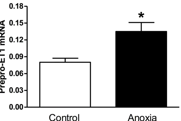
Newborn anoxia treatment increased pre-proET-1 mRNA in the heart of
P4 neonate
Neonatal rats were exposed to anoxia twice a day from postnatal day 1 to 3, and hearts were isolated at P4. As seen inFig. 1, there was a significant increase in prepro-ET-1 mRNA abun-dance in neonatal hearts exposed to anoxia (<0.2% O2), as compared to the normoxic control (21% O2).
Fig 1. Effect of newborn anoxia on prepro-ET-1 mRNA in the neonatal heart.Hearts were isolated from day 4 neonatal rats treated with control or anoxia. mRNA abundance of prepro-ET-1 was determined by real-time RT-PCR. Data are means±SEM.*P<0.05, anoxiavs. control. n = 3–5.
Newborn anoxia treatment increased HIF-1
α
protein abundance in the
heart of the P4 neonate
Hearts from P4 rats treated with anoxia were collected and protein isolated. Neonatal hearts ex-posed to anoxia had significantly increased levels of the HIF-1αprotein (Fig. 2).
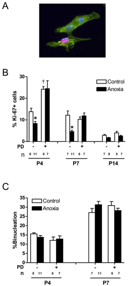
Newborn anoxia treatment decreased cardiomyocyte proliferation
As shown inFig. 3, there is a development-dependent decrease in cardiomyocyte proliferation at the critical window of the heart development during the first two weeks of life in rodents,Fig 2. Effect of newborn anoxia on HIF-1αprotein abundance in the neonatal heart.Hearts were isolated from day 4 neonatal rats treated with control or anoxia. Protein abundance of HIF-1αwas determined by Western immunoblotting. Data are means±SEM.*P<0.05, anoxiavs. control. n = 4.
and myocyte proliferation reduces to minimal levels at postnatal day 14. Anoxia treatment of newborns caused a significant decrease in the proliferation of neonatal cardiomyocytes at both postnatal days 4 and 7 (Fig. 3B). Treatment of newborn rats with a selective ETA-receptor an-tagonist, PD156707, caused a significant increase in cardiomyocyte proliferation in P4 neonatal rats (Fig. 3B). In addition, PD156707 abrogated the anoxia-induced effects in the developing hearts (Fig. 3B). In contrast to proliferation, there is a development-dependent increase in car-diomyocyte binucleation in the developing heart (Fig. 3C). Neither anoxia nor PD156707 treat-ments had significant effects on cardiomyocyte binucleation (Fig. 3C).
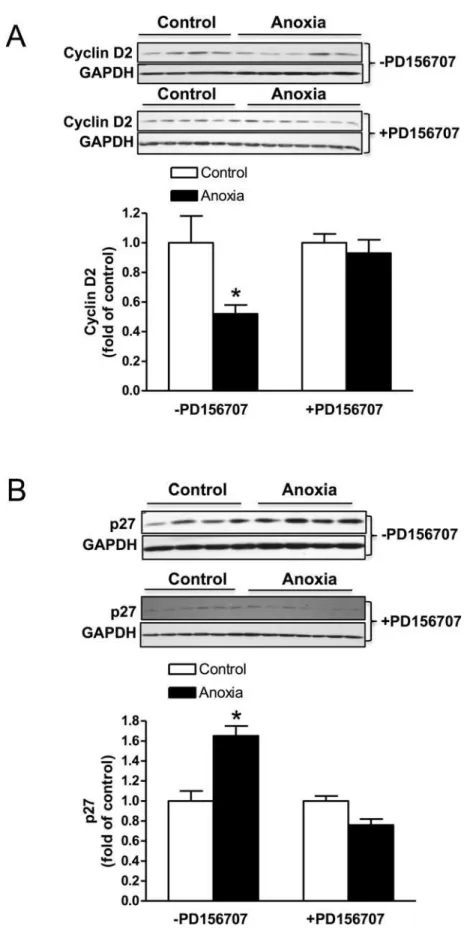
Newborn anoxia treatment decreased cyclin D2 and increased p27
expression in the heart of the P4 neonate
The protein expression of cyclin D2 was decreased due to anoxia treatment and this effect was abolished in the presence of PD156707 (Fig. 4A). On the contrary, p27 expression in the neo-natal heart was significantly increased due to anoxia treatment, and PD156707 blocked the ef-fect of anoxia (Fig. 4B).
Newborn anoxia treatment decreased cardiomyocyte number by day 14
There was no significant change in cardiomyocyte number due to newborn anoxia treatment at day 4 and 7. However, results for day 14 show that anoxia leads to a significant decrease in cardiomyocyte number per heart weight (Fig. 5). PD156707 alone caused a significant increase in cardiomyocyte number in the day 7 neonate (Fig. 5). In the presence of PD156707, the anox-ia-mediated effects at day 14 were blocked (Fig. 5).Cell size was increased in the presence of PD156707
Anoxia had no effect on mononucleate or binucleate cell size at either day 4 or 7 (Fig. 6). How-ever, PD156707 treatment was able to increase both mononucleate and binucleate cell size at postnatal day 7 (Fig. 6).
PD156707 increased heart to body weight ratio
There was no significant effect of anoxia on the heart to body weight ratio for any day (Fig. 7). However, PD156707 treatment significantly increased the heart to body weight ratio in day 4 and 7 neonates (Fig. 7). Heart and body weight averages in the presence and absence of anoxia and PD156707 are listed inTable 1.
Neonatal anoxia treatment had no effect on ET-receptor density
Hearts from postnatal day 4 rats that were treated with anoxia were collected and protein iso-lated. There was no significant change in protein abundance of either ETAR or ETBR, due to anoxia treatment (Fig. 8).
control or anoxia, in the absence or presence of PD156707. Cells from P4 and P7 rats were stained withα -actinin and 67, and nuclei were stained using Hoechst staining. P14 cardiomyocytes were stained with Ki-67 and analyzedviaFACS.Panel Ashows a representative image of cardiomyocytes stained withα-actinin (green), Ki-67 (red), and Hoescht (blue).Panel Bshows percent of Ki-67 expressing cells.Panel Cshows percent of binucleate cells. Data are means±SEM.*P<0.05, anoxiavs. control.†P<0.05, -PD156707vs. +PD156707. PD: PD156707; n: animal numbers.
Prenatal hypoxia decreased cardiomyocyte proliferation in the fetal heart
To investigate the comparative effect of prenatal hypoxia, pregnant rats were treated with ei-ther normoxic control or 10.5% O2from gestational day 15 to 21, and hearts were isolated from postnatal day 4 and 7 neonatal rats. Similar to the findings in newborn anoxia treatment, prenatal hypoxia resulted in a significant decrease in the proliferation of cardiomyocytes at postnatal day 7 (Fig. 9A), but had no significant effects on percent binucleation (Fig. 9B) or the heart to body weight ratio (Fig. 9C).Discussion
In the present study, we provide evidence showing thatin vivonewborn anoxia leads to a de-crease in the proliferation of cardiomyocytes in the developing heart. Furthermore, our results 4) of PD156707. Protein abundance of cyclin D2 in the absence and presence of PD156707 (A), and p27 in the absence and presence of PD156707 (B) was determined by Western immunoblotting. Data are means±
SEM.*P<0.05, anoxiavs. control.
doi:10.1371/journal.pone.0116600.g004
Fig 5. Effect of newborn anoxia and PD156707 on number of cardiomyocytes per heart weight.Cardiomyocytes were isolated from day 4, 7, and 14 neonatal rats that were treated with control or anoxia, in the absence or presence of PD156707. Hearts were weighed and cardiomyocytes counted by hemacytometer. Data are expressed as cardiomyocyte number/g heart weight, and are means±SEM.*P<0.05, anoxiavs. control.†P<0.05, -PD156707
vs. +PD156707. PD: PD156707; n: animal numbers.
suggest that anoxia treatment leads to a significant reduction in number of cardiomyocytes per heart weight of the day 14 neonate, which is terminally differentiated. The findings that anoxia increased ET-1 production in the heart and the anoxia-induced changes in proliferation and cardiomyocyte number were reversed with PD156707, suggest a mechanism mediated by the ETA-receptor. In addition, basal ET-1 was also found to play a role in cardiomyocyte prolifera-tion, as well as the heart to body weight ratio.
Cardiomyocytes undergo a terminal differentiation process that reaches completion by the first two weeks of neonatal life in rats [2,6]. After this, cardiomyocytes in the heart have negli-gible proliferative capacity and further growth is mainlyviahypertrophy. Thus the number of cardiomyocytes that will reside in the adult heart is determined during this early stage and if al-tered may result in life-long consequences. Hypoxic stress during perinatal development has Fig 6. Effect of newborn anoxia and PD156707 on cardiomyocyte size in the neonatal heart.Cardiomyocytes were isolated from day 4, 7, and 14 neonatal rats that were treated with control or anoxia, in the absence or presence of PD156707. Mononucleate (A) and binucleate (B) cell size was measured usingImageJ. Data are means±SEM.†P<0.05, -PD156707vs. +PD156707. PD: PD156707; n: animal numbers.
doi:10.1371/journal.pone.0116600.g006
Fig 7. Effect of newborn anoxia and PD156707 on heart to body weight ratio of neonatal rats.Body and heart weights were taken from day 4, 7, and 14 neonatal rats that were treated with control or anoxia, in the absence or presence of PD156707. Data are means±SEM.†P<0.05, -PD156707vs.
+PD156707. PD: PD156707; n: animal numbers.
been shown by previous studies to diminish the proliferation of cardiomyocytes [8,9,26]. Fur-thermore, fetal hearts exposed to hypoxia have fewer [7,14] and larger cardiomyocytes [7], and adult male rats that were exposed to hypoxiain uterowere more susceptible to ischemic in-jury as seen by increased myocardial infarction and reduced recovery [33].
Preterm birth is a complex clinical problem that is highly associated with episodes of severe hypoxia and even anoxia, which can be so severe that the infant must be mechanically ventilat-ed [34]. Preterm infants have an immature respiratory system [35] that is unable to provide ad-equate oxygen at times and thus ventilatory support is frequently needed. However, several studies have shown that episodic airway obstruction and hypoxemia commonly occur in these infants [36,37]. Given that the rodent heart is relatively immature at birth, the present study with episodic anoxia treatments of newborn rats provides a reasonable animal model to study the effects of anoxia/hypoxia on the heart development in preterm infants. Anoxia itself has been shown to alter proliferation, and, in rat fibroblasts, leads to arrest of the cell cycle at the G1 and S phase [38].
To confirm the extent of hypoxic exposure to the neonatal hearts used in our study, the pro-tein levels of hypoxia-inducible factor 1 alpha (HIF-1α) were evaluated. The results show that
HIF-1αprotein abundance is significantly increased in neonatal hearts exposed toin vivo
an-oxia; furthermore increased levels of HIF-1αin the heart have also been observed in the
prena-tal hypoxia model [7]. In agreement with previous work, we showed that cardiomyocyte proliferation was decreased followingin vivoneonatal anoxia treatment at postnatal day 4 and 7. The cardiomyocytes take approximately 24 hours for them to fully attach to the plate before the immunocytochemical staining of Ki-67 can be performed. While the potential effect of this attachment process on the rate of proliferation may not be excluded in the present study, the same procedure applied to all treatment groups. By postnatal day 14, there was a trend for an-oxia to decrease proliferation however this trend was not significant in our data. The heart is thought to be fully mature and essentially an adult phenotype of cardiomyocytes by day 14 in rats, therefore the rate of myocyte proliferation is normally very low at this point and anoxia had no significant effect on lowering it further. Similarly, our results from the prenatal hypoxia model showed a decrease in the proliferation of neonatal cardiomyocytes at postnatal day 7. In-terestingly, newborn anoxia had no significant effect on the binucleation of cardiomyocytes. Table 1. Effect of newborn anoxia and PD156707 on body and heart weight of neonatal rats.
Body weight (grams) Heart weight (grams) Heart to Body weight ratio
P4 P7 P14 P4 P7 P14 P4 P7 P14
-PD156707 Control 8.51±
0.374 (8) 14.44± 0.550 (8) 22.45± 1.090 (7) 0.047± 0.0023 (8) 0.083± 0.0046 (8) 0.123± 0.0076 (7) 0.0055± 0.0002 (8) 0.0058± 0.0003 (8) 0.0055± 0.0002 (7) Anoxia 8.06±
0.317 (12) 13.65± 0.472 (12) 22.28± 0.879 (7) 0.044± 0.0023 (12) 0.083± 0.0025 (12) 0.120± 0.0072 (7) 0.0054± 0.0002 (12) 0.0061± 0.0002 (12) 0.0054± 0.0002 (7) +PD156707 Control 9.57±
0.186 (6) 15.05± 0.289 (6) 21.68± 0.960 (5) 0.062± 0.0029 (6) 0.103±
0.0024*(6)
0.114±
0.0053 (5)
0.0065±
0.0003 (6)
0.0068±
0.0002*(6)
0.0053±
0.0001 (5) Anoxia 7.57±
0.498 (7) 12.75± 0.302 (7) 23.90± 0.779 (8) 0.054± 0.0042 (7) 0.084± 0.0034 (7) 0.127± 0.0081 (8) 0.0072± 0.0004 (7) 0.0066± 0.0002 (7) 0.0053± 0.0002 (8)
Body and isolated hearts were weighed from day 4, 7, and 14 neonatal rats that were treated with control or anoxia, in the absence or presence of PD156707. Heart to body weight ratio values are also represented. Data are means±SEM.
*P<0.05, control-PD156707vs. +PD156707. Number of animals is represented in parentheses.
Previous work has shown that maternal hypoxia leads to an increase in the amount of binucle-ate cardiomyocytes in the fetal heart [7], thus indicating a development stage-specific effect.
Furthermore, we investigated two proteins that are closely involved in the regulation of the cell cycle: cyclin D2 and p27. These proteins have previously been studied and found to be dif-ferentially expressed in the hypoxia-treated fetal heart [8]. Cyclin D2 is associated with other cell cycle regulators that work to promote cell cycle activity, while p27 is a cyclin-dependent ki-nase inhibitor and thus inhibits cell cycle activity. Therefore the expression of these two pro-teins should be inversely related, as our results indicate. Cyclin D2, a cell cycle promoter, is significantly decreased during anoxia treatment, while the cell cycle inhibitor, p27, is upregu-lated under these conditions. These results are consistent with our finding that anoxia treat-ment decreases cardiomyocyte proliferation. In addition, we tested the role of ET-1 acting through its ETAR on the expression of cyclin D2 and p27. In the presence of PD156707, an ETAR antagonist, anoxia had no effect on cyclin D2 expression. However, p27 expression was significantly decreased in the presence of PD156707 compared to control conditions. These findings suggest that ET-1 and the ETAR are key mediators in the anoxia-induced effects on cy-clin D2 and p27. Ultimately, these results may help to explain the overall decrease in cardio-myocyte proliferation due to anoxia treatment.
Although a gradual decrease in proliferation in the critical window of the heart development is a normal developmental process, hypoxia and anoxia appear to accelerate this progression, particularly during the early development. The endpoint of cardiomyocyte number is a metric to measure the consequence of altering the proliferative capacity. Our results suggest that anox-ia reduces cardiomyocyte endowment at postnatal day 14, when the heart is presumed to be fully mature and cardiomyocytes have terminally differentiated. Anoxia reduced proliferation at days 4 and 7, resulting in fewer cardiomyocytes in the differentiated heart seen at day 14. Given that cardiomyocytes are the functional contractile units of the heart, this decreased car-diomyocyte endowment in the heart may have negative impact in cardiac function and become more susceptible to injury later in life. While our results suggest a significant reduction in car-diomyocyte endowment due to anoxia at the critical window of the heart development, future studies using unbiased and random stereology will be needed to provide conclusive evidence of this effect.
Previous studies from our laboratory and others have shown that hypoxia regulates prolifer-ation of cardiomyocytes and vascular muscle [8,10,26,39]. However the downstream regula-tors of this response have yet to be identified. Our previous work in anex vivomodel showed that primary fetal cardiomyocytes exhibited a similar decrease in proliferation when treated with endothelin-1 (ET-1) [26]. It is known that ET-1 expression is induced under hypoxic con-ditionsviaa HIF-binding site on its promoter [15–20], specifically in cardiomyocytes [21]. ET-1 itself has also been shown to regulate proliferation, having a mitogenic effect on vascular smooth muscle [24,27,28]. Moreover our results showed an increase in prepro-ET-1 mRNA in the P4 neonatal heart when exposed to anoxia. Previous work has also shown an increase in prepro-ET-1 mRNA in the fetal heart exposed to maternal hypoxia [26]. These studies taken together implicate a role for ET-1 in mediating the hypoxia- and anoxia-induced decrease in cardiomyocyte proliferation.
A selective ET-receptor antagonist was used to study the role of both basal and anoxia-in-duced ET-1 in the present study. ET-1 can activate two receptor subtypes: the ETA- and ETB -Fig 8. Effect of newborn anoxia on ETA- and ETB-receptor protein abundance in the neonatal heart. Hearts were isolated from day 4 neonatal rats treated with control or anoxia. Protein abundance of ETAR (A)
and ETBR (B) was determined by Western immunoblotting. Data are means±SEM. n = 4–5.
receptor. Activation of the ETA-receptor leads to vasoconstriction and is primarily found in vascular muscle [40]. In contrast, the ETB-receptor can provide a vasodilation effect as well as vasoconstriction depending on the receptor location, in endothelial cells [41] or vascular mus-cle [41–44], respectively. The ETB-receptor also plays a role in the clearance of endothelin from tissues [45]. In cardiomyocytes, the ETA-receptor is the predominant subtype [23], and has been implicated in regulating proliferation [24,27,28]. Therefore, our study evaluated the ef-fects of PD156707, a selective antagonist for the ETA-receptor [46], on cardiomyocyte prolifer-ation. Due to the short half-life of PD156707 of about one hour [47], it was given twice a day just prior to anoxia exposure in the present study. We also evaluated the protein expression of the ET-receptors, both ETAR and ETBR. Interestingly, the results showed no change in the ex-pression of either receptors due to anoxia treatment, suggesting that a change in receptor densi-ty is not contributing to the effects of anoxia or ET-1. The finding that PD156707 ameliorated the anoxia-induced decrease in proliferation of cardiomyocytes at day 4 and 7 implicates the ETA-receptor as a key mediator. Furthermore, the addition of PD156707 alone elicited an in-crease in proliferation at day 4 beyond that of the control. This observation was not seen at day 7 or day 14, suggesting that the regulation of basal ET-1 function in the heart is dependent on the stage of development. At an earlier stage, basal ET-1 levels play a key role in regulating car-diomyocyte proliferation. The effect of basal ET-1 in regulating carcar-diomyocyte endowment in the developing heart is intriguing. The treatment of newborn rats with ETA-receptor antagonist led to an increase in cardiomyocyte number per heart weight at day 7, suggesting that an ap-propriate level of basal ET-1 is necessary to optimize cardiomyocyte endowment in the heart.
Anoxia treatment had no significant effect on mononucleate and binucleate cell size, howev-er inhibition of ETAR by PD156707 caused an increase in cell size at day 7. This may suggest that basal ET-1 plays a role in maintaining cell size and, and if activation of the ETA-receptor is blocked, the cell undergoes hypertrophy. The change in binucleate cell size is likely more rele-vant because the mononucleate cells still have the capacity to divide and are not yet
terminally differentiated.
The heart to body weight ratio was unchanged with anoxia treatment for all age groups. However by blocking basal ET-1 with PD156707, the heart to body weight ratio was increased at postnatal day 7. These results suggest that the heart is increasing in size, which agrees with the results of increased cell size, proliferation, and cardiomyocyte number in the presence of PD156707. In the present study, the cardiomyocyte number were counted in freshly isolated myocytes, and thein vivoPD156707 treatment increased the cardiomyocyte number by about 65% in day 7 hearts. The heart is made up by cardiac fibroblasts, myocytes, endothelial cells, and vascular smooth muscle cells, with the majority being fibroblasts and myocytes. There are significant differences in cell populations of the heart among various species. In rats, the heart is composed of about 60–70% nonmyocytes and 30–40% cardiac myocytes [48–53]. In neona-tal rats, cardiac fibroblasts made up about 64% of the toneona-tal heart, whereas the myocyte popula-tion was 30%, with the nonmyocyte and nonfibroblast cell populapopula-tions comprising the remainder of the heart [54]. In the same study [54], it was found that neonatal and adult mouse hearts contained around 60% cardiac myocytes. However, in a more recent study, 20–
30% cardiac myocytes were demonstrated in the mouse heart [55]. Because of the variability of Fig 9. Effect of prenatal hypoxia on neonatal cardiomyocyte proliferation, binucleation, and heart to body weight ratio.Cardiomyocytes were isolated from day 4 and 7 neonatal rats that were treated with control or maternal hypoxia. Cells were stained withα-actinin and Ki-67, and nuclei stained with Hoechst.
Panel Ashows percent of Ki-67 expressing cells (n = 3–4).Panel Bshows percent of binucleate cells (n = 4). Panel Cshows the heart to body weight ratio (n = 8–9). Data are means±SEM.*P<0.05, hypoxia
vs. control.
the myocyte maturity in near-term fetuses and neonates among different species, there is also a dramatic difference in the myocyte volume density of the heart between various species. The near-term heart myocyte volume density, for example, was 53–55% in sheep [56] with highly matured heart at birth, but was much lower in rats of 21–30% [57] and rabbits of 22% [58], the hearts of which were much immature at birth and continued the maturity in the first two weeks of postnatal life. However, a study reported that the myocyte volume density in fetal and neonatal rats was around 80–94% [59]. This was a somewhat surprising finding and it is un-likely that rats would have much higher myocyte volume density than lambs given that ma-tured myocytes in lambs should have larger volume than those of immature myocytes in neonatal rats. In the present study, if the PD156707 treatment induced proportional changes in the nonmyocyte composition of the heart, it would increase the cardiomyocyte composition in the heart to around 50%, albeit the proliferation of nonmyocyte cells in the heart could be dif-ferentially regulated. It is important to note that although changes in cardiomyocyte size mea-sured in cells that were attached to plates suggest a physiological difference due to the
PD156707 treatment, they are not necessarily representative of what’s happeningin vivo. The possibility that the PD156707 treatment may cause more rapidly flatten out of myocytes, giving a larger area reading in the 24 hour-period of culture, may not be excluded in the present study. It is likely that the increases of both cardiomyocyte number and cell size may contribute to significantly increased heart weight observed in the PD156707-treated animals. The finding that the heart to body weight ratio is unchanged at day 14 even though anoxia treatment de-creases cardiomyocyte endowment implies that the cardiomyocytes may increase their size to compensate for the loss of cells and maintain the size of the heart. However, we were unable to measure cardiomyocyte size from the day 14 hearts due to technical limitations and the diffi-culty of cardiomyocytes in the attachment to the plate at this stage. Another possibility includes an increase in non-cardiomyocyte cell number and size in the heart after anoxia treatment.
The present study evaluated not only the effects of newborn anoxia treatment on the termi-nal differentiation of neonatal cardiomyocytes but also the role of basal ET-1 on this process. We identified a mechanism through which neonatal anoxia exposure induces an accelerated loss of cardiomyocyte proliferationviathe ETA-receptor, which subsequently results in re-duced cardiomyocyte endowment in the fully differentiated heart. Our study also demonstrat-ed the role that basal ET-1 plays in regulating cardiomyocyte size, proliferation, and number in the developing heart. Given the clinical implications of these findings in understanding the ef-fects of hypoxia on the heart development in preterm infants, further investigation into the mechanisms involved is needed.
Acknowledgments
The authors would like to acknowledge Dr. David Baylink and Dr. Xiaobing Zhang for the use of the FACSAria, as well as Amanda Neises for her technical assistance.
A portion of this research used the Loma Linda University School of Medicine Advanced Imaging and Microscopy Core, a facility supported in part by the National Science Foundation through the Major Research Instrumentation program of the Division of Biological Infrastruc-ture Grant No. 0923559 and the Loma Linda University School of Medicine.
Author Contributions
References
1. Barker DJ (1995) Fetal origins of coronary heart disease. BMJ (Clinical research ed) 311: 171–174. PMID:7613432
2. Ahuja P, Sdek P, MacLellan WR (2007) Cardiac myocyte cell cycle control in development, disease, and regeneration. Physiological reviews 87: 521–544. PMID:17429040
3. Li F, Wang X, Capasso JM, Gerdes AM (1996) Rapid transition of cardiac myocytes from hyperplasia to hypertrophy during postnatal development. Journal of molecular and cellular cardiology 28: 1737–1746. PMID:8877783
4. Bugaisky L, Zak R (1979) Cellular growth of cardiac muscle after birth. Texas reports on biology and medicine 39: 123–138. PMID:162244
5. Bergmann O, Bhardwaj RD, Bernard S, Zdunek S, Barnabe-Heider F, et al. (2009) Evidence for cardio-myocyte renewal in humans. Science (New York, NY) 324: 98–102. doi:10.1126/science.1164680 PMID:19342590
6. Clubb FJ Jr, Bishop SP (1984) Formation of binucleated myocardial cells in the neonatal rat. An index for growth hypertrophy. Laboratory investigation; a journal of technical methods and pathology 50: 571–577. PMID:6232423
7. Bae S, Xiao Y, Li G, Casiano CA, Zhang L (2003) Effect of maternal chronic hypoxic exposure during gestation on apoptosis in fetal rat heart. American journal of physiology Heart and circulatory physiolo-gy 285: H983–990. PMID:12750058
8. Tong W, Xiong F, Li Y, Zhang L (2013) Hypoxia inhibits cardiomyocyte proliferation in fetal rat hearts via upregulating TIMP-4. American journal of physiology Regulatory, integrative and comparative physiology.
9. Tong W, Xue Q, Li Y, Zhang L (2011) Maternal hypoxia alters matrix metalloproteinase expression pat-terns and causes cardiac remodeling in fetal and neonatal rats. American journal of physiology Heart and circulatory physiology 301: H2113–2121. doi:10.1152/ajpheart.00356.2011PMID:21856922 10. Zhang FX, Chen ML, Shan QJ, Zou JG, Chen C, et al. (2007) Hypoxia reoxygenation induces
prema-ture senescence in neonatal SD rat cardiomyocytes. Acta pharmacologica Sinica 28: 44–51. PMID: 17184581
11. Bubb KJ, Cock ML, Black MJ, Dodic M, Boon WM, et al. (2007) Intrauterine growth restriction delays cardiomyocyte maturation and alters coronary artery function in the fetal sheep. The Journal of physiol-ogy 578: 871–881. PMID:17124269
12. Louey S, Jonker SS, Giraud GD, Thornburg KL (2007) Placental insufficiency decreases cell cycle ac-tivity and terminal maturation in fetal sheep cardiomyocytes. The Journal of physiology 580: 639–648. PMID:17234700
13. Morrison JL, Botting KJ, Dyer JL, Williams SJ, Thornburg KL, et al. (2007) Restriction of placental func-tion alters heart development in the sheep fetus. American journal of physiology Regulatory, integrative and comparative physiology 293: R306–313. PMID:17428893
14. Botting KJ, McMillen IC, Forbes H, Nyengaard JR, Morrison JL (2014) Chronic hypoxemia in late gesta-tion decreases cardiomyocyte number but does not change expression of hypoxia-responsive genes. Journal of the American Heart Association 3.
15. Hashiguchi K, Takagi K, Nakabayashi M, Takeda Y, Sakamoto S, et al. (1991) Relationship between fetal hypoxia and endothelin-1 in fetal circulation. Journal of cardiovascular pharmacology 17 Suppl 7: S509–510. PMID:1725427
16. Ostlund E, Lindholm H, Hemsen A, Fried G (2000) Fetal erythropoietin and endothelin-1: relation to hyp-oxia and intrauterine growth retardation. Acta obstetricia et gynecologica Scandinavica 79: 276–282. PMID:10746842
17. Yamada J, Fujimori K, Ishida T, Sanpei M, Honda S, et al. (2001) Plasma endothelin-1 and atrial natri-uretic peptide levels during prolonged (24-h) non-acidemic hypoxemia in fetal goats. The Journal of ma-ternal-fetal medicine 10: 409–413. PMID:11798452
18. Yamashita K, Discher DJ, Hu J, Bishopric NH, Webster KA (2001) Molecular regulation of the endothe-lin-1 gene by hypoxia. Contributions of hypoxia-inducible factor-1, activator protein-1, GATA-2, AND p300/CBP. The Journal of biological chemistry 276: 12645–12653. PMID:11278891
19. Hu J, Discher DJ, Bishopric NH, Webster KA (1998) Hypoxia regulates expression of the endothelin-1 gene through a proximal hypoxia-inducible factor-1 binding site on the antisense strand. Biochemical and biophysical research communications 245: 894–899. PMID:9588211
21. Kakinuma Y, Miyauchi T, Yuki K, Murakoshi N, Goto K, et al. (2001) Novel molecular mechanism of in-creased myocardial endothelin-1 expression in the failing heart involving the transcriptional factor hyp-oxia-inducible factor-1alpha induced for impaired myocardial energy metabolism. Circulation 103: 2387–2394. PMID:11352889
22. Kedzierski RM and Yanagisawa M (2001) Endothelin system: the double-edged sword in health and disease. Annual review of pharmacology and toxicology 41: 851–876. PMID:11264479
23. Kohan DE, Rossi NF, Inscho EW, Pollock DM (2011) Regulation of blood pressure and salt homeosta-sis by endothelin. Physiological reviews 91: 1–77. doi:10.1152/physrev.00060.2009PMID:21248162 24. Agapitov AV, Haynes WG (2002) Role of endothelin in cardiovascular disease. Journal of the
renin-an-giotensin-aldosterone system: JRAAS 3: 1–15. PMID:11984741
25. Rubanyi GM, Polokoff MA (1994) Endothelins: molecular biology, biochemistry, pharmacology, physiol-ogy, and pathophysiology. Pharmacological reviews 46: 325–415. PMID:7831383
26. Paradis A, Xiao D, Zhou J, Zhang L (2014) Endothelin-1 Promotes Cardiomyocyte Terminal Differentia-tion in the Developing Heart via Heightened DNA MethylaDifferentia-tion. InternaDifferentia-tional journal of medical sciences 11: 373–380. doi:10.7150/ijms.7802PMID:24578615
27. Goldie RG (1999) Endothelins in health and disease: an overview. Clinical and experimental pharma-cology & physiology 26: 145–148.
28. Komuro I, Kurihara H, Sugiyama T, Yoshizumi M, Takaku F, et al. (1988) Endothelin stimulates c-fos and c-myc expression and proliferation of vascular smooth muscle cells. FEBS letters 238: 249–252. PMID:3139457
29. Patterson AJ, Xiao D, Xiong F, Dixon B, Zhang L (2012) Hypoxia-derived oxidative stress mediates epi-genetic repression of PKCepsilon gene in foetal rat hearts. Cardiovascular research 93: 302–310. doi: 10.1093/cvr/cvr322PMID:22139554
30. Xue Q, Dasgupta C, Chen M, Zhang L (2011) Foetal hypoxia increases cardiac AT(2)R expression and subsequent vulnerability to adult ischaemic injury. Cardiovascular research 89: 300–308. doi:10.1093/ cvr/cvq303PMID:20870653
31. Xiao Y, He J, Gilbert RD, Zhang L (2000) Cocaine induces apoptosis in fetal myocardial cells through a mitochondria-dependent pathway. The Journal of pharmacology and experimental therapeutics 292: 8–14. PMID:10604926
32. Chlopcikova S, Psotova J, Miketova P (2001) Neonatal rat cardiomyocytes—a model for the study of morphological, biochemical and electrophysiological characteristics of the heart. Biomedical papers of the Medical Faculty of the University Palacky, Olomouc, Czechoslovakia 145: 49–55. PMID:12426771 33. Li G, Xiao Y, Estrella JL, Ducsay CA, Gilbert RD, et al. (2003) Effect of fetal hypoxia on heart
suscepti-bility to ischemia and reperfusion injury in the adult rat. Journal of the Society for Gynecologic Investiga-tion 10: 265–274. PMID:12853087
34. Bolivar JM, Gerhardt T, Gonzalez A, Hummler H, Claure N, et al. (1995) Mechanisms for episodes of hyp-oxemia in preterm infants undergoing mechanical ventilation. The Journal of pediatrics 127: 767–773. PMID:7472834
35. Martin RJ, Wang K, Koroglu O, Di Fiore J, Kc P (2011) Intermittent hypoxic episodes in preterm infants: do they matter? Neonatology 100: 303–310. doi:10.1159/000329922PMID:21986336
36. Dimaguila MA, Di Fiore JM, Martin RJ, Miller MJ (1997) Characteristics of hypoxemic episodes in very low birth weight infants on ventilatory support. The Journal of pediatrics 130: 577–583. PMID:9108856 37. Dransfield DA, Spitzer AR, Fox WW (1983) Episodic airway obstruction in premature infants. American
journal of diseases of children (1911) 137: 441–443.
38. Gardner LB, Li F, Yang X, Dang CV (2003) Anoxic fibroblasts activate a replication checkpoint that is bypassed by E1a. Molecular and cellular biology 23: 9032–9045. PMID:14645516
39. Cooper AL, Beasley D (1999) Hypoxia stimulates proliferation and interleukin-1alpha production in human vascular smooth muscle cells. The American journal of physiology 277: H1326–1337. PMID: 10516167
40. Hosoda K, Nakao K, Hiroshi A, Suga S, Ogawa Y, et al. (1991) Cloning and expression of human endothelin-1 receptor cDNA. FEBS letters 287: 23–26. PMID:1652463
41. Sakurai T, Yanagisawa M, Takuwa Y, Miyazaki H, Kimura S, et al. (1990) Cloning of a cDNA encoding a non-isopeptide-selective subtype of the endothelin receptor. Nature 348: 732–735. PMID:2175397 42. Kawanabe Y, Nauli SM (2011) Endothelin. Cellular and molecular life sciences: CMLS 68: 195–203.
doi:10.1007/s00018-010-0518-0PMID:20848158
44. Yanagisawa M (1994) The endothelin system. A new target for therapeutic intervention. Circulation 89: 1320–1322. PMID:8124823
45. Wilkes BM, Susin M, Mento PF (1993) Localization of endothelin-1-like immunoreactivity in human pla-centa. The journal of histochemistry and cytochemistry: official journal of the Histochemistry Society 41: 535–541. PMID:8450193
46. Reynolds EE, Keiser JA, Haleen SJ, Walker DM, Olszewski B, et al. (1995) Pharmacological character-ization of PD 156707, an orally active ETA receptor antagonist. The Journal of pharmacology and ex-perimental therapeutics 273: 1410–1417. PMID:7791115
47. Coe Y, Haleen SJ, Welch KM, Liu YA, Coceani F (2002) The endothelin A receptor antagonists PD 156707 (CI-1020) and PD 180988 (CI-1034) reverse the hypoxic pulmonary vasoconstriction in the perinatal lamb. The Journal of pharmacology and experimental therapeutics 302: 672–680. PMID: 12130731
48. Baudino TA, Carver W, Giles W, Borg TK (2006) Cardiac fibroblasts: friend or foe? American journal of physiology Heart and circulatory physiology 291: H1015–1026. PMID:16617141
49. Camelliti P, Borg TK, Kohl P (2005) Structural and functional characterisation of cardiac fibroblasts. Cardiovascular research 65: 40–51. PMID:15621032
50. Harvey PR RN (1999) Heart Development. New York: Academic.
51. Nag AC (1980) Study of non-muscle cells of the adult mammalian heart: a fine structural analysis and distribution. Cytobios 28: 41–61. PMID:7428441
52. Rubart M, Field LJ (2006) Cardiac regeneration: repopulating the heart. Annual review of physiology 68: 29–49. PMID:16460265
53. Zak R (1974) Development and proliferative capacity of cardiac muscle cells. Circulation research 35: suppl II:17–26. PMID:4276486
54. Banerjee I, Fuseler JW, Price RL, Borg TK, Baudino TA (2007) Determination of cell types and numbers during cardiac development in the neonatal and adult rat and mouse. American journal of physiology Heart and circulatory physiology 293: H1883–1891. PMID:17604329
55. Walsh S, Ponten A, Fleischmann BK, Jovinge S (2010) Cardiomyocyte cell cycle control and growth es-timation in vivo—an analysis based on cardiomyocyte nuclei. Cardiovascular research 86: 365–373. doi:10.1093/cvr/cvq005PMID:20071355
56. Smolich JJ, Walker AM, Campbell GR, Adamson TM (1989) Left and right ventricular myocardial mor-phometry in fetal, neonatal, and adult sheep. The American journal of physiology 257: H1–9. PMID: 2750930
57. Anversa P, Vitali-Mazza L, Loud AV (1975) Morphometric and autoradiographic study of developing ventricular and atrial myocardium in fetal rats. Laboratory investigation; a journal of technical methods and pathology 33: 696–705. PMID:1202286
58. Smith HE, Page E (1977) Ultrastructural changes in rabbit heart mitochondria during the perinatal peri-od. Neonatal transition to aerobic metabolism. Developmental biology 57: 109–117. PMID:863101 59. Mandarim-de-Lacerda CA, Pessanha MG (1995) Stereology of the myocardium in embryos, fetuses