Iranian Journal of Basic Medical Sciences
ijbms.mums.ac.ir
Effect of thoracic epidural blockade on hypoxia-induced
pulmonary arterial hypertension in rats
Shi-huan YU
1, 2, Jing-ying CHEN
2, Yi-mei ZHANG
2, Gui-wei JIAO
2, Feng-qi LIU
2, Ling-fei KONG
2*
1 Institute of Pulmonary Disease, the First Hospital of China Medical University, Shenyang, China, 110001 2 First Hospital of Harbin Medical University, Harbin, China, 150001
A R T I C L E I N F O A B S T R A C T
Article type: Original article
Objective(s):The present study was aimed to investigate the influence of thoracic epidural blockade on hypoxia-induced pulmonary hypertension in rats.
Materials and Methods:Forty eight Wistar rats were randomly divided into 4 equal groups, named normoxia hypoxia hypoxia/ ropivacaine and hypoxia/saline. Animals were placed in a hypoxia chamber and instrumented with epidural catheters at the thoracic level. Rats were injected with saline or ropivacaine. Haemodynamic measurements included pulmonary artery pressure and right ventricular hypertrophy. Degree of pulmonary vascular remodeling was determined by Hematoxylin and Eosin (HE) staining. Serum cyclic GMP (cGMP) and TNF- were measured using radioimmuno assay. Real-time PCR and western boltting were employed to examine the expression of cAMP responding-element binding protein (CREB).
Results: We found that the thoracic epidural blockade significantly decreased chronic hypoxia-induced pulmonary hypertension and vascular remodeling in rats. Ropivacaine-treated rats exhibited significantly lower mean pulmonary artery pressure (mPAP), ratio of right ventricular weight to left ventricular plus septal weight (RV/(LV+S)) and wall thickness of pulmonary artery compared with those of control rats. Hypoxia-induced increase in levels of serum cGMP and TNF-was reversed by thoracic epidural blockade. Moreover, hypoxia increased expression of CREB at mRNA and protein levels which could be suppressed by thoracic epidural blockade.
Conclusion:Thoracic epidural blockade reduced mPAP and serum level of TNF- and increased cGMP. The treatment reversed upregulated expression of CREB at mRNA and protein production.
Article history: Received: Jul 11, 2013 Accepted: Dec 25, 2013
Keywords:
CREB Hypoxia
Pulmonary arterial hypertension
Thoracic epidural blockade TNF-α
►
Please cite this paper as:YU Sh, CHEN J, ZHANG Y, JIAO G, LIU F, KONG L.Effect of thoracic epidural blockade on hypoxia-induced pulmonary arterial hypertension in rats. Iran J Basic Med Sci 2014; 17:710-715.
Introduction
Pulmonaryarterialhypertension (PAH) is character-
ized by profound increase in pulmonary vascular resistance in association with a degree of vascular proliferation and remodeling, vasoconstriction, and in situ thrombosis. The disorder progressively leads to right heart failure and high mortality. The cellular and molecular mechanisms underlying the development of PAH are investigated intensively and continued to develop new treatment. Current therapies for PAH include anticoagulation, lung transplantation, atrial septostomy, and pulmonary endarterectomy (1, 2). Therapeutic agents under investigation include calcium-channel blockers, prostacyclin analogues, endothelin-1 receptor antagonists, phosphodiesterase
inhibitors and L-arginine (3-5). In addition,
experimental treatments including genetic therapy, stem cell therapy, and anti-proliferative therapies are of interest for the management of PAH (6).
Hypoxia is one of typically frequent causative
factors inducing PAH, leading to pulmonary vasoconstriction and vascular remodeling (7). Hypoxia-associated vascular remodeling is attributed to aberrant proliferation of endothelial cells, smooth muscle cells and adventitial fibroblasts, resulting in increased resistance in the pulmonary circulation
accompanying right ventricular failure (8).
Additionally, hypoxia exposure leads to sympathetic nervous system (SNS) activation which acts as a compensating mechanism to assure oxygen supply for critical organs (9, 10, 13, 14). It has been reported that chronic hypoxia increased systemic arterial pressure and massive activation of the SNS (11, 12). It is suggested that enhanced SNS activity in response to hypoxia plays a critical role in pathogenesis of hypertension.
Thoracic epidural blockade (TEB) is a standard procedure that is commonly exercised for pain management after a variety of surgeries. In addition to brilliant analgesic properties, it has been shown to
*Corresponding author: Ling-fei KONG. Institute of Pulmonary Disease, the First Hospital of China Medical University, Shenyang, China 110001.
have effects far beyond pain management (15). TEB has been demonstrated to reduce post-operative pulmonary complications, and to attenuate the peri-operative stress response (16, 17). It is evident that TEB constitutes cardioprotective effect through cardiac sympathetic denervation (18-20). It is suggested that TEB is beneficial to PAH in a hypoxic setting. Current understanding of the effect of TEB on PAH-changes in cardiophysiology such as haemodynamic parameters- is resulted from limited animal models and clinical studies. However, the molecular mechanism underlying the effect of TEB on vasculature in hypoxia-induced PAH setting remains vague. Additionally, inflammatory responses to have been considered to contribute to the pathogenesis of PAH. It is of interest to elucidate the role of inflammation in PAH pathogenesis and the effect of TEB on this disorder.
In this regard, we hypothesized that TEB may ameliorate pulmonary arterial hypertension and vascular remodeling induced by chronic hypoxia.
Materials and Methods
Animal and experimental design
Male Wistar rats (obtained from China medical University, China) weighing 150 ~ 190 grams were used. All experimental protocols were reviewed and approved by the animal care committee. Animal were randomly assigned to one of 4 groups: (1) Normoxia: rats exposed to ambient air with no treatment (n=12); (2) Hypoxia: rats exposed to hypoxia with no treatment (n=12); (3) Hypoxia/ ropivacaine: rats exposed to hypoxia and administered with ropivacaine, (n=12); (4)
Hypoxia/saline: rats exposed to hypoxia and
administered with saline, (n=12).
Hypoxia exposure was conducted as previously
described (2). In brief, animals were either exposed to
ambient air or placed in a tightly sealed hypoxia chamber for 21 days. Oxygen concentration was
maintained at 10% and the ratio of O2 to CO2 in
chamber was monitored daily.
Assessment of pulmonary artery pressure
The mean pulmonary artery pressure (mPAP) was measured as described previously (2). Briefly, after 21 days normoxic or hypoxic exposure, the animals were anesthetized with intraperitoneal injection of pentobarbital sodium (10 mg/kg). mPAP was measured through introducing a catheter passed through the right external jugular vein and right ventricle into pulmonary artery. After measurement of mPAP, the animals were sacrificed and used
immediately for further experiments including
evaluation of right ventricular hypertrophy,
immunoblotting and RT-PCR.
Measurement of right ventricular hypertrophy The ventricles and septum were harvested, weighed
and dehydrated for 24 hr at 60°C. Then a ratio of right
ventricle to left ventricle plus septum weight (RV/(LV+S)) was calculated for determination of right ventricular hypertrophy (2).
Measurement of pulmonary vascular remodeling The thickness of pulmonary arteries was measured as previously described (2). Pulmonary arteries were harvested and stained for assessment of medial wall thickness. The percentage of medial wall thickness of pulmonary arteries was used to determine pulmonary artery remodeling. The percentage of wall thickness was calculated as average diameter of the external elastic lamina minus the average diameter of internal elastic lamina divided by the average diameter of external elastic lamina.
RT-PCR
Total RNA was extracted using TRIzol reagent (Ambion, CA). The amount of each RNA sample was determined by Qubit fluorometer (Invitrogen, CA).
Reverse transcription was performed in a 20 μl
reaction system with 200 ng total RNA using high capacity cDNA reverse transcription kits. Relative quantification of inflammation mediators and apoptosis indicators were assessed by real-time PCR using ABI
(T AB), CA system. A housekeeping gene -actin
was used as an internal control. Sequences of primers
used were -acti 5′-GTCAGGTCATCACTA
TCGGCAAT-3′ and 5′- AGAGGTCTTTACGGATGTCAACG-3′ CREB:
′-TTGTT GTTCAAGCTGCCTCTG- ′ and ′-
ACGAACCTCTCTCTTTCGTGC- ′.
Western blotting
Tissue samples were harvested and lysed using RIPA buffer (Sigma-Aldrich, CA). Samples were centrifuged at 12000 rpm for 15 min at 4°C.
The supernatants were collected to perform
immunoblotting. Thirty microgram of protein samples were subjected to 10% SDS-PAGE, then transferred to PVDF (Millipore, MA) for 1 hr at 100 V and 4°C. Membranes were blocked with 5% BSA in TBST and subsequently incubated with rabbit antiCREB and -actin (Santa Cruz Biotechnology, CA) for 1 hr at room temperature. The blots were washed by TBST and incubated with secondary antibody for 30 min at room temperature. The blots were assessed using ECL system (Amersham, UK).
Statistical analysis
All quantitative data were expressed as mean±SD. Statistical analysis was performed using Prism software package (GraphPad v3). One-way ANOVA
was conducted to compare each parameter and Post
hoc t test comparisons were performed to identify
which group differences accounted for significant overall ANOVA results. Results were considered
A B
C D
Figure 1. Effects of TEB on chronic hypoxia-induced pulmonary vascular structure remodeling of rats. Hematoxylin and Eosin staining of
pulmonary arterioles. (A) Normoxia (B) Hypoxia (C) Hypoxia/ ropivacaine (D) Hypoxia/ saline
Results
TEB decreased hypoxia-induced pulmonary artery pressure and right ventricular hypertrophy in rats After 3 weeks of hypoxia exposure, rats exhibited pulmonary hypertension characteristics, showing a significant increase in (mPAP) comparing with that of
the normoxia group (Table 1, P<0.05). In the hypoxia/
ropivacaine group, the pulmonary artery pressure was significantly decreased compared with that of
hypoxia/saline group (P<0.05) and their difference
with, normoxia group was negligible. There was no
significant difference in the mPAP between hypoxia and hypoxia/saline groups. TEB also significantly ameliorated right ventricular hypertrophy, showing a decrease in the ratio of RV/(LV+S) in the hypoxia/ ropivacaine group compared with the hypoxic ones (Table 1).
Vascular remodeling was induced in hypoxic animals, showing an increase in medial wall thickness of pulmonary arteries as compared with the normoxia group. The percentage of wall thickness (WT%) of arterioles, which is the index of pulmonary artery
Table 1. Comparison of hemodynamic data and right ventricle hypertrophy index between 4 groups
Normoxia Hypoxia Hypoxia/ ropivacaine
Hypoxia/
saline P pH 7.324±0.034 7.305±0.086 7.315±0.055 7.308±0.074 n.s. PaO2 (mmHg) 90.94±7.35 49.16±11.58 87.58±8.05 51.04±10.95 <0.05
PaCO2 (mmHg) 41.55±6.32 43.21±7.25 42.01±7.09 43.52±6.98 n.s.
HR (beats/min) 447±21 428±11 439±14 430±12 n.s.
mSAP (mmHg) 96±4 101± 3 98±5 102±4 n.s.
CO (ml/min) 142±18 102±15 136±21 105±14 <0.05 mPAP (mmHg) 17.00±1.84 29.75±2.79 20.50±2.07 27.85±3.13 <0.05 RV/(LV+S) 19.90±1.39 29.25±2.42 20.00±1.42 29.40±2.63 <0.05 WA % 59.67±3.20 76.58±2.98 61.08±5.75 79.53±4.71 <0.05 WT % 4.12±0.20 8.00±0.55 4.36±0.34 7.68±0.53 <0.05 All values are expressed as mean ± SD. PaO2: arterial pressure of o2; PaCO2: arterial pressure of CO2; HR: heart rate; mSAP: mean systemic
arterial pressure; CO: cardiac output; mPAP: mean pulmonary arterial pressure; RV/(LV+S): right ventricular hypertrophy index; WA%: wall area ratio; WT%: wall thickness ratio. n.s.: not significant
Table 2. Effect of TEB on serum levels of cyclic GMP and TNF- in hypoxia-exposed rats
Normoxia Hypoxia Hypoxia/ ropivacaine Hypoxia/ saline P
cGMP (pmol/ml) 5.05±1.06 2.75±0.85 5.10±0.76 3.25±0.58 <0.005 TNF- (pmol/ml) 0.613±0.137 1.184±0.283 0.585±0.120 0.923±0.213 <0.005
remodeling, was significantly increased in response to chronic hypoxia exposure in comparison with the normoxia group (Table 1). Treatment of rats with the ropivacaine significantly ameliorated the increment of the wall thickness of pulmonary arteries induced by hypoxia. There was no significant difference in the WT% between hypoxia and hypoxia/saline groups. The microscopic results revealed the incidence of smooth muscle cell proliferation and hypertrophy, and inflammatory cell infiltration (Figure 1). In accordance with the wall thickness, the wall area percentage (WA%) in the hypoxia group was also significantly higher than in the normoxia group
(Table 1, P<0.05).
TEB modulated serum levels of cyclic GMP and TNF-α
It is documented that hypoxic pulmonary hypertension is associated with decreased cyclic GMP
(cGMP) level and an elevated production of TNF- in
serum (3, 4). In consistency with previous studies, chronic hypoxia caused a significant decrease in cGMP
level and a 2-fold increase in TNF- serum level
compared with those of normoxia group (Table 2). The deceased level of cGMP in hypoxia-exposed rat was
significantly ameliorated by administration of
ropivacaine, whereas cGMP level was unaffected in hypoxia/ saline group. Our results show that TEB significantly ameliorated hypoxia-mediated elevation of
TNF- level from . ±0.283 to 0.585±0.120 pmol/ml
(Table 2). There was no difference in TNF- level in the
group of hypoxia/ saline.
The effect of TEB on cAMP responding-element binding protein (CREB)
Hypoxia
The expression of CREB in lung tissue of hypoxia-exposed rats was determined in transcriotnal and translational levels. Using RT-PCR, mRNA expression of CREB was upregulated in lung tissue of rats
exposed to chronic hypoxia (Table 3). Treatment of hypoxia-exposed rats with TEB resulted in a
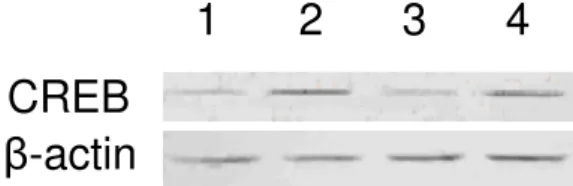
significant suppression of CREB expression in lung tissue at mRNA level. Consistently, protein level of CREB in lung tissue of hypoxia-exposed rats was significantly reduced by TEB compared with that of hypoxia/saline group (Figure 2).
Discussion
Pulmonary arterial hypertension is commonly complicated with many lung diseases such as cardiac obstructive pulmonary disease (COPD). It is evident
that the presence of chronic hypoxia is also associated with PAH, which plays a critical role in the pathogenesis of PAH with respect to myocardial dystrophy. Chronic hypoxia leads to excessive pulmonary vasoconstriction and right ventricular hypertrophy and thickening peripheral pulmonary vasculature. In the present study, rats exposed to hypoxic atmosphere showed an increase in mPAP accompanying with remodeling of vasculature. The findings are consistent with results of pervious preclinical and clinical researches supporting the statement that hypoxia induces high blood pressure and vascular remodeling response (5-8). Responding to hypoxia, a variety of cell residing in pulmonary artery such as smooth muscle cells and fibroblasts undergoes rapid expansion and contributes to vessel wall thickening (9, 10). Our histological data also support the idea that hypoxia induces inflammation and infiltration of leukocyte which contributes to the vascular thickening. We also found that serum level
of TNF- was elevated in response to chronic
hypoxia exposure, suggesting the presence of systemic inflammation. This finding is in agreement with previous studies in which hypoxia stimulated the release of inflammatory mediators such as nitric oxide and cytokines. Moreover, we observed that the changes in pulmonary vasculature have significant influence on pulmonary blood flow and right heart function resulting in hypertension and heart failure, in consistent with previous studies.
Cardiac sympathetic nerves are involved in the regulation of cardiac performance and peripheral circulation (11, 12). It is documented that the pulmonary vasculature is innervated by the autonomic nervous system. There is accumulating evidence indicating that chronic hypoxia induce sympathetic activity leading to high vasoconstriction
1
2
3
4
β
-actin
CREB
Figure 2. Effect of TEB on CREB expression in lung tissue of
hypoxia-exposed rats.Animals were exposed to either normoxia or hypoxia atmosphere for 3 weeks and treated with TEB or saline. After treatments, total protein extraction was carried out to determine CREB by immunoblotting.. Lane 1: normoxia; lane 2: hypoxia; Lane 3: hypoxia/ ropivacaine and lane 4: hypoxia/ saline. -actin was used as internal control. CREB: cAMP responding-element binding protein
Table 3. Effect of TEB on mRNA expression of CREB in lung tissue of hypoxia-exposed rats
Normoxia Hypoxia Hypoxia/ ropivacaine Hypoxia/ saline P
CREB mRNA 0.792±0.044 1.209±0.046 0.874±0.066 1.137±0.111 <0.005
(13-17). During hypoxia, oxygen sensitive chemoreceptors in the carotid body are stimulated leading to increased efferent sympathetic outflow in
humans (17, 18)). The resulting rise in
vasoconstrictor drive is considered to counteract hypoxia-induced vasodilatation and maintain arterial blood pressure (19, 20). Chronic exposure to hypoxia is attributed to persistent chemoreflex activation. Unlike hypobaric hypoxia to which human body is physically acclimated, chronic hypoxia associated with lung disease contributes to PAH due to a vicious cycle of sympathoexcitation in pulmonary arteries. Cardiac sympathetic blockade with thoracic epidural anesthesia has been reported to intervene the vicious cycle of myocardial dystrophy (18-20). Ishibe et al reported that sympathetic blockage by thoracic epidural anesthesia enhanced the haemodynamic changes in dog with lobar hypoxia (24). Similar findings have been reported using different models by other researchers (25, 26). Our study, for the first time showed that TEB improves not only hemodynamic changes but also the progression of vasculature remodeling. Our data revealed that TEB significantly improved mPAP and pulmonary vascular remodeling in rats exposed to chronic hypoxic conditions. It is suggested that TEB reduces hypoxia-induced sympathetic activation, resulting in an attenuation of PAH progression. In addition, we observed that TEB ameliorated changes in serum
levels of cGMP and TNF- . )t is indicated that TEB
treatment improves hypoxia-induced pulmonary vasoconstriction and inflammation contributing to worsening of PAH.
Cyclic AMP responding-element binding protein is a hypoxia-responsive transcription factor and the expression of CREB has been considered to indicate the degree of PAH regarding SMC proliferation. CREB is required for transcriptional activation of hypoxia inducible factor (HIF) in hypoxic background. Our results showed that CREB was upregulated in response to chronic hypoxia at mRNA and protein levels. The result showing elevated expression of CREB in lung tissue of hypoxic rats was ameliorated in the presence of ropivacaine, suggesting that TEB abolishes hypoxia-induced upregualtion of CREB with result of attenuated vascular remodeling.
Conclusion
By acting on the autonomic nervous system, TEB not only attenuates blood pressure but also reverses
vascular remodeling in hypoxia-induced PAH in vivo.
In addition, TEB can affect hypoxia-associated
inflammation. Collectively, we have presented in vivo
findings strongly suggesting TEB as a beneficial therapy with great potential for treating patients with hypoxic PAH.
Conflict of interests
No external funding and no competing interests declared.
References
1. Lin MJ, Leung GP, Zhang WM, Yang XR, Yip KP, Tse CM, Sham JS: Chronic hypoxia-induced upregulation of store-operated and receptor-operated ca2+ channels in pulmonary arterial smooth muscle cells: A novel mechanism of hypoxic pulmonary hypertension. Circ Res 2004; 95:496-505. 2. Yu L, Quinn DA, Garg HG, Hales CA: Cyclin-dependent kinase inhibitor p27kip1, but not p21waf1/cip1, is required for inhibition of hypoxia-induced pulmonary hypertension and remodeling by heparin in mice. Circ Res 2005; 97:937-945.
3. Soon E, Holmes AM, Treacy CM, Doughty NJ, Southgate L, Machado RD, Trembath RC, Jennings S, Barker L, Nicklin P, Walker C, Budd DC, Pepke-Zaba J, Morrell NW: Elevated levels of inflammatory cytokines predict survival in idiopathic and familial pulmonary arterial hypertension. Circulation 2010; 122:920-927.
4. Murray F, MacLean MR, Pyne NJ: Increased expression of the cgmp-inhibited camp-specific (pde3) and cgmp binding cgmp-specific (pde5) phosphodiesterases in models of pulmonary hypertension. Br J Pharmacol 2002; 137:1187-1194. 5. Yu L, Hales CA: Effect of chemokine receptor cxcr4 on hypoxia-induced pulmonary hypertension and vascular remodeling in rats. Respir Res 2011; 12:21. 6. Kanazawa H, Asai K, Nomura S: Vascular endothelial growth factor as a non-invasive marker of pulmonary vascular remodeling in patients with bronchitis-type of copd. Respir Res 2007; 8:22. 7. Calbet JA: Chronic hypoxia increases blood pressure and noradrenaline spillover in healthy humans. J Physiol 2003; 551:379-386.
8. Arias-Stella J, Saldana M: The terminal portion of the pulmonary arterial tree in people native to high altitudes. Circulation 1963; 28:915-925.
9. Stenmark KR, Fagan KA, Frid MG: Hypoxia-induced pulmonary vascular remodeling: Cellular and molecular mechanisms. Circ Res 2006; 99:675-691. 10. Frid MG, Brunetti JA, Burke DL, Carpenter TC, Davie NJ, Reeves JT, Roedersheimer MT, van Rooijen N, Stenmark KR: Hypoxia-induced pulmonary vascular remodeling requires recruitment of circulating mesenchymal precursors of a monocyte/macrophage lineage. Am J Pathol 2006; 168:659-669.
11. Fahim M: Cardiovascular sensory receptors and their regulatory mechanisms. Indian J Physiol Pharmacol 2003; 47:124-146.
12. Noll G, Wenzel RR, Binggeli C, Corti C, Luscher TF: Role of sympathetic nervous system in hypertension and effects of cardiovascular drugs. Eur Heart J 1998; 19 Suppl F:F32-38.
13. Ainslie PN, Ogoh S: Regulation of cerebral blood flow in mammals during chronic hypoxia: A matter of balance. Exp Physiol 2010; 95:251-262.
15. Neubauer JA, Sunderram J: Oxygen-sensing neurons in the central nervous system. J Appl Physiol 2004; 96:367-374.
16. Xie A, Skatrud JB, Puleo DS, Morgan BJ: Exposure to hypoxia produces long-lasting sympathetic activation in humans. J Appl Physiol 2001; 91:1555-1562.
17. Velez-Roa S, Ciarka A, Najem B, Vachiery JL, Naeije R, van de Borne P: Increased sympathetic nerve activity in pulmonary artery hypertension. Circulation 2004; 110:1308-1312.
18. Missant C, Rex S, Claus P, Derde S, Wouters PF: Thoracic epidural anaesthesia disrupts the protective mechanism of homeometric autoregulation during right ventricular pressure overload by cardiac sympathetic blockade: A randomised controlled animal study. Eur J Anaesthesiol 2011; 28:535-543. 19. Premkumar DR, Mishra RR, Overholt JL, Simonson MS, Cherniack NS, Prabhakar NR: L-type ca(2+) channel activation regulates induction of c-fos transcription by hypoxia. J Appl Physiol 2000; 88:1898-1906.
20. Rowell LB, Johnson DG, Chase PB, Comess KA, Seals DR: Hypoxemia raises muscle sympathetic activity but not norepinephrine in resting humans. J Appl Physiol 1989; 66:1736-1743.
21. Missant C, Rex S, Claus P, Derde S, Wouters PF: Thoracic epidural anaesthesia disrupts the protective mechanism of homeometric autoregulation during
right ventricular pressure overload by cardiac sympathetic blockade: A randomised controlled animal study. Eur J Anaesthesiol 2011; 28:535-543. 22. Svorkdal N: Treatment of inoperable coronary disease and refractory angina: Spinal stimulators, epidurals, gene therapy, transmyocardial laser, and counterpulsation. Semin Cardiothorac Vasc Anesth 2004; 8:43-58.
23. Kock M, Blomberg S, Emanuelsson H, Lomsky M, Stromblad SO, Ricksten SE: Thoracic epidural anesthesia improves global and regional left ventricular function during stress-induced myocardial ischemia in patients with coronary artery disease. Anesth Analg 1990; 71:625-630.
24. Ishibe Y, Shiokawa Y, Umeda T, Uno H, Nakamura M, Izumi T: The effect of thoracic epidural anesthesia on hypoxic pulmonary vasoconstriction in dogs: An analysis of the pressure-flow curve. Anesth Analg 1996; 82:1049-1055.
25. Rex S, Missant C, Segers P, Wouters PF: Thoracic epidural anesthesia impairs the hemodynamic response to acute pulmonary hypertension by deteriorating right ventricular-pulmonary arterial coupling. Crit Care Med 2007; 35:222-229.