Printed version ISSN 0001-3765 / Online version ISSN 1678-2690 http://dx.doi.org/10.1590/0001-3765201820180106
www.scielo.br/aabc | www.fb.com/aabcjournal
Effects of Aliskiren, an RAAS inhibitor, on a carrageenan-induced pleurisy model of rats
YASINBAYIR1, HARUN UN2, ELIF CADIRCI3, EROL AKPINAR3, BUSRA DIYARBAKIR3, ILKNUR CALIK4 and ZEKAI HALICI3
1
Faculty of Pharmacy, Department of Biochemistry, Ataturk University Campus, 25240, Erzurum, Turkey
2
Faculty of Pharmacy, Department of Biochemistry, Agri Ibrahim Cecen University Campus 04100, Agri, Turkey
3
Faculty of Medicine, Department of Pharmacology, Ataturk University Campus 25240, Erzurum, Turkey
4
Department of Pathology, Erzurum Region Education and Research Hospital, Center 25100, Erzurum, Turkey
Manuscript received on February 6, 2018; accepted for publication on May 15, 2018
ABSTRACT
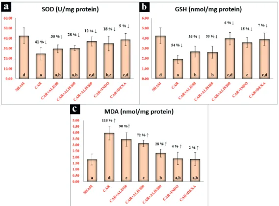
Our aim is to investigate the potentially preventive effects of Aliskiren in a carrageenan-induced lung pleurisy model and to compare the standard anti-inflammatory agents, indomethacin and dexamethasone. The pleurisy model was induced through the injection of carrageenan (0.2 ml-%2) into the pleural cavity. After the experiment, serum and lung tissues were collected and biochemical, molecular and pathological examinations were performed. In our study, pleural inflammation decreased superoxide dismutase activity and the glutathione level and increased the malondialdehyde level in the lung of rats, while Aliskiren increased the superoxide dismutase activity and glutathione level and decreased the malondialdehyde level. In addition, carrageenan-induced pleurisy caused a significant increase in pro-inflammatory cytokines mRNA expressions (TNF-α, IL-1β, and NF-KB), while Aliskiren administration decreased their expressions as well as the standard treatments, indomethacin and dexamethasone, did. Aliskiren administration at the 200 mg/kg dose protected the lungs in the pathological evaluation, especially against inflammatory cell infiltration and edematous lesions. It appears that Aliskiren protects the lung from carrageenan-induced pleurisy damage by regulating inflammation and antioxidant-oxidant balance via Renin Angiotensin Aldosterone System inhibition.
Key words: Aliskiren, cytokines, lung injury, oxidative stress, pleurisy, rat.
Correspondence to: Yasin Bayir E-mail: [email protected]
INTRODUCTION
Pleurisy is one the most prevalent inflammatory diseases and is characterized by an accumulation of an abnormal amount of pleural fluid in the pleural cavity (Hsieh et al. 2012). Various life-threatening diseases, such as tuberculosis, adenocarcinomas, pneumonia, and myocardial infarction, are
exudate and cellular migration with changes in other inflammatory parameters (Moore 2003), and is frequently used to detect the anti-inflammatory effects of pharmaceutical agents (Cuzzocrea et al. 2004).
The renin-angiotensin-aldosterone system (RAAS) is a hormone system, which plays a role in the regulation of arterial blood pressure (Te Riet et al. 2015). When blood flow is reduced, renin is secreted from the kidney into the circulation. The plasma renin level is directly responsible for the level of angiotensin I (Ag I), into which angiotensinogen is converted (Te Riet et al. 2015). Ag I is then converted into Ag II by the angiotensin converting enzyme (ACE) in the pulmonary surface and lung capillaries (Yee et al. 2010). Ag II plays major roles in the RAAS. These include arteriole constriction and multiple biological functions (Te Riet et al. 2015).
The expression of RAAS-related parameters in lung diseases, including a high level of ACE, activates the pulmonary RAAS (Hsieh et al. 2012, Yang et al. 2016). Previous studies examined the effects of the RAAS on pleurisy (Hsieh et al. 2012, Kim et al. 2016, Yang et al. 2016). Research showed that ACE levels played a pivotal role in the development of pleural effusions (Hsieh et al. 2012). RAAS blockers have anti-inflammatory effects and alleviate acute lung injury (Raiden et al. 2002).
Aliskiren (ALIS), an inhibitor of the renin, is currently used in antihypertensive treatment due to its effects on the rate-limiting step of the RAAS. A previous study reported that ALIS-induced inhibition of renin reduced organ failure in sepsis-induced lung injury (Akpinar et al. 2014). As RAAS blockers have anti-inflammatory effects and offer protection against pleurisy, the role of ALIS in pleurisy should be explored. However, no previous studies have investigated the effects of ALIS on CAR-induced pleurisy and lung injury.
We hypothesized that ALIS would inhibit the RAAS during pleurisy-induced lung injury and that it would concomitantly ameliorate inflammation and oxidative stress. Therefore, the aim of this study was to investigate the potential protective effects of ALIS in a model of CAR-induced lung injury and to compare its effects with that of standard anti-inflammatory drug treatments (i.e., indomethacin [INDO] and dexamethasone [DEXA]).
MATERIALS AND METHODS
ANIMALS
The study consisted of 42 male, albino Wistar rats aged 11 weeks old, each weighing 200–220 g. The animals were obtained from Ataturk University’s Experimental Animal Laboratory at the Medicinal and Experimental Application and Research Centre. The Institutional Animal Care and Use Ethics Committee of Ataturk University approved the study (Protocol number: 42190979-01-02/2232).
EXPERIMENTAL DESIGN
The rats were separated into the following seven groups, and each group consisted of six rats:
Group 1: Sham-operated control group
(SHAM)
Group 2: CAR-only treated group (CAR)
Group 3: CAR plus 50 mg/kg of ALIS, administered orally (CAR+ALIS 50)
Group 4: CAR plus 100 mg/kg of ALIS, administered orally (CAR+ALIS 100)
Group 5: CAR plus 200 mg/kg of ALIS, administered orally (CAR+ALIS 200)
Group 6: CAR plus 5 mg/kg of INDO,
administered orally (CAR+INDO)
Group 7: CAR plus 0.5 mg/kg of DEXA,
administered intraperitoneally (CAR+DEXA)
PLEURISY MODEL
(Sun et al. 1988), glutathione levels (GSH) (Sedlak and Lindsay 1968), and malondialdehyde levels (MDA) (Ohkawa et al. 1979) from each sample supernatant and standards were measured at room temperature in duplicate according to the modified methods with ELISA reader (Bayir et al. 2012). The results for the SOD, GSH, and MDA levels in the tissues were expressed as U/mg protein, nmol/ mg protein, and nmol/mg protein, respectively. All the data were presented as the mean ± standard deviation results per mg of protein.
PROTEIN DETERMINATION
The protein concentrations were determined by the Lowry Method, using commercial protein standards (Sigma Aldrich, Total protein kit-TP0300-1KT-(USA)).
MOLECULAR INVESTIGATIONS
Total RNA Extraction and cDNA Synthesis
Total RNA extraction and cDNA synthesis were performed according to our previous data (Albayrak et al. 2013). Tissues (20 mg) were briefly stabilized in an RNA stabilization reagent (RNAlater, Qiagen) and then disrupted using the TissueLyser II (2 x 2 minutes for lung tissues). Total RNA was purified using RNeasy Mini Kit Qiagen according to the instructions of the manufacturer in Qiaqube (Qiagen, Hilden, Germany). The RNA samples were reverse-transcribed into complementary DNA using a high-capacity cDNA reverse transcription kit (Applied Biosystem). From 10 µl, the total RNA was treated with 2 μl 10 X RT buffer, 0.8 μl 25 X dNTPs mix, 2 μl 10X RT random primers, 1 μl MultiScribe reverse transcriptase, and 4.2 μl DEPC-H2O. Reverse transcription was carried out at 25°C for 10 minutes, then at 37°C for 120 minutes, and, finally, at 85°C for 5 minutes using a Veriti 96 Well Thermal Cycler (Applied Biosystem). The cDNA concentration and quality were assessed Anesthesia was induced through intraperitoneal
administration of thiopental (20 mg/kg) and inhalation of 5% sevoflurane. ALIS (suspended in sterile saline) was given three times by oral gavage 3 days before the surgical procedure, and INDO (suspended in sterile saline) (1 ml, 5 mg/kg, oral) and DEXA (dissolved in sterile saline) (2 ml, 0.5 mg/kg, intraperitoneal) were given 30 min before the surgical procedure.
After anesthesia, the rats were subjected to a skin incision at the right sixth intercostal space. The underlying muscle was dissected, and CAR (0.2 ml, 2% prepared in sterile distilled water) was injected into the pleural cavity. The control group received vehicle administration for all procedures. The pleural incision was then closed using a 4/0 sterile synthetic suture in two layers. For all experimental protocols all animals received metamizole sodium, an analgesic, intramuscular injection at the dose of 50 mg/kg after operation. Four hours after the CAR injection, the animals were killed with an overdose of a general anesthetic (thiopental sodium, 50 mg/kg), and blood samples were collected from their hearts. The serum samples were immediately separated by centrifugation at 4,000 rpm for 10 min at 4°C and stored at -80° C until they were assayed. The lung tissues were then quickly removed and washed in ice-cold saline. Half the tissues were transferred to a biochemistry laboratory and stored at -80°C for biochemical and molecular analyses. The remaining tissues were fixed in a 10% formalin solution for histopathological analyses.
BIOCHEMICAL INVESTIGATION OF LUNG TISSUE
and quantified using the Epoch Spectrophotometer System and Take3 Plate (Biotek).
RELATIVE QUANTIFICATION OF GENE
EXPRESSION
Relative TNF-α, IL-1β, and NF-KB expression analyses were performed with StepOne Plus Real Time PCR System technology (Applied Biosystem) using synthesized cDNA from rat lung RNA. A qPCR was run using TaqMan Probe mix, Taqman Probe-based technology (Applied Biosystem). A real-time PCR was performed using primers generated for rat TNF-α Rn00562055_m1, rat IL-1β Rn00580432_m1, rat NF-KB Rn0399583_m1, and rat β-actin Rn00667869_m1. The results were expressed in relation to those associated with control animals. The expression data for β-actin in each tissue were used as endogenous controls. Primers and probes for β-actin were designed by Primer Design, Southampton, UK. For each tissue, triplicate determinations were performed in a 96-well optical plate for both targets using 9 µl of cDNA (100 ng), 1 µl of Primer Perfect Probe mix, and 10 µl of QuantiTect Probe PCR Master mix (Qiagen, Hilden, Germany) in each 20 µl reaction. The plates were heated for 2 minutes at 50°C and for 10 minutes at 95 °C. Subsequently, 40 cycles of 15 seconds at 94 °C and of 60 seconds at 60 °C were applied. All data were expressed as fold changes in expression compared to expression in other animal groups using the 2-ΔΔCt method (Livak and Schmittgen 2001).
HISTOLOGICAL PROCEDURES
Lung tissue samples were taken at necropsy and fixed in a 10% formaldehyde solution. Routine pathological processing was then performed, and the tissues were embedded in paraffin wax. From the paraffin wax block of each lung sample, 4–5 µm thick pieces were cut. After hematoxylin and eosin staining (H&E), a pathologist examined the prepared slides using light microscopy
using a digital camera (Olympus BX51, DP25 Olympus Optical, Tokyo, Japan). The following histopathological parameters were used for scoring: edema, emphysema, perivascular/peribronchiolar inflammation, thickness of the interalveolar septum, vascular congestion, and inflammatory cell infiltration. For each lung slide, a minimum of five fields was evaluated at 100× magnification, and the severity of changes was classified using a scoring system.
STATISTICAL ANALYSIS
For statistical analysis, SPSS, version 20.0 software was used. The results were presented as means ± standard deviation (SD). Comparisons between groups were performed using a one-way analysis of variance and Duncan’s multiple comparison tests. Statistical significance was accepted at p < 0.05.
RESULTS
BIOCHEMICAL RESULTS
activity and GSH level as compared with the CAR group. However, it was not possible to completely recover these parameters as in the sham group (p
< 0.05).
MOLECULAR RESULTS
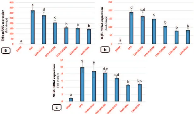
The expression of TNF-α, IL-1β, and NF-KB mRNA in lung tissues is shown in Figure 2. In the CAR group, TNF-α (Fig. 2a) and IL-1β (Fig. 2b ) mRNA expression significantly increased as compared with that in the sham group (p < 0.05). All doses of ALIS decreased both TNF-α and IL-1β mRNA expression as compared with that in the CAR group. The CAR+INDO, CAR+DEXA, and CAR+ALIS 200 treatments induced anti-inflammatory effects and significantly decreased TNF-α and IL-1β mRNA expression as compared with that in the CAR group (p < 0.05).
In the CAR-only group, NF-KB, expression increased significantly as compared with that in the sham group (Fig. 2c) (p < 0.05). While there was no significant difference between the CAR, CAR+ALIS50, and CAR+ALIS100 groups, ALIS200 treatments significantly decreased NF-KB expression compared to the CAR group (p < 0.05). In the NF-KB mRNA expressions, the most significant effects were shown in the CAR+INDO and CAR+DEXA groups compared to the CAR group (p<0.05).
HISTOPATHOLOGICAL RESULTS
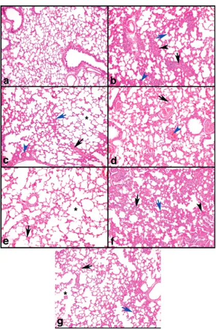
Lung tissues in the sham group showed a normal histological structure (Fig. 3a). The results of the histopathological assessment of the lungs in the different groups are presented in Table I. The CAR group showed severe emphysema, perivascular/ Figure 1 - Effects of Aliskiren treatments on SOD activity, GSH and MDA levels in rats’ lungs. SHAM:
Sham-operated, ALIS: Aliskiren 50-100-200 mg/kg, CAR: Carrageenan, INDO: Indomethacine, DEXA:
Dexamethasone. Means in the same column by the same letter are not significantly different to the test of
peribronchiolar inflammation, thickness in the interalveolar septum, inflammatory cell infiltration, moderate edema, and vascular congestion (Fig. 3b). The CAR+DEXA group had fewer lesions (Fig. 3c) than the CAR group. The CAR+INDO group showed a low to moderate number of lesions (Fig. 3d). In the treatment groups, the CAR+ALIS 200 group had fewer lesions (mild to moderate) (Fig. 3g) than the CAR+ALIS 50 (Fig. 3e) and CAR+ALIS 100 groups (Fig. 3f).
DISCUSSION
The present study revealed protective effects of ALIS in a CAR-induced pleurisy rat model. ALIS significantly alleviated lung injury of rats after CAR-induced pleurisy, and its effects were comparable to those of INDO and DEXA, standard anti-inflammatory agents. The findings suggest
that ALIS could be used to prevent lung injury by decreasing tissue damage, overexpression of tissue proinflammatory cytokines, and oxidative stress caused by pleurisy. Furthermore, they indicate that the protective effects of ALIS could result in RAAS blockage.
Previous studies of the association of the RAAS with the inflammatory process showed that modulating RAAS via renin inhibition prevented various conditions, such as ischemia-reperfusion and sepsis, associated with metabolic disorders (Akpinar et al. 2014, Un et al. 2015, Karcioglu et al. 2016). However, neither the role of renin inhibition in acute inflammation nor the role of inflammatory cytokines in inflammation-induced lung injury has been studied. During the inflammatory process, vascular permeability and leucocyte migration increase, followed by changes Figure 2 - Effects of Aliskiren treatments on relative mRNA expression levels of TNF-α, IL-1β, and NF-KB in rats’
lungs. Expressions of mRNAs were detected by quantitative real time PCR analysis. β-actin was used as the reference
gene. Results are expressed as relative-fold compared to sham animals. Gene-specific probes were used as outlined under Material and Methods. The relative expression levels were calculated by the ΔΔCT method. SHAM: Sham-operated, ALIS: Aliskiren 50-100-200 mg/kg, CAR: Carrageenan, INDO: Indomethacine, DEXA: Dexamethasone.
Means in the same column by the same letter are not significantly different to the test of Duncan (p<0.05). Results are
Figure 3 - a-g: Effects of Aliskiren treatments on light micrographs in rats’ lung tissues. a) SHAM group animal lung section
showed normal histologic features. Haematoxylin and eosin staining. Magnification, 100X. b) CAR group animal lung section
has severe edema (black arrow), Perivascular /Peribronchiolar inflammation (black arrow head), thickness in interalveolar septum and inflammatory cell infiltration (blue arrow) and vascular congestion (blue arrow head). Haematoxylin and eosin staining. Magnification, 100X. c) CAR+DEXA group animal lung section showed mild emphysema (asterisk), moderate edema (black arrow),
mild thickness in interalveolar septum and inflammatory cell infiltration (blue arrow) and mild vascular congestion (blue arrow head). Haematoxylin and eosin staining. Magnification, 100X. d) CAR+INDO group animal lung section showed moderate edema
in levels of proinflammatory cytokines (e.g., TNF-α and IL-1β), free radicals (e.g., superoxide), and inflammatory mediators (e.g., neutrophil-mediated reactive oxygen species) (Cadirci et al. 2016). Tissue activation by phagocytic cells results in the uncontrolled release of pathophysiologically proinflammatory cytokines, such as TNF-α (Zanotti et al. 2002, Cadirci et al. 2013). Previous research demonstrated that the aforementioned process aggravated inflammation and multisystem organ failure in an experimentally induced inflammation model (Cadirci et al. 2016). Studies also showed that Ag II, the most important effector of the RAAS, contributed to tissue repair and leucocyte accumulation from the blood circulation in interstitial tissues during the inflammation process (Touyz 2005). In a clinical study, researchers demonstrated increased levels of ACE, which converts Ag I into Ag II, in pleural effusions (Hsieh et al. 2012). Another study showed that increased Ag II levels were directly related to pleural inflammation and that losartan, an Ag II type-1 (AT1) receptor blocker, reduced the adverse effects of Ag II in pleural mesothelial cell culture (Yang et al. 2016). The same study showed that Ag II induced the activation of the NF-KB signaling pathway through pleural inflammation and that losartan decreased NF-KB mRNA expression in
pleural cells. In parallel with these results, the current study showed that the ALIS treatments decreased NF-KB levels in lung tissue in a CAR-induced pleurisy model. When the rats were treated with low- and middle-level doses of ALIS (50 and 100 mg/kg), there was no significant decrease in NF-KB mRNA expression in lung tissues. In contrast, the administration of 200 mg/kg of ALIS to rats with CAR-induced pleurisy decreased NF-KB expression in tissues to those of sham levels. These findings suggest that a high dose of ALIS can prevent CAR-induced lung injury.
The activation of NF-KB involves a major signal transduction pathway, which regulates the expression of genes related to inflammation. NF-KB regulates the expression of many cytokines, such as TNF-α and IL-1β, related to inflammatory responses, with TNF-α playing a major role (Schreiber et al. 1999, He et al. 2007). A previous study showed that losartan decreased TNF-α and IL-1β levels (Imai et al. 2005, Yuksel et al. 2015). Research also demonstrated that ALIS treatment reduced levels of TNF-α and IL-1β in acute lung injury (Akpinar et al. 2014). In accordance with these results, we showed that ALIS treatment decreased TNF-α and IL-1β mRNA expression in CAR-induced lung injury, with the strongest effect apparent in the 200 mg/kg treatment group. TABLE I
Histopathological assessments of lungs.
SHAM CAR CAR+ DEXA CAR+ INDO ALIS50CAR+ ALIS100CAR+ ALIS200CAR+
Edema 0 +2 +2 +2 +2 +2 +1
Emphysema 0 +3 +1 +1 +2 +1 +2
Perivascular / Peribronchiolar
inflammation 0 +3 +1 +1 +2 +3 +1
Thickness in interalveolar
septum 0 +4 +1 +1 +2 +2 +1
Vascular congestion 0 +2 +1 +1 +2 +2 +1
Inflammatory cell
The results of the present study also showed that the RAAS had an important impact on pleural inflammation and inflammatory mediators, in contrast to the findings of two studies in the literature (Raghavendra and Kulkarni 2000, Carvalho et al. 2006). In the first study, losartan treatment increased inflammation (edema volume). Researchers showed that TNF-α and IL-1β levels increased in the CAR-induced paw edema model, but they could not show the protective effect of losartan on TNF-α and IL-1β levels (Carvalho et al. 2006). In the second study, the researchers showed that losartan potentiated acute inflammation (Raghavendra and Kulkarni 2000). They attributed this potentiation to the stimulation of inflammatory pathways by unknown mechanisms. As demonstrated in the aforementioned studies, the inflammatory process includes many complex pathways and mechanisms, some of which remain unknown. Thus, the present study examined the potential involvement of Tα, IL-1β, and NF-KB in mechanisms underlying the inflammatory process.
Free oxygen radicals play important roles in the inflammation mechanism and tissue injury (Fink 2002, Un et al. 2015). Previous research showed that free radicals, such as oxygen radicals, hydrogen peroxide, and hydroxyl anions, increased during the inflammatory process (Tanas et al. 2010). Antioxidant molecules and drug treatments showed protective effects through the elimination of these free radicals (Halici et al. 2007, Karakus et al. 2013). Antioxidant defense systems include many enzymes, such as SOD and GSH, which limit the cytotoxic effects of free oxygen radicals (Fink 2002). Many previous studies demonstrated that levels of antioxidants decreased in CAR-induced pleurisy (Nardi et al. 2007, Cai et al. 2014). A number of studies also demonstrated a relationship between the RAAS and oxidative stress (Ziypak et al. 2015, Bayir et al. 2016, Halici et al. 2016). For example, Akpinar et al. showed
that an excessively high level of Ag II due to RAAS activation increased the level of MDA, which is the end product of oxidative damage of cell membrane phospholipids, and decreased SOD activity and GSH levels (Akpinar et al. 2014). In line with these results, in the present study, a high dose of ALIS (200 mg/kg) significantly decreased SOD activity and GSH levels in the CAR-induced inflammation group as compared with that in the sham group. In addition, MDA levels increased significantly in the high-dose ALIS treatment group. No significant differences in SOD activity, GSH level or MDA levels were apparent between the ALIS 200 group and INDO and DEXA groups. These results suggest that renin inhibition by ALIS provided protection against oxidative stress damage.
In the present study, the histopathological analysis of lung tissues revealed increased inflammatory cell infiltration, edema, vascular congestion, and vascular and interalveolar inflammation following CAR administration. In contrast, ALIS treatment decreased these effects, with the optimum protective effects observed at an ALIS dose of 200 mg/kg. treatment. In the current study, both the histopathological findings and cytokine mRNA levels pointed to protective effects of ALIS on lungs during inflammation.
RAAS inhibition in the first step in the suppression of inflammation, oxidative stress, and tissue injury in CAR-induced pleurisy in a rat model.
ACKNOWLEDGMENTS
This research supported by funding of Ataturk University (BAP-2014/170) scientific research project. The authors thank the Ataturk University for the opportunity to undertake this study, and allowing the use of the facilities within the university. Also all authors thank to School of Medicine, Molecular Pharmacology Laboratory team for their hard work on the development, data analysis and preparation of this article.
REFERENCES
AKPINAR E, HALICI Z, CADIRCI E, BAYIR Y, KARAKUS E, CALIK M, TOPCU A AND POLAT B. 2014. What is
the role of renin inhibition during rat septic conditions: preventive effect of aliskiren on sepsis-induced lung injury. Naunyn Schmiedebergs Arch Pharmacol 387: 969-978.
ALBAYRAK A, HALICI Z, CADIRCI E, POLAT B, KARAKUS E, BAYIR Y, UNAL D, ATASOY M AND DOGRUL A. 2013. Inflammation and peripheral 5-HT7
receptors: the role of 5-HT7 receptors in carrageenan induced inflammation in rats. Eur J Pharmacol 715: 270-279.
BAYIR Y ET AL. 2016. Aliskiren - a promising strategy for
ovarian ischemia/reperfusion injury protection in rats via
RAAS. Gynecol Endocrinol 32: 675-683.
BAYIR Y, KARAGOZ Y, KARAKUS E, ALBAYRAK A, SENGUL O, CAN I, YAYLA N, KUSKUN U AND KELES MS. 2012. Nigella sativa reduces tissue damage
in rat ovaries subjected to torsion and detorsion: oxidative stress, proinflammatory response and histopathological
evaluation. Gynecol Obstet Invest 74: 41-49.
CADIRCI E ET AL. 2013. Peripheral 5-HT7 receptors as a
new target for prevention of lung injury and mortality in septic rats. Immunobiology 218: 1271-1283.
CADIRCI E, HALICI Z, YAYLA M, TOKTAY E, BAYIR Y, KARAKUS E, TOPCU A, BUYUK B AND ALBAYRAK
A. 2016. Blocking of urotensin receptors as new target for treatment of carrageenan induced inflammation in rats. Peptides 82: 35-43.
CAI C, CHEN Y, ZHONG S, JI B, WANG J, BAI X AND SHI G. 2014. Anti-inflammatory activity of N-butanol extract from Ipomoea stolonifera in vivo and in vitro. PLoS One
9: e95931.
CARVALHO RF, RIBEIRO RA, FALCAO RA, LIMA RC, LEITAO RF, ALCANTARA C, SOUZA MH, CUNHA FQ AND BRITO GA. 2006. Angiotensin II
potentiates inflammatory edema in rats: Role of mast cell degranulation. Eur J Pharmacol 540: 175-182.
CUZZOCREA S ET AL. 1999. Role of IL-6 in the pleurisy
and lung injury caused by carrageenan. J Immunol 163: 5094-5104.
CUZZOCREA S ET AL. 2004. Rosiglitazone, a ligand of
the peroxisome proliferator-activated receptor-gamma, reduces acute inflammation. Eur J Pharmacol 483: 79-93. FINK MP. 2002. Reactive oxygen species as mediators of organ
dysfunction caused by sepsis, acute respiratory distress syndrome, or hemorrhagic shock: potential benefits of resuscitation with Ringer’s ethyl pyruvate solution. Curr Opin Clin Nutr Metab Care 5: 167-174.
HALICI Z, BILEN H, ALBAYRAK F, UYANIK A, CETINKAYA R, SULEYMAN H, KELES ON AND UNAL B. 2009. Does telmisartan prevent hepatic fibrosis
in rats with alloxan-induced diabetes? Eur J Pharmacol 614: 146-152.
HALICI Z, DENGIZ GO, ODABASOGLU F, SULEYMAN H, CADIRCI E AND HALICI M. 2007. Amiodarone
has anti-inflammatory and anti-oxidative properties: an experimental study in rats with carrageenan-induced paw edema. Eur J Pharmacol 566: 215-221.
HALICI Z, POLAT B, CADIRCI E, TOPCU A, KARAKUS E, KOSE D, ALBAYRAK A AND BAYIR Y. 2016.
Inhibiting renin angiotensin system in rate limiting step by aliskiren as a new approach for preventing indomethacin induced gastric ulcers. Chem Biol Interact 258: 266-275.
HE X, HAN B, MURA M, XIA S, WANG S, MA T, LIU M AND LIU Z. 2007. Angiotensin-converting enzyme
inhibitor captopril prevents oleic acid-induced severe acute lung injury in rats. Shock 28: 106-111.
HSIEH WY ET AL. 2012. ACE/ACE2 ratio and MMP-9
activity as potential biomarkers in tuberculous pleural effusions. Int J Biol Sci 8: 1197-1205.
IMAI Y ET AL. 2005. Angiotensin-converting enzyme 2
protects from severe acute lung failure. Nature 436: 112-116.
KARAKUS E, HALICI Z, ALBAYRAK A, BAYIR Y, AYDIN A, UNAL D, CADIRCI E, FERAH I AND ODACI E.
2013. Beneficial pharmacological effects of levosimendan on antioxidant status of acute inflammation induced in paw of rat: involvement in inflammatory mediators. Basic Clin Pharmacol Toxicol 112: 156-163.
KARCIOGLU SS, PALABIYIK SS, BAYIR Y, KARAKUS E, MERCANTEPE T, HALICI Z AND ALBAYRAK
KASS SM, WILLIAMS PM AND REAMY BV. 2007.
Pleurisy. Am Fam Physician 75: 1357-1364.
KIM J, LEE JK, HEO EY, CHUNG HS AND KIM DK. 2016.
The association of renin-angiotensin system blockades and pneumonia requiring admission in patients with COPD. Int J Chron Obstruct Pulmon Dis 11: 2159-2166.
LIVAK KJ AND SCHMITTGEN TD. 2001. Analysis of
relative gene expression data using real-time quantitative PCR and the 2(T)(-Delta Delta C) method. Methods 25: 402-408.
MOORE AR. 2003. Pleural models of inflammation: immune and nonimmune. Methods Mol Biol 225: 123-128.
NARDI GM, SIQUEIRA JUNIOR JM, DELLE MONACHE F, PIZZOLATTI MG, CKLESS K AND RIBEIRO-DO-VALLE RM. 2007. Antioxidant and anti-inflammatory
effects of products from Croton celtidifolius Bailon on carrageenan-induced pleurisy in rats. Phytomedicine 14: 115-122.
OHKAWA H, OHISHI N AND YAGI K. 1979. Assay for lipid
peroxides in animal tissues by thiobarbituric acid reaction. Anal Biochem 95: 351-358.
RAGHAVENDRA V AND KULKARNI SK. 2000. AT1
receptor antagonism enhances angiotensin-II-facilitated carrageenan-induced paw edema. Methods Find Exp Clin Pharmacol 22: 633-636.
RAIDEN S, NAHMOD K, NAHMOD V, SEMENIUK
G, PEREIRA Y, ALVAREZ C, GIORDANO M AND GEFFNER JR. 2002. Nonpeptide antagonists of AT1
receptor for angiotensin II delay the onset of acute respiratory distress syndrome. J Pharmacol Exp Ther 303: 45-51.
SCHREIBER S, NIKOLAUS S, HAMPE J, HAMLING J, KOOP I, GROESSNER B, LOCHS H AND RAEDLER A.
1999. Tumour necrosis factor alpha and interleukin 1beta
in relapse of Crohn’s disease. Lancet 353: 459-461. SEDLAK J AND LINDSAY RH. 1968. Estimation of total,
protein-bound, and nonprotein sulfhydryl groups in tissue with Ellman’s reagent. Anal Biochem 25: 192-205.
SIPAL S ET AL. 2012. Comparative study of three angiotensin
II type 1 receptor antagonists in preventing liver fibrosis
in diabetic rats: stereology, histopathology, and electron microscopy. J Mol Histol 43: 723-735.
SUN Y, OBERLEY LW AND LI Y. 1988. A simple method
for clinical assay of superoxide dismutase. Clin Chem 34: 497-500.
TANAS S, ODABASOGLU F, HALICI Z, CAKIR A, AYGUN H, ASLAN A AND SULEYMAN H. 2010. Evaluation of
anti-inflammatory and antioxidant activities of Peltigera rufescens lichen species in acute and chronic inflammation models. J Nat Med 64: 42-49.
TE RIET L, VAN ESCH JH, ROKS AJ, VAN DEN
MEIRACKER AH AND DANSER AH. 2015. Hypertension: renin-angiotensin-aldosterone system alterations. Circ Res 116: 960-975.
TOUYZ RM. 2005. Molecular and cellular mechanisms in
vascular injury in hypertension: role of angiotensin II. Curr Opin Nephrol Hypertens 14: 125-131.
UN H, BAYIR Y, HALICI Z, AKPINAR E, KARAKUS E, ORAL A, ZIYPAK T AND SELLI J. 2015. The Effects of RAAS Inhibition in Rate Limiting Step by Aliskiren
on Testicular Torsion Injury in Rats. J Urol 194: 828-833.
YANG J ET AL. 2016. Activation of calpain by
renin-angiotensin system in pleural mesothelial cells mediates
tuberculous pleural fibrosis. Am J Physiol Lung Cell Mol Physiol 311: L145-153.
YEE AH, BURNS JD AND WIJDICKS EF. 2010. Cerebral salt wasting: pathophysiology, diagnosis, and treatment. Neurosurg Clin N Am 21: 339-352.
YUKSEL TN, HALICI Z, DEMIR R, CAKIR M, CALIKOGLU C, OZDEMIR G AND UNAL D. 2015.
Investigation of the effect of telmisartan on experimentally induced peripheral nerve injury in rats. Int J Neurosci 125: 464-473.
ZANOTTI S, KUMAR A AND KUMAR A. 2002. Cytokine
modulation in sepsis and septic shock. Expert Opin Investig Drugs 11: 1061-1075.
ZIYPAK T ET AL. 2015. Renoprotective effect of aliskiren