other proteins contain a similar intriguing knot-like structure as in leptin and discovered 11 structurally homologous proteins in the PDB. We call this new helical family class the Pierced Lasso Bundle (PLB) and the knot-like threaded structural motif a Pierced Lasso (PL). In the current study, we use structure-based simulation to investigate the threading/folding mechanisms for all the PLBs along with three unthreaded homologs as the covalent loop (or lasso) in leptin is important in folding dynamics and activity. We find that the presence of a small covalent loop leads to a mechanism where structural elements slipknot to thread through the covalent loop. Larger loops use a piercing mechanism where the free terminal plugs through the covalent loop. Remarkably, the position of the loop as well as its size influences the native state dynamics, which can impact receptor binding and biological activity. This previously unrecognized complexity of knot-like proteins within the helical bundle family comprises a completely new class within the knot family, and the hidden complexity we unraveled in the PLBs is expected to be found in other protein structures outside the four-helix bundles. The insights gained here provide critical new elements for future investigation of this emerging class of proteins, where function and the energetic landscape can be controlled by hidden topology, and should be take into account inab initiopredictions of newly identified protein targets.
Citation:Haglund E, Sulkowska JI, Noel JK, Lammert H, Onuchic JN, et al. (2014) Pierced Lasso Bundles Are a New Class of Knot-like Motifs. PLoS Comput Biol 10(6): e1003613. doi:10.1371/journal.pcbi.1003613
Editor:Marco Punta, Wellcome Trust Sanger Institute, United Kingdom
ReceivedJanuary 23, 2014;AcceptedMarch 26, 2014;PublishedJune 19, 2014
Copyright:ß2014 Haglund et al. This is an open-access article distributed under the terms of the Creative Commons Attribution License, which permits unrestricted use, distribution, and reproduction in any medium, provided the original author and source are credited.
Funding:This work was supported by the Center for Theoretical Biological Physics sponsored by the NSF (Grant PHY-1308264) and by NSF-MCB-1214457. JNO is a CPRIT Scholar in Cancer Research sponsored by the Cancer Prevention and Research Institute of Texas. PAJ is supported by NSF PHY-1212312. JIS is funded by Foundation for Polish Science Homing Plus grant co-financed by the EU European Regional Development Fund. The funders had no role in study design, data collection and analysis, decision to publish, or preparation of the manuscript.
Competing Interests:The authors have declared that no competing interests exist. * Email: [email protected]
Introduction
The four-helix bundle is a common motif in nature [1,2,3,4] often used as a target for designed pharmaceutical and nutritional biomolecules [5,6,7]. The cytokine subfamily is a family of four-helix bundles that are soluble proteins secreted from different organs/tissues. Cytokines act as chemical messengers important in intercellular communication. They regulate differentiation, prolif-eration, activation and death of many cell types, with particular involvement in the regulation of the circulatory system and production of immunity and inflammatory responses [8]. Most four-helix bundles also have conserved cysteines within the motif, whose disulphide bonds help maintain their structure and stability [2]. Every protein containing a disulphide bridge forms a covalently closed loop. When the N- and C-termini are covalently linked you have the simplest knotted topology in mathematics, termed a ‘‘zero knot’’ [9]. The ‘‘zero knot’’ is present in the cytokine Interleukin-36 [10], theh-defensins as well as other lower
Figure 1A). This threading is reminiscent of the more common knotted proteins, where a protein terminal must thread across a twisted loop [15,16,17]. In the case of leptin, the structure is analogous to slipknotted proteins, where part of the protein adopts a hairpin-like configuration that threads across the covalent loop (Figure 1A). A slipknotted polypeptide geometry (topology) adds folding complexity that was unrecognized until recently [13,14,15,18,19,20,21]. Since the PL in leptin is distinct from knotted/slipknotted proteins, where the protein backbone ties a knot, and from the cystine knot that is created by at least three disulphide bonds [22,23,24,25], we called this new motif a Pierced Lasso Bundle (PLB, Figure 2A) [12]. This new class of proteins is distinct from previously classified cystine knotted proteins. In the PLB case, a closed covalent loop is created from a single disulphide bridge enclosing one of the terminals with one of the loops where part of the amino acid chain threads through and pierces the covalent loop. In the cystine knotted class, the added complexity beyond a closed loop is created by an additional side-chain mediated chemically cross-linked knot through the covalent loop [28].
PLBs, unlike cystine knots, are able to unfold their threaded elements. Furthermore, unlike knotted/slipknotted proteins, PLBs can modulate their complex topology based on the oxidation conditions of the disulphide bridge. Thus, breaking the bond/ contact between the two cysteines also breaks the covalent loop and thereby unthreads the structure. Because of the exciting functional consequences these dynamics may have, we searched for other proteins containing a similar PLB topology. A comprehensive search of the Protein Data Bank (PDB) found 11 structures with a similar threaded motif. Leptin has many structurally homologous proteins where disulphide bridges create a covalent loop, but only 11 had a threaded element through the covalent loop. Interestingly, there is a difference between leptin and the other threaded structures in terms of the location of the closed loop. The covalent loop in leptin is located at the C-terminal end while all other structures, found to date, are knotted at the N-terminal end (Figure 2A and B).
The threaded structure of leptin influences the Native State Dynamics (NSD) and thus the biological activity [12]. Here, we explore the effects of a C- versus N-terminal pierced lasso as well as the folding and the NSD in the related structures. Additionally,
we investigate the threading mechanism as the effect(s) of loop size. Structure Based Models (SBMs) were used to study thehumanand
murine interleukin 3and two zebra fish interferons(Figure 2B and C). Additionally, we compare the folding mechanism for three of the unthreaded four-helix bundles (the G-CSF, LIF and hGH, Figure 2B and C), which are members of the leptin family of long-chain helical cytokines. The results show that all PLB proteins stabilize the covalent loop as an initial step in folding (independent of an N- or C-terminal lasso). The disulphide bridge helps stabilize the secondary structure formation that builds the base of the lasso. Remarkably, leptin and mIL-3 mainly slipknot structural compo-nents through their lassos, whereas the remaining PLBs thread the C-terminal helix through the N-terminal lasso by a so-called plugging mechanism [26,27]. We provide, for the first time, direct evidence that the size of the covalent loop influences the threading mechanism. A small loop primarily uses a slipknotting route while the bigger loops are preferentially pierced by a plugging mechanism. In all cases, the N-terminal receptor-binding helix (helix A) is the last element to fold. All PLBs found to date, save leptin, have an N-terminal lasso that pins down the canonical helix A via a covalent linkage, while leptin’s helix A has freedom to
Figure 1. The topology of covalently linked loops in proteins.
Closed loops occur in proteins under oxidizing conditions when two cysteines are close enough in space to form a disulphide bridge. The size of the loop depends on the sequence separation between the cysteines forming the closed loop. The simplest knot in mathematics is called a ‘‘zero knot’’ (a closed circle). This occurs in proteins under oxidizing conditions when the N- and C-terminal residues are cysteines. In this case a complete circle will be formed by the polypeptide backbone. When the two cysteines are positioned in the middle of the sequence a so-called ‘‘cinch’’ is formed leaving the two terminals open. When one of the terminal residues is a cysteine a lasso like structure is formed. If there are any parts of the polypeptide chain piercing the covalent loop a pierced lasso is formed. The pierced lasso is colored blue in this figure. The white fragment demonstrates the threaded polypeptide chain and the red part is the terminal in front of the covalent loop.
doi:10.1371/journal.pcbi.1003613.g001
Author Summary
reorient and fray in the C-terminal PLB. This permutation from the more common N-terminal to C-terminal linkage of the PLBs results in an intriguing switch of the receptor binding helix A from tethered to dynamic and suggests that while the functional landscapes are shared in PLBs, variations in protein-receptor interface dynamics may be needed for signaling activity.
Results and Discussion
The cytokine family
Cytokines are soluble proteins secreted from different organs/ tissues that act as chemical messengers important in intercellular communication. All cytokines bind to a subset of homologous membrane bound receptors, activating similar intercellular signaling pathways [29,30,31]. The conserved cytokine motif, a four-helix bundle, indicates that the helical cytokines may have evolved from the same ancestral origin (Supporting Figure S1 and Supporting Table S1). Despite the structural identities, there are little or no sequence similarities within the family due to co-evolution, where each ligand and its specific receptor have diverged in sequence from its ancestors. Therefore, recognition by commonly used sequence homology methods is not possible
[32]. Instead, structural methods are used to classify these four-helix bundles as cytokines. Furthermore, all cytokines share a characteristic up-up-down-down fold, forming a two-layer packing of anti-parallel helix pairs were helix A and D packs against helix C and B. The superfamily of helical cytokines is divided into three families: long-chain helical cytokines, short-chain helical cytokines
and interferons/interleukin 10 (Figure 3) [32]. While the overall
geometry of the cytokines is conserved, there are differences in structure such as chain length and secondary structural elements (Supporting Table S1).
The PLB protein motif in leptin is a unique fold for proteins in general. A search of the PDB lead to the discovery of an additional 11 proteins with a similar threaded motif. Here, we compare leptin dynamics and threading to four PLBs, two Zebra fish interferons,
human (hIL-3) and murine (mIL-3) interleukin 3 (Supporting Table
S1). Additionally, three unthreaded four-helix bundles were investigated as controls, namely Granulocyte colony-stimulating factor
(G-CSF),Leukemia inhibitory factor(LIF) and human Growth Hormone
(hGH). Figure 2 displays the various structures as well as a cartoon describing the position of cysteines (yellow) creating the two types of lassos, i.e. the N-terminal loop (light blue) and the C-terminal loop (dark blue). The four canonical helices making up the core of
Figure 2. The unique topology of a Pierced Lasso Bundles (PLBs).(A) The native structure of leptin (PDB code 1AX8 [79]). A disulphide bridge is located between residue C96 and the C-terminal cysteine (yellow), creating a covalent C-terminal loop (dark blue). Consequently, a helical hairpin of helix C and half of helix B (white), have to thread through the covalent loop to reach the native state. The N-terminal part stays in front of the covalent loop (red). Hence, the overall conformation of the protein creates a Pierced Lasso Bundle (PLB) topology [12]. (B) Four of the new PLBs that we discovered in the PDB plus three unthreaded four-helix bundles are shown in ribbon diagram format. These new PLB proteins have their covalent loop at the opposite end from leptin, creating an N-terminal lasso (light blue). The threaded element (white) is behind the loop from this view while the C-terminal end (red) is in front of the covalent loop. (C) A cartoon defining the elements of the different PLBs showing the PL in blue (dark blue for
a C-terminal lasso and light blue for an N-terminal lasso). The threaded element, behind the covalent loop, is white and the terminal in front of the loop is red. The unthreaded foud-helix bundles are shown in white.
each protein are labeled A–D. Additional helices are numbered from the position in sequence; for example, the extra helix in leptin is the fourth helix in sequence and is labeled 49.
Structural alignment of the helical cytokines and comparison to leptin
The long-chain helical proteins. All four-helix bundles
within the long-chain helical cytokines have high structural similarity, from 2.45–3.78 A˚ rmsd compared to leptin. These proteins have low or no sequence identity/similarity (from 14.7/ 23.2% to 20.8/27.8%). All structures have an extra small helix in one of the loops outside the four-helix bundle. Leptin has the extra helix in the C–D loop while G-CSF, LIF and hGH have an extra helix in the A–B loop. Additionally, helix D, the longest helix, has a characteristic bend which is also seen in helix A, B and D for G-CSF, LIF and hGH. Leptin shows a small kink in helix D while the other helices are straight. G-CSF has five cysteines creating two disulphide bridges and one free cysteine, LIF has four cysteines creating one disulphide bond and two free cysteines and hGH has four cysteines forming two disulphide bridges. In all three cases the covalent loop created by the disulphides is ‘‘empty’’, i.e. without any threaded parts through the loop. The size of the ‘‘empty’’ loop varies from 6 residues in G-CSF to 126 residues in LIF. Overall, all the long-chain helical bundles are structurally similar even though leptin is smaller than the other members and has a threaded motif.
The Pierced Lasso Bundle (PLB) proteins. The PLBs have
a disulphide-mediated lasso where part of the structure is threaded through the covalent loop. This new class of proteins has low or no sequence identity/similarity (from 10.9/22.6% to 15.1/33.3%, Supporting Table S1) but high structural agreement, from 2.76– 4.10 A˚ (Supporting Table S1). We use four of the 11 new helical bundles (IFNw-1, IFNw-2, hIL-3 and mIL-3) to investigate the folding and threading route. The interferons are larger molecules with two extra helices, one short helix in the A–B loop and a long helix in the C–D loop, while theinterleukinsare shorter and have the extra helix in the A–B loop (Figure 2).Interferonsandinterleukins
are part of the cytokine subfamily but are classified as members of
the interferons/interleukin 10 family (Figure 3). These proteins are
part of the immune system acting as inflammatory response agonists/antagonists upon pathogen invasion or inflammatory response modulation [33]. There are four significant differences between leptin and all other PLBs (Figure 2): (1) The lasso is
formed by the N-terminal end in all PLBs save leptin, which uses the C-terminal end. (2) The size of the lasso varies from 50 residues seen in leptin to 122 in one of the LIFs (Supporting Table S1). (3) The threaded element has different lengths, varying from 10 residues in IFNw-1 to 45 residues in leptin. (4) Helix D is the threaded element in all PLBs save leptin, where the helix D is part of its covalent loop. The threaded element in leptin is instead a helical hairpin formed by helix C and part of helix B (Figure 2).
Folding mechanisms
Structure-based simulations were used to investigate the folding mechanism of the PLB proteins. Two different oxidation states were investigated for the disulphide bridge involved in the lasso, i.e. the reduced state (blue) and the oxidized sate (red) (details in Section Methods). Three unthreaded helical bundles were used as controls and their reduced states (DD, details in Section Methods) are plotted in black. Additionally, both the reduced and oxidized state of hGH were plotted as a control for an unthreaded structure, as it has a large ‘‘empty’’ covalent loop, where nothing is threaded through this element. The folding transition is monitored by the fraction of native contacts formed (Q) along the folding trajectory. A native contact is a contact formed between two residues that are close in the native state.Qvaries from 0, completely denatured, to 1, completely native. The folding mechanism is monitored via
q(segment), the fraction of native contacts formed by a secondary
structure element.q(segment)versusQshows the average number of
contacts a segment makes as a function of the overall folding progress, and therefore discerns the average order of events during folding. The results are plotted in Figure 4 and Supplementary Figure S2, S3, S4. The diagonal dashed gray line shows where q(segment) is tracking the overall folding progress. The boxes
represent the actual positioning of the covalent loop from Figure 2, where light blue represents the N-terminal loop, dark blue the C-terminal loop and gray the unthreaded structures.
Comparison of reduced and oxidized PLBs. The PLBs
have two different oxidation states of the disulphide bridge controlled by solvent conditions and the distance/space between the cysteines. In the case of PLBs, the structures lose the knot-like topology when the disulphide bond/contact is broken, meaning that the reduced state is actually an unknotted version of the same protein. Interestingly, the order of folding events (mechanism) of leptin is conserved between the two states despite the added complexity in the folding landscape for piercing the C-terminal
Figure 3. Schematic view of the helical cytokine sub-family.The cytokine sub-family (red) is divided into three families: The long-chain helical cytokines (pink), the short-chain helical cytokines (orange) and interferons/interleukin 10 (yellow). The overall topology within the cytokines is conserved with differences in length and topology, where theinterferonsandinterleukin10 have two extra helices outside the four-helix bundle and
the long-chain and short-chain helical cytokines have one extra helix. The figure shows the proteins discussed in the text, where threaded motifs (PLBs) are circled and unthreaded proteins are boxed. We note that there are many unthreaded examples as well as a few PLB proteins in each family, not shown in this figure.
lasso requirement [12]. The final step in folding of leptin is the formation of helix A (Figure 4). In contrast, the folding of the N-terminal PLBs is altered in the oxidized versus the reduced states (Figure 4 and Supporting Figure S2, S3, S4). For instance, the formation of helix A occurs earlier in the oxidized N-terminal PLBs (Figure 4). This change in kinetics is likely a result of the disulphide bridge pinning helix A to the four-helix bundle in the N-terminal PLBs, adding native contacts in the unfolded state at low values of Q. It is important to note that while the formation of helix A occurs earlier in the oxidized state, it is still the last element to be fully formed in all PLBs. A careful comparison of all the structural elements in the folding of the PLBs indicates that there are also changes in helix C as a function of oxidation state in the N-terminal PLBs (Supporting Figure S3, and S2 and S4 for helix B and D) although they are much smaller than the observed shift in Helix A upon oxidation of the covalent loop. As both helix A and C are part of the closed loop in the N-terminal PLBs, but only part of the threaded element in the C-terminal PLB leptin, the effect of oxidation/reduction is specific to the N-terminal PLBs.
Reduced PL bundles versus the unthreaded helical
bundles. The PLBs and unthreaded helical bundles are all
structurally similar upon reduction. That is, once the additional complexity afforded by the presence of threaded elements is removed by the breaking of the covalent loop, the proteins all
display same native topologies (Figure 2). In addition, while the absolute stabilities of the individual proteins vary depending on the size and overall buried surface area, all reduced proteins exhibit decreased stability relative to their oxidized counterparts. The unthreaded and the reduced four-helix bundles also form Helix A last, similar to what is observed for the N- and C-terminal PLBs. Taken together, these results indicate that unlike the b-trefoil cytokines which protect their functional motifs early in folding [34], the helical cytokines fold their receptor engaging Helix A in a late event, likely allowing remodeling during receptor engagement.
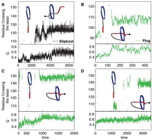
Piercing the lasso. Threading of a closed loop can occur by
two distinct mechanisms: (1) Slipknotting where a hairpin or loop threads through the closed loop or (2) Plugging the free end of the thread through the closed loop. To determine the dominant routes for threading of the oxidized PLBs, we monitored the residue(s) that crosses the covalent loop during folding. Leptin mainly uses a slipknotting mechanism [15,18] where the helical hairpin of helix C and part of helix B slipknots through the covalent loop [12]. The other possibility of a plugging mechanism is a rare event (less than 1% folding transitions) where the N-terminal plugs through the covalent loop (Supplementary Figure S5). All N-terminal PLBs have a larger covalent loop than leptin (from 63 residues in mIL-3 to 95 in the IFNs, Supporting Table S1). Similar to the dominant slipknotting event in the C-terminal PLB leptin, the N-terminal
Figure 4. Probability of the formation of helix A.The plot shows data for Q (a measure of nativeness, between 0–1)versusq(segment)(formation of a particular secondary structure, in this case helix A). The plots display data for reduced (blue) and oxidized (red) protein in the case of the PLBs and the reduced state (black, DD) for the unthreaded proteins (see method section for a full description). The plots are grouped in three boxes according to position in sequence of the covalent loop, dark blue for the C-terminal covalent loop, light blue for the N-terminal covalent loop and grey for the unthreaded proteins. The plots show that the formation of helix A is a late event on the folding landscape as it starts to build its contacts at high Q. The diagonal dashed grey line shows where q(segment)is tracking the overall folding progress. A significant shift is seen between reduced and oxidized N-terminal PLBs where the oxidized state folds faster than the reduced state.
PLB mIL-3 predominantly uses a slipknotting mechanism (Figure 5A) [35]. However, we observe that the N-terminal PLBs with covalent loops$68 residues, predominantly use a plugging mechanism where the C-terminal inserts itself through its covalent loop. Hence, as the loop becomes larger/bigger, the mechanism switches from a slipknot- to a plugging mechanism. This is the first evidence, to date, that directly demonstrates a change in threading mechanism based on covalent loop length in structurally homologous proteins. Smaller covalent loops form more local native contacts with hairpins or turns because these structures allow for building up interaction surfaces that provide the driving force to pierce the lasso via a slipknotting mechanism. Therefore, slipknotting is more likely to occur when threading smaller covalent loops. Increasing the size of the covalent loop increases the ‘‘open’’ space inside the lasso. This leads to a larger number of possible conformations capable of threading a terminal and there are more possible entry encounters with the larger loop that will be productive. As the size of the loop becomes even bigger the terminal is no longer restricted by topological constraints. However in contrast to a polymer [17,36], which will perform a random motion without forming any native contacts, the protein terminal will make native contacts on the other side of loop and be stabilized, leading to a knot-like native conformation. We use a native-centric model although threading also involves many native interactions. It has indeed been shown that including non-native interactions in the model can facilitate knotting [37] and
enhance the probability of threading via the entropically unfavorable plugging mechanism [38]. In the case of the smallest knotted protein it was shown by explicit solvent simulation that the energetic roughness along the slipknotting route is not greatly increased by the non-native interactions [39].
Native State Dynamics (NSD), N-loop versus C-loop The NSD for leptin together with in vitro activity assays revealed that the disulphide-bond plays an important role in controlling receptor binding [40] and thus biological activity by controlling local motions on distal receptor-binding sites far removed from the disulphide-bridge (Figure 6). These shifts are seen, for example, in helix A as well as in loop four, despite leptin is a C-terminal PLB [12]. To quantify the NSD for the PLBs we performed all-atom structure- based simulations far below the folding temperature, where the protein is effectively always in the folded basin. We calculated the essential dynamics, of the backbone, by projecting the trajectory onto the first four principle components. Oxidation has a significant effect on the amplitude of fluctuation of individual amino acids along the sequence for the PLBs due to topological constraints introduced by the threaded element [41] (Figure 7 and Supporting Figure S5). These modulations in fluctuations are not limited to the regions in the vicinity of the disulphide. Since the disulphide bridge mobilizes helix A, the dynamics of the N-terminal PLBs show the largest shifts. Both interferons show additional small
Figure 5. The threading mechanism of the N-terminal PLB proteins.(A) The threading mechanism for mIL-3 where slipknotting is the major
event (black). We also observe cases where the C-terminal region remains random and no apparent lasso crossing is observed (see cartoon). Panels B-D show the predominance of the plugging mechanism for hIL-3, IFNw-1 and IFNw-2, respectively. In this mechanism the C-terminal pierces through the N-terminal loop as is shown in the cartoon. As in (A) we also observe cases where the C-terminal region remains random and no apparent lasso crossing is observed (see cartoon).
increased NSD in the reduced state around the helical hairpin (loop 2, helix B and C), which is part of the closed loop. The
interleukins show increased dynamics in both terminals in the
reduced state. Indeed, in hIL-3 helix A even unfolds completely up on reduction. Taken together, the NSD data indicate that both the interferons and interleukins are more dynamic in the reduced state. However, leptin is unique in that it introduces increased dynamics in the oxidized state. As a control, we performed NSD for the empty covalent loop homolog protein, hGH. This protein shows no significant changes between the oxidized and reduced protein, with the exception of the expected increased local dynamics in the vicinity of the disulphide bridge upon reduction. Taken together, this data indicates that the observed long-range changes in dynamics in the PLBs are a direct result of the effect of a closed loop in the presence of a threaded element of the polypeptide chain. In the case of leptin, increased dynamics distal to the knot in the oxidized state is likely a consequence of the inability to relax the tension introduced into the vicinity of the small closed loop in the lasso. In the other cases discovered to date, local motions are enhanced by increasing the size of the loop and the expected reduction in dynamics upon disulphide bond formation are observed. That is, the simple formation of the closed loop effects only the local but not the global dynamics in the helical bundles, while the presence of a PL alters the global NSDs in both expected and unexpected ways.
Fraying of helix A and receptor binding
Frustrated surface regions have been proposed as sites relevant for allostery [42]. NSD and frustration in proteins have shown to be essential to protein function [43,44]. The conserved region for receptor binding within the cytokine family is helix A [45,46,47,48,49,50,51]. The late formation of helix A as well as the fraying of helix A in the reduced state of the PLBs implies that the dynamics is important to receptor binding and activity of the PLBs [12]. As an example, we show the receptor complex of leptin and the changed dynamics between the reduced and oxidized state in Figure 6. This suggests that the malleability of
the substrate and receptor interface has been conserved throughout evolution (Supporting Figure S1). More importantly, leptin has kept the malleability in helix A by forming its covalent loop at the C-terminal end. Interestingly, comparing the oxidized and reduced NSD data for leptin reveals a altered dynamics of helix A where the oxidized protein of leptin has greater dynamics then the reduced state.In vitroactivity assays showed that oxidized leptin is more active than the reduced protein, and supports that a dynamic structure is of importance for biological activity [12]. Nevertheless, all unthreaded helical bundles show reduced dynamic of helix A in general than the PLBs suggesting an overall more rigid structure. Our results suggest that a threaded topology is an important factor designating function. Here we focus on the knot-like four helix bundle class of proteins, however based on the current results we expect that other proteins will have a similar complex topology.
Future directions
We have found a new class of knotted proteins, namely the Pierced Lasso Bundles (PLBs). The PLB topology is defined as a four-helix bundle where a disulphide bridge closes a covalent loop, and part of the polypeptide chain is slipknotted/plugged through this covalent loop. All PLBs discovered, save leptin, have their covalent loop at the N-terminal end and plug the C-terminal helix through the covalent loop. In contrast, leptin mainly slipknots a helical-hairpin through its C-terminal loop. The closed loop also changes the dynamics of helix A which is important for activity and receptor interaction [45,46,47, 48,49,50,51]. The NSD reveals that oxidized leptin is more flexible than the reduced state, implying that dynamics in helix A is biologically important for leptin. Interestingly, sequence alignments of leptin homologs reveal that chicken and turkey leptin has three cysteines, one at the C-terminal end, one at position 100 and one at the N-terminal end [52,53,54]. Additionally, natural genetic polymorphism in bovine leptin has developed a sequence with a single cysteine substitution at the N-terminal end (R4C) [52,53,54]. This actually allows for three different combinations of covalent loops: (1) Closure of the
terminal covalent loop, as seen in wild-type leptin, forming a disulphide bridge between residue C96 and C146. (2) The formation of an almost completely circularized protein where residue C4 and C146 form a disulphide bridge, creating a ‘‘zero knot’’. (3) The formation of an N-terminal covalent loop where residue C4 binds to residue C96. In the latter case, the C-terminal helix could either slipknot or plug through the closed loop. Futurein vitro experiments can distinguish the three states from each other and the effect of a long N-terminal PLB (90 residues)versusthe shorter C-terminal PLB as well as investigate fully circularized ‘‘zero knot’’ protein as a template for understanding knot formation and threading control of function in the PLBs (Figure 1). The folding landscape of these three states of leptin could additionally be studied by traditional mutagenic analyses [55,56,57]. However, the analysis of the full landscape is complicated by the early threading of the covalent loop that occurs at the level of the transition state. While threading mechanisms have previously been investigated through Fluores-cence Resonance Energy Transfer (FRET) [58] this is not an
optimal technique in the case of leptin as the loop is 50 residues long and big probes could compromise the threading event. On the other hand, leptin is an optimal system for pulling experiments. For example, a major issue in the field is the inability to untie knots with denaturant [59]. In the case of leptin, simply reducing the disulphide bridge unthreads the structure and we are assured that we are comparing the energetics of the fully threaded and fully unthreaded states. Additionally, revers-ibly knotting proteins with pulling experiments is extremely complicated [60], while for leptin the experiment is straightfor-ward and can be used to investigate rate of loop threading. Nevertheless, understanding the topological constraints will lead to a broader understanding of the exotic shapes of the free energy landscapes in the growing class of knot-like PLB proteins. Moreover, one should point out that there are probably other undiscovered PL structures deposited in the PDB, where they proteins are bold up of b-strands and/or mixed a/b proteins where the loop is a cinch instead of a lasso. Finally, knowledge about topological constraints in the PLBs could increase the
Figure 7. Native state dynamics of leptin, hGH, IFN -1 and hIL-3.Structure based all-atom simulations were performed to obtain NSD. Data for reduced and oxidized protein are shown in blue and red, respectively. The overall fluctuations are shown as bar graphs and the difference between the two states is plotted as a yellow line. The protein sequence is displayed at the top of the graphs and a cartoon of secondary structures is displayed at the bottom (indicating the position of the N- versus the C-terminal loop in light blue and dark blue respectively). Leptin shows an interesting shift in dynamics where the oxidized state is more dynamic than the reduced state over the majority of the structure. The disulphide bridge, not only closes the covalent loop, but also acts as a point of tension inducing dynamics, far from the disulphide bridge. Helix A in leptin shows increased dynamics in the oxidized state, opposite to what is observed in the other PLBs. The decreased dynamics in helix A for the other PLBs is a result of the disulphide bride pining down helix A, thus restricting its dynamics. Interestingly helix A in hIL-3 completely unfolds in the native basin in the reduced state (the data dwarfs the effects on the rest of the sequence and is not included in this figure for clarity). hGH is has an ‘‘empty’’ covalent loop (a ‘‘cinch’’ like structure) of about 100 residues. The plot for hGH shows no significant change of the overall dynamics between reduced and oxidized protein, except for the expected local effect around the disulphide bridge. This implies that that the formation of the covalent loop alone has no effect on the NSD while the presence of a threaded topology piercing the lasso changes the entire protein dynamics.
doi:10.1371/journal.pcbi.1003613.g007
four-helix bundles (1RHG, 1EMR and 1HGU). Each amino acid is represented as a single bead and attractive interactions are given to residue pairs close in the native state. These native interactions are identified based on a shadow map [68,69]. The basic Hamiltonian is,
V(rij)~ X
bondsfijg
kb(rij{rNij)2z X
anglesfijkg
ka(hijk{hNijk)
2z
X
dihedralsfijklg
k1d 1{cos(Qijkl{QNijkl)2
h i
zk3d 1{cos(3(Qijkl{QNijkl))2
h i
z
X
native contacts
R(rij)zGij(rij)zR(rij)Gij(rij)z X
nonnative contacts R(rij)
R(rij)~e s
rij 12
Gij(rij)~{eexp { rij{rN ij 2
2w2
Native interactions have a repulsive term plus an attractive Gaussian term. The R(rij)Gij(rij) term is a correction that anchors the minimum of each contact -e(where the last two corresponds respectively to attractive and repulsive non-bonded interactions [70].rN
ij denotes the native distance between atomsiandjalong the sequence. The local topology of the chain is described by the native angles hNijk between the bonds connecting residue pairs ij
andjk, and by the native dihedralswNijklor torsional angles between the planes defined by atoms ijk and jkl. The strengths of the interactions are given in reduced energy units by the constants kb= 26104e/nm2, ka= 40e/rad2, k1d=eand k2d= 0.5e, wheree
is the reduced energy unit.S= 4 A˚ . The details of the model are characterised elsewhere [70,71].
Molecular dynamics
We used the web server SMOG (http://smog-server.org/) to create the input files for our simulations [66,68,69]. The GROMACS 4.5.3 package was used to perform the molecular dynamics simulations [72]. The integration steps were t = 0.005, stochastic dynamics with coupling constant 2 was used to maintain temperature. The apparent folding temperatures are estimated from each maximum peak in each specific heat curve. For a formed native contact the energy gain is measured by epsilon (e), and thus the temperatures and energies reported in this paper are
and LIF, black in all plots), see Haglundet alfor a full description of the states of the disulphide bridge [12]. The hGH was simulated as a control for covalent loop formation, as one of the disulphide bridges (C53–C163, forming a so called ‘‘cinch’’) forms a large ‘‘empty’’ covalent loop of 112 residues. This loop is classified as ‘‘empty’’ as no part of the polypeptide chain is threaded through the loop. This construct can help show the effects of the threaded element in PLBs.
Native State Dynamics (NSDs)
All-atom structure-based simulations [66,76] were performed to characterize the NSDs. To investigate the contribution of the threaded topology we performed simulations of both reduced and oxidized states for all PLBs as well as for hGH. A reduced state was simulated for the unthreaded structures (G-CSF and LIF). The slow component of the dynamics described by the first four eigenvector was analysed as described in Haglundet al[12].
Structural and sequence alignment
Supporting Information
Figure S1 Phylogenetic tree of the cytokines discussed
in the text. The figure shows the average distance tree using percentage identity (PID) on region from ClustalWS alignment of Retrieved from Uniprot. Data analyses indicates that these proteins emerged from a common ancestor (IFN, 2KZ1). The C-terminal PLB leptin is the newest member of the family. (TIF)
Figure S2 Probability of the formation of helix B. The
plot shows the same data as Figure 4 where reduced protein is shown in blue and oxidized protein in red. The unthreaded proteins show the reduced state in black (full description in method section). The plots are boxed from the position of the covalent loop with a threaded element, dark blue for the C-terminal loop, light blue for the N-terminal loop and grey for the unthreaded protein (same colours are used as in Figure 4). The formation of helix B is not affected by the threaded topology where all plots shows similar trends.
(TIF)
Figure S3 Probability of the formation of helix C.Plotted
in the same way as Supporting Figure S2. Helix C seems to be influenced by the formation of the N-loop for the N-terminal PLBs where helix C forms native contacts at lower values of Q. (TIF)
Figure S4 Probability of the formation of helix D.Plotted
in the same way as Supporting Figure S2. Helix D shows no significant changes between the oxidized and reduced states. (TIF)
Figure S5 The threading mechanism of C-terminal PLB
leptin.(Top) The two possible threading mechanisms are observed in simulations and shown in cartoon format. Slipknotting is the major event (black). We also observe a rare event where the N-terminal plugs through the covalent loop (green). (Bottom) A plot of the progress from unfolded to native (plotted as Q, 0 to 1, respectively) versus time indicates that a slip knotting event progresses more readily to the native state than a plugging mechanism.
(TIF)
Figure S6 Native state dynamics of IFNw-2, mIL-3 and
two unknotted four-helix bundles.Structure based all-atom simulations were performed to obtain NSD. Reduced and oxidized protein is shown in blue and red respectively. The overall fluctuations are shown as bar graphs and the difference between the two states is plotted as a yellow line. The protein sequence is displayed at the top the graphs and a cartoon of secondary structures is displayed at the bottom (indicating the position of the N- versus the C-terminal loop in light blue and dark blue respectively). All PLBs show an excepted shift around the disulphide bridge with additional increased dynamics in the reduced state, when you break the disulphide bridge. The N-terminal PLBs pin down helix A in the oxidized state which decreasing the dynamics significantly in this region.
(TIF)
Table S1 Structural alignment of leptin compared to
PLBs and unthreaded helical cytokines.We discovered 11 new slipknotted proteins in the cytokine subfamily and compared the structures to leptin: both slipknotted and unthreaded structures were aligned to leptin using the PDB-tool jFATCAT. The results are shown in Supporting Table S1. The cytokines share structural similarities such as the four-helix bundle and the up-up-down-down topology but they have no significant sequence similarity (from 8.7–15.2% sequence similarity compared to leptin). (DOCX)
Acknowledgments
We would like to thank Spencer Bliven for his help with structural alignment. Also, we want to thank Lannie Nguyen and Elizabeth Baxter for helpful discussions about the work and native state dynamic simulations (EB).
Author Contributions
Conceived and designed the experiments: EH JIS JKN HL JNO PAJ. Performed the experiments: EH. Analyzed the data: EH JIS JKN HL. Contributed reagents/materials/analysis tools: EH JIS JKN HL. Wrote the paper: EH JIS JKN HL JNO PAJ.
References
1. Beldarrain A, Lopez-Lacomba JL, Furrazola G, Barberia D, Cortijo M (1999) Thermal denaturation of human gamma-interferon. A calorimetric and spectroscopic study. Biochemistry 38: 7865–7873.
2. Vaz DC, Rodrigues JR, Sebald W, Dobson CM, Brito RM (2006) Enthalpic and entropic contributions mediate the role of disulfide bonds on the conformational stability of interleukin-4. Protein Sci 15: 33–44.
3. Brems DN, Brown PL, Becker GW (1990) Equilibrium denaturation of human growth hormone and its cysteine-modified forms. J Biol Chem 265: 5504–5511. 4. Bishop B, Koay DC, Sartorelli AC, Regan L (2001) Reengineering granulocyte colony-stimulating factor for enhanced stability. J Biol Chem 276: 33465–33470. 5. H K (2009) Modification of seed composition to promote health and nutrition. 6. Hill RB, Raleigh DP, Lombardi A, DeGrado WF (2000) De novo design of helical bundles as models for understanding protein folding and function. Acc Chem Res 33: 745–754.
7. Wei Y, Liu T, Sazinsky SL, Moffet DA, Pelczer I, et al. (2003) Stably folded de novo proteins from a designed combinatorial library. Protein Sci 12: 92–102. 8. Dinarello CA (2000) Proinflammatory cytokines. Chest 118: 503–508. 9. Adams CC (1994) The Knot Book: An Elementary Introduction to the
Mathematical Theory of Knots. New York: W. H. Freeman.
10. Dunn EF, Gay NJ, Bristow AF, Gearing DP, O’Neill LA, et al. (2003) High-resolution structure of murine interleukin 1 homologue IL-1F5 reveals unique loop conformations for receptor binding specificity. Biochemistry 42: 10938– 10944.
11. Craik DJ, Allewell NM (2012) Thematic minireview series on circular proteins. J Biol Chem 287: 26999–27000.
12. Haglund E, Sulkowska JI, He Z, Feng GS, Jennings PA, et al. (2012) The unique cysteine knot regulates the pleotropic hormone leptin. PLoS One 7: e45654. 13. King NP, Yeates EO, Yeates TO (2007) Identification of rare slipknots in
proteins and their implications for stability and folding. J Mol Biol 373: 153–166.
14. Sikora M, Sulkowska JI, Cieplak M (2009) Mechanical strength of 17,134 model proteins and cysteine slipknots. PLoS Comput Biol 5: e1000547.
15. Sulkowska JI, Sulkowski P, Onuchic J (2009) Dodging the crisis of folding proteins with knots. Proc Natl Acad Sci U S A 106: 3119–3124.
16. Mohazab AR, Plotkin SS (2013) Polymer uncrossing and knotting in protein folding, and their role in minimal folding pathways. PLoS One 8: e53642. 17. Virnau P, Mirny LA, Kardar M (2006) Intricate knots in proteins: Function and
evolution. PLoS Comput Biol 2: e122.
18. Sulkowska JI, Sulkowski P, Onuchic JN (2009) Jamming proteins with slipknots and their free energy landscape. Phys Rev Lett 103: 268103.
19. Norcross TS, Yeates TO (2006) A framework for describing topological frustration in models of protein folding. J Mol Biol 362: 605–621.
20. Yeates TO, Norcross TS, King NP (2007) Knotted and topologically complex proteins as models for studying folding and stability. Curr Opin Chem Biol 11: 595–603.
21. Sulkowska JI, Rawdon EJ, Millett KC, Onuchic JN, Stasiak A (2012) Conservation of complex knotting and slipknotting patterns in proteins. Proc Natl Acad Sci U S A 109: E1715–1723.
22. Craik DJ, Simonsen S, Daly NL (2002) The cyclotides: novel macrocyclic peptides as scaffolds in drug design. Curr Opin Drug Discov Devel 5: 251–260. 23. Trabi M, Craik DJ (2002) Circular proteins–no end in sight. Trends Biochem
Sci 27: 132–138.
24. Daly NL, Clark RJ, Craik DJ (2003) Disulfide folding pathways of cystine knot proteins. Tying the knot within the circular backbone of the cyclotides. J Biol Chem 278: 6314–6322.
35. Sulkowska JI, Noel JK, Ramirez-Sarmiento CA, Rawdon EJ, Millett KC, et al. (2013) Knotting pathways in proteins. Biochem Soc Trans 41: 523–527. 36. Raymer DM, Smith DE (2007) Spontaneous knotting of an agitated string. Proc
Natl Acad Sci U S A 104: 16432–16437.
37. Skrbic T, Micheletti C, Faccioli P (2012) The role of non-native interactions in the folding of knotted proteins. PLoS Comput Biol 8: e1002504.
38. a Beccara S, Skrbic T, Covino R, Micheletti C, Faccioli P (2013) Folding pathways of a knotted protein with a realistic atomistic force field. PLoS Comput Biol 9: e1003002.
39. Noel JK, Onuchic J.N. and sulkowska J.I. (2013) Knotting a Protien in Explicit Solvent. Journal of Physical Chemistry Letters 4: 3570–3573.
40. Carpenter B, Hemsworth GR, Wu Z, Maamra M, Strasburger CJ, et al. (2012) Structure of the human obesity receptor leptin-binding domain reveals the mechanism of leptin antagonism by a monoclonal antibody. Structure 20: 487– 497.
41. Sulkowska JI, Sulkowski P, Szymczak P, Cieplak M (2008) Tightening of knots in proteins. Phys Rev Lett 100: 058106.
42. Ferreiro DU, Hegler JA, Komives EA, Wolynes PG (2007) Localizing frustration in native proteins and protein assemblies. Proc Natl Acad Sci U S A 104: 19819– 19824.
43. Frauenfelder H, Sligar SG, Wolynes PG (1991) The energy landscapes and motions of proteins. Science 254: 1598–1603.
44. Marianayagam NJ, Jackson SE (2005) Native-state dynamics of the ubiquitin family: implications for function and evolution. J R Soc Interface 2: 47–54. 45. de Vos AM, Ultsch M, Kossiakoff AA (1992) Human growth hormone and
extracellular domain of its receptor: crystal structure of the complex. Science 255: 306–312.
46. Hill CP, Osslund TD, Eisenberg D (1993) The structure of granulocyte-colony-stimulating factor and its relationship to other growth factors. Proc Natl Acad Sci U S A 90: 5167–5171.
47. Smith DK, Treutlein HR, Maurer T, Owczarek CM, Layton MJ, et al. (1994) Homology modelling and 1H NMR studies of human leukaemia inhibitory factor. FEBS Lett 350: 275–280.
48. McDonald NQ, Panayotatos N, Hendrickson WA (1995) Crystal structure of dimeric human ciliary neurotrophic factor determined by MAD phasing. EMBO J 14: 2689–2699.
49. Feng Y, Klein BK, McWherter CA (1996) Three-dimensional solution structure and backbone dynamics of a variant of human interleukin-3. J Mol Biol 259: 524–541.
50. Yao S, Young IG, Norton RS, Murphy JM (2011) Murine interleukin-3: structure, dynamics, and conformational heterogeneity in solution. Biochemistry 50: 2464–2477.
51. Hamming OJ, Lutfalla G, Levraud JP, Hartmann R (2011) Crystal structure of Zebrafish interferons I and II reveals conservation of type I interferon structure in vertebrates. J Virol 85: 8181–8187.
52. Dridi S, Raver N, Gussakovsky EE, Derouet M, Picard M, et al. (2000) Biological activities of recombinant chicken leptin C4S analog compared with unmodified leptins. Am J Physiol Endocrinol Metab 279: E116–123.
8194.
60. Sulkowska JI, Sulkowski P, Szymczak P, Cieplak M (2010) Untying knots in proteins. J Am Chem Soc 132: 13954–13956.
61. Tutone M, Lauria A, Almerico AM (2014) Leptin and the OB-receptor as anti-obesity target: recent in silico advances in the comprehension of the protein-protein interaction and rational drug design of anti- obesity lead compounds. Curr Pharm Des 20: 136–145.
62. Garofalo C, Surmacz E (2006) Leptin and cancer. J Cell Physiol 207: 12–22. 63. Bluher S, Mantzoros CS (2007) Leptin in reproduction. Curr Opin Endocrinol
Diabetes Obes 14: 458–464.
64. Coppari R, Bjorbaek C (2012) Leptin revisited: its mechanism of action and potential for treating diabetes. Nat Rev Drug Discov 11: 692–708.
65. Friedman JM, Halaas JL (1998) Leptin and the regulation of body weight in mammals. Nature 395: 763–770.
66. Whitford PC, Noel JK, Gosavi S, Schug A, Sanbonmatsu KY, et al. (2009) An all-atom structure-based potential for proteins: bridging minimal models with all-atom empirical forcefields. Proteins 75: 430–441.
67. Andrews BT, Gosavi S, Finke JM, Onuchic JN, Jennings PA (2008) The dual-basin landscape in GFP folding. Proc Natl Acad Sci U S A 105: 12283–12288. 68. Noel JK, Whitford PC, Onuchic JN (2012) The shadow map: a general contact definition for capturing the dynamics of biomolecular folding and function. J Phys Chem B 116: 8692–8702.
69. Noel JK, Whitford PC, Sanbonmatsu KY, Onuchic JN (2010) SMOG@ctbp: simplified deployment of structure-based models in GROMACS. Nucleic Acids Res 38: W657–661.
70. Lammert H, Schug A, Onuchic JN (2009) Robustness and generalization of structure-based models for protein folding and function. Proteins 77: 881–891. 71. Clementi C, Nymeyer H, Onuchic JN (2000) Topological and energetic factors: what determines the structural details of the transition state ensemble and ‘‘en-route’’ intermediates for protein folding? An investigation for small globular proteins. J Mol Biol 298: 937–953.
72. Van Der Spoel D, Lindahl E, Hess B, Groenhof G, Mark AE, et al. (2005) GROMACS: fast, flexible, and free. J Comput Chem 26: 1701–1718. 73. Lammert H, Noel JK, Onuchic JN (2012) The dominant folding route
minimizes backbone distortion in SH3. PLoS Comput Biol 8: e1002776. 74. Ferrenberg AM, Swendsen RH (1988) New Monte Carlo technique for studying
phase transitions. Phys Rev Lett 61: 2635–2638.
75. Ferrenberg AM, Swendsen RH (1989) Optimized Monte Carlo data analysis. Phys Rev Lett 63: 1195–1198.
76. Baxter EL, Zuris JA, Wang C, Vo PL, Axelrod HL, et al. (2013) Allosteric control in a metalloprotein dramatically alters function. Proc Natl Acad Sci U S A 110: 948–953.
77. Narcis Fernandez-Fuentes JZ, Andra´s Fiser (2006) ArchPRED: a template based loop structure prediction server. Nucleic Acids Research 34: W173–6 78. Prlic A, Bliven S, Rose PW, Bluhm WF, Bizon C, et al. (2010) Pre-calculated
protein structure alignments at the RCSB PDB website. Bioinformatics 26: 2983–2985.
![Figure 1A). This threading is reminiscent of the more common knotted proteins, where a protein terminal must thread across a twisted loop [15,16,17]](https://thumb-eu.123doks.com/thumbv2/123dok_br/16426978.195702/2.918.469.790.89.492/figure-threading-reminiscent-knotted-proteins-protein-terminal-twisted.webp)