Assemblage B Isolate GS: Is Human Giardiasis Caused by
Two Different Species?
Oscar Franze´n1, Jon Jerlstro¨m-Hultqvist2, Elsie Castro3, Ellen Sherwood1, Johan Ankarklev2, David S. Reiner4, Daniel Palm3, Jan O. Andersson5, Bjo¨rn Andersson1, Staffan G. Sva¨rd2*
1Department of Cell and Molecular Biology, Karolinska Institutet, Stockholm, Sweden,2Department of Cell and Molecular Biology, BMC, Uppsala University, Uppsala, Sweden,3Centre for Microbiological Preparedness, Swedish Institute for Infectious Disease Control, Solna, Sweden,4The Burnham Institute for Medical Research, La Jolla, California, United States of America,5Department of Evolution, Genomics and Systematics, EBC, Uppsala University, Uppsala, Sweden
Abstract
Giardia intestinalisis a major cause of diarrheal disease worldwide and two majorGiardiagenotypes, assemblages A and B, infect humans. The genome of assemblage A parasite WB was recently sequenced, and the structurally compact 11.7 Mbp genome contains simplified basic cellular machineries and metabolism. We here performed 454 sequencing to 166
coverage of the assemblage B isolate GS, the onlyGiardiaisolate successfully used to experimentally infect animals and humans. The two genomes show 77% nucleotide and 78% amino-acid identity in protein coding regions. Comparative analysis identified 28 unique GS and 3 unique WB protein coding genes, and the variable surface protein (VSP) repertoires of the two isolates are completely different. The promoters of several enzymes involved in the synthesis of the cyst-wall lack binding sites for encystation-specific transcription factors in GS. Several synteny-breaks were detected and verified. The tetraploid GS genome shows higher levels of overall allelic sequence polymorphism (0.5 versus ,0.01% in WB). The genomic differences between WB and GS may explain some of the observed biological and clinical differences between the two isolates, and it suggests that assemblage A and BGiardiacan be two different species.
Citation:Franze´n O, Jerlstro¨m-Hultqvist J, Castro E, Sherwood E, Ankarklev J, et al. (2009) Draft Genome Sequencing ofGiardia intestinalisAssemblage B Isolate GS: Is Human Giardiasis Caused by Two Different Species? PLoS Pathog 5(8): e1000560. doi:10.1371/journal.ppat.1000560
Editor:William Petri, Jr., University of Virginia Health System, United States of America
ReceivedApril 3, 2009;AcceptedJuly 27, 2009;PublishedAugust 21, 2009
Copyright:ß2009 Franze´n et al. This is an open-access article distributed under the terms of the Creative Commons Attribution License, which permits unrestricted use, distribution, and reproduction in any medium, provided the original author and source are credited.
Funding:This study was supported by the Swedish National Research Agencies FORMAS (www.formas.se) and VR (www.vr.se). The funders had no role in study design, data collection and analysis, decision to publish, or preparation of the manuscript.
Competing Interests:The authors have declared that no competing interests exist.
* E-mail: [email protected]
Introduction
Giardia intestinalis (syn G. duodenalis and G. lamblia) is a major contributor to the enormous burden of diarrheal diseases, as causes of morbidity and mortality worldwide. The human prevalence rates range from 2–7% in developed countries to 20– 30% in most developing countries [1]. Infection of young farm animals is a major economical problem and G. intestinalis is a potentially zoonotic organism [2]. Nonetheless, the mechanism of giardial disease is poorly understood [3]. It is not invasive and secretes no known toxin and there is no general consensus on the cause of symptoms. However, recent data suggest that there is induction of apoptosis in intestinal epithelial cells during acute human giardiasis and that diarrhea is partly a result of increased intestinal permeability due to the apoptosis [4]. Chronic infections are common and in a hyperendemic area, 98% of drug-cured children are reinfected within six months [5]. On the other hand, about half of the infections are asymptomatic and frequently the infection spontaneously resolves [3]. Thus, both the duration and symptoms of giardiasis are highly variable.
Currently, there are seven defined genotypes (assemblages) ofG. intestinaliswith only assemblages A and B being known to infect humans. Although assemblage B is the most prevalent worldwide [2], it is inconclusive whether the various genotypes are associated
with different disease outcomes [6,7,8]. Difficulties with growth of
Giardia in vitroand the tetraploid genome [9] divided between two nuclei, have precluded the efficient use of biochemical, genetic and molecular biology approaches to experimentally correlate geno-typic differences with virulence. Only two Giardiaisolates (WB-assemblage A and GS-(WB-assemblage B) have been successfully cultured and studied in any detail at the molecular levelin vitro[1]. Early studies suggested large sequence differences between the genes of WB and GS since the nucleotide sequence in the coding region of the triose phosphate isomerase (tpi) gene showed only 81% identity between the WB and GS isolates and the non-coding regions were too different to be aligned [10]. Genetic differences between WB and GS have been confirmed in several other genes in more recent studies [11,12,13]. Several biological differences have also been identified between the WB and the GS isolates [14] and GS is currently the only Giardiaisolate that has been used successfully in experimental infections in humans [15] and adult mice [16]. It has even been suggested that assemblage A and B parasites should be considered as two differentGiardiaspecies [17]. Genome sequencing and comparative genomics can be used to identify genetic characteristics that are either unique or shared by all G. intestinalis assemblages and this approach has been used successfully for other protozoan parasites (e.g. Plasmodium and
recently published and it was shown to have a highly streamlined genome [20]. In order to understand in greater details the differences between Giardiaassemblage A and B we decided to produce a draft genome sequence of the GS isolate. We chose to use the 454 sequencing technology (Roche) to characterize the genome of GS, due to the rapidness of data generation and a read length long enough to enablede novoassembly of sequence reads. Since its launch in 2005, the 454 technology has [21] been successfully used in a number of genome sequencing projects, most notably in the resequencing of the human genome [22], the sequencing of a Neanderthal mitochondrial genome [23] and several bacterial species [24]. However, to our knowledge this is the first study to use the 454 technology to sequence the genome of a protozoan parasite.
We have produced a draft genome sequence of the assemblage B isolate GS, using a combination of de novo sequencing and resequencing, and compared it to the genome of WB. Our findings show only a few assemblage-specific genes, except for the Variable Surface Protein (VSP) gene family where the repertoires of the two isolates are completely different. This study has improved the annotation of the WBGiardiagenome and provided a framework for further experimental investigations of clinical and biological differences between assemblage A and BGiardiaisolates. Results
General features of theG. intestinalisGS genome The publishedG. intestinalis WB genome is 11.7 Mbp in size, distributed in 306 contigs on 92 scaffolds [20]. Originally, 6470 open reading frames (ORFs) were identified in the WB genome but only 4,787 were shown to be associated with transcription [20]. This is slightly less than the number of protein coding sequences found in the yeastSaccharomyces cerevisiae[25] but more than the number of coding sequences found in the intestinal, eukaryotic microbial pathogens Encephalitozoon cuniculiand Crypto-sporidium parvum [26,27]. Three rounds of sequencing of the GS genome using the 454 FLX sequencer generated 808,181
high-quality reads with an average length of 227 bp (182 Mbp; 166 coverage). The final assembled sequence was distributed in 2,931 contigs with an average length of 3,753 bp (Table S1). Automated ORF prediction identified 6,768 ORFs and manual curation of the data (see Methods) resulted in a final set of 4,470 intact (mean length 1,836 bp) and 221 interrupted protein coding genes. The mean intergenic distance was 130 bp. The number of protein coding genes is similar to what was observed in the WB genome and the intergenic distance is smaller due to annotation of an additional 754 open reading frames that were never annotated in the pioneering genome project. A small number of genes (64) were fragmented and in these cases specific primers were designed and the genes were amplified by PCR and sequenced. Fifty-eight of the fragmented genes were found to be disrupted by frame-shifts caused by single-base indels due to 454 sequencing errors and only 6 genes had actual frame-shifts (Table S2). Approximately 75% of the assembly is annotated as coding. However, the coding content is 90% in contigs with two or more genes annotated. The two genomes showed 77%65% nucleotide and 78%614% amino-acid identity in protein coding regions. The average GC-content was 46.5% in coding regions and 37.8% in intergenic regions. The codon usage was similar in the two isolates, but the codons in the GS ORFs have a higher level of A/T in the third positions compared to WB. We identified 124 small RNA genes in the GS genome, including tRNAs (Table S3 and Text S1). The GS isolate contains 69 tRNAs of the same number of isotypes (45) as the WB genome (Text S1). Most tRNAs are encoded by one gene, but the tRNAGln(TGG)gene, containing an intron, was found in 7 copies plus one copy without the intron.
Unique genes in WB and GS
It has been suggested that the WB and GS isolates belong to differentGiardia species [17] so we decided to study the protein coding capacities of the two isolates in order to address this issue. Comparisons between the sets of predicted protein coding genes showed that 673 WB genes lacked significant sequence similarity to any of the predicted GS genes. Searches at the nucleotide level identified conserved ORFs in GS corresponding to orthologs of 80 of the WB protein coding genes and these were therefore subsequently added to the GS annotation. Five WB protein coding genes showed sequence similarity to chromosomal GS regions without any corresponding full-length ORFs, which indicated the presence of pseudogenes in GS. Of the remaining 588 genes that lacked GS orthologs, 585 coded for proteins shorter than 200 amino-acids (aa) and lacked similarity to sequences present in the public databases. This suggests that these predicted proteins are most likely erroneous annotations, rather than unique WB proteins. Thus, surprisingly, only three WB genes coding for proteins longer than 200 aa are completely absent from the GS genome, all of which code for hypothetical proteins (Table 1). However, it should be noted that for this analysis members of largeGiardia-specific gene families, such as the VSPs, the ankyrin-repeat domain containing Protein 21.1, High Cysteine Membrane Proteins (HCMPs) and NIMA-Related Kinases (NEKs) were excluded.
For 754 of the predicted GS genes no annotated orthologs could be detected in the WB genome (www.giardiadb.org). However, searches against the WB scaffolds revealed that these have conserved ORFs, which were not annotated as coding sequences in the WB genome project. An additional four genes corresponded to WB chromosomal regions with putative pseudogenes. The remaining 59 protein coding genes did not show any significant sequence similarity to the WB genome, of these 23 code for proteins longer than 200 aa and are likely coding sequences (Table 1). An additional 4 sequences code for shorter proteins, but Author Summary
Giardia intestinalisis a major contributor to the enormous burden of diarrheal diseases with 250 million symptomatic infections per year, and it is part of the WHO neglected disease initiative. Nonetheless, there is poor insight into howGiardiacauses disease; it is not invasive, secretes no known toxin and both the duration and symptoms of giardiasis are highly variable. Currently, there are seven defined variants (assemblages) ofG. intestinalis, with only assemblages A and B being known to infect humans. Although assemblage B is the most prevalent worldwide, it is inconclusive whether the various genotypes are associated with different disease outcomes. We have used the 454 sequencing technology to sequence the first assemblage B isolate, and the genome was compared to the earlier sequenced assemblage A isolate. Large genetic differences were detected in genes involved in survival of the parasite during infections. The genomic differences between assemblage A and B can explain some of the observed biological and clinical differences between the two assemblages. Our data suggest that assemblage A and BGiardiacan be two different species. The identification of genomic differences between assemblages is indeed very important for further studies of the disease and in the development of new methods for diagnosis and treatment of giardiasis.
are located in proximity to longer unique genes, and a single gene shorter than 200 aa encodes a conserved hypothetical protein. We kept these and annotated them as functional proteins (Table 1). The remaining 31 short proteins without sequence similarity in the WB genome were deemed unlikely to represent functional genes and were therefore not considered further.
Thus, 28 protein-coding genes were found to be unique for the GS isolate. Although the majority of these genes code for hypothetical proteins, eight showed sequence similarity to genes present in the public databases. Four of these appear to be of
bacterial origin, since the best BLAST matches were to bacterial sequences, and they branched with bacterial genes in phylogenetic trees (Table 1; Fig. 1B; Fig. S1A,B,C). Another four showed similarity to a gene family associated with rolling circle replication and mostly found in viruses (Table 1; Fig. S1D). Homologs to these proteins of putative viral origin have previously been shown to be present in aG. intestinalisisolate BRIS/92/HEPU/1541 [28], but they are not present in the WB genome (Table 1). Interestingly, three additional gene families were detected among the unique genes coding for hypothetical proteins, resulting in a total of 19 Table 1.Unique genes in GS and WB.
Gene Isolate Gene family1 length/aa Contig E-value2 Annotation2 Organism2 Tree3
GL50803_3386 WB 528 AACB020000025
GL50803_4447 WB 259 AACB020000145
GL50803_101423 WB 248 AACB020000015
GL50581_2613 GS 270 2512 6E-69 Beta-lactamase domain
protein
Desulfatibacillum alkenivoransAK-01
S1A
GL50581_2037 GS 354 2425 4E-26 Conserved hypothetical
protein
Clostridium perfringens
B str. ATCC 3626
S1B
GL50581_2038 GS A 230 2425
GL50581_2039 GS A 158 2425
GL50581_2040 GS B 198 2425
GL50581_2041 GS 138 2425
GL50581_2042 GS 663 2425
GL50581_3192 GS 257 2785 1E-88 Conserved hypothetical
protein
Clostridium ramosum
DSM 1402
S1C
GL50581_3340 GS C 307 3001 2E-66 Putative
replication-accociated protein REP1
Giardia intestinalis
BRIS/92/HEPU/15414 S1D
GL50581_3339 GS D 244 3001
GL50581_3637 GS 1936 3065
GL50581_1168 GS 250 1703
GL50581_1169 GS 355 1703
GL50581_1170 GS 274 1703
GL50581_1171 GS 311 1703
GL50581_1633 GS D 244 2481
GL50581_1632 GS C 306 2481 4E-69 Putative
replication-accociated protein REP1
Giardia intestinalis
BRIS/92/HEPU/15414 S1D
GL50581_4567 GS 721 2481
GL50581_3038 GS A 252 2663
GL50581_3039 GS C 392 2663 2E-67 Putative
replication-accociated protein REP1
Giardia intestinalis
BRIS/92/HEPU/15414
S1D
GL50581_3319 GS A 222 2914
GL50581_180 GS B 182 2914
GL50581_3321 GS 150 2921 7E-42 Hypothetical protein
MS1399
Mannheimia succiniciproducens
MBEL55E
1B
GL50581_3333 GS C 335 2936 2E-18 Master replication protein Faba bean necrotic yellows virus (isolate SV292-88)
S1D
GL50581_3342 GS 304 3020
GL50581_7 GS 305 38
GL50581_62 GS 245 42
GL50581_100 GS D 237 120
1Indicates internal sequence similarity within the set of unique proteins.
2The best matches in BLAST searches against the non-redundant protein database at NCBI. 3Maximum likelihood phylogenetic trees found in the indicated figures.
4The best
non-Giardiamatches are against viral sequences.
5Contig names and from GiardiaDB, release 1.1 (http://www.giardiadb.org/giardiadb/).
genes and gene families that were only present in the GS/M-H7 genome (Table 1).
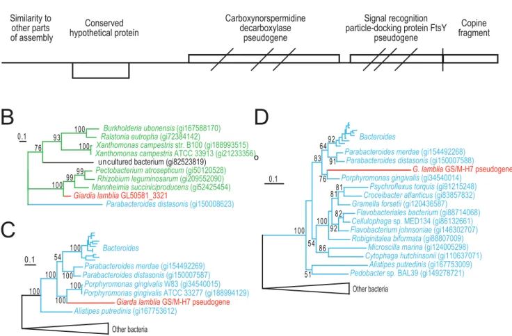
Recent introduction of a bacterial genomic fragment into the GS/M-H7 genome
To get a more detailed view of the process of gene acquisition in
Giardia genomes, we examined one short contig in more detail (contig 2921). This contig contained a single unique gene of bacterial origin (GL50581_3321, Fig. 1A). The 39 end of contig 2921 terminates in a truncated Copine-1 (CPNE1) gene, which suggested that the bacterial fragment has been inserted in the CPNE1 genomic environment (Fig. 1A). PCR and subsequent sequencing demonstrated that the rest of the truncated CPNE1 gene (GL50581_2716) is located in the end of contig 2545, which is syntenic with scaffold CH991778 in the WB genome. Thus, contig 2921 is linked to chromosomal regions showing strong similarity to the WB genome. Searches using the 59end of contig 2921 revealed strong similarities to multiple parts of the assembly (Fig. 1A), suggesting that the shortness of the contig is due to assembly errors caused by repetitive sequences.
Searches against protein databases using the sequence outside the annotated conserved hypothetical protein revealed sequence similarity to two bacterial genes coding for carboxynorspermidine decarboxylase and signal recognition particle-docking protein (FtsY), respectively (Fig. 1A). However, there are three frame-shift mutations in the carboxynorspermidine decarboxylase and theftsY
genes. Inspection of the assembled reads did not reveal any sequence ambiguities that could explain the apparent frame-shifts in these two genes; resequencing using the Sanger method confirmed this observation. In addition, the 39 part of the ftsY
gene is missing from the GS genome, indicating that it is a truncated pseudogene.
Phylogenetic analysis of the intact gene (GL50581_3321) within contig 2921 showed it nested within mostly proteobacterial sequences, most likely indicating a recent bacterial origin (Fig. 1B). Similarly, phylogenetic analyses of the reconstructed putative protein sequences for the two pseudogenes show that the
Giardia sequences group with Porphyromonas gingivalis sequences nested within members of the bacterial Bacteroidetes group (Fig. 1C,D). Indeed, these two genes are found in the same gene
o
Figure 1. Characterization of a recently introduced chromosomal region in GS.(A) Overview of contig 2921 showing identified genes and pseudogenes. Diagonal bars indicate approximate positions of frameshift mutations in the putative proteins. Vertical bar indicate putative deletion causing loss of the 39end of the FtsY gene and 59end of a gene coding for Copine-1. Arrows indicate positions of primers used in PCR reactions to connect contig 2921 with contig 2545. Protein maximum likelihood tree based on 125 unambiguously aligned position between the conserved hypothetical protein and all available homologs (B). Giardia sequence is shown in red, proteobacterial sequences in green, and sequences belonging to Bacteroidetes in blue. Protein maximum likelihood trees of carboxynorspermidine decarboxylase (C) and signal recognition particle-docking protein FtsY (D) based on 322 and 297 unambiguously aligned positions, respectively. The Giardia sequences were reconstructed based on alignments of the translation in all three reading frames to functional homologs of the pseudogenes. Only bootstrap support values above 50% are shown.
doi:10.1371/journal.ppat.1000560.g001
order as in the Porphyromonas genomes. This strongly suggests a recent transfer toGiardiaof these two genes in a single event from a close relative ofPorphyromonas, a genus of bacteria frequently found associated with humans [29]. The relatively long branches leading to theG. intestinalissequences suggest that the pseudogenes have accumulated several mutations in addition to the frameshifts observed (Fig. 1C,D). These observations show that contig 2921 of the G. intestinalis GS genome encodes three genes of recent bacterial origin, two of which likely became pseudogenes after the introduction into the genome.
Inspection of the assembly of contig 2921 showed that the average coverage was about half of the expected 16 times coverage. This could be due to random variations in genome coverage but it can also indicate that this region of the genome may not be present in all four copies of the genome in the cell. To test the latter hypothesis we designed PCR primers covering the part of contig 2921 harboring bacterial genes (Fig. 1A). Quanti-tative PCR analyses showed an approximate copy number of 0.3 for this part of the genome, compared to the single-copy gene beta-giardin (data not shown). This is in agreement with presence of the bacterial gene and pseudogenes in only 1 or 2 of the 4 chromosomal copies in the cell, which provide additional support for a recent introduction into the GS genome.
Higher frequency of allelic sequence heterozygosity in GS
Giardia is a tetraploid organism with two diploid nuclei [9]. Sequence divergence is expected to accumulate in polyploid organisms in the absence of genetic exchange. For example, extensive sequence divergence has been observed between the two former haplotypes in asexual bdelloid rotifers [30]. However, a surprisingly low level of allelic sequence divergence, less than 0.01%, was reported in the WB genome [20]. PCR analyses of genes from patient samples containing assemblage B isolates often show a high degree of sequence divergence in certain positions [2,31]. These observations could be caused by frequently mixed infections of different assemblage B lineages, or by a higher level of allelic sequence divergence. There are two different haplotypes of
tpicoding for triose phosphate isomerase in the GS isolate in the public databases. A comparison of these sequences with the sequence reads in the genome show the presence of two distinct classes of sequence reads (Fig. 2A), which strongly suggest allelic sequence variation in thetpigene. This allelic sequence divergence was also verified using PCR amplification and Sanger sequencing of individual clones (data not shown).
A genome-wide analysis of the presence of allelic sequence variation was performed on 8,618,167 positions with 106 coverage or more. A position was defined to contain an allelic sequence variation if two or more independent reads contained an alternative base compared to the consensus. The reads were classified as independent if they started at different positions. Insertion and deletion variation were not included because of the relatively high frequency of such sequencing errors using the 454 technology. Using these criteria, we detected 45,153 positions with two different bases, of which 22,655 were in coding regions and 22,498 were in non-coding regions. A strong bias towards transitions was observed (Fig. 2B). The average coverage for all positions with more than 10 independent reads and positions identified as variable are 16.3 and 18.8, respectively, which shows that the variation was not caused by the collapse of repeated genes in the assembly.
Allowing for a third variant with two or more independent reads detected 106 positions with three different bases. The average coverage for these positions is slightly higher (23.9), which suggests
that a fraction of these indeed could represent misassembled duplicated regions. No positions with all four nucleotides represented were found. Thus, the overall level of allelic sequence divergence was 0.53% in the GS genome, compared to less than 0.01% in the WB genome [20]. As expected, the allelic sequence divergence is higher in non-coding than in coding regions, 1.25% and 0.3%, respectively. This high frequency of allelic sequence variations suggests that the ‘‘double-peaks’’ observed in genotyping studies [31] could be explained by allelic sequence variation within a single infecting Giardia. The presence of a large number of positions with allelic sequence variation suggests that gene sequences may differ between the two nuclei in the cell. However, analysis of the ratio between the major and minor nucleotide in the variable positions indicate that ratios that deviate from 1:1 are common (Fig. 2C), which indicates that the two chromosomes within a single nucleus may differ.
Interestingly, the allelic sequence variations were not homo-genously distributed among the genes (Fig. 2D). Analyses of the distribution of the allelic variations along the contigs show large regions with very low sequence divergence with high divergence regions scattered within them (Fig. 2E–H). This indicates that a large part of the GS genome is identical between the four copies distributed in the two nuclei, while some parts remain divergent. Thirty-eight percent of the polymorphisms change the protein sequence and these are distributed in 1,962 genes, suggesting that GS has an extended proteome of almost 2000 proteins with a slightly altered primary structure. This can be important for understanding the biology and virulence of the parasite.
Synteny breaks and super-contigs
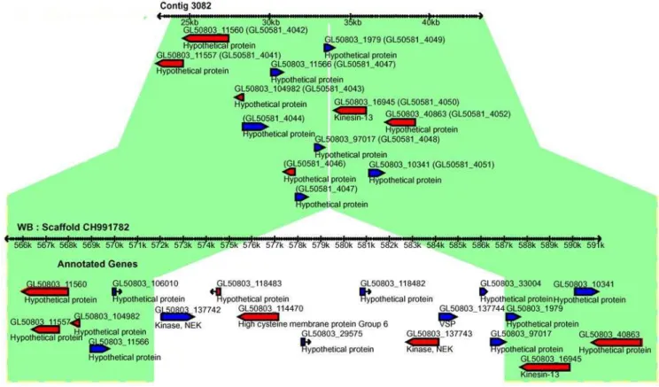
Several breaks of genome synteny (41) were identified when the GS contigs were aligned with the WB genome scaffolds (Table S4). Twenty-four of these were verified using PCR and sequencing and 21 of the breaks were between regions in the same WB-scaffold, with 3 occurring between scaffolds. The most common class among the synteny breaks (16 cases) was insertions or deletions of a region in WB or GS. One example of a synteny break in this class is shown in Fig. 3 where a 15 kb region containing 7 genes, among them one VSP, two NEKs and one HCMP are missing in GS. Indeed, VSPs, NEKs, HCMPs and also protein 21.1 were often found associated with insertions or deletions (12 cases). Addition-ally, VSP fragments were detected at contig edges in GS where the syntenic region in WB is devoid of such proteins. These two observations suggest that VSPs are not confined to certain genomic locations. Another interesting observation is that regions missing from GS but present in WB had a higher GC-content than the average for the genome (Fig. S2). The GC%-profiles of WB scaffolds reveal that the genomic regions where VSPs localize have a higher GC content than the surrounding genome and that these high GC% islands may be important for VSP regulation.
Intra-scaffold rearrangements were the second most prominent category of synteny breaks with 8 detected events. Of these, we detected a recombination event between two dipeptidyl-peptidase I precursors (GL50803_28651 and GL50803_22553) that creates ‘‘hybrid proteases’’. However, we could also detect non-recombi-nant variants by PCR analyses indicating that there are two different variants in the GS genome; one similar to WB and one due to recombination between the two protease genes.
This likely reflects a limitation due to the relatively short read-length obtained by 454 FLX sequencing (250 bp) compared to traditional Sanger sequencing.
The draft GS genome sequence is highly fragmented with 2,931 contigs of an average length of 3,753 bp. In order to see if it is feasible to generate larger contigs we designed primers against Figure 2. Characterization of sequence variations within GS.(A) Individual sequence reads of a portion of the triosephosphate isomerase (tpi) gene aligned to two previously reported sequences from the GS isolate. (B) Distribution in the six possible two-base combinations for the 45153 variable positions with two alternative bases represented among the individual sequence reads. (C) Analysis of the ratio between the numbers of independent reads for the major and minor base in the positions with two alternative bases. (D) Analysis of the frequency of positions with variations for individual genes. Analysis of the distribution of positions with variations along the four largest contigs in the assembly; contig 2890 (E), contig 2435 (F), contig 540 (G), and contig 1134 (H). Sliding windows of 2000 bp were analyzed in steps of 200 bp.
doi:10.1371/journal.ppat.1000560.g002
truncated genes and contig ends that were predicted to be close according to the synteny analysis. Twelve super-contigs of total size 1,363,697 bp, (corresponding to around 10% of the total genome) were produced after running 28 PCR reactions followed by Sanger sequencing of the products (Table S5). This shows that synteny analysis is useful for the generation of super-contigs and importantly that it is technically feasible to close the sequence gaps.
Promoters
Promoters in Giardiaare short (around 50 bp) and the main feature is an initiator-like AT-rich sequence around the ATG start codon, which is enough to drive transcription [32]. AT-rich stretches around the ATG start codon could be found in most GS genes (data not shown), but apart from these, there is very little intergenic sequence conservation, consistent with earlier observa-tions of a few GS genes. Encystation-specific promoters in WB are also short, with 65 bp found to be sufficient for a developmentally regulated promoter [33]. An alignment of the promoters (2100 to +3) from the three major cyst-wall proteins CWP 1–3 from WB with the orthologs from GS showed a high degree of conservation in the 65 bp directly upstream of the start codon (see alignment of CWP 1 and 2, Fig. 4). The transcription factor Myb2 has been shown to bind to the CWP promoters and its own promoter [34]. The GS Myb2 protein is well conserved (77% amino acid identity) and so is the 65 bp directly upstream of the ATG start-codon, including the Myb2 binding site (Fig. 4). This suggests that the cyst-wall proteins and Myb2 protein are regulated in the same way during encystation in the two isolates. The key-regulatory enzyme in WB, glucosamine-6 phosphate isomerase, has a promoter that is similar to the cyst-wall promoters [33]. However, the promoter of
this enzyme in GS is not similar to the promoters of the cyst wall proteins (Fig. 4) and, most importantly, it lacks a typical Myb2 binding sequence. The same was found to be true for the last enzyme in the pathway, UDP-N-acetylglucosamine 49 epimerase [35] (Fig. 4). The GS isolate is known for its poor encysting ability
in vitro[36] and may suggest that the regulation of cyst-wall sugar synthesis during early encystation is different in GS.
Introns and splicing
Four mRNA introns were identified in the WB genome after combining cDNA and genome sequences [20]. The introns are variable in length (32 to 220 bp) with conserved 59and 39motifs. Introns of similar sizes were found in the corresponding genes in GS and the 59(consensusG/CTAT GT)and 39motifs (consensus A/C CT A/G ACA/C CACAG) were conserved, whereas the sequences in the rest of the introns were highly diverged. Interestingly, we found three unique contigs containing the intron corresponding to the intron of ORF 35332 in WB. The longest contig (c2890) has a consensus 39motif, while the two other contigs (c299 and c305) have an A to G mutation in the branchpoint A that is crucial for splicing. Thus, these are three allelic variants of the intron and potentially pseudo-introns. Putative spliceosomal RNAs were recently predicted in the WB genome [37]. Only the U4 and U5 RNAs showed high sequence identity (92 and 85%, respectively) with GS, whereas the U1, U2 and U6 RNAs showed less than 80% identity (Table S3). This is surprising, considering that all other verified small RNAs showed a high level of sequence identity and suggesting that some of the predicted U RNAs are not true spliceosomal RNAs or that there are less constrains in the nucleotide sequence of the giardial spliceosomal RNAs.
Figure 3. Synteny-break in the GS genome.Comparison of genomic synteny between contig 3082 (upper panel) of the GS assembly and the syntenic region of scaffold CH991782 of the WB genome (lower panel). A 15 kb region is missing from the GS genome. This region contains 7 genes including one VSP, two NEK kinases and one HCMP. Syntenic regions in the two isolates are indicated in green.
Gene families: Variant-specific surface proteins (VSPs) Earlier studies using Southern blot analyses suggested that the GS genome contains approximately 150 VSP genes [38]. A search of all the reads from the GS data set using TBLASTN with the conserved C-terminal VSP region gave 3,183 hits with more than 80% identity over 30 aa. With a sequence coverage of 166we could estimate that 200 VSP genes are present in the GS genome. In our study only 16 complete VSP genes were obtained and most of them were short. The low number of identified full-length VSP genes is most likely due to assembly problems caused by the repetitive nature of these genes. The higher levels of allelic sequence divergence in GS most likely also caused problems in the assembly of the VSP genes. We used the 16 ORFs coding for complete GS VSP genes and 188 VSP genes in WB to search for VSP genes in the GS genome. In this search, 15,249 reads over 100 nt were identified, corresponding to 1.9% of all reads. None of these GS VSPs showed a high degree of similarity to VSPs from WB (30–70%, average 55%), except in the conserved CXXC motifs and the C-terminal region. This is in agreement with earlier studies that have suggested that the two isolates have unique VSP repertoires [38,39]. It is clear that more data using other sequencing platforms is needed to get a view of the complete VSP repertoire in GS.
Gene families: Kinases, HCMPs and alpha-giardins The largest gene family in the WB genome was the kinases with 276 putative members [20]. No histidine- or tyrosine-specific kinases were identified and only four giardial kinases contain membrane-spanning regions. The core kinome in Giardia is the smallest among eukaryotes thus far with more than 70% of the kinases in WB belonging to the NEK kinase group [20]. We found that certain NEK kinases were highly conserved between the two isolates, whereas others were highly diverged or missing. We identified 360 ankyrin motif-containing proteins in GS (Table S6),
with the number of repeats/protein ranging from 1–28. The ankyrin-repeats are commonly localized downstream of the kinase domain in NEKs or in multiple repeats in the 21.1 protein family, indicating that this is an important protein/protein interaction domain inGiardia.
The HCMP gene family [40] was discovered during the analysis of the WB genome and is similar to the VSP gene family, except for the absence of the conserved C-terminal CRGKA sequence. Twenty-one full-length HCMPs were identified in GS (Table S6) and many were not complete due to assembly problems. Similar to the NEK gene family, some of the HCMP proteins were highly conserved between WB and GS, while others are highly diverged or absent.
Alpha-giardins is a cytoskeleton gene family that is unique to
Giardiabut it is related to annexins [41]. All the 21 alpha-giardin genes in WB were conserved in GS along with the genome synteny. The amino acid identity of the alpha-giardins between the two genomes is between 70 to 95%, with the exception of alpha-7 giardin, which only displays a 57% identity. Alpha-2 giardin was recently proposed to be assemblage A specific [42]. We found an alpha-2 giardin-like gene in GS with 92% aa identity in a syntenic position but the alpha-1 giardin was less conserved with 87% aa identity and most of the changes were found in the N-terminal region.
Major cellular processes inGiardia
Giardia is a micro-aerophilic intestinal parasite with a very limited metabolic repertoire [43], containing no classical mito-chondrion, no Krebs cycle or nucleotide and amino acid synthesis genes or enzymes required forde novosynthesis of lipids [20]. Many key metabolic enzymes are bacterial-like, including the arginine metabolic pathway that is used for energy production in trophozoites [43]. Phylogenetic analyses indicate that these genes were acquired by the diplomonad lineage via lateral gene transfers Figure 4. Comparison of encystation-specific promoters. The promoters of the cyst-wall proteins-1 and 2 (CWP-1 and CWP-2), the encystation-specific transcription factor (gMyb2), the key enzymes involved in the synthesis of cyst wall sugars, glucosamine-6 phosphate isomerase (G6PI-B) and UDP-glucosamine-4 epimerase (UG4E) were aligned using the program CLUSTALW. Poly A rich initiator regions and gMyb2 binding-sites are underlined. Note that the G6PI-B and UG4E promoters are missing the gMyb2 binding-sites, whereas they are found in the corresponding positions in the promoters of CWP-1 and –2 and gMyb2.
doi:10.1371/journal.ppat.1000560.g004
from bacteria relatively recently, rather than being retained from a bacterial-like eukaryotic ancestor [20,44,45,46]. We found no differences in the metabolic gene content between the WB and GS isolates. There are 149 genes in the WB genome that have been identified as good drug targets [20] as defined by Hopkins and Groom [47], all these genes are also found in the GS genome. This suggests that drugs directed against these particular genes can have an effect on parasites from both assemblages.
One of the main observations of the WB genome project was the simplicity of major cellular machineries [20]. We investigated the same cellular processes studied in detail in the WB genome (e.g. DNA replication, transcription, polyadenylation and actin cytoskel-eton) [20], and came to the same conclusion for the GS genome, i. e.
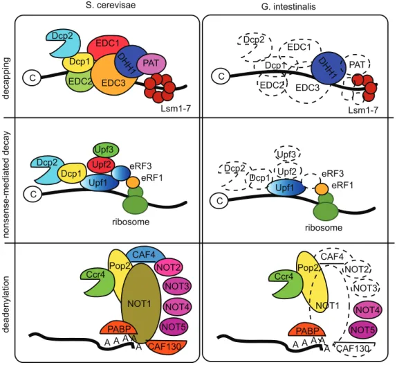
Giardiahas a minimal and simplified cellular composition. We also extended these analyses to several more basic cellular processes and we found the same pattern. One example is the rudimentary composition of the mRNA degradation system (Fig. 5). No decapping enzymes are present, but two typical 59 to 39 exoribonucleases were detected. The PARN and Pan 2–3 dead-enylation complexes were not detected, but weak homologs for a few proteins in the CCR4-Not complex were identified (Fig. 5). The catalytically important proteins Rpr-4 and -40 of the exosome
complex were identified, but only Rpr-45 of the ring structure. It was recently shown [48] that nonsense-mediated decay of mRNAs is present inGiardiabut at lower efficiency. The Upf-1 protein of the nonsense-mediated decay machinery was identified in both genomes, but Upf-2 and –3 and the other proteins found to be important for nonsense mediated decay in yeast and humans were not (Fig. 5), which may explain the low efficiency of the process in
Giardia. It was recently shown that the Upf-1 protein is an important regulator of the stability of the cyst-wall protein transcripts during encystations [48] and it will be informative to see how the nonsense mediated decay machinery is composed inGiardia.
An analysis of the WB genome showed no evidence of true myosin genes [20], suggesting that cytokinesis is not performed by an actomyosin ring inGiardia. However, homologs to actin and the mitotic cyclins A and B were detected in both Giardia isolates. Furthermore, we identified giardial homologs to several Mitotic Exit Network (MEN) proteins; Tem1, Cdc5, Cdc14, Cdc15, Bub2 and Mob1 as well as two members of the related FEAR complex; the kinase Cdc5 and the protease Esp1 (see Fig. S3). Our results suggest that the regulation of cytokinesis inGiardiais similar to the process in other eukaryotes, even if no strong myosin ortholog has been identified [20].
Figure 5. mRNA degradation pathways in Giardia and yeast. The major mRNA degradation pathways in Giardia compared to the corresponding pathways in yeast. Decapping: Decapping in S. cerevisiae involves the decapping enzymes Dcp1–2, EDC1–3, DHH1, PAT and Lsm1-7. In Giardia only DHH1 and three Lsm proteins were identified. Nonsense-mediated decay: The process involves Dcp1-2, Upf1-3 and eRF1 and 3 in yeast. Giardia has only orthologs to Upf1 and eRF1. Deadenylation: Giardia lacks the PARN and Pan2/Pan3 systems. In the CCR4-NOT complex only Ccr4, Pop2, NOT4 and 5 and PABP are found.
Discussion
In this study, we have produced a draft genome sequence of the assemblage BGiardiaisolate GS. The sequence has numerous gaps, which makes it less complete than the published genome ofGiardia
WB. Given that approximately 12–13% of the WB genome is composed of repeated gene families dispersed over the genome, and that such regions are difficult to assemble with short reads, it is likely that most of these gaps represent repeat regions. This is also supported by comparing the genomic synteny of the two data sets. We found that most contig ends had an interrupted gene sequence belonging to one of the large gene families inGiardia(Protein 21.1, NEKs, VSPs or HCMPs). However, our strategy has resulted in reasonably long contigs that provide sufficient information for extraction of the core gene content. Our proof of concept for this shows that we indeed identified orthologs to all annotated protein-coding genes from non-repeated WB gene families in the GS assembly, except 3 (Table 1). This study further supports the concept that for most purposes, a ‘‘quick and dirty’’ approach is sufficient for comparative genomics to be highly informative [49]. This type of strategy is particularly useful for resequencing closely related strains and isolates where a high quality reference sequence is available. Using this approach, we identified several genetic and genomic differences between the GS isolate and the previously sequenced assemblage A isolate WB: (1) unique, isolate-specific proteins, (2) unique patterns of allelic sequence divergence, (3) differences in genome synteny, (4) differences in the promoter regions of encystation-specific genes and (5) differences in the VSP repertoires. These multiple variations may help explain many of the known biological differences between the GS and WB isolates.
Unique proteins
Lateral gene transfer is increasingly appreciated as an evolutionary mechanism in microbial eukaryotes [50,51], and metabolic adaptations via gene acquisitions have been shown to occur in diplomonads on longer evolutionary timescales [20,44,45,46]. The comparative study identified 3 unique WB and 28 unique GS proteins, suggesting that gene loss or gain is ongoing withinGiardia. This is in agreement with earlier findings, although the rate of the process appears relatively low. Although most unique proteins are hypothetical, there are also examples of recently introduced bacterial genes in the GS genome. One example of this is a protein coding gene flanked by two bacterial pseudogenes, indicating a very recent introduction of a member from the Bacteroidetes group. To our knowledge, this is the first report of a ‘‘dead-on-arrival’’ prokaryotic pseudogene incorporat-ed into a eukaryotic nucleus. Until now no isolate-specific genes have been identified inGiardia, and further studies will show what functions these genes have in the parasite. These unique genes may be of importance for the development of new tools for diagnosis and typing ofGiardia. If strain-specific genes from each assemblage are expressed at relatively high levels in trophozoites and cysts in all isolates, it may be feasible to develop antibody-based genotyping/detection assays. The role of these unique genes during host-parasite interactions will also be of great interest for future studies.
Allelic sequence divergence
One surprising result from the WB genome project was the low level of allelic sequence divergence (,0.01%) [20]. We detected a dramatically higher level of allelic sequence divergence in the GS isolate (average 0.5%). Our analyses indicate that the sequence divergence between haplotypes for most genes is much smaller than the divergence between the genes from the two isolates
(Fig. 2D). This contradicts a recent report where genes classified into both assemblage A and B were found in the same Giardia
isolate [11]. In that study, the GS isolate was shown to contain actin genes from both assemblages and certain intergenic regions showed.99% identity to the corresponding region in WB. We failed to identify any of these reported assemblage A-type GS sequences [11] in our 166 coverage whole genome shotgun sequencing dataset. Unfortunately, the level of genetic exchange between Giardia assemblages cannot be determined until more genomic datasets fromGiardiaisolates become available.
The observed allelic variations are likely the result of interactions between mechanisms that create and reduce variation between the four copies of the genome in the cell. The major sources of variations are probably mutations and DNA recombi-nation, which has been proposed to occur between different
Giardiaisolates [52]. It is not likely that the mutations have been induced by genetic drift during asexual mitotic growthin vitrosince this is the original clone isolated by Nash et al. [17] and it has been grown relatively few generations in vitro. There are also other mechanisms that could change the level of genetic variation between the four copies of the genome. Diplomixis, a recombi-nation process betweenGiardia’s two nuclei, shown to occur during encystation [53], could be an important mechanism. This is a unique process forGiardiawith its two nuclei and genes related to meiotic processes in other organisms were suggested to be important in this process [53,54]. All identified meiosis-related genes identified in WB [55,56] can be found in GS. Several are well conserved (Spo11–78% aaID, Dmc1–92% aaID, Msh6–78% aaID), others are not as conserved (Mre11–62%, Rad50–60%) and some even show deletions (15 aa in Rad52) or insertions (14 aa in Mlh1) in important regions. The higher levels of allelic sequence divergence in GS could suggest that diplomixis is less efficient in the GS isolate compared to the WB isolate, although further investigations are needed to separate the effects of different mechanisms creating and reducing allelic variation in Giardia
isolates.
Synteny breaks
We identified and confirmed several breaks of gene synteny when the GS contigs were aligned with the WB genome scaffolds. Twenty one of the synteny breaks occur between regions in the same WB-scaffold with 3 occurring between different WB scaffolds. The most common class of synteny breaks was insertions or deletions of chromosomal regions containing members of the large Giardia-specific gene families VSPs, NEKs, HCMPs and Protein 21.1. In at least two cases we detected two different variants of gene synteny; one identical to WB and one unique for GS. These recombination events occurred between two very similar genes localized within 15 to 30 kbp of each other. It is possible that these kinds of inversions between similar regions are more common than what we have detected here and it is also possible that there are differences between the two nuclei.
Encystation-specific promoters
potentially explain the poor encystation observed in GS, and furthermore suggests that the encystation stimuli may be different between assemblage A and B parasites. In order to further understand the epidemiology of giardiasis, it will be important to determine if there is a correlation betweenGiardiaassemblages and cyst production in either infected humans or animals.
VSP repertoires
The most well characterized virulence factors inGiardiaare the VSP proteins [14,59]. The genome sequencing of WB found approximately 200 genes encoding different VSPs spread over the 5 chromosomes [20]. Our results showed that the VSP repertoires are very different in GS compared to WB. Certain VSPs are known to have toxin-like motifs [60] and it is possible that the differences in symptoms seen duringGiardiainfections are not due to assemblage differences, but rather because of differences in VSP repertoires and expression.
Are humans infected by two different Giardiaspecies? One major issue in the Giardia research field has been the identification of genetic differences between the two human-associated Giardia assemblages A and B that would explain the observed phenotypic differences. Early genetic studies suggested that the levels of genetic diversity between the assemblage A and B parasites parasites are sufficient to recognize them as different species [17,61]. It is difficult to define a general species concept in eukaryotic microbes and there are more than a dozen of alternate eukaryote-specific ‘‘species concepts’’ used currently [62]. The biological species concept emphasizes the property of reproductive isolation but it is not applicable for organisms that multiply far more often by asexual than sexual reproduction [62]. The ecological species concept emphasizes occupation of a distinct niche or adaptive zone whereas one version of the phylogenetic species concept emphasizes diagnosability and another version requires monophyly of members of the species in phylogenetic trees [63,64,65]. The WB and GS isolates can be considered as separate species according to the phylogenetic species concept because they group into difference assemblages in genotyping studies and our data show an extensive primary sequence divergence across the majority of the genes. However, not enough data is available to define them as separate species according to many of the other species concepts, e.g. they both infect humans. Nevertheless, several biological differences have been detected between WB and GS and/or assemblage A and B isolates. WB is more easily stably transfected by episomal plasmids than GS [66]. Cytogenetic studies showed that certain assemblage A and B isolates differ in the number of chromosomes in each nucleus [67] and pulse-field analysis detected differences in chromosome size [68]. The repertoire of VSP proteins is very different in the two different isolates [68]. The GS isolate can readily infect mice, whereas the WB is cleared before it can establish an infection [16]. The GS isolate gave more severe symptoms than the assemblage A isolate Isr in experimental human infections [15]. Here we present data that connect phenotypic differences between the WB and GS isolates (poor encystation and no cross-protection) to genetic differences (differences in encystations-specific promoters and VSP repertoire). Our results supports the recent suggestion of a revised
Giardiataxonomy [69]. However, more data is needed in order to determine if the differences detected between WB and GS is true for all assemblage A and B isolates. This study has provided the tools to do this type of studies, which is important in further studies of giardiasis since the uncertain taxonomy has had a negative effect on the understanding of the disease.
Materials and Methods
Reagents and cell culture
Unless otherwise indicated, the reagents were obtained from Sigma Chemical Co, USA.Giardia intestinalisstrain GS, clone H7 (ATCC50581) trophozoites were grown as described [70]. The GS strain was isolated from a human patient infected in Alaska [17] and H7 is a clone of the original isolate.
454 sequencing and assembly
Genomic DNA was extracted fromG. intestinalisGS trophozoites using the Easy-DNA kit for genomic DNA isolation (Invitrogen, Carlsbad, CA, US, Cat. no. K1800-01). The genomic DNA was sequenced using a Genome Sequencer FLX instrument (Roche). Preparation and sequencing of the sample was performed according to the manufacturer’s instructions. Base-calling was performed using the bundled 454 software. The quality of the generated sequence reads was evaluated using the Phred-like [71] quality scores associated with the sequence reads. Only 2.98% of the bases in the pool of sequence reads were shown to have quality values less than 20, which corresponds to 5,445,574 bases. If each one of these bases would have a score equal to 10 this would correspond to a probability of 1 in 10, or 544,557 incorrectly called bases- equal to 0.29% of the total number of sequenced bases. Thus, low quality sequence data is not a problem in the data set.
The 808,181 reads generated from the 454 instrument were clustered using the Newbler sequence assembler from 454 Life Sciences (version 1.1.03) and the reads that were successfully clustered were extracted and reclustered using the MIRA sequence assembler (version 2.9.2663 for 64 bit Linux) [72]. This combined assembly strategy was required because of the tendency of the Newbler assembler to misinterpret polymorphisms as sequencing errors and introduce artifactual gaps into the sequence. The default parameters were used for both programs. A contamination control of the assembly was performed using BLAST searches against the GenBank non-redundant nucleotide database and contaminating sequences were removed. The clustering and final analysis formed 2,931 contigs over 200 bp with a total assembly size of 11 Mbp (Table S1).
Prediction of protein coding genes
Gene prediction was performed on the 2,931 contigs using Glimmer version 3.02 [73] and CRITICA [74] using training genes (6,500) from the publishedGiardiagenome (isolate WB). The programs have overlapping prediction patterns, therefore dupli-cated ORFs were removed, as were ORFs without proper start and/or stop codons. The genomic data from this study has been deposited in GenBank with accession number ACGJ00000000 and the genome sequence is reported as recommended by the Genome Standards Consortium [75] (Table S7).
WB and GS orthologs
Orthologous relationships between putative coding sequences in GS and WB-C6 (ATCC50803) were determined using NCBI BLASTP. The predicted ORFs from GS were queried against a database containing putative coding sequences from WB. The reciprocal best hit was used to identify orthologs. The top hit from each BLAST report was required to have an E-value,10210, amino acid identity above 50% and the high scoring pair had to have a length at least 60% of the CDS length in WB.
Annotation and curation
the Artetmis Comparison tool [76] and SynBrowse [77]. Additional orthologs were identified by examination of the conserved gene order. Conserved, non-overlapping GS ORFs with no annotation in WB were kept in the GS annotation, and their annotations were added to the WB genome. Similarity searches with annotated WB genes with no GS ortholog assignment were used to evaluate differences in genomic gene content. For certain genes, unambiguous ortholog assignments were not possible because of their divergent nature. Truncated genes present at contig ends were listed separately.
Synteny analysis
Gene synteny was analyzed using SynBrowse and the Artemis Comparison Tool. Synteny information combined with transla-tional BLAST searches provided evidence for orthologous genes located at contig ends. Synteny breaks were verified using PCR. Contigs where PCR did not support a synteny break were split into two separate sequences. Interrupted ORFs were also amplified using PCR and 66 were sequenced using Sanger sequencing. Verification of frame-shifted genes, synteny breaks and joining of contigs were also performed by PCR and Sanger sequencing of the resulting PCR products. Primers were designed manually according to recommendations in the Phusion HotStart polymer-ase instruction manual (Tm 60uC and 22–25 bp in length) and synthesized by Sigma-Genosys (Text S2). The targets were amplified in a mixture containing 1xPhusion HF buffer with 1.5 mM MgCl2 , 200mM dNTPs, 0.5mM of the forward and
reverse primers, 10 ng GS/M-H7 genomic DNA and 0.8 U Phusion HS DNA polymerase (Finnzymes) in a total volume of 40ml. The reactions were incubated for 2 min at 98uC followed by (98uC for 15 sec, 55uC for 30 sec, 72uC for 30 sec/1 kb of expected amplicon)635 cycles and were subsequently held at 4uC. The PCR products were purified using the QIAquick PCR purification kit according to the manufacturer’s recommendations and eluted in 30ml ddH2O. The purified PCR products were
sequenced with their respective forward and/or reverse primers at the Uppsala Genome Center using the BigDyeHTerminator v3.1 (Applied Biosystems) chemistry followed by capillary electropho-resis on an ABI3730XL sequencer (Applied Biosystems).
Production of super-contigs
Synteny analysis identified GS contigs that were predicted to be close to but not joined in the assembly. Super-contigs were produced by PCR amplification of the missing regions between GS contigs and the PCR products were sequenced in both directions to obtain paired-read coverage. Primer design, PCR conditions and sequencing were performed as in the synteny analysis section.
Alignment of ortholog pairs
Pairwise nucleotide and amino acid alignments were created for the ortholog pairs using the blastn and blastp programs in the wublast package and the sequence identity for each alignment were subsequently extracted.
Codon usage
We used the EMBOSS [78,79] program cusp to examine codon usage in putative coding sequences in bothGiardiaisolates. Analysis of allelic sequence variation
In-house Perl scripts were developed to identify sequence variation in the ace file generated by the MIRA sequence assembler and the 8,618,167 positions in 3,118 assembled contigs
with at least 10-fold coverage and lacking indels and ambiguous nucleotides (‘n’) were examined for sequence variations. For a position to be classified as a polymorphism an alternative nucleotide had to be present in at least 2 reads with different start positions.
Prediction of RNA genes
The prediction of tRNA genes was performed using tRNAScan (version 1.23) [80] with default parameters. Ribosomal and small RNAs were identified by sequence comparison of published RNA genes fromGiardiaWB.
Phylogenetic analyses
Sets of homologous sequences from the public databases were compiled and aligned using CLUSTALW [81]. Only unambig-uously aligned regions identified by manual inspection were used in the phylogenetic analyses. The optimal substitution models for each dataset were determined using MODELGENERATOR, version 0.84 [82] Maximum likelihood analyses were performed using PHYML, version 2.4.5 [83]. In addition, bootstrap analyses with 100 replicates were performed for each dataset with the same parameters.
Supporting Information
Figure S1 Phylogenetic trees of the identified unique GS genes with homologs in the public databases. Maximum likelihood tree of (A) beta-lactamase (GL50581_2613), two conserved hypothet-ical proteins: (B) GL50581_2037 and (C) GL50581_3192, and (D) a replication-associated protein (GL50581_3340, GL50581_1632, GL50581_3039 and GL50581_3333).
Found at: doi:10.1371/journal.ppat.1000560.s001 (0.41 MB PDF)
Figure S2 A sliding-window GC content analysis of a chromo-somal region in the WB genome (CH991782) compared to the corresponding region in the GS genome. A custom script was used with a windowsize of 5000 bp and 500 bp steps.
Found at: doi:10.1371/journal.ppat.1000560.s002 (0.20 MB PDF)
Figure S3 Regulatory cytokinesis proteins inG. intestinalisandS. cerevisae. White indicates non-identified proteins in Giardia and green identified orthologs. A signaling cascade results in the assembly of an actinomyosin ring and cell division.
Found at: doi:10.1371/journal.ppat.1000560.s003 (0.47 MB PDF)
Table S1 Distribution of contig sizes in the assembled GS genome.
Found at: doi:10.1371/journal.ppat.1000560.s004 (0.04 MB PDF)
Table S2 Fragmented GS ORFs and results from PCR analyses. Found at: doi:10.1371/journal.ppat.1000560.s005 (0.01 MB XLS)
Table S3 Small RNAs identified inGiardiaGS and WB. Found at: doi:10.1371/journal.ppat.1000560.s006 (0.12 MB PDF)
Table S4 Identified synteny breaks in the GS compared to the WB genome.
Found at: doi:10.1371/journal.ppat.1000560.s007 (0.09 MB PDF)
Table S5 GS supercontigs generated using PCR and Sanger sequencing.
Found at: doi:10.1371/journal.ppat.1000560.s008 (0.04 MB PDF)
Table S6 Analysis of HCMP and ankyrin repeat proteins in GS compared to WB.
Found at: doi:10.1371/journal.ppat.1000560.s009 (0.11 MB XLS)
Table S7 SupplementaryGiardiaGS MIGS report.
Text S1 Summary of tRNA genes identified in the GS genome. Found at: doi:10.1371/journal.ppat.1000560.s011 (0.06 MB PDF)
Text S2 Oligonucleotides used to connect GS contigs, amplify truncated ORFs and sequence different regions of the genome. Found at: doi:10.1371/journal.ppat.1000560.s012 (0.06 MB XLS)
Author Contributions
Conceived and designed the experiments: ES JOA BA SGS. Performed the experiments: OF JJH EC JA DSR JOA. Analyzed the data: OF JJH EC ES JA DSR DP JOA BA SGS. Contributed reagents/materials/analysis tools: OF JJH DP JOA. Wrote the paper: OF JJH EC ES JA DSR DP JOA BA SGS.
References
1. Adam RD (2001) Biology ofGiardia lamblia. Clin Microbiol Rev 14: 447–475. 2. Caccio SM, Ryan U (2008) Molecular epidemiology of giardiasis. Mol Biochem
Parasitol 160: 75–80.
3. Farthing MJ (1997) The molecular pathogenesis of giardiasis. J Pediatr Gastroenterol Nutr 24: 79–88.
4. Buret AG (2007) Mechanisms of epithelial dysfunction in giardiasis. Gut 56: 316–317.
5. Gilman RH, Marquis GS, Miranda E, Vestegui M, Martinez H (1988) Rapid reinfection byGiardia lambliaafter treatment in a hyperendemic Third World community. Lancet 1: 343–345.
6. Homan WL, Mank TG (2001) Human giardiasis: genotype linked differences in clinical symptomatology. Int J Parasitol 31: 822–826.
7. Read C, Walters J, Robertson ID, Thompson RC (2002) Correlation between genotype ofGiardia duodenalisand diarrhoea. Int J Parasitol 32: 229–231. 8. Haque R, Roy S, Kabir M, Stroup SE, Mondal D, et al. (2005) Giardia
assemblage A infection and diarrhea in Bangladesh. J Infect Dis 192: 2171–2173.
9. Bernander R, Palm JE, Svard SG (2001) Genome ploidy in different stages of the
Giardia lamblialife cycle. Cell Microbiol 3: 55–62.
10. Lu SQ, Baruch AC, Adam RD (1998) Molecular comparison ofGiardia lamblia
isolates. Int J Parasitol 28: 1341–1345.
11. Teodorovic S, Braverman JM, Elmendorf HG (2007) Unusually low levels of genetic variation amongGiardia lambliaisolates. Eukaryot Cell 6: 1421–1430. 12. von Allmen N, Bienz M, Hemphill A, Muller N (2005) Quantitative assessment
of sense and antisense transcripts from genes involved in antigenic variation (vsp genes) and encystation (cwp 1 gene) ofGiardia lamblia clone GS/M-83-H7. Parasitology 130: 389–396.
13. Bienz M, Siles-Lucas M, Muller N (2001) Use of a novel DNA melting profile assay for the identification of PCR-amplified genomic sequences encoding for variant-specific surface proteins from the clonal GS/M-83-H7 line ofGiardia lamblia. Parasitol Res 87: 1011–1015.
14. Nash TE (2002) Surface antigenic variation inGiardia lamblia. Mol Microbiol 45: 585–590.
15. Nash TE, Herrington DA, Losonsky GA, Levine MM (1987) Experimental human infections withGiardia lamblia. J Infect Dis 156: 974–984.
16. Byrd LG, Conrad JT, Nash TE (1994)Giardia lambliainfections in adult mice. Infect Immun 62: 3583–3585.
17. Nash TE, McCutchan T, Keister D, Dame JB, Conrad JD, et al. (1985) Restriction-endonuclease analysis of DNA from 15Giardiaisolates obtained from humans and animals. J Infect Dis 152: 64–73.
18. El-Sayed NM, Myler PJ, Bartholomeu DC, Nilsson D, Aggarwal G, et al. (2005) The genome sequence ofTrypanosoma cruzi, etiologic agent of Chagas disease. Science 309: 409–415.
19. Carlton JM, Escalante AA, Neafsey D, Volkman SK (2008) Comparative evolutionary genomics of human malaria parasites. Trends Parasitol 24: 545–550.
20. Morrison HG, McArthur AG, Gillin FD, Aley SB, Adam RD, et al. (2007) Genomic minimalism in the early diverging intestinal parasiteGiardia lamblia. Science 317: 1921–1926.
21. Droege M, Hill B (2008) The Genome Sequencer FLX System–longer reads, more applications, straight forward bioinformatics and more complete data sets. J Biotechnol 136: 3–10.
22. Wheeler DA, Srinivasan M, Egholm M, Shen Y, Chen L, et al. (2008) The complete genome of an individual by massively parallel DNA sequencing. Nature 452: 872–876.
23. Green RE, Malaspinas AS, Krause J, Briggs AW, Johnson PL, et al. (2008) A complete Neandertal mitochondrial genome sequence determined by high-throughput sequencing. Cell 134: 416–426.
24. Pearson BM, Gaskin DJ, Segers RP, Wells JM, Nuijten PJ, et al. (2007) The complete genome sequence ofCampylobacter jejunistrain 81116 (NCTC11828). J Bacteriol 189: 8402–8403.
25. Goffeau A, Barrell BG, Bussey H, Davis RW, Dujon B, et al. (1996) Life with 6000 genes. Science 274: 546563–547.
26. Katinka MD, Duprat S, Cornillot E, Metenier G, Thomarat F, et al. (2001) Genome sequence and gene compaction of the eukaryote parasiteEncephalitozoon cuniculi. Nature 414: 450–453.
27. Abrahamsen MS, Templeton TJ, Enomoto S, Abrahante JE, Zhu G, et al. (2004) Complete genome sequence of the apicomplexan,Cryptosporidium parvum. Science 304: 441–445.
28. Gibbs MJ, Smeianov VV, Steele JL, Upcroft P, Efimov BA (2006) Two families of rep-like genes that probably originated by interspecies recombination are represented in viral, plasmid, bacterial, and parasitic protozoan genomes. Mol Biol Evol 23: 1097–1100.
29. Kinane DF, Galicia JC, Gorr SU, Stathopoulou PG, Benakanakere M (2008)P. gingivalisinteractions with epithelial cells. Front Biosci 13: 966–984.
30. Mark Welch D, Meselson M (2000) Evidence for the evolution of bdelloid rotifers without sexual reproduction or genetic exchange. Science 288: 1211–1215.
31. Lebbad M, Ankarklev J, Tellez A, Leiva B, Andersson JO, et al. (2008) Dominance ofGiardiaassemblage B in Leon, Nicaragua. Acta Trop 106: 44–53. 32. Elmendorf HG, Singer SM, Pierce J, Cowan J, Nash TE (2001) Initiator and upstream elements in the alpha2-tubulin promoter of Giardia lamblia. Mol Biochem Parasitol 113: 157–169.
33. Knodler LA, Svard SG, Silberman JD, Davids BJ, Gillin FD (1999) Developmental gene regulation in Giardia lamblia: first evidence for an encystation-specific promoter and differential 59 mRNA processing. Mol Microbiol 34: 327–340.
34. Huang YC, Su LH, Lee GA, Chiu PW, Cho CC, et al. (2008) Regulation of cyst wall protein promoters by Myb2 in Giardia lamblia. J Biol Chem 283: 31021–31029.
35. Lopez AB, Sener K, Trosien J, Jarroll EL, van Keulen H (2007) UDP-N-acetylglucosamine 49-epimerase from the intestinal protozoanGiardia intestinalis
lacks UDP-glucose 49-epimerase activity. J Eukaryot Microbiol 54: 154–160. 36. von Allmen N, Bienz M, Hemphill A, Muller N (2004) Experimental infections
of neonatal mice with cysts ofGiardia lambliaclone GS/M-83-H7 are associated with an antigenic reset of the parasite. Infect Immun 72: 4763–4771. 37. Chen XS, White WT, Collins LJ, Penny D (2008) Computational identification
of four spliceosomal snRNAs from the deep-branching eukaryote Giardia intestinalis. PLoS ONE 3: e3106. doi:10.1371/journal.pone.0003106. 38. Nash TE, Mowatt MR (1992) Characterization of aGiardia lamblia
variant-specific surface protein (VSP) gene from isolate GS/M and estimation of the VSP gene repertoire size. Mol Biochem Parasitol 51: 219–227.
39. Bienz M, Siles-Lucas M, Wittwer P, Muller N (2001) vsp gene expression by
Giardia lambliaclone GS/M-83-H7 during antigenic variationin vivoandin vitro. Infect Immun 69: 5278–5285.
40. Davids BJ, Reiner DS, Birkeland SR, Preheim SP, Cipriano MJ, et al. (2006) A new family of giardial cysteine-rich non-VSP protein genes and a novel cyst protein. PLoS ONE 1: e44. doi:10.1371/journal.pone.0000044.
41. Weiland ME, McArthur AG, Morrison HG, Sogin ML, Svard SG (2005) Annexin-like alpha giardins: a new cytoskeletal gene family inGiardia lamblia. Int J Parasitol 35: 617–626.
42. Steuart RF, O’Handley R, Lipscombe RJ, Lock RA, Thompson RC (2008) Alpha 2 giardin is an assemblage A-specific protein of human infectiveGiardia duodenalis. Parasitology 135: 1621–1627.
43. Brown DM, Upcroft JA, Edwards MR, Upcroft P (1998) Anaerobic bacterial metabolism in the ancient eukaryote Giardia duodenalis. Int J Parasitol 28: 149–164.
44. Field J, Rosenthal B, Samuelson J (2000) Early lateral transfer of genes encoding malic enzyme, acetyl-CoA synthetase and alcohol dehydrogenases from anaerobic prokaryotes toEntamoeba histolytica. Mol Microbiol 38: 446–455. 45. Andersson JO, Sjogren AM, Davis LA, Embley TM, Roger AJ (2003)
Phylogenetic analyses of diplomonad genes reveal frequent lateral gene transfers affecting eukaryotes. Curr Biol 13: 94–104.
46. Andersson JO, Sjogren AM, Horner DS, Murphy CA, Dyal PL, et al. (2007) A genomic survey of the fish parasiteSpironucleus salmonicidaindicates genomic plasticity among diplomonads and significant lateral gene transfer in eukaryote genome evolution. BMC Genomics 8: 51.
47. Hopkins AL, Groom CR (2002) The druggable genome. Nat Rev Drug Discov 1: 727–730.
48. Chen YH, Su LH, Huang YC, Wang YT, Kao YY, et al. (2008) UPF1, a conserved nonsense-Mmediated mRNA decay factor, regulates cyst wall protein transcripts inGiardia lamblia. PLoS ONE 3: e3609.
49. Blakesley RW, Hansen NF, Mullikin JC, Thomas PJ, McDowell JC, et al. (2004) An intermediate grade of finished genomic sequence suitable for comparative analyses. Genome Res 14: 2235–2244.
50. Keeling PJ, Palmer JD (2008) Horizontal gene transfer in eukaryotic evolution. Nat Rev Genet 9: 605–618.
51. Andersson JO (2009) Gene transfer and diversification of microbial eukaryotes. Ann Rev Microbiol 63: in press.
52. Cooper MA, Adam RD, Worobey M, Sterling CR (2007) Population genetics provides evidence for recombination inGiardia. Curr Biol 17: 1984–1988. 53. Poxleitner MK, Carpenter ML, Mancuso JJ, Wang CJ, Dawson SC, et al. (2008)
54. Melo SP, Gomez V, Castellanos IC, Alvarado ME, Hernandez PC, et al. (2008) Transcription of meiotic-like-pathway genes in Giardia intestinalis. Mem Inst Oswaldo Cruz 103: 347–350.
55. Ramesh MA, Malik SB, Logsdon JM Jr (2005) A phylogenomic inventory of meiotic genes; evidence for sex inGiardiaand an early eukaryotic origin of meiosis. Curr Biol 15: 185–191.
56. Malik SB, Pightling AW, Stefaniak LM, Schurko AM, Logsdon JM Jr (2008) An expanded inventory of conserved meiotic genes provides evidence for sex in
Trichomonas vaginalis. PLoS ONE 3: e2879.
57. Reiner DS, Ankarklev J, Troell K, Palm D, Bernander R, et al. (2008) Synchronisation ofGiardia lamblia: identification of cell cycle stage-specific genes and a differentiation restriction point. Int J Parasitol 38: 935–944.
58. Karanis P, Ey PL (1998) Characterization of axenic isolates ofGiardia intestinalis
established from humans and animals in Germany. Parasitol Res 84: 442–449. 59. Svard SG, Meng TC, Hetsko ML, McCaffery JM, Gillin FD (1998) Differentiation-associated surface antigen variation in the ancient eukaryote
Giardia lamblia. Mol Microbiol 30: 979–989.
60. Chen N, Upcroft JA, Upcroft P (1995) AGiardia duodenalisgene encoding a protein with multiple repeats of a toxin homologue. Parasitology 111(Pt 4): 423–431.
61. Mayrhofer G, Andrews RH, Ey PL, Chilton NB (1995) Division ofGiardia
isolates from humans into two genetically distinct assemblages by electrophoretic analysis of enzymes encoded at 27 loci and comparison withGiardia muris. Parasitology 111(Pt 1): 11–17.
62. De Queiroz K (2007) Species concepts and species delimitation. Syst Biol 56: 879–886.
63. Cracraft J (1983) Species concepts and speciation analysis. Curr Ornithol 1: 159–187.
64. Nixon KC, W QD (1990) An amplification of the phylogenetic species concept. Cladistics 6: 211–223.
65. Rosen DE (1979) Fishes from the uplands and intermontane basins of Guatemala: revisionary studies and comparative geography. BullAmMusNatH-ist 162: 267–376.
66. Singer SM, Yee J, Nash TE (1998) Episomal and integrated maintenance of foreign DNA inGiardia lamblia. Mol Biochem Parasitol 92: 59–69.
67. Tumova P, Hofstetrova K, Nohynkova E, Hovorka O, Kral J (2007) Cytogenetic evidence for diversity of two nuclei within a single diplomonad cell ofGiardia. Chromosoma 116: 65–78.
68. Adam RD (1992) Chromosome-size variation inGiardia lamblia: the role of rDNA repeats. Nucleic Acids Res 20: 3057–3061.
69. Monis PT, Caccio SM, Thompson RC (2009) Variation inGiardia: towards a taxonomic revision of the genus. Trends Parasitol 25: 93–100.
70. Ringqvist E, Palm JE, Skarin H, Hehl AB, Weiland M, et al. (2008) Release of metabolic enzymes byGiardiain response to interaction with intestinal epithelial cells. Mol Biochem Parasitol 159: 85–91.
71. Ewing B, Green P (1998) Base-calling of automated sequencer traces using phred. II. Error probabilities. Genome Res 8: 186–194.
72. Chevreux B, Wetter T, Suhai S (1999) Genome Sequence Assembly Using Trace Signals and Additional Sequence Information Computer Science and Biology. pp 45–56.
73. Delcher AL, Harmon D, Kasif S, White O, Salzberg SL (1999) Improved microbial gene identification with GLIMMER. Nucleic Acids Res 27: 4636–4641.
74. Badger JH, Olsen GJ (1999) CRITICA: coding region identification tool invoking comparative analysis. Mol Biol Evol 16: 512–524.
75. Field D, Garrity G, Gray T, Morrison N, Selengut J, et al. (2008) The minimum information about a genome sequence (MIGS) specification. Nat Biotechnol 26: 541–547.
76. Carver TJ, Rutherford KM, Berriman M, Rajandream MA, Barrell BG, Parkhill J (2005) ACT: the Artemis Comparison Tool. Bioinformatics 21: 3422–3423.
77. Pan X, Stein L, Brendel V (2005) SynBrowse: a synteny browser for comparative sequence analysis. Bioinformatics 21: 3461–3468.
78. Rice P, Longden I, Bleasby A (2000) EMBOSS: the European Molecular Biology Open Software Suite. Trends Genet 16: 276–277.
79. Olson SA (2002) EMBOSS opens up sequence analysis. European Molecular Biology Open Software Suite. Brief Bioinform 3: 87–91.
80. Lowe TM, Eddy SR (1997) tRNAscan-SE: a program for improved detection of transfer RNA genes in genomic sequence. Nucleic Acids Res 25: 955–964. 81. Thompson JD, Gibson TJ, Higgins DG (2002) Multiple sequence alignment
using ClustalW and ClustalX. Curr Protoc Bioinformatics Chapter 2: Unit 2 3. 82. Keane TM, Naughton TJ, McInerney JO (2004)ModelGenerator: amino acid and
nucleotide substitution model selection.
83. Guindon S, Gascuel O (2003) A simple, fast, and accurate algorithm to estimate large phylogenies by maximum likelihood. Syst Biol 52: 696–704.