http:// ijp.mums.ac.ir Original Article
Clinical and Serological Findings in Juvenile Patients with
Idiopathic Arthritis in South West of Iran
Soheila Alyasin 1, 2, *Mozhgan Moghtaderi1,Mansour Rahimi 3,4, Reza Amin1, 2,Sara Kashef 1,2
1
Allergy Research Center,Shiraz University of Medical Sciences, Shiraz, Iran. 2
Department of Pediatrics, Shiraz University of Medical Sciences, Shiraz, Iran. 3
Department of Ophthalmology, Shiraz University of Medical Sciences, Shiraz, Iran.
4
Poostchi Eye Research Center, Shiraz University of Medical Sciences, Shiraz, Iran.
Abstract
Introduction
The purpose of this study was to describe clinical features and serological findings of children with idiopathic arthritis in South West of Iran.
Materials and Methods
This descriptive study included 60 patients with juvenile idiopathic arthritis who were referred to a pediatric rheumatology clinic at a university hospital during 6-month period. Initial manifestations, first laboratory tests and clinical course of patients were reviewed.
Results
Sixty children (32 boys and 28 girls) with idiopathic arthritis ranged in age from 1.5 to 16 years. The mean age at the first presentation was 4.92+3.68 years. Oligoarthritis was the most common subtype in 27 (45%), followed by systemic-onset in 17 (28.3%) and polyarthritis in 16 (26.7%) of patients. The most
commonly involved joints were knee 53(88.3%), ankle 28(46.6%) and wrist 27(45%). Uveitis was
detected in two patients, and positivity for Antinuclear antibody (ANA) titer was revealed in one patient.
Conclusions
In this study, the pattern of most clinical features in different subtypes of juvenile idiopathic arthritis resembles to other studies. Positive ANA was less; however, the low numbers of Iranian patients with uveitis was noteworthy.
Key words: Arthritis, Children, Juvenile idiopathic arthritis, Iran, Uveitis.
Corresponding Author:
Mozhgan Moghtaderi, MD, Allergy Research Center, Namazee Hospital, Shiraz University of Medical Sciences, Shiraz, Iran.
E-mail: moghtadery @sums.ac.ir
Introduction
Juvenile idiopathic arthritis (JIA) is a chronic inflammatory condition of joints characterized by arthritis for at least 6 weeks. The arthritis results from inflammation of synovium, the lining tissue of the joints. Although the cause and pathogenesis of JIA are poorly understood, the current studies implicates that it arises in a genetically susceptible individual due to environmental factors (1-3).
The diagnosis of disease continues to depend on distinctive clinical characteristics in the face of supportive, nonspecific laboratory findings. The clinicians must exclude other disorder associated with arthritis and joint pain, including septic arthritis, malignancies and other rheumatic diseases for diagnosing JIA (4).
There are several classifications for standardizing nomenclature of JIA; the International League of Associations for Rheumatology (ILAR) is inclusive of all subtypes of chronic juvenile arthritis. The major subtypes of JIA are oligoarticular (up to 4 involved joints within the first 6 months of disease), polyarticular (more than 4 involved joints) and systemic-onset (5). The major extra- articular manifestation of JIA is uveitis and growth disturbance (6). It is noticeable that the most common cause of pediatric uveitis is uveitis associated with JIA (7). Positivity of antinuclear antibody (ANA) correlates with more chance for developing uveitis (8), regular screening by an ophthalmologist is required, as well. Growth failure is reported either generalized or localized in children with idiopathic arthritis (9).
The most life- threatening complication of systemic JIA is a rare syndrome that is termed the Macrophage activation syndrome (MAS). It is associated with
hepatosplenomegaly, diffuse intravascular coagulation and hematocytopenia. The etiology of MAS is unknown, but it often follows an infection (10).
A big number of patients with JIA respond to Non-steroidal anti-inflammatory drugs (NSAID), corticosteroids and modifying anti-rheumatic drugs (Methotrexate and leflunomide). Furthermore, a relative minority of patients who are resistant to standard therapy needs to use newer biologic treatment (11).
Numerous studies conducted in different countries have reported incidence and prevalence of JIA, there are a few studies from Iranian children with JIA. This study was designed to describe three major different subtypes, clinical course and serological findings in patients with JIA in South West of Iran.
Materials and Methods
Juvenile idiopathic arthritis (JIA) was diagnosed according to the International League of Associations for Rheumatology (ILAR) (12), in patients who were referred to the pediatric rheumatology clinic at Nemazee Hospital affiliated to Shiraz University of Medical Sciences, Iran, during a six- month period from April to November 2013. The study protocol was approved by our University Ethics Committee and an informed consent was obtained from patients, parents.
Demographic data including gender, date of birth, age of first presentation, age of diagnosis, duration of disease, family history of rheumatic disease, clinical symptoms, involvement of different joints, medical course, and detailed information of treatment was obtained by reviewing of medical records.
Information about eye, kidney, neurological system, cardiovascular, lungs and gastroenterology tract in the course of disease was collected by medical chart. All patients were examined by an ophthalmologist for detection of any eye abnormality on the day of interview, too. The results of laboratory parameters including cell blood count, Erythrocyte sedimentation rate (ESR), C-reactive protein (CRP), urinalysis, Antistreptolysin O (ASO) titer, Serum glutamic oxaloacetic transaminase (SGOT), Serum glutamic pyruvic transaminase (SGPT), Rheumatic factor (RF), Antinuclear antibodies (ANA), Complement 3(C3) and complement 4 (C4)
at the beginning of disease diagnosis were also
gathered. ANA was measured using the
Enzyme-linked immunosorbent assay
(ELISA) method. Result of bone marrow aspiration and biopsy was observed in medical chart. Disease activity of patients was defined as the criteria A of European League Against Rheumatism into three
levels: (1) Complete remission is clinical remission without disease activity that was achieved without treatment, (2) Partial remission is discrete disease activity without abnormal joints can be controlled by drugs, and (3) Non-remission is continuous or repeated disease activity together joint arthritis (13).
By a digital scale, weight and height were measured at the time of study. Body mass index (BMI) was calculated as weight (kilograms) divided by height (meters) squared, too.
The clinical and laboratory characteristics were analyzed by descriptive statistics. Results were reported as mean ± standard deviation (SD) for the quantitative variables.
Results
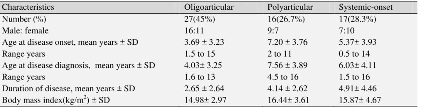
A total of 60 children with JIA ranging in age from 1.5 to 16 years (mean 8.41 ± 4.49 years) were studied, and the male/female ratio was 32/28. All children were from Southwestern of Iran. The mean age at disease onset was 4.92 years (range: 0.5 to 14 years) and the mean age at identification was 5.39 years (range: 0.8 to 14.1 years) in JIA patients. The mean interval from initial symptoms to the JIA diagnosis was 4.90 ± 7.23 months. (Table.1) displays JIA characteristics in three various subtypes.
Table 1: Characteristics of patients with various subtypes of JIA
Characteristics Oligoarticular Polyarticular Systemic-onset
Number (%) 27(45%) 16(26.7%) 17(28.3%)
Male: female 16:11 9:7 7:10
Age at disease onset, mean years ± SD Range years
3.69 ± 3.23 1.5 to 15
7.20 ± 3.76 2 to 11
5.37± 3.93 0.5 to 14 Age at disease diagnosis, mean years ± SD
Range years
4.03± 3.25 1.6 to 13
7.56 ± 3.89 4.5 to 16
6.03± 4.11 1.5 to 16 Duration of disease, mean years ± SD 2.65 ± 2.64 4.14 ± 2.62 4.91± 4.46 Body mass index(kg/m2) ± SD 14.98± 2.97 16.44± 3.61 15.87± 4.67
27(33.3%) oligoarthritis patients, nine out of 16 (56.2%) polyarthritis patients and in nine out of 17(52.9%) systemic-onset patients. All patients with systemic-onset had a daily or twice-daily fever; it was accompanied by a transient rash in 6 out of 17 systemic-onset patients (35.3%).
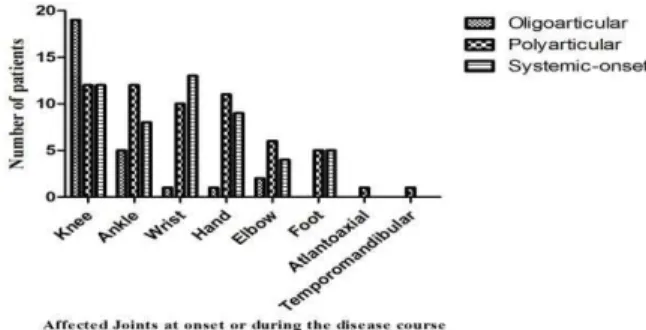
Any number of joints can be affected at onset or during the disease course of JIA. Figure.1 shows the rate of joint involvement in patients with different subtypes of JIA at onset or during the disease course. As shown, the most frequent affected joints were knee (88.3%) and ankle (46.6%) in JIA patients.
Pericardial effusion was detected in two patients with systemic-onset JIA, only one of whom also had pleural effusion. One patient with oligoarthritis showed mild tricuspid regurgitation by echocardiography. By endoscopy, gastritis and esophagitis was documented each in one patient during the course of disease. Two patients had hepatomegaly and one of whom was complicated with Mycobacterium avium complex (MAC). Depression was appeared in one patient during the course of JIA. Leukocytosis was seen in 25 (41.6%), thrombocytosis in 22(36.6%) and anemia in 20 (33.3%) patients with JIA at the beginning of disease diagnosis. High ESR and elevated CRP was detected in in 50 (83.3%) and 33(55%) of patients, respectively. The results of urinalysis were normal at the time of diagnosis and follow-up of
the all patients. Laboratory findings in
different subtypes of JIA at the first presentation show in (Table.2).
Fig.1: Rate of joint involvement in patients with different subtypes of JIA
Table 2: Early laboratory findings in 60 individuals with Juvenile idiopathic arthritis
Laboratory data Oligoarticular
n=20
Polyarticular n=14
Systemic-onset n=16
Overall n=60 Hemoglobin(g/dL) , mean±
SD
11.03±1.58 10.52±1.96 9.77±1.76 10.54±1.79
White blood cell(mm3) Mean ±SD
9372.72 ±2372.29
9185.25 ±3154.93
15113.33 ±8097.78
10939.62 ±5460.18 Platelet
Mean ±SD
462173.91 ±165168.68
428733.33 ±197918.98
437086.92 ±285846.83
449269.23 ±206056.92 ESR level (mm/hour), mean±
SD
52.70±32.81 65.06±32.14 65.33±27.21 59.64±31.22
SGOT, mean± SD 27.06±9.16 25.61±10.11 33.76±15.57 28.73±12.05 SGPT, mean± SD 16.06±5.06 15.30±9.38 21.23±20.60 17.46±13.02
Rheumatic factor 2 3 0 5
ANA 0 1 0 1
C3, mean± SD 1.43±0.31 1.38±0.41 1.65±0.21 1.50±0.32
C4, mean± SD 0.38±0.18 0.45±0.11 0.34±0.14 0.39±0.15
High ASO 1 0 0 1
The normal results of bone marrow aspiration and biopsy was seen in 40 patients with JIA.
One of the most devastating complications of JIA is eye involvement. Uveitis detected in two patients and cataract in other 2 patients with JIA. One patient with
systemic-onset JIA developed MAC during the course of disease who responded to medication well. Moreover, no amyloidosis was observed in any of the JIA patients. The goals of JIA treatment are to prevent pain and joint damage. Among 27 JIA patients with oligoarthritis, use of non-steroidal anti-inflammatory drugs (NSAID) used in 26 patients (as monotherapy in 3 patients), methotrexate in 25 and intra-articular glucocorticoid injection in 11. All patients with polyarthritis (n=16) took methotrexate and low dose of corticosteroid, 4 patients treated with leflunomide and 2 of whom with biologic agents (infliximab). Treatment of systemic-onset JIA (n=17) was done with NSAID in 16, methotrexate in 15, systemic glucocorticoid in 14, intra-articular glucocorticoid injection in 3, leflunomide in 2 and biologic agents (infliximab) in 1 patient.
Among our patients, complete remission was observed in 3(5%), partial remission in 51(85%) and non-remission in 6(10%) of them.
Discussion
JIA is the most common rheumatic disease of childhood; it is classified to three major subtypes: oligoarticular, polyarticular and systemic-onset. This study showed oligoarticular subtype in 45%, polyarticular in 26.7% and systemic-onset in 28.3% of JIA patients. Increased rate of oligoarticular subtype is consistent with those from most
cohorts of JIA (14,15). Among 60 patients with JIA, male was predominant in a few numbers. It is unusual for children to develop JIA before 6 months of age (4), similar to our study that was less 6 months. Growth retardation is distributed throughout the subtypes, frequently associated with polyarticular and systemic type secondary to immobilization, poor nutrition, prolonged inflammation, medication toxicity and maybe psychosocial factors among JIA patients (16).
Like other reports, inflammation of knee joints followed by the ankle joints was the main type of arthritis among our patients with oligoarthritis (17,18). The ratio male/female of 16/11 in oligoarticular subtype was in similar to Asian children report (19). Positive RF was detected in 2 patients, RF likely more related to non-specific immune complex formation such as viral diseases (20). ANA positivity was never detected in our patients with oligoarthritis, it is completely different from the incidence of ANA positivity in Western studies (4). No positivity of ANA can be explained by technical inaccuracy in lab, too low level of ANA in patient’s serum, and bounding ANA in the form of immune complex (21). Furthermore, genetic studies might be helpful to determine this dissimilarity.
involvement in an extended prospective follow up.
Valvular heart disease is less reported in JIA (25). Just one of the patients with oligoarticular subtype presented mild tricuspid regurgitation; this finding was not diagnostic of rheumatic carditis in any patient.
RF positive poly-JIA accounted for 18.8% and RF negative poly-JIA for 81.2%. The sex distribution within our polyarticular patients showed no gender disparity while female is typically seen predominant (26). In addition to peripheral joint disease, propensity to develop arthritis of atlantoaxial and temporomandibular joint was seen each two in our patients. The positive rate of Human leukocyte antigen (HLA) B27 was detected in one patient with polyarthritis; it can indicate susceptibility to the development of axial arthritis in future (6). The rate of uveitis in this subtype was low (one patient), too.
The most prominent feature of systemic-onset was a high spiking fever, and arthritis associated with this type involved small and large joints. Sex ratio was nearly equal in accordance with other studies (4,20). Erythrocyte sedimentation rate (ESR) and C-reactive protein (CRP) levels as well as leukocyte counts were also significantly higher than normal ranges among all of our patients with systemic-onset JIA at onset of disease. It appears that these indexes maybe helpful in diagnosis.
Among systemic-onset patients no positivity to ANA and RF was observed in any of patients because this subtype is closer to the group of auto-inflammatory diseases with uncommon positive autoantibodies (27,28). Bone marrow aspiration and biopsy were done in suspicion of malignancies such as leukemia and lymphoma in all patients with
systemic-onset, which was reported normal. The results of present study showed pericardial effusion in 2 (3.3%) of JIA patients in agreement with 3% to 9% in other reports (29,30).
Cataract is the most commonly described complication related to the chronic inflammatory process and corticosteroid therapy (8); it was seen in two patients with systemic-onset JIA. No patient with systemic-onset was diagnosed as having uveitis in agreement with other reports (2). MAS is a severe and life-threatening complication (10), it was appeared in one of our patients, resulting in persistent fever, intravascular coagulation, prolonged Prothrombin time (PT) and Partial
thromboplastin time (PTT),
hypofibrinogenemia, and increased liver enzymes. Additionally, MAS in the patient was treated vigorously with intravascular methylprednisolone. While amyloidosis is one of complication of JIA, none of our patients had clinical or laboratory evidence of amyloidosis.
Gasterointestinal tract is rarely involved in children with chronic arthritis, but it can see after using medications in treatment of JIA (31).
Management of JIA should be individualized according to the patient’s specific problems. Medication is needed to minimize joint inflammation and treatment of extra-articular manifestations of disease. Biologic agents are well established in the treatment of JIA (11) when other medications have failed to control the disease, three our patients should be taken these medications.
None of our patients had family history of JIA, however, lack of a reliable and complete family history should be considered.
southwestern Iranian JIA patients. In this study, the pattern of most clinical features in different subtypes of JIA resembles to other studies. Positive ANA was less; however, the low numbers of Iranian patients with uveitis was noteworthy. As genetic background of Iranian patients with JIA is important, it will be interesting to see if there is increased uveitis in an extended prospective follow- up.
Conflict of interest
The authors declare that they have no conflict of interest.
Acknowledgments
we thank Ms. Nasiri and Ms. Babaii for their assistance with data collection.
References
1. Hyrich KL, Lal SD, Foster HE, Thornton J, Adib N, Baildam E. Disease activity and disability in children with juvenile idiopathic arthritis one year following presentation to paediatric rheumatology. Results from the
Childhood Arthritis Prospective Study.
Rheumatology (Oxford), 2010; 49(1):116-22. 2. Prakken B, Albani S, Martini A. Juvenile idiopathic arthritis. Lancet 2011; 9783(377): 2138-49.
3. Hahn YS, Kim JG. Pathogenesis and clinical manifestations of juvenile rheumatoid arthritis. Korean J Pediatr 2010; 53(11): 921-30. 4. Cassidy JT, Petty RE. Juvenile rheumatoid
arthritis. In: Textbook of Pediatric
Rheumatology, 4th ed, Cassidy JT, Petty RE (Eds), WB Saunders Company, Philadelphia 2001;218.
5. Foeldvari I, Bidde M. Validation of the proposed ILAR classification criteria for juvenile idiopathic arthritis. International League of Associations for Rheumatology. J Rheumatol 2000; 27(4): 1069-72.
6. Boros C, Whitehead B. Juvenile idiopathic arthritis. Aust Fam Physician 2010; 39(9): 630-6.
7. Tugal-Tutkun I, Havrlikova K, Power WJ, Foster CS. Changing patterns in uveitis of childhood. Ophthalmolog 1996; 103(3): 375-83.
8. Sendagorta E, Peralta J, Romero R, García-Consuegra R, Abelairas J. Uveitis and
idiopathic juvenile arthritis in Spain.
Epidemiological and therapeutic aspects. Arch Soc Esp Oftalmol 2009; 84(3): 133-8.
9. Minden K. Adult outcomes of patients with juvenile idiopathic arthritis. Horm Res 2009; 72( suppl 1): 20-5.
10. Sawhney S, Woo P, Murray KJ. Macrophage activation syndrome: a potentially fatal complication of rheumatic disorders. Arch Dis Child 2001; 85(5): 421-6.
11. McCann LJ, Woo P. Biologic therapies in juvenile idiopathic arthritis: why and for whom?. Acta Reumatol Port 2007; 32(1): 15-26.
12. Petty RE, Southwood TR, Manners P, Baum J, Glass DN, Goldenberg J. International League
of Associations for Rheumatology
classification of juvenile idiopathic arthritis: second revision, Edmonton, 2001. J Rheumatol 2004;31(2):390-2.
13. Smolen JS, Landewé R, Breedveld FC, Buch M, Burmester G, Dougados MSmolen JS. EULAR recommendations for the management of rheumatoid arthritis with synthetic and biological disease-modifying antirheumatic drugs. Ann Rheum Dis 2010; 69(6): 964-75. 14. Thomas E, Barrett JH, Donn RP, Thomson W,
Southwood TR. Subtyping of juvenile idiopathic arthritis using latent class analysis.
British Paediatric Rheumatology Group.
Arthritis Rheum 2000; 43(7): 1496-1503. 15. Yilmaz M, Kendirli SG, Altintas DU,
Karakoc GB, Inal A, Kilic M. Juvenile idiopathic arthritis profile in Turkish children. Pediatr Int 2008; 50(2): 154-8.
boys with juvenile rheumatoid arthritis. Rheumatol Int 2011; 31(5): 635-40.
17. Algergawy S, Haliem T, Al-Shaer O. Clinical, laboratory, and ultrasound assessment of the knee in juvenile rheumatoid arthritis. Clin Med Insights Arthritis Musculoskelet Disord 2011; 25(4): 21-7.
18. El-Miedany YM, Housny IH, Mansour HM, et al. Ultrasound versus MRI in the evaluation of juvenile idiopathic arthritis of the knee. Joint Bone Spine 2001; 68(3): 222-30.
19. See Y, Kidon M, Koh E. More males and less uveitis in Asian children with juvenile idiopathic arthritis in Singapore. Clin. Exp. Rheumatol 2003; 21: 536.
20. Kahn P. Juvenile idiopathic arthritis: an update for the clinician. Bull NYU Hosp Jt Dis 2012; 70(3): 152-66.
21. Mashhadi MA, Bari Z. Thrombotic
thrombocytopenic purpura and deep vein thrombosis as the presenting manifestations of systemic lupus erythematosus: A case report and review of literature. J Res Med Sci 2011; 16(8): 1082-8.
22. Kotaniemi K, Kaipiainen-Seppänen O, Savolainen A, Karma A. A population-based study on uveitis in juvenile rheumatoid arthritis. Clin Exp Rheumatol 1999; 17(1): 119-22.
23. Yu HH, Chen PC, Wang LC, Lee JH, Lin YT,
Yang YH. Juvenile idiopathic
arthritis-associated uveitis: a nationwide
population-based study in Taiwan. PLoS One 2013; 8(8): e70625.
24. Sircar D, Ghosh B, Ghosh A, Haldar S. Juvenile idiopathic arthritis. Indian Pediatr 2006; 43(5): 429-33.
25. Svantesson H, Björkhem G, Elborgh R. Cardiac involvement in juvenile rheumatoid arthritis. A follow-up study. Acta Paediatr Scand 1983; 72(3): 345-50.
26. Moroldo MB, Chaudhari M, Shear E, Thompson SD, Glass DN, Giannini EH. Juvenile rheumatoid arthritis affected sibpairs: extent of clinical phenotype concordance. Arthritis Rheum 2004; 50(6): 1928-34.
27. Ramanan AV, Grom AA. Does systemic-onset juvenile idiopathic arthritis belong under juvenile idiopathic arthritis? Rheumatology (Oxford)2005;44(11):1350-3.
28. Frosch M, Roth J. New insights in systemic
juvenile idiopathic arthritis--from
pathophysiology to treatment. Rheumatology (Oxford), 2008; 47(2): 121-5.
29. Alukal MK, Costello PB, Green FA. Cardiac tamponade in systemic juvenile rheumatoid arthritis requiring emergency pericardiectomy. J Rheumatol 1984; 11(2): 222-5.
30. Goldenberg J, Ferraz MB, Pessoa AP, Fonseca AS, Carvalho AC, Hilario MO. Symptomatic cardiac involvement in juvenile rheumatoid arthritis. Int J Cardiol 1992; 34(1): 57-62. 31. Snape WJ Jr. Pseudo-obstruction and other