FACULDADE DE ODONTOLOGIA
PROGRAMA DE PÓS-GRADUAÇÃO EM ODONTOLOGIA
MARIANA PASSOS DE LUCA
EFEITO DE VERNIZ DE PRÓPOLIS EM Streptococcus
mutans E EM MODELO EXPERIMENTAL DE CÁRIE
EFEITO DE VERNIZ DE PRÓPOLIS EM Streptococcus
mutans E EM MODELO EXPERIMENTAL DE CÁRIE
Te se a pr e se n t a da a o Pr ogr a m a de Pós-Gr a du a çã o da Fa cu lda de de Odont ologia da Un iv e rsida de Fe de r a l de M in a s Ge r a is com o pa rt e dos re quisit os pa ra a obt e n çã o do t ít u lo de Dou t or e m Odon t ologia
Ár e a de Con ce nt r a çã o : Odon t ope d ia t ria
Or ie n t a dor : Prof. Dr. Vagner Rodrigues Sant os Co- or ie nt a dor a : Profa. Dra. Miriam Pim ent a Vale
Belo Horizonte Faculdade de Odontologia Universidade Federal de Minas Gerais
FICHA CATALOGRÁFICA
D278e 2015 T
De Luca, Mariana Passos
Efeito de verniz de própolis em Streptococcus mutans e em modelo experimental de cárie / Mariana Passos De Luca. – 2015. 72f. : il.
Orientador: Vagner Rodrigues Santos
Coorientadora: Miriam Pimenta Parreira do Vale
Tese (Doutorado) – Universidade Federal de Minas Gerais, Faculdade de Odontologia.
1. Cárie dentária. 2. Própole– uso terapêutico.
3. Streptococcus mutans. I. Santos, Vagner Rodrigues. II. Vale Miriam Pimenta Parreira do. III. Universidade Federal de Minas Gerais. Faculdade de Odontologia. IV. Título.
BLACK D047
difíceis da minha trajetória.
Ao meu marido Paulo por ter sempre me dado condições de seguir os meus sonhos, mesmo sacrificando os nossos! Você é o meu grande exemplo de
homem, professor e pesquisador!
À minha filha Isabela, por ter me ensinado tantas coisas desde que veio ao mundo! Você é a razão pela qual eu luto para ser uma pessoa melhor todos os
dias!
À Deus, pela oportunidade de aprendizado durante esses anos, pelas
conquistas e inspiração para realizar este trabalho;
Agradeço aos meus orientadores Prof. Dr. Vagner Rodrigues Santos e Profa.
Dra. Miriam Pimenta Vale pela amizade, ajuda e suporte no doutorado;
Ao Prof. Dr. Pedro Luiz Rosalen por ter me acolhido em seu grupo de
pesquisa e permitido que eu desenvolvesse o experimento em animais em seu
laboratório;
Ao Prof. Dr. Frederico Marianetti Soriani pela amizade e valorosa
colaboração no experimento com PCR em tempo real;
Aos amigos que colaboraram em várias fases da pesquisa na UFMG: Patrícia
Corrêa-Faria, Alfonso Gala-García, Elizete Maria Rita Pereira, Suzane Gonçalves Paixão, Marieta Torres Abreu Assis;
Aos colegas da Faculdade de Odontologia de Piracicaba (UNICAMP) que me
acolheram e me ajudaram durante a minha passagem por Piracicaba: Carina,
Bruna, Irlan, Laila, Luciana, Lívia, Luiz, Marcelo, Jonny, Camila, Ana Paula, Bruno, Luciano, Cleiton, Talita e Juliana;
Aos demais colegas de doutorado, pela convivência durante esta jornada, em
especial à Maria Luiza da Matta Felisberto Fernandes (Malú) pela amizade;
À técnica do Laboratório de Microbiologia da FOUFMG, Silvana Maria de
Sousa, pela ajuda e companheirismo durante todas as fases da pesquisa;
Aos técnicos do Laboratório de Farmacologia da FOP/UNICAMP Eliane Melo e
especial aos coordenadores Saul Martins Paiva e Maria Cássia Ferreira
Aguiar por todo o apoio e auxílio durante o doutorado;
Aos Professores de Odontopediatria da FOUFMG pelo apoio;
A todos os pacientes que participaram da pesquisa e às coordenadoras
dos abrigos que permitiram a participação das crianças na pesquisa, minha
gratidão pela disponibilidade e pela colaboração;
À todos aqueles que participaram e ajudaram de alguma forma o
desenvolvimento desta pesquisa,
Que eu continue a acreditar no outro mesmo sabendo de alguns
valores esquisitos que permeiam o mundo; que eu continue otimista,
mesmo sabendo que o futuro que nos espera nem sempre é tão
alegre; que eu continue com a vontade de viver, mesmo sabendo que
a vida é, em muitos momentos, uma lição difícil de ser aprendida; que
eu permaneça com a vontade de ter grandes amigos, mesmo
sabendo que com as voltas do mundo, eles vão partindo...
Que eu realimente sempre a vontade de ajudar as pessoas, mesmo
sabendo que muitas delas são incapazes de ver, sentir, ou entender
esta ajuda; que eu exteriorize a vontade de amar, entendendo que
amar não é sentimento de posse, é sentimento de doação; que eu
alimente minha garra, mesmo sabendo que a derrota e a perda são
ingredientes tão fortes quanto o sucesso e a alegria; que eu atenda
sempre mais à minha intuição, que sinaliza o que de mais autêntico
possuo; que eu pratique sempre mais o sentimento de justiça,
mesmo em meio à turbulência dos interesses; que eu manifeste o
amor por minha família, mesmo sabendo que ela muitas vezes me
exige muito para manter sua harmonia; que eu acalente a vontade de
ser grande, mesmo sabendo que minha parcela de contribuição no
mundo é pequena; e, acima de tudo...
Que eu lembre sempre que todos nós fazemos parte desta
maravilhosa teia
chamada vida, criada por alguém bem superior a todos nós!
Produtos naturais são fontes de agentes terapêuticos eficazes e inovadores,
oferecendo diversos princípios ativos a serem testados, favorecendo a
descoberta e desenvolvimento de novos produtos. A própolis tem efeito
antimicrobiano comprovado sobre bactérias cariogênicas, atraindo muitos
estudos para este fim. Este trabalho avaliou o efeito do verniz experimental de
própolis em modelo experimental de cárie dentária e na redução de
Streptococcus mutans (SM) na saliva e em biofilme de crianças de 8 a 11 anos
utilizando uma única aplicação do produto. No experimento em modelo animal
de alto desafio cariogênico, 56 ratas wistar foram infectadas com SM e alocadas
em quatro grupos (n=14): G1 – verniz de própolis; G2 – verniz de quitosana
(veículo); G3 - Duraphat; e G4 – sem tratamento. Após a aplicação do verniz
nos molares, os animais foram submetidos a dieta rica em sacarose (Dieta 2000,
56% sacarose, ad libitum) por 4 semanas. Depois desse período, os animais
foram eutanaziados e suas mandíbulas removidas para quantificar a microbiota
total, SM e lesões de cárie. Os dados foram analisados utilizando-se o teste de
Tukey-Kramer HSD. A microbiota total e a porcentagem de SM não foram
diferentes entre os grupos (p<0,05). Não houve diferença estatística entre o total
de lesões de cárie em superfície lisa entre os grupos G1 e G3 (p <0.05). Em
lesões de cárie mais severas, G1 não teve diferença estatística com o G4. Em
lesões de cárie de sulco, Duraphat® teve melhores resultados, apresentando
diferença estatística em todos os índices de severidade entre os grupos (p
<0.05). O estudo clínico seguiu o protocolo de aplicação única no início do
tratamento (baseline), seguindo com coletas diárias até o 5º dia e nos 10º, 15º e
30º dia após a aplicação do verniz. As colônias com morfologia típica foram
contadas e o número de CFU/mL foi comparado utilizando-se o teste t para
amostras independentes com nível de significância de 5%. Houve diferença
estatística significativa entre o baseline e o 2º dia, onde foi verificada uma
redução drástica nos níveis de SM, e entre o 2º dia e o 30º dia, mas não houve
diferença entre o 4º dia de coleta e os dias subsequentes. Conclui-se que o
verniz de própolis teve efeito em lesões cariosas superficiais com uma única
impacto na incidência de cárie precisa ser confirmada.
Natural products are sources of effective and innovative therapeutic agents,
offering various active principles to be tested, enabling the discovery and
development of new products. Propolis has proven antimicrobial effect on
cariogenic bacteria, attracting many studies for this purpose. This work evaluated
the effect of the experimental propolis varnish in a caries experimental model and
in the reduction of Streptococcus mutans (SM) in saliva and in biofilm of children
from 8 to 11 years using a single application of the product. In the experiment
with animal model under high cariogenic challenge, fifty-six female SPF wistar
rats, previously infected with SM, were assigned to 4 groups (n=14) and treated
as follows: G1 - propolis varnish; G2 - chitosan varnish/vehicle; G3 - Duraphat
and G4 - no treatment. Animals received a single varnish application on their
molars and were then submitted to a high cariogenic challenge (Diet 2000, 56%
sucrose, ad libitum) for 4 weeks. After this period, animals were euthanized by
CO2 asphyxiation and their jaws removed to quantify the total cultivable
microbiota, recovered S. mutans, and caries lesions. Data were analyzed using
the Tukey-Kramer HSD test. Total microbiota and the percentage of SM were not
different between the experimental groups (p<0.05). There was no statistical
difference in total smooth surface caries lesions between G1 and G3 (p <0.05).
However, in more severe caries lesions, G1 did not differ statistically from G4. In
sulcal caries, Duraphat® had better results, showing statistical differences in all
severity indexes among all groups (p <0.05). The clinical study followed the single
application protocol at the beginning of treatment (baseline), following with daily
collections until the 5th day and on the 10th, 15th and 30th day after applying the
varnish. The colonies with typical morphology were counted and the number of
CFU/ml was compared using the t test for independent samples with 5%
significance level. There was no statistically significant difference between the
baseline and the 2nd day and between 2nd day and 30th day, but there was no
difference from 4th day forward. We conclude that propolis varnish had effect in
superficial caries lesions with a single application in animal model and
suppressed the number of salivary SM for 3 days in children. The possibility of
these results have an impact on the incidence of caries needs to be confirmed.
FOP Faculdade de Odontologia de Piracicaba
UNICAMP Universidade Estadual de Campinas SWP Doutorado Sanduíche no País
CNPq Conselho Nacional de Pesquisa
SPF Specific Pathogen Free
SM Streptococcus mutans
CO2 Gás carbônico
HSD Honest Standard Deviation
pH Potencial de hidrogênio
CEMIB Centro Multidisciplinar para Investigação Biológica
SP São Paulo
CEUA Comissão de Ética no uso de Animais
MI Michigan
USA United States of America
MSB Mitis Salivarius Bacitracin
w/v Weight/volume
mL Mililitro
W Watts
Inc Incorporation
Ds Slight dentinal caries
Dm Moderate dentinal caries
Dx Extensive dentinal caries
NaCl Cloreto de Sódio
µL Microlitros
CFU Colony Forming Units
ANOVA Analysis of variance
MG Minas Gerais
PCR Polymerase Chain Reaction
s Segundos
°C Grau Celsius
h Horas
IA Iowa
SPSS Statistical Package for Social Sciences
SD Standard Deviation
min Minutes
Min Minimum
Artigo 1
Table 1 Analysis of the animals’ microbiota (mean and standard deviation/M±SD) submitted to cariogenic challenge for four weeks with propolis varnish and its controls.
28
Table 2 Effect of propolis varnish and their controls (mean and standard deviation/M±SD) in the development of smooth surface caries and severity (E - enamel caries; Ds - slight dentinal caries; Dm - moderate dentinal caries; Dx - extensive dentinal caries) in rats using classification Keyes' modified by Larson.
29
Table 3 Effect of propolis varnish and their controls (mean and standard deviation) in the development of sulcal caries and severity (E - enamel caries; Ds - slight dentinal caries; Dm - moderate dentinal caries; Dx - extensive dentinal caries) in rats using classification Keyes' modified by Larson.
29
Artigo 2
Table 1 Mutans streptococci colony forming units in saliva (Mean; SD: standard deviation), in different collection times.
1. INTRODUÇÃO
2. REVISÃO DA LITERATURA
14
3. JUSTIFICATIVA E RELEVÂNCIA 17
4. OBJETIVOS 19
4.1 Objetivos gerais 19
4.2 Objetivos específicos 19
5. ARTIGOS
5.1 Artigo do experimento em modelo animal 5.2 Artigo da pesquisa clínica
21 21 33 6. CONSIDERAÇÕES FINAIS
7.CONCLUSÃO
44
8. REFERÊNCIAS GERAIS 48
9. ANEXOS
Anexo A 51
Anexo B 59
Anexo C 60
Anexo D 65
8. APÊNDICE 68
1. INTRODUÇÃO
A cárie é uma doença de alta prevalência (ISMAIL et al., 2013), causada
pela presença de um biofilme polimicrobiano maduro na superfície dentária
(HESSE et al., 2014). A atividade metabólica deste biofilme confere um processo
dinâmico de desmineralização e remineralização do esmalte dentário, iniciando
o desenvolvimento desta doença (HESSE et al., 2014).
Múltiplos fatores, incluindo bactéria, açúcar, saliva e flúor, podem afetar o
processo de desmineralização, podendo ser manipulados de maneira a pender
a balança para a doença (desmineralização) ou para a saúde (remineralização).
Durante o desenvolvimento da cárie, bactérias acidúricas e acidogênicas são
predominantes no biofilme dental, sendo o Streptococcus mutans a mais
fortemente associada à cárie dentária. Quando fatores ambientais possibilitam a
seleção dessas bactérias no biofilme, o processo da doença começa
(TAKAHASHI & NYVAD, 2008; BUENO-SILVA et al., 2013; GUO & SHI, 2013;
SEGURA et al., 2014).
Para auxiliar no restabelecimento do equilíbrio saúde-doença na cavidade
bucal, suprimindo o número de bactérias cariogênicas e restabelecendo a
integridade do esmalte dentário (BORGES et al., 2011), diversos materiais vêm
sendo desenvolvidos com diferentes princípios ativos. Além do flúor e da
clorexidina (MARECHAL, 1991; FERREIRA et al., 2009; FURIGA et al., 2013),
produtos naturais contém agentes terapêuticos eficazes, oferecendo uma
diversidade de princípios ativos a serem testados, favorecendo a descoberta e
desenvolvimento de novos medicamentos (KOEHN & CARTER, 2005; JEON et
al., 2011). Setenta por cento dos agentes terapêuticos descobertos e aprovados
entre 1981 e 2010 são de origem natural (NEWMAN & CRAGG, 2012). Muitos
destes foram submetidos a pesquisas in vivo e a ensaios clínicos (BUTLER,
2005),porém, poucos estudos vêm sendo feitos para avaliar a aplicabilidade
destes produtos no tratamento preventivo e terapêutico anti-biofilme e anti-cárie
dentária (JEON et al., 2011).
A própolis é um produto natural produzido pelas abelhas, contendo várias
antifúngica, antitumoral, dentre outras. A própolis tipo 12, derivada
predominantemente da Baccharis dracunculifolia e também conhecida como
própolis verde, é comumente encontrada na região Sudeste do Brasil (PARK et
al., 2002; BUENO-SILVA et al., 2008) se destaca por sua propriedade
antimicrobiana, atraindo diversas pesquisas na área Odontológica, mais
precisamente na prevenção da cárie dentária (HAYACIBARA et al., 2005).
Aplicações tópicas de extrato etanólico de própolis foram eficazes na redução da
incidência e severidade de lesões de cárie em animais (IKENO et al., 1991; KOO
et al., 1999; HAYACIBARA et al., 2005; DUARTE et al., 2006). No entanto, seus
efeitos cariostáticos podem variar de acordo com sua composição química e
origem geográfica (KOO et al., 1999).
Nesse sentido, foi desenvolvido um verniz experimental à base de
quitosana e extrato etanólico de própolis, com o intuito de testar a possibilidade
de manter a atividade antimicrobiana da própolis numa base polimérica
(quitosana) por um período prolongado.
A quitosana, produto derivado de uma substância encontrada nas cascas
de insetos e crustáceos (RINAUDO, 2006), possui propriedades biocompatíveis,
biodegradáveis e antibacterianas (PILLAI et al., 2009), além de sua propriedade
biopolimérica permitir a liberação lenta e prolongada de princípios ativos
incorporados a ela (LIU et al., 2009).
Estudos in vitro demonstraram que o verniz de própolis possui baixa
citotoxicidade, atividade antimicrobiana contra diversas bactérias da microbiota
bucal e liberação em meio aquoso por 8 semanas (DE LUCA et al., 2014). Este
estudo avaliará se estes resultados serão reproduzidos em modelo de lesões de
cárie experimental em animais e na redução de Streptococcus mutans na saliva
2. REVISÃO DA LITERATURA
2.1 CÁRIE
A cárie é a doença crônica mais comum da infância, afetando 60 a 90%
das crianças de 2 a 11 anos no mundo todo (BOYCE et al., 2010).
Apesar do aumento e subsequente declínio desta doença desde 1950 até
o começo dos anos 90 na maioria dos países desenvolvidos, observou-se que
ela se tornou fortemente polarizada, mostrando uma distribuição desigual em
diversas populações (OLIVEIRA et al., 2006; PESSAN et al., 2008).
Informações sobre saúde geral, dieta, higiene oral, exposição ao flúor e
experiência de cárie podem ajudar no diagnóstico de fatores de risco para o
desenvolvimento da doença num indivíduo (PETERSSON & TWETMAN, 2015),
tornando possível o seu manejo e prevenção (SAJJAM ET AL.,2013).
As medidas preventivas têm como alvo, direta ou indiretamente, a
microbiota bucal (SAJJAM ET AL.,2013), já que a cárie dentária está associada
ao aumento na frequência de consumo de açúcar e rápida conversão desses
carboidratos em produtos ácidos pelas bactérias, numa superfície dentária
susceptível (MARSH, 2003; JEON et al., 2011). Condições repetidas de baixo
pH no biofilme fazem o número de micro-organismos acidogênicos e acidúricos
prevalecer (p.e. estreptococos mutans e lactobacilos) (MARSH et al., 2000),
formando uma matriz rica em polissacarídeo extracelular (EPS) na interface
dente-biofilme (BOWEN & KOO, 2011; JEON et al., 2011).
Portanto, apesar dos biofilmes serem compostos de flora mista, os
Streptococcus mutans são reconhecidos como os produtores primários da matriz
2.1.2 Streptococcus mutans
Considerado o mais cariogênico de todos os estreptococos, S. mutans é
o colonizador inicial das superfícies dentárias, produzindo quantidades
significativas de EPS, sendo responsável pelo estágio inicial da formação de
biofilme e lesões de cárie (FREIRES et al., 2015). Possui uma importante
capacidade produção de glucanos na presença de sacarose, contribuindo para
a formação de um biofilme rico em polissacarídeos e altamente acidogênico,
causando a desmineralização do esmalte dental (BOWEN & KOO, 2011).
Os fatores de virulência do S. mutans tem sido intensamente estudados,
destacando-se a tolerância ao meio ácido e a síntese de glucanos através das
enzimas glucosiltransferases (Gtfs) (BUENO-SILVA et al., 2013; BOWEN &
KOO, 2011). Os glucanos são os principais fatores de aderência e acúmulo de
estreptococos na superfície dental (SCHILLING & BOWEN 1992). Aliado ao
consumo frequente de sacarose, produzem uma matriz extracelular rica em
polissacarídeos, formando um biofilme altamente coeso e adesivo, que protege
as bactérias ali embebidas (WOLFF et al., 2013). Além disso, o metabolismo dos
açúcares levam a acidificação do microambiente do biofilme, contribuindo para
o predomínio de bactérias acidúricas. Assim, estreptococos do grupo mutans e
glucanos são considerados fatores críticos no desenvolvimento do biofilme
dental cariogênico (BUENO-SILVA et al., 2013).
Apesar dos biofilmes ricos em EPS serem microambientes extremamente
coesos, agentes bioativos podem penetrar e desorganizar essa estrutura,
interrompendo dessa forma a patogênese da cárie dentária de uma maneira
eficaz e precisa (NGUYEN et al., 2014).
2.2 PRÓPOLIS
O termo própolis deriva do grego pro, de “em frente de, na entrada de”, e
polis, “comunidade ou cidade” (CASTALDO & CAPASSO, 2002; SALATINO et
al., 2005). Ela é utilizada pelas abelhas como proteção contra a entrada de
de luz e umidade em seu interior. É utilizada também para forrar os favos, de
modo a permitir a deposição de ovos pela rainha, e para embalsamar pequenos
animais mortos, evitando sua putrefação (MARCUCCI et al., 1995; BURDOCK
et al., 1998; SALATINO et al., 2005).
A própolis é uma substância natural tóxica coletada por abelhas Apis
mellifera em várias espécies de plantas, determinando a origem botânica
predominante de cada tipo de própolis. A classificação da própolis brasileira em
treze tipos foi feita de acordo com as propriedades físico-químicas e relatos da
localização geográfica (PARK et al., 2002; BUENO-SILVA et al., 2008). A
principal origem botânica dos tipos Sul (três), Nordeste (seis) e Sudeste (doze)
da própolis brasileira são originadas de resinas de Populus sp., Hyptis divaricata
e Baccharis dracunculifolia, respectivamente.
O Cerrado brasileiro é uma das áreas mais ricas em Baccharis sp. Essas
plantas são um grupo de arbustos lenhosos perenes, com inflorescências
masculinas e femininas que aparecem nas plantas separadamente. Das várias
espécies de Baccharis, a Baccharis dracunculifolia (“alecrim do campo”) é a
origem dominante de própolis verde, ou do tipo 12, no sudeste do Brasil (Estado
de São Paulo e área de cerrado de Minas Gerais), onde a maioria dos produtos
comercializados a base dessa própolis são produzidos (PARK et al., 2004).
Os típicos constituintes da própolis verde são ácido cafeoquínico e
derivados prenilatados de ácido cinâmico, tais como artepilin C e baccharina
(MOURA et al., 2009). Apesar da composição química da própolis ser muito
complexa, pode-se observar atividade antibacteriana conferida pela presença de
flavonóides, ácidos aromáticos e ésteres em sua composição; ação bactericida,
decorrente da presença dos ácidos cinâmico e cumarínico; atividade antiviral, in
vitro, (herpes simplex, influenza), em função da ação de flavonóides e derivados
de ácidos aromáticos; e ação cicatrizante, imunoestimuladora, hipotensiva e
citostática (SFORCIN, 2007; SFORCIN & BANKOVA, 2010).
Na Odontologia, tem-se estudado a atividade farmacológica da própolis
lesões de cárie em animais (GERALDINI et al., 2000). Também vem sendo
empregada em curativos pré e pós-cirúrgicos, em tratamentos da candidose,
herpes labial, além de produtos de higiene bucal. Dessa forma, a própolis se
mostra um produto de grande aplicabilidade para o tratamento de afecções
bucais (MANARA et al.,1999).
2.3 QUITOSANA
A quitosana (FIGURA 1) é um polímero biodegradável oriundo da
desacetilação da quitina (FIGURA 2), derivada de exoesqueletos de insetos e
crustáceos (RODRIGUES et al, 2009). Sua atividade antimicrobiana e
antifúngica já foi relatada na literatura, além de suas propriedades
biodegradáveis e biocompatíveis. O mecanismo que lhe confere a capacidade
antimicrobiana é devido à sua carga positiva que, interagindo com a carga
negativa da parede celular bacteriana, destrói os constituintes intracelulares e a
permeabilidade de sua membrana. Outra propriedade importante é a sua
capacidade de adesão em superfícies bucais e assim, agir como um veículo para
a liberação de agentes terapêuticos (DECKER et al., 2005).
Fig. 2: Estrutura molecular da quitosana.
FIG.1: Estrut ura molecular da quitosana.
A quitosana tem a capacidade de se aderir tanto aos cristais de
hidroxiapatita como à parede celular bacteriana, inibindo a adsorção de
Streptococcus sobrinus, Streptococcus sanguinis e Streptococcus mitis à
hidroxiapatita, conferindo-lhe ação bacteriostática (SHIBASAKI et al, 1995).
Além disso, ela tem uma forte tendência de se adsorver a película adquirida, o
que impede a colonização bacteriana na superfície dentária e,
consequentemente a formação do biofilme dental cariogênico (VAN DER MEI et
al; 2007).
Os derivados de quitosana estão sendo usados para melhorar a
biocompatibilidade de superfícies, inibir a proliferação de fibroblastos, estimular
a migração de polimorfonucleares, promover o crescimento de células nervosas,
angiogênese e melhorar a formação óssea, cartilaginosa e tecidual (HSIEH et al,
2005; BUMGARDNER et al, 2003; KITTUR et al., 2005).
Na Odontologia, um dentifrício a base de quitosana já foi testado na
prevenção da desmineralização ao redor dos brackets ortodônticos,
demonstrando diferença significativa com os dentifrícios fluoretados de uso
comum (UYSAL et al., 2011). A quitosana interfere no processo de
desmineralização do esmalte dentário inibindo a perda de fósforo e agindo como
uma barreira contra a penetração de ácidos nessa estrutura. Essa proteção
depende da concentração de quitosana e do tempo de contato do polímero ao
dente (ARNAUD et al., 2010).
Um estudo piloto avaliou o efeito sinergístico de quitosana e clorexidina
sobre S. sanguinis na forma planctônica e aderida. Essa combinação promoveu
uma redução significativa dessas bactérias, melhorando o efeito bactericida e
bacteriostático da clorexidina sobre esses micro-organismos (DECKER et al,
3. JUSTIFICATIVA E RELEVÂNCIA
Vernizes contendo flúor ou clorexidina já são empregados na prevenção
da cárie dentária, possuindo eficácia comprovada em diversos estudos
realizados. No entanto, vernizes de ação antimicrobiana, como o de clorexidina,
não são comercializados no Brasil, impedindo o uso destes produtos para esta
finalidade.
A atividade antimicrobiana dos extratos etanólicos de própolis já é
cientificamente comprovada contra diversas bactérias da microbiota bucal. A
incorporação desta substância a um biopolímero poderia aumentar essa
atividade, devido ao maior tempo de permanência na cavidade bucal.
A redução do número de microrganismos é importante, aliada a outras
medidas de controle da cárie. Enquanto outros fatores não são controlados, a
aplicação do verniz de própolis pode ser uma abordagem interessante por
diminuir a microbiota cariogênica.
Os resultados favoráveis dos estudos realizados in vitro demonstram o
potencial do uso da própolis na forma de verniz (DE LUCA et al., 2014). Esses
resultados precisam ser testados na prática clínica, o que possibilitaria o
preenchimento da lacuna existente no mercado com a falta de um produto com
propriedades antimicrobianas de aplicação profissional e de baixo custo se
4. OBJETIVOS
4.1. OBJETIVOS GERAIS
Avaliar o efeito do verniz de própolis na incidência e severidade de lesões
cariosas em modelo animal. Avaliar o efeito do verniz de própolis a redução de
Streptococcus mutans em saliva e em biofilme em pacientes com idades
variando entre oito e onze anos em um estudo clínico de fase I.
4.2 OBJETIVOS ESPECÍFICOS
4.2.1 Artigo 1: “Effect of an experimental propolis varnish on caries development in rats”
1. Avaliar o efeito do verniz de própolis sobre o desenvolvimento de lesões
cariosas no modelo de alto desafio cariogênico em animais, após única
aplicação do produto.
2. Avaliar o efeito do verniz de própolis na microbiota total e em
Streptococcus mutans no modelo de alto desafio cariogênico em animais.
3. Comparar a eficácia do verniz de própolis com o verniz de flúor e verniz
de quitosana (veículo) na incidência e severidade das lesões cariosas em
modelo animal.
4.2.2 Artigo 2: “Evaluation of reduction in salivary levels of Streptococcus mutans after propolis varnish single application”
1. Avaliar a eficácia do verniz de própolis na redução dos níveis salivares de
Streptococcus mutans em crianças por um período de 30 dias após única
aplicação do produto.
2. Avaliar a presença de efeitos colaterais nos indivíduos que receberam
5. MATERIAL E MÉTODOS
A metodologia utilizada neste estudo será abordada nos artigos
científicos. A seguir, serão apresentados os artigos relacionados ao experimento
em modelo animal e ao estudo clínico de fase I, respondendo aos objetivos
específicos propostos.
5.1 ARTIGO 1
Os experimentos em modelo animal foram realizados na Faculdade de
Odontologia de Piracicaba (FOP/UNICAMP) sob a orientação do Prof. Dr. Pedro
Rosalen, durante o período de Doutorado Sanduíche no país (Bolsa SWP/Cnpq
nº 300165/2013-7).
O embasamento para a realização deste experimento, assim como a
metodologia completa e resultados, encontram-se no artigo intitulado:”Effect of
an experimental propolis varnish on caries development in rats”, a ser submetido
para a revista Caries Research.
“Effect of an experimental propolis varnish on caries development in rats”
Mariana Passos De Lucaa, Vagner Rodrigues Santosb, Miriam Pimenta Valea,
Pedro Luiz Rosalen c
a Department of Paediatric Dentistry and Orthodontics, Faculty of Dentistry,
Federal University of Minas Gerais, Av. Antonio Carlos, 6.627, Pampulha,
31.270-901, Belo Horizonte, MG, Brazil
b Department of Oral Pathology and Oral Surgery, Faculty of Dentistry, Federal
University of Minas Gerais, Av. Presidente Antônio Carlos, 6.627, 31.270-901
Belo Horizonte, MG, Brazil
c Department of Physiological Sciences, Piracicaba Dental School, University of
Campinas, Av. Limeira, 901, Areião, 13414-903, Piracicaba, SP, Brazil
Key words: propolis, Streptococcus mutans, dental caries
Corresponding author:
Mariana Passos De Luca
Avenida Presidente Antônio Carlos 6627, 31.270-901 Belo Horizonte, MG, Brazil
Tel: +55 31 34092497 / FAX: +55 31 34092430
DECLARATION OF INTERESTS
The authors report no relationships, financial or otherwise, with any entity that
may influence the objectivity of this manuscript.
ABSTRACT
The objective of this study was to evaluate the anti-caries potential of a propolis varnish using high cariogenic challenge experimental model. Fifty-six female SPF wistar rats, previously infected with Streptococcus mutans (SM), were assigned to 4 groups (n=14) and treated as follows: G1 - propolis varnish; G2 - chitosan varnish (varnish base); G3 - Duraphat (gold standard varnish, positive control); and G4 - no treatment (negative control). Animals received a single varnish application on their molars and were then submitted to a high cariogenic challenge (Diet 2000, 56% sucrose, ad libitum) for 4 weeks. After this period, animals were euthanized and their jaws removed to quantify the total cultivable microbiota, recovered S. mutans, and dental caries. Data were analyzed using the Tukey-Kramer HSD test. Total microbiota and the percentage of SM were not different among treated groups (p<0.05). There was no statistical difference in total smooth surface caries between propolis varnish and Duraphat® (p <0.05).
However, as caries lesions became more severe, propolis varnish did not differ from the untreated group. In sulcal caries, Duraphat® had better results, differing
INTRODUCTION
Dental caries is still a major public health problem worldwide due to its high
prevalence and significant social impact [Petersen et al., 2005]. It is the result of
a demineralization process in which acidogenic and acidophilic bacteria
embedded in a mature and well-arranged biofilm degrade tooth substance,
leading to cavitation [Wolff et al., 2013].
Although many recent studies relate the participation of other bacteria in
the pathogenesis of dental caries [Beighton, 2005; Choi et al., 2009; Wolff et al.,
2013; Head et al., 2014], Streptococcus mutans plays an important role in the
development of cariogenic biofilm, mainly for its acid tolerant and acidogenic
characteristics, using dietary sucrose to synthetize extracellular polyssacharides
(EPS). EPS are structures that allows its adherence to the tooth surface, creating
a low-pH environment leading to enamel demineralization [Jeon et al., 2011].
Natural products are major sources of research on pharmaceutical agents
and new drugs development [Mishra & Tiwari, 2011]. Propolis is one such product
that stands out for its pharmaceutical properties, mainly the antimicrobial ones,
with direct application in Dentistry [Libério et al., 2009, Bueno-Silva et al., 2013].
It acts decreasing the number of cariogenic microorganisms, and specially
Streptococcus mutans. It also reduces the synthesis of insoluble glucans,
inhibiting glucosyltransferase enzyme. Moreover, the production of acids and the
tolerance of microorganisms to acid pH is reduced by fatty acids contained in
propolis [Ikeno et al., 1991; Duarte et al., 2006; Kumar, 2014].
Several biological activities are attributed to different types of propolis,
among them, antimicrobial, antifungal, antitumor, and others. The propolis type
12 (green propolis) stands out for its antimicrobial property, attracting several
studies in the Dentistry, specifically the prevention of dental caries [Hayacibara
et al., 2005]. Topical application of propolis ethanolic extract were effective in
reducing the incidence and severity of dental caries in animals [Ikeno et al., 1991;
Koo et al., 1999; Hayacibara et al., 2005;. Duarte et al., 2006]. However, the
cariostatic effects of propolis may vary according to their geographical origin and
chemical composition [Koo et al., 1999].
Varnishes are materials widely used as a professional treatment for the
are incorporated to these formulations in order to promote and prolong their
anticaries effect. Fluoride varnish, with remineralizing action [Marinho et al.,
2013], and chlorhexidine varnish, with antimicrobial action [Jayabal & Mahesh,
2014], are the most common products used in dental practice.
Propolis varnish was developed as a new product with antimicrobial
activity that has the ability to adhere to the tooth, allowing sustained release of
propolis. In vitro results have shown good antimicrobial activity against cariogenic
bacteria, low cytotoxicity and a very satisfactory release of the propolis varnish
[De Luca et al., 2014]. These informations have encouraged us to investigate the
anti-caries activity of this product using an animal model under a high cariogenic
challenge.
MATERIALS AND METHODS Animals
Fifty-six specific pathogen-free (SPF) female Wistar rats were obtained
from CEMIB (Multidisciplinary Center for Biological Research, University of
Campinas, SP, Brazil) and maintained at the animal facility of Piracicaba Dental
School. The animal experiment was approved by the Ethics Committee on Animal
Use (CEUA - State University of Campinas, SP, Brazil; protocol #3142-1)
(ANEXO A).
Experimental model
Animals were screened for indigenous Streptococcus mutans on mitis
salivarius agar (Difco Laboratories, Detroit, MI, U.S.A.) and mitis salivarius agar
plus bacitracin (MSB/Sigma Chemical Co., St Louis, MO, U.S.A.). At the age 21
days, the rats were infected with an actively growing overnight culture of
Streptococcus mutans UA159 by cotton swab and they were fed with pellet chow,
diet 2000 [Keyes, 1959] and 5% sucrose in drinking water ad libitum [Murata,
2010]. Oral infection was confirmed at age 25 days by plating on MSB agar. At
the age of 26 days, the rats were randomly assigned to 4 groups (n=14),
anesthetized with chloral hydrate (440 mg/Kg) and submitted to the following
treatments: G1 - topical application of propolis varnish (propolis ethanolic extract
15% w/v); G2 - topical application of chitosan varnish (varnish base, vehicle
2.26%, w/v – positive control); and G4 - no treatment (negative control). The
varnishes were applied on molars using one micro brush per hemi-arcade. After
topical application, the animals fasted for 2 hours and were kept in individual
cages until the end of the experiment. Cariogenic diet 2000 containing 56% of
sucrose and 5% sucrose water were provided ad libitum. The animals were
weighed weekly, and their behavior and physical appearance was noted daily.
The experiment proceeded for 4 weeks, at the end of which the animals were
euthanized.
Microbiological analyses
The lower left jaw was aseptically dissected, suspended in 5.0 mL of sterile
saline solution (0.9 %, w/v), and sonicated using three 10 second pulses at 5
second intervals, at 30 W (Vibracell, Sonics & Material Inc.); providing the
maximum recoverable viable counts. A spiral plater (Whitley Automatic Spiral
Plater, DW Scientific®) was used to streak the suspensions obtained (50 l) on
blood agar (5% sheep's blood) and mitis salivarius agar containing 100 g/mL
streptomycin sulfate (MSB, Sigma®) to determine the number of total microorganisms and the S. mutans recovered (CFU/mL), respectively. The
suspension was plated on MSB to estimate the S. mutans UA159 populations
and on blood agar to determine the total cultivable microorganisms [Bowen et al.,
1988]. The percentage of S. mutans with respect to total microorganisms (% S.
mutans/total microorganisms) was calculated and recorded. The smooth-surface
and sulcal caries and their severity (E, enamel lesion; Ds, dentin exposed; Dm,
3/4 of the dentin affected; Dx, all dentin affected) were evaluated according to
Larson’s modification of Keyes’ system [Larson, 1981]. The determination of the
caries score was blinded by codification of the jaws and performed by one
calibrated examiner.
Statistics
Data were submitted to analysis of variance (ANOVA) followed by the
Tukey-Kramer HSD (Honest Standard Deviation) test for all pairs using JMP
Version 3.1 software for statistical visualization at a significance level of 5%.
Smooth-surface, sulcal caries scores, and severities were expressed as
RESULTS
All rats gained weight and remained apparently healthy and active until the
end of the experiment. The average weight gains for individual groups of rats
were not significantly different from one another (p > 0.05, data not shown).
Regarding the percentage of Streptococcus mutans, there was no statistically
significant difference between the tested groups (p >0.05), as shown in Table 1.
Caries scoring (Table 2) showed no significant difference (p >0.05) concerning
total smooth surface for the positive control (Duraphat ®). In slight dentinal caries
(Ds), there was no significant difference between propolis varnish and the positive
and negative control groups. Regarding moderate dentinal caries (Dm), there
was no statistically significant difference between propolis varnish, chitosan
varnish (vehicle control) and the untreated group (negative control), as well as
between the chitosan varnish and Duraphat®. In extensive dentinal caries (Dx),
there was no statistically significant difference between any groups.
With respect to total sulcal caries score, no significant differences were
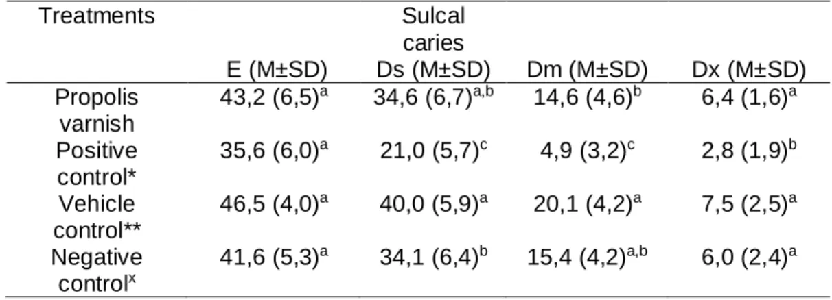
observed. However, regarding the severity, Duraphat® had better results, showing statistically significant difference (p <0.05) between all groups (Table 3).
No macroscopic tissue changes or abnormalities in the animals of all groups were
observed.
DISCUSSION
Propolis has many biological activities that attracts many researches in
Dentistry field. The deepening of scientific research in the chemical composition
of various types of propolis helped to envision their potential use and application
in dental practice [Libério et al., 2009; Więckiewicz et al., 2013; Kumar, 2014].
Previous studies have demonstrated the antimicrobial activity of propolis
on several bacteria of the oral microbiota, including Streptococcus mutans
(ANAUATE NETO et al., 2013; BARRIENTOS et al., 2013; TULSANI et al., 2014;
MOHSIN et al., 2015). In this research, this activity had no difference compared
to the other groups. While this result may mean that the propolis varnish had no
satisfactory antimicrobial activity, one must take into consideration that this effect
was only evaluated at one endpoint, when probably the material was no longer
us informations about the Streptococcus mutans recovery levels, how long
propolis antimicrobial activity lasted and re-evaluate the need for the product’s
reapplication.
Propolis varnish had low anti-caries activity in the experiment with high
cariogenic challenge in a rodent model. The results of this study diverge from
previous research of propolis anti-caries effect in rats [Koo et al., 1999; Koo et
al., 2002; Hayacibara et al., 2005; Duarte et al., 2006; Bueno-Silva et al., 2013],
which have achieved a significant reduction in caries activity. In these studies,
propolis ethanolic extracts, or its bioactive fractions, were applied topically on
rats’ teeth twice a day for a period of 4 to 5 weeks, resulting in an anti-caries
activity similar to Duraphat®. This result suggests that the continuing presence of
propolis enabled a more effective antimicrobial activity, leading to the reduction
of caries lesions.
Chitosan, which is the vehicle of this product, is a biocompatible and
biodegradable polymeric material [Uysal et al., 2011] with non-toxic and
antimicrobial properties [Busscher et al., 2008]. In this study, this vehicle was
inert as the severity of dental caries increased. This may be due to the medium
molecular weight of the chitosan used in this formulation, which is different from
the low molecular weight chitosan generally used in other studies that proven the
antimicrobial activity in other dental products [No et al., 2002; Decker et al., 2005;
Salamat-Miller et al., 2005; Uysal et al., 2011; Chen & Chung, 2012; Costa et al.,
2013; Costa et al., 2014]. Changes in the molecular weight of chitosan used in
the varnish formulation preclude the polymeric film formation, which is the feature
that allows the adhesion of product to the teeth.
Fluoride, considered the gold standard in caries prevention, has little
antimicrobial effect, acting on the synthesis of glucan and on the acid tolerance
of S. mutans [Jeon et al., 2011]. Duraphat®, a fluoride varnish used as positive control in this experiment, showed no statistical difference in the decrease of total
microbiota between the groups, but significantly reduced the severity of dental
caries (p<0.05). The role of this product in the development of caries is interfering
of incipient lesions [Dawes & ten Cate 1990]. It could clearly be seen in our study
because, as the severity of carious lesions were increasing, the difference
between the groups became remarkable.
On the other hand, despite propolis antimicrobial properties [Libério et al.,
2009; Barrientos et al., 2013], as tooth decay was becoming more severe,
propolis varnish was not effective to reduce incidence of caries lesions. Its lack
of residual effect shows that propolis activity occurs while it is in direct contact
with oral tissues. Repeated and prolonged applications of this new product could
reduce cariogenic bacteria, and thus, achieve effective anti-caries activity.
Changes in varnish application protocol should be based on the
microbiological evaluation of the recovery of the initial number of the targeted
bacteria. Only at the end of the study these bacteria were quantified, which is a
limitation. If this quantification was made in regular times, we could have
information about the need of reapplication of the product, which could enhance
the anti-caries results of all groups.
Further studies are needed to develop an antimicrobial supression
protocol that enables the maintenance of a longer antimicrobial activity and thus,
the effective prevention of dental caries lesions.
ACKNOWLEDGMENTS
The authors are grateful to CNPq for scholarship; Eliane Franco and José
Carlos for technical support; Bruna Benso, Carina Denny, Irlan Almeida Freires,
Laila Facin, Lívia Galvão, Camila Batista, Luiz Eduardo Ferreira and Cleiton P.
LEGENDS
Table 1: *Duraphat®; Vehicle **(chitosan varnish); and xwithout treatment. All
values showed no significant difference ANOVA, comparison between all pairs
using Tukey-Kramer HSD (p <0.05). Different superscript letters indicate
significant differences between treatments.
Table 2: *Duraphat®; Vehicle **(chitosan varnish); and xwithout treatment. All
values showed no significant difference ANOVA, comparison between all pairs
using Tukey-Kramer HSD (p <0.05). Different superscript letters indicate
significant differences between treatments.
Table 3: *Duraphat®; Vehicle **(chitosan varnish); and xwithout treatment. All values showed no significant difference ANOVA, comparison between all pairs
using Tukey-Kramer HSD (p <0.05). Different superscript letters indicate
significant differences between treatments.
TABLES
Table 1 - Analysis of effects on the total oral microbiota and Steptococcus mutans (mean and standard deviation/M±SD) submitted to cariogenic challenge for four weeks with propolis varnish and its controls.
Treatments Total microbiota (CFU/mL) (M±SD) Total S. mutans (CFU/mL) (M±SD)
% S. mutans (M±SD)
Propolis varnish
75.4 (7,8)a 37.6 (4,2)a 40.7 (11,3)a
Positive control*
43.5 (6,0)a 24.4 (3,7)a 46.2 (15,5)a
Vehicle control**
74.3 (8,1)a 38.7 (4,7)a 47.1 (7,8)a
Negative controlx
Table 2 - Effect of propolis varnish (mean and standard deviation/M±SD) in the development of smooth surface caries and severity (E - enamel caries; Ds - slight dentinal caries; Dm - moderate dentinal caries; Dx - extensive dentinal caries) in rats using classification Keyes' modified by Larson.
Treatments Smooth-surface caries
E (M±SD) Ds (M±SD) Dm (M±SD) Dx (M±SD) Propolis
varnish
19,8 (1,5)b 13,4 (5,2)b,c 1,4 (1,4)a 0,1 (0,3)a
Positive control*
16,5 (1,6)b 10,3 (4,6)c 0,1 (0,4)b 0,0 a
Vehicle control**
33,5 (1,6)a 19,8 (5,9)a 1,2 (1,4)a,b 0,1 (0,3)a
Negative controlx
28,3 (1,5)a 16,5 (4,5)a,b 1,1 (1,0)a 0,3 (0,6)a
Table 3 - Effect of propolis varnish (mean and standard deviation) in the development of sulcal caries and severity (E - enamel caries; Ds - slight dentinal caries; Dm - moderate dentinal caries; Dx - extensive dentinal caries) in rats using classification Keyes' modified by Larson.
Treatments Sulcal
caries
E (M±SD) Ds (M±SD) Dm (M±SD) Dx (M±SD) Propolis
varnish
43,2 (6,5)a 34,6 (6,7)a,b 14,6 (4,6)b 6,4 (1,6)a
Positive control*
35,6 (6,0)a 21,0 (5,7)c 4,9 (3,2)c 2,8 (1,9)b
Vehicle control**
46,5 (4,0)a 40,0 (5,9)a 20,1 (4,2)a 7,5 (2,5)a
Negative controlx
REFERENCES
Petersen PE, Bourgeois D, Ogawa H, Estupinan-Day S, Ndiaye C: The global burden of oral diseases and risks to oral health. Bull World Health Organ 2005, 83:661-69.
Wolff D, Frese C, Maier-Kraus T, Krueger T, Wolff B: Bacterial biofilm composition in caries and caries-free subjects. Caries Res DOI: 10.1159/000344022.
Beighton D: The complex oral microflora of high-risk individuals and groups and its role in the caries process. Community Dent Oral Epidemiol 2005; 33: 248–55.
Choi EJ, Lee SH, Kim YJ: Quantitative real-time polymerase chain reaction for
Streptococcus mutans and Streptococcus sobrinus in dental plaque samples and
its association with early childhood caries. Int J Paediatr Dent DOI: 10.1111/j.1365-263X.2008.00942.x.
Head DA, Marsh PD, Devine DA: Non-lethal control of the cariogenic potential of an agent-based model for dental plaque. PLoS ONE DOI:10.1371/journal.pone.0105012.
Jeon JG, Rosalen PL, Falsetta ML, Koo H. 2011: Natural products in caries research: current (limited) knowledge, challenges and future perspective. Caries Res DOI: 10.1159/000327250.
.
Mishra BB, Tiwari VK: Natural products: An evolving role in future drug discovery. Eur J Med Chem 2011; 46:4769-4807.
Libério SA, Pereira AL, Araújo MJ, Dutra RP, Nascimento FR, Monteiro-Neto V, Ribeiro MN, Gonçalves AG, Guerra RN: The potential use of propolis as a cariostatic agent and its actions on mutans group streptococci. J Ethnopharmacol DOI:10.1016/j.jep.2009.04.047.
Ikeno, K.; Ikeno, T.; Miyazawa, C: Effects of propolis in dental caries in rats.
Caries Res Basel 1991;25: 347-351.
Duarte S, Rosalen PL, Hayacibara MF, Cury JA, Bowen WH, Marquis RE, et al.: The influence of a novel propolis on mutans streptococci biofilms and caries development in rats. Arch Oral Biol 2006;51:15-22.
Kumar, LSV: Propolis in dentistry and oral cancer management. N Am J Med Sci 2014;6(6):250-9. DOI: 10.4103/1947-2714.134369.
Koo H, Rosalen PL, Cury JA, Park YK, Ikegaki M, Sattler A: Effect of Apis mellifera propolis from two Brazilian regions on caries development in desalivated rats. Caries Res 1999;33(5):393-400.
Petersson LG, Twetman S, Dahlgren H, Norlund A, Holm AK, Nordenram G, et
al.: Professional fluoride varnish treatment for caries control: a systematic review
of clinical trials. Acta Odontol Scand 2004;62(3):170-6.
Marinho VCC, Worthington HV, Walsh T, Clarkson JE. Fluoride varnishes for preventing dental caries in children and adolescents. Cochrane Database Syst Rev DOI: 10.1002/14651858.CD002279.pub2.
Jayabal J, Mahesh R: Current state of topical antimicrobial therapy in
management of early childhood caries. ISRN Dent DOI: 10.1155/2014/762458.
De Luca MP, Franca JR, Macedo FA, Grenho L, Cortes ME, Faraco AA, Moreira AN, Santos VR: Propolis varnish: antimicrobial properties against cariogenic bacteria, cytotoxicity, and sustained-release profile. Biomed Res Int 2014. DOI: 10.1155/2014/348647.
Keyes PH. Dental caries in the Syrian hamster. VIII. The induction of rampant caries activity in albino and golden animals. J Dent Res 1959;38:525-33.
Murata RM, Branco-de-Almeida LS, Franco EM, Yatsuda R, Santos MH, Alencar SM, Koo H, Rosalen PL: Inhibition of Streptococcus mutans biofilm accumulation and development of dental caries in vivo by 7-epiclusianone and fluoride. Biofouling DOI:10.1080/08927014.2010.527435.
Bowen WH, Madison KM, Pearson SK: Influence of desalivation in rats on incidence of caries in intact cagemates. J Dent Res 1988;67:1316-18.
Larson RM. Merits and modifications of scoring rat dental caries by Keyes’ method. In: Animal Models in Cariology. Washington DC, IRL Press, 1981.195-203.
Bueno-Silva B, Koo H, Falsetta ML, Alencar SM, Ikegaki M, Rosalen PL: Effect of neovestitol–vestitol containing Brazilian red propolis on accumulation of biofilm
in vitro and development of dental caries in vivo, Biofouling DOI:
10.1080/08927014.2013.834050.
Koo H, Pearson SK, Scott-Anne K, Abranches J, Cury JA, Rosalen PL, Park YK, Marquis RE, Bowen WH: Effects of apigenin and tt-farnesol on glucosyltransferase activity, biofilm viability and caries development in rats. Oral Microbiol Immunol 2002;17(6):337-43.
Busscher HJ, Engels E, Dijkstra RJ, van der Mei HC: Influence of a chitosan on oral bacterial adhesion and growth in vitro. Eur J Oral Sci DOI: 10.1111/j.1600-0722.2008.00568.x.
No HK, Park NY, Lee SH, Meyers SP. Antibacterial activity of chitosans and chitosan oligomers with different molecular weights. Int J Food Microbiol 2002;74:65-72.
Decker EM, Von Ohle C, Weiger R, Wiech I, Brecx M. A synergistic chlorhexidine/chitosan combination for improved antiplaque strategies. J Periodont Res 2005; 40:373–377.
Salamat-Miller N, Chittchang M, Johnston TP: The use of mucoadhesive polymers in buccal drug delivery. Adv Drug Deliv Rev 2005; 57(11):1666-91.
Chen CY, Chung YC: Antibacterial effect of water-soluble chitosan on representative dental pathogens Streptococcus mutans and Lactobacilli brevis. J Appl Oral Sci 2012;20(6):620-7.
Costa EM, Silva S, Tavaria FK, Pintado MM: Study of the effects of chitosan upon Streptococcus mutans adherence and biofilm formation. Anaerobe2013; 20:27-31.
Costa EM, Silva S, Madureira AR, Cardelle-Cobas A, Tavaria FK, Pintado MM: A comprehensive study into the impact of a chitosan mouthwash upon oral microorganism’s biofilm formation in vitro. Carbohydr Polym 2014;101: 1081– 1086.
Dawes C, ten Cate JM: International symposium on fluorides:
mechanisms of action and recommendations for use. J Dent Res1990; 69:505– 836.
Barrientos L, Herrera CL, Montenegro G, Ortega X, Veloz J, Alvear M, et al.: Chemical and botanical characterization of Chilean propolis and biological activity on cariogenic bacteria Streptococcus mutans and Streptococcus sobrinus. Braz J Microbiol2013; 44(2):577-585.
5.2 ARTIGO 2
A avaliação do efeito do verniz na redução de Streptococcus mutans na
saliva de crianças de 8 a 11 anos foi realizada na cidade de Belo Horizonte-MG.
A aplicação do verniz foi realizada na clínica de Odontopediatria da FO-UFMG e
a inclusão na pesquisa foi feita a partir da assinatura do consentimento livre
esclarecido (APÊNDICE 1). A pesquisa clínica seguiu a aplicação única do
produto no início do tratamento, seguindo com coletas diárias até o 5º dia e no
10º, 15º e 30º dia após a aplicação do verniz. Neste artigo intitulado: “Evaluation
of reduction in salivary levels of Streptococcus mutans after propolis varnish
single application.”, comparamos a quantidade de SM na saliva e em biofilme em
diferentes tempos. Este estudo foi aprovado pelo Comitê de Ética em Pesquisa
da UFMG (parecer nº 168.394) (ANEXO B) e registrado na base de dados de
ensaios clínicos (Clinical Trials ID: 10726612.8.0000.5149) (ANEXO C).
“Evaluation of reduction in salivary levels of Streptococcus mutans after propolis varnish single application”
Mariana Passos De Luca1, Miriam Pimenta Parreira Vale1, Patrícia Corrêa-Faria1, Suzane Gonçalves Paixão1, Vagner Rodrigues Santos2
1
Department of Paediatric Dentistry and Orthodontics, Faculty of Dentistry, Federal University of Minas Gerais, Av. Antonio Carlos, 6.627, Pampulha, 31.270-901, Belo Horizonte, MG, Brazil
2 Department of Oral Pathology and Oral Surgery, Faculty of Dentistry, Federal
University of Minas Gerais, Av. Presidente Antônio Carlos, 6.627, 31.270-901 Belo Horizonte, MG, Brazil
Corresponding author:
Prof. Dr. Vagner Rodrigues Santos
Avenida Presidente Antônio Carlos 6627, 31.270-901 Belo Horizonte, MG,
ABSTRACT
The aim of this study was to evaluate the efficacy of an experimental
propolis varnish containing 15% of propolis ethanolic extract in reducing salivary
levels of Streptococcus mutans (SM). Caries-free patients with age varying from
8 to 11 years were recruited from a preventive program of the pediatric dentistry
clinic of UFMG. After profilaxis, propolis varnish was applied in all tooth surface
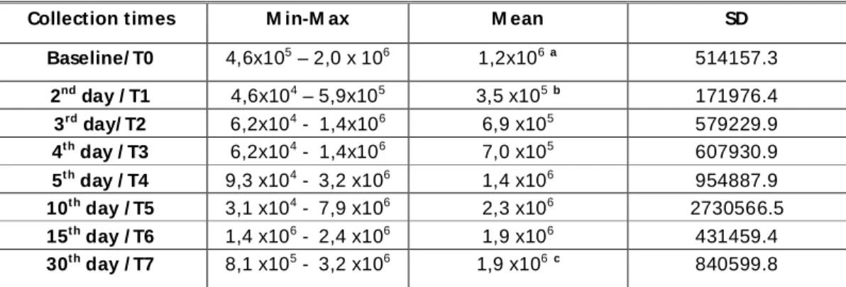
and saliva samples were collected in regular times (Baseline/T0, 2nd day/T1, 3rd
day/T2, 4th day/T3, 5th day/T4, 10th day/T5, 15th day/T6 and 30rd day/T7). At each
sampling, subjects chewed a 1 cm piece of a sterilized latex tube to stimulate
whole saliva, which was homogenized for 30 s and ten-fold serial dilutions in
saline were plated in duplicate on a petri dish containing
mitis-salivarius-bacitracin (MSB) agar. The plates were incubated at 37ºC for 48 h. Colonies with
typical morphology were counted and the number of CFU/mL was compared
using independent sample test t. There was significant difference between T0 /
T1 (baseline and 2nd day), and T1 / T7 (2nd day/30th day), but no difference was verified from the 4th day forward. Based on the results, the varnish enabled the maintainance of reduction of SM salivary levels for 3 days. If these findings would
INTRODUCTION
Dental caries is a disease with identified etiology and able to be prevented and controlled (Pitts, 2004). It is proven that caries development is dependent on the stagnation of an organized biofilm on the dental surface, and its metabolic activity is the fuel for the development of this disease (Kidd, 2004; Hesse et al., 2014). The predominance of bacteria with aciduric and acidogenic characteristcs, using sucrose as a substrate for synthesis of exopolysaccharide, lead to the development of virulent biofilms on susceptible tooth surfaces (Bowen & Koo, 2011; Bueno-Silva et al., 2013).
Many researches suggest that Streptococcus mutans (SM) are the major pathogens of human dental caries, often associated with the virulence of the biofilm (Jeon et al., 2011). This bacteria is usually isolated from caries lesions and is generally used in animals experiments for induction of caries formation (Hamada & Slade, 1980; Loesche, 1986). In addition, SM has the ability to produce water-insoluble glucan, promoting its adhesion to the tooth surface and to other bacteria (Hamada & Slade, 1980; Takahashi & Nyvad, 2008).
Antimicrobial treatments have been an important part of preventive dentistry for a long time and often the main purpose of these treatments is the reduction of Streptococcus mutans in the oral cavity (Ekenbäck et al., 2000). This bacteria is very sensitive in treatments with chlorhexidine, and the formulation as a varnish reduces its side effects and allows prolonged release of its components (Jentsch et al., 2014).
Dental varnishes have been designed with the intention of being adhered to the tooth surface and thereby, through this intimate contact, enable the main active ingredient present in the formulation to be more easily incorporated, and maintain its activity for a longer period. These preparations differ in the polymeric matrix, pharmaceutical additives and therapeutical agents, being the most common fluoride and chlorhexidine (Steinberg et al., 2001).
Other varnishes formulations can be found for preventive purposes (Steinberg et al., 2001; Cardoso et al., 2014). Here in Brazil, only fluoride varnishes are commercialized, although there is little evidence that fluoride at levels that can occur naturally in dental biofilms could significantly change the number or proportions of species found in this dynamic microbiome, not effectively addressing the infectious character of the disease (Killian et al., 1979; Ekenbäck et al., 2000; Bueno-Silva et al., 2013).
Natural products are being tested as a potential source of new and active therapeutic agents (Newman & Cragg, 2012). Propolis is one of the main source of biologically active compounds that exhibit proven antimicrobial activity, with potential anti-biofilm and anticaries activities (Koo, et al. 2002; Bueno-Silva et al., 2013). Although this product has shown promising results in studies in vitro, only a few studies were performed with the objective of evaluating its possible use in anti-biofilm and anticaries chemotherapy using a clinical treatment protocol in
vivo (Jeon et al.,2011). In this study, we have used propolis type 12, also known
as “green propolis”, found in southeastern Brazil (Park et al., 2002; Bueno-Silva
Due to the paucity of studies in dental literature in this regard, this clinical study aimed to investigate the efficacy of an experimental varnish containing 15% of propolis type 12 (green propolis) ethanolic extract on the reduction of salivary levels of Streptococcus mutans in children for a 30 days period.
MATERIALS AND METHODS
The present study was approved by the ethics committee of Federal University of Minas Gerais (report nº 168.394) and registered in Clinical Trials data base (ID: 10726612.8.0000.5149). This clinical study was conducted to evaluate the effect of propolis varnish application on S. mutans counts in saliva of children aged 8-11 years, from both genders, who were recruited from public shelters registered and attended by the project of prevention and oral health maintenance of UFMG, totaling 11 children. Those responsible for the shelters received an invitation letter explaining the purpose of the study and signed their informed consent. The inclusion criteria were: (i) children 8-11 years in mixed dentition, with at least eight permanent teeth, (ii) residing in the greater area of Belo Horizonte, MG (iii) good health condition and (iv) children not making use of mouthwashes or medications, especially antibiotics, until three weeks prior to the beginning of the experiment.Children with active caries lesions at the time of clinical examination were excluded from the study. The difference in the level of ‘colony-forming units’ (CFU) of S. mutans at different time intervals such as at the baseline, after 1 day to the 5th day, at the 10th day, at the 15th day and at the end
of 30 days was compared.
Saliva samples were obtained from each individual initially prior to the start of the experiment to establish baseline SM levels. Volunteers were informed to refrain from oral hygiene procedures for 12 h prior to collection. The total volume of 360 µL of propolis varnish was applied in all tooth surfaces (volume set in a prior pilot study). At each sampling, subjects chewed a 1 cm piece of a sterilized latex tube to stimulate whole saliva, and the samples were collected in sterile bottles with no eating/drinking for two hours prior to the sampling. No transport medium was used as culturing was done within half an hour of collection of samples (Rupesh et al., 2014). Stimulated saliva was homogenized in a Thermolyne mixer (Barnsted International, Dubuque, IA, USA) for 30 s and ten-fold serial dilutions in saline were plated in duplicate on a petri dish (Inlab, Diadema, SP, Brazil) containing mitis-salivarius-bacitracin (MSB) agar (Difco Laboratories, Detroit, MI, USA), by the drop-counting technique (Miles et al., 1938). The plates were incubated in microaerophilic condition at 37ºC for 48 h. Only colonies with typical morphology were counted and the number of CFU/ml of saliva was calculated by multiplying the colony count of each plate with its respective dilution fator (Tenuta et al., 2003).
STATISTICAL ANALYSIS
All data were processed by the SPSS 19.0 software. The comparisons between groups with normal distribution were tested using independent sample t test, and the Wilcoxon test when the distribution was not normal (10th day). The