Abstract
Submitted: June 23, 2017
Modification: August 28, 2017 Accepted: September 25, 2017
Analysis of the antimicrobial and
anti-caries effects of TiF
4
varnish under
microcosm biofilm formed on enamel
Titanium tetrafluoride (TiF4) is known for interacting with enamel reducing
demineralization. However, no information is available about its potential antimicrobial effect. Objectives: This study evaluated the antimicrobial and anti-caries potential of TiF4 varnish compared to NaF varnish, chlorhexidine gel (positive control), placebo varnish and untreated (negative controls) using a dental microcosm biofilm model. Material and Methods: A microcosm biofilm was produced on bovine enamel previously treated with the varnishes, using inoculum from human saliva mixed with McBain saliva, under 0.2% sucrose exposure, for 14 days. All experiments were performed in biological triplicate (n=4/group in each experiment). Factors evaluated were: bacterial viability (% dead and live bacteria); CFU counting (log10 CFU/
mL); and enamel demineralization (transverse microradiography – TMR). Data were analysed using ANOVA/Tukey’s test or Kruskal-Wallis/Dunn’s test (p<0.05). Results: Only chlorhexidine significantly increased the number of dead bacteria (68.8±13.1% dead bacteria) compared to untreated control (48.9±16.1% dead bacteria). No treatment reduced the CFU counting (total microorganism and total streptococci) compared to the negative controls. Only TiF4 was able to reduce enamel demineralization (ΔZ 1110.7±803.2 vol% μm) compared to both negative controls (untreated: ΔZ 4455.3±1176.4 vol% μm). Conclusions: TiF4 varnish has no relevant antimicrobial effect.
Nevertheless, TiF4 varnish was effective in reducing enamel demineralization under this model.
Keywords: Dental biofilm. Enamel caries. Fluoride. Titanium. Varnish. Beatriz Martines de SOUZA1
Constantino FERNANDES NETO1
Priscila Maria Aranda SALOMÃO1
Layla Reginna Silva Munhoz de
VASCONCELOS2
Flaviana Bombarda de ANDRADE2
Ana Carolina MAGALHÃES1
1Universidade de São Paulo, Faculdade de Odontologia de Bauru, Departamento de Ciências
Biológicas, Bauru, São Paulo, Brasil.
2Universidade de São Paulo, Faculdade de Odontologia de Bauru, Departamento de Dentística,
Endodontia e Materiais Odontológicos, Bauru, São Paulo, Brasil. Corresponding address:
Ana Carolina Magalhães Departamento de Ciências Biológicas - Faculdade de Odontologia de Bauru - Universidade de São Paulo. Al. Dr. Octávio Pinheiro Brisolla, 975
Introduction
Dental biofilm is defined as a community of
microorganisms that colonizes the oral cavity,
dimensionally arranged and enclosed into an
extracellular matrix rich in polysaccharides, proteins/ amino acids, environmental DNA (eDNA) and
minerals. The exposure to sucrose from diet may
favor the development of a cariogenic biofilm rich
in acidogenic and aciduric bacteria and extracellular
polysaccharides25. Hygiene habits, diet, salivary flow and antimicrobial agents may modulate the quantity
and quality of the dental biofilm19. Considering the
protective factors, fluorides and antimicrobials are
among the most studied agents5,6,12,14,21-23.
Fluoride has anticaries effect mainly due to its
action reducing demineralization and improving
remineralization of the tooth structure. Secondarily,
it can provide some antimicrobial effect by reducing
bacterial metabolism and interfering in protons
extrusion27. Studies have shown that certain
concentrations of fluoride (10, 50 and 125 ppm F-, 5
min/day) are effective in reducing acid production and
acid tolerance as well as extracellular polysaccharide
formation of Streptococcus mutans (S. mutans)
biofilm22. Recently, Pandit et al.20 (2015) showed that
1 min of application of ≥300 ppm F was able to control
cariogenic biofilm through inhibition of virulence
properties. All previous studies have been done using
a monospecies biofilm (46-h to 74-h-old biofilm) and
testing NaF as F- source20-22. Generally, NaF affects
the virulence factors, but not the bacteria viability20.
The antimicrobial effect of fluoride depends on
its concentration20,21. Varnish is the highest fluoride
concentrated vehicle, with the advantage of having
resinous base, which allows a long contact time with
the tooth surface16. Most varnishes contain NaF as active agent, which has shown to be able to protect the
teeth against dental caries when applied twice a year
(46% of preventive fraction in permanent dentition)16. On the other hand, our research group has tested
the anticariogenic effect of an experimental 4% TiF4
varnish compared to 5.42% NaF varnish under abiotic
environment15. Our results have shown greater effect
of TiF4 varnish compared to NaF varnish due to its
chemical reaction with enamel surface, promoting
deposition of Ti compounds with high acid resistance.
However, none of our studies have tested its potential
as antimicrobial agent, another possible mechanism
of action related to fluorides. We expected that the
glaze layer produced by TiF4 varnish could alter the
microorganism adhesion and, consequently, the biofilm
growth and viability.
The use of microcosm biofilm, produced from microorganisms in human saliva, can benefit studies with monospecies or multispecies biofilms, allowing
the presence of high number of microorganisms
and interactions between them in the presence of
fluoride or antimicrobial agents. Considering that 1) most studies on antimicrobial action of fluoride have
been done using monospecies or dual-species4,12,20-22
and 2) the lack of knowledge on the antimicrobial
effect of TiF4, the aim of this study was to compare
the antimicrobial and anticariogenic effects of TiF4
varnish with NaF varnish, chlorhexidine gel (positive control), placebo varnish (without any active agent) and untreated specimens (negative controls) using a microcosm biofilm model on bovine enamel.
This research tested the following null hypotheses:
1) There is no significant difference between the
fluoride varnishes and positive control on the
microbial viability; 2) There is no significant difference between the fluoride varnishes and positive control on CFU counting for total microorganisms and total
streptococci; 3) There is no significant difference
between the fluoride varnishes and positive control
in reducing enamel demineralization.
Material and methods
Saliva collection
This study was firstly approved by the local Ethical Committee (CEEA 38143714.7.0000.5417). Saliva was collected from 2 healthy donors, who fit the following inclusion criteria: 1) normal salivary flow (stimulated saliva flow >1 mL/min and non-stimulated saliva flow >0.3 mL/min), 2) with previous history of caries, but no current active caries (no active white spot and/ or cavitated lesions), 3) with no gingivitis (red or
blooding gingival tissue) and 4) with no history of
antibiotic intake in the last 3 months. Prior to the day
of collection, the donors did not brush their teeth.
Furthermore, they were not allowed to ingest food
or drinks in the last 2h before saliva collection. The
saliva was collected under stimulation by chewing a
rubber material for 10 min during the morning. After
and 30% glycerol). Aliquots of 1 mL were stored in -80°C23.
Tooth sample preparation and treatment
One hundred twenty (60 for viability assay and 60 for CFU counting) enamel samples (4 mm x 4
mm) were prepared from bovine teeth, using a
semi-precision cutting machine (Buehler; Lake Bluff,
Illinois, USA). The samples were fixed in acrylic discs with wax and polished in a metallographic polishing
machine (Arotec; Cotia, São Paulo, Brazil) using water-cooled silicon-carbide discs (600-grade papers ANSI grit. Buehler, USA) to achieve a flat surface and to
standardize the surface roughness of approximately
0.133±0.029 µm. The average surface roughness (Ra) was assessed using a contact profilometer and Mahr Surf XCR 20 software (Mahr; Göttingen, Lower
Saxony, Germany). Two thirds of the enamel surfaces
were protected with nail varnish to obtain control areas
for the transverse microradiography (TMR) analysis. Enamel samples were randomly divided among study groups according to the Ra values: A) 4.0% TiF4
varnish (pH 1.0, 2.45% F-); B) 5.42% NaF varnish
(pH 5.0, 2.45% F-); C) 2% chlorhexidine gel – CHX
(pH 6.0) – Positive control; D) placebo varnish (pH
5.0) and E) untreated specimens – Negative controls.
The varnishes were produced by FGM Produtos Odontológicos LTDA (Joinville, Santa Catarina, Brazil) and contained artificial resin as base and ethanol as solvent. During 6 h-treatment7,15, the samples were immersed in remineralizing solution13. Thereafter, the
varnishes and gel were removed using scalpel blade
and the samples were cleaned with swab soaked
in acetone-water solution (1:1). Two-thirds of the
samples surfaces were protected again and they were
then stored in artificial saliva overnight, until they were used for the microcosm biofilm formation.
Microcosm biofilm formation
The human saliva was defrosted and mixed with
McBain artificial saliva18 in a proportion of 1:50. The
McBain saliva contained 2.5 g/L mucin from porcine stomach (type II), 2.0 g/L bacteriological peptone, 2.0 g/L tryptone, 1.0 g/L yeast extract, 0.35 g/L NaCl, 0.2 g/L KCl, 0.2 g/L CaCl2, 0.1 g/L cysteine
hydrochloride, 0.001 g/L hemin, 0.0002 g/L vitamin K1, at pH 7.0. All reagents were from Sigma-Aldrich (St. Louis, Missouri, USA). The solution of human saliva and McBain saliva was added to each well containing a treated enamel sample (v=1.5 mL/well) in a 24-well
plate, which was incubated at 5% CO2 and 37°C, for 8
h. Subsequently, the enamel samples were transferred
to wells containing fresh McBain saliva with 0.2%
sucrose and incubated at the same conditions. After
16 h, the samples were again transferred to new wells containing fresh McBain saliva with 0.2% sucrose
and incubated for 24 h at the same conditions30. This
procedure was repeated each 24 h, for a total time
of 14 days.
Bacterial viability analysis
After 14 days, the samples were immersed in
phosphate-buffered saline (PBS) solution (twice for 5
s) under stirring to remove unattached bacteria. The
biofilm was stained using the nucleic acid markers diluted in PBS (1 mL PBS + 1 µL SYTO9 + 1 µL propidium iodide, 10 µL/well) (Kit Live & Dead® cells
viability assay, Thermo Fisher Scientific; Waltham, Massachusetts, USA) for 15 min in a dark environment. Live bacteria were stained with SYTO9 producing a green fluorescence and dead lysed bacteria were stained with propidium iodide/SYTO9 producing a red fluorescence10. Biofilm was examined using
confocal laser scanning microscope (Leica TCS SPE, Leica Mannheim; Wetzlar, Hesse, Germany) and Leica
Application Suite-Advanced Fluorescence software
(LAS AF, Leica Mannheim; Wetzlar, Hesse, Germany). Three images (275 μm2) were captured from each
sample surface and analysed using BioImage L 2.0 software, to quantify the live and dead bacteria (%).
Colony forming unit (CFU) counting
The samples were immersed in PBS solution (twice for 5 s) under stirring to remove unattached bacteria.
The samples were then transferred to microtubes
containing 1 mL of Brain Heart Infusion (BHI, Difco; Lawrence, Kansas, USA) and vortexed at 2400 rpm for 30 s (vortex 251, Fanem; Guarulhos, São Paulo, Brazil). The bacterial suspension was then diluted to
10-4 and spread on petri dishes (50 µL/dish) containing
two different types of agar for CFU counting: 1) tryptic soy blood agar with 5% sheep blood for total
microorganisms5 and 2) mitis salivarius agar (MSA)
containing 15% sucrose and 1% potassium tellurite for
total streptococci14. The dishes were stored at 5% CO 2
and 37°C. After 72 h, the CFU numbers were counted
and transformed in log10 CFU/mL.
Transverse microradiography (TMR)
at the center of the surface, perpendicularly to the
orientation of the protective nail varnish, allowing all
enamel areas (sound and demineralized) to be included in the TMR specimens. The specimens were polished to obtain slices with 80-100 µm of thickness. Enamel slices were fixed in a sample-holder together with an
aluminium calibration step wedge with 14 steps. A
microradiograph was taken using an x-ray generator
(Softex; Tokyo, Japan) on the glass plate at 20 kV and 20 mA (at a distance of 42 cm) for 13 min. The glass plates were developed for 7 min, rinsed in deionized water, fixed for 7 min in a dark environment, and then rinsed in running water for 10 min and air-dried (all procedures were done at 20°C). The developed plate
was analysed using a transmitted light microscope
fitted with a 20x objective (Zeiss, Oberkochen; Baden-Württemberg, Germany), a CCD camera (Canon; Tokyo, Japan), and a computer. Two images per sample
were taken using data-acquisition (version 2012) and interpreted using calculation (version 2006) software from Inspektor Research System bv (Amsterdam,
North Holland, The Netherlands). The mineral content
was calculated considering the density of the mineral
to be 3.15 kg L-1 and 87 vol% of mineral content
for the sound enamel. The lesion depth (LD, µm), the integrated mineral loss (∆Z, vol% µm) and the average mineral loss over the lesion depth (R, vol%)
were calculated7.
Statistical number and analysis
All biofilm analyses were done in biological triplicate (n=4/each experiment, final number=12) while all enamel samples from both analyses (% dead and
log10 CFU/mL) were applied for TMR (final number=24,
∆Z, LD and R). The sample number calculation for biofilm analysis was based on previous work30. Data were statistically analysed using the software
Graph Pad Instat for Windows (GraphPad Software; La Jolla, California, USA). Normal distribution and homogeneity were checked using Kolmogorov-Smirnov and Bartlett’s tests, respectively. Ordinary ANOVA followed by Tukey’s test were applied to compare the
different treatments for all analyses except LD. In case
of LD, Kruskal-Wallis followed by Dunn’s test were performed. A significance level of 5% was considered
for all statistical tests.
Results
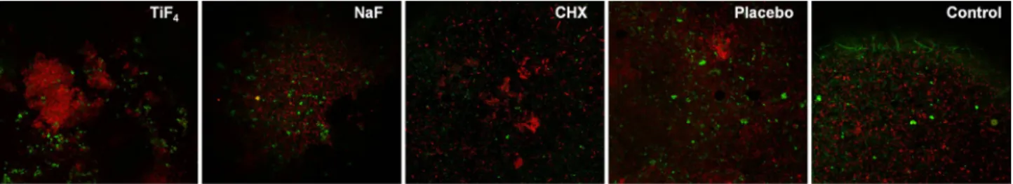
In respect to the biofilm viability, only chlorhexidine was able to significantly increase the number of dead bacteria compared to untreated control (p<0.002), but it did not significantly differ from placebo. No significant differences were found between fluoride treatments or among fluoride treatments and negative
or positive control. Figure 1 shows a representative
confocal picture of the biofilm from each treatment group and Figure 2 shows the viability data. CFU
Figure 1- Representative confocal images of the biofilm formed on enamel samples. *CHX=chlorhexidine
counting for total microorganism and total streptococci
showed no significant differences among treatments (p>0.05) (Table 1).
TiF4 and NaF varnishes were similarly able to
significantly reduce the integrated mineral loss
and the average mineral loss compared to the
untreated group and chlorhexidine, while only TiF4
was significantly different from placebo varnish.
Both TiF4 and NaF reduced lesion depth compared to
control, but only TiF4 was significantly different from placebo. Treatment with chlorhexidine did not reduce
enamel demineralization (Table 2). Figure 3 shows a representative TMR picture of demineralized enamel
from each treatment group.
Discussion
This experimental model allowed the formation of
a biofilm based on saliva, reproducing the complex
relationship between salivary components and
bacterial species, as proposed in the microcosm biofilm
model. A sucrose supplementation was provided to
favor the proliferation of cariogenic species in the
biofilm, which can induce tooth demineralization28.
Microcosm model is a validated method to test the effects of complex biofilms on tooth24. The donors
were submitted to a complete screening for a better
selection of the source of microorganisms (saliva).
However, a recent study investigated the effect of
different types of inoculum (saliva and dental biofilm)
from caries-active and caries-free individuals on the
cariogenic potential of biofilm produced in vitro. The
Treatment Total microorganism Total Streptococci
TiF4 7.04±0.20a 6.83±0.26a
NaF 7.02±0.18a 6.97±0.22a
Chlorhexidine 6.97±0.30a 6.92±0.25a
Placebo 7.14±0.22a 6.93±0.25a
Control 7.03±0.09a 7.04±0.17a
Similar letters show no significant differences among the treatments (per column).
ANOVA and Tukey’s test (n=12, p>0.05)
Table 1- Mean and standard deviation (SD) of colony forming units (CFU) counting (log10 CFU/mL) for total microorganisms and total streptococci in the biofilm formed on enamel after applying the treatments tested
Treatment ΔZ (%vol. μm) R (%vol) LD (μm)
TiF4 1100.7±803.2c 19.9±3.7c 68.3±33.4c
NaF 1680.0±538.3bc 20.9±4.9bc 86.9±29.1bc
Chlorhexidine 3262.7±909.6a 33.9±6.4a 104.3±29.2abc
Placebo 3374.6±1636.9ab 28.9±6.5ab 162.3±64.7ab
Control 4455.3±1176.4a 29.4±5.3a 156.1±27.1a
Different letters show significant differences among the treatments (per column).
ΔZ and R-values are displayed as mean ± SD (ANOVA and Tukey’s test, p<0.0001). LD is presented as median ± CI (Kruskal-Wallis and Dunn’s test p<0.0001).
From 24 samples, the final number was: TiF4 (n=14), NaF (n=12), CHX (n=15), placebo (n=11) and control (n=15). The samples were lost during the preparation for TMR.
Table 2- Integrated mineral loss (ΔZ, vol% μm), average mineral loss (R, vol%) and lesion depth (LD, μm) of enamel from each treatment group
authors found that the cariogenic potential of the
biofilms, produced under identical conditions in vitro,
is similar regardless of the microorganism’s source25.
Therefore, the criteria applied for subjects’ selection
may be negligible for this model.
The metabolic activity of the microorganisms,
a determinant of the development of the disease,
is influenced by the conditions (the atmospheric
condition as well as the type of nutrient) of the
environment during the biofilm formation26. In our
study, the microcosm biofilm was created as previously
described30 and grown under sucrose exposure, at
5% CO2 and 37°C for 14 days, allowing the formation
of a thicker biofilm and the production of an artificial caries lesion with LD of 150 µm. As positive control,
we applied a commercial chlorhexidine gel that was
able to reduce the microorganism viability in our study.
However, chlorhexidine had no effect on CFU counting. We believe that chlorhexidine affects the viability
of microorganisms not directly involved with dental
caries, which are in lower quantity in our microcosm
biofilm and, therefore, it did not have significant influence on the total microorganisms CFU counting.
Other possible explanation is that the bacteria affected
by chlorhexidine in the biofilm recovered its viability
under favorable conditions provided during the
cultivation (fresh medium with nutrients) for the CFU
counting. The result suggests that chlorhexidine had
no residual effect on the bacteria after their growth
in a specific medium for 72 h. If chlorhexidine had
been applied daily as done by other authors17, its
residual effect could have been seen. However, it
induces some side effects as tooth discoloration and
astringent taste under uninterrupted use2. Therefore,
considering the side effects and allowing comparison
with the varnishes, we applied chlorhexidine once at
the beginning of the experiment.
We already know that TiF4 can reduce enamel
demineralization6,7,15 mainly due to its reaction with
hydroxyapatite producing an acid resistant layer. This
layer is composed of hydrated titanium phosphate,
titanium oxide and calcium fluoride and behave
significantly better (more acid resistant) in protecting enamel than CaF2
-, such as the layer produced by
NaF6. We expected that this layer would interfere in
the microorganism adhesion and biofilm growth and activity; but we found no antimicrobial effect of TiF4
varnish under this model. Recently, Eskandarian, et al.9
(2017) showed antimicrobial effect of TiF4, however,
using a model and a fluoride preparation that are
far to simulate the real conditions. They applied a
planktonic model (broth dilution and disk diffusion) and
neutralized TiF4 solution to achieve a pH of 7.2, which is shown to negatively impact its effect on the tooth29.
Furthermore, the minimum inhibitory concentration
(MIC) and minimum bactericidal concentration (MBC)
values for S. mutans were extremely high for TiF4
(12.5% and 25%, respectively, inapplicable in the oral cavity), and not significantly different from NaF.
Regarding the antimicrobial effect of fluorides,
the literature is restricted to NaF. The understanding
about the antimicrobial effect of NaF is mainly based
on studies using a short-term S. mutans biofilms20-22.
Generally, the aforementioned studies have shown
that NaF can reduce acid production and tolerance
of S. mutans, a dose-dependent effect (mainly in a
range of 10 and 100 ppm F-). Biomass and viability
are only affected when NaF is often applied (two times
a day) and extracellular polysaccharide production is
only disturbed when high F- concentrations are tested
(>300 ppm F-). Recently, Dang, et al.8 (2016)showed
that a short fluoride treatment (1-8 min, representing an exposure to fluoride mouthrinse or toothpaste) does not sustain anti-acidogenic activity of NaF
(0-2000 ppm F-) against S. mutans biofilm, since the
acid production recovers with time. They also showed
that the bacteria viability is not affected by fluoride,
in agreement with our results.
Additionally, Jung, et al.12 (2016)demonstrated
that NaF (0-100 ppm F-) reduced the proportion and
bio-volume of S. mutans biofilm, but did not decrease those of S. oralis biofilm under a short-term and
dual-species biofilm model; a result that was attributed to the inhibitory effect of fluoride on extracellular
polysaccharide production. There is no study using a
complex biofilm model, as microcosm biofilm, to test the antimicrobial effect of fluorides so far. There is only
one in situ study that tested the effect of AmF/NaF
mouthrinse on the adhesion of bacteria to enamel and
dentin, which showed some inhibition only for dentin,
but not for enamel10, in agreement with our study.
Our study is the first one dealing with microcosm biofilm to test the antimicrobial effect of NaF and
TiF4 varnishes. It is important to highlight that all
aforementioned studies only analyzed biofilm and
not the tooth alterations, because almost all works
produced biofilm on hydroxyapatite discs. Therefore,
it combined the biofilm analysis with the enamel demineralization quantification.
In opposition to the study by Chau, et al.4 (2014),
we removed the fluoride varnish before the biofilm
formation to better simulate the oral environment,
since varnishes do not permanently stay on the tooth.
We wanted to check if the layer produced by NaF (rich in CaF2) or TiF4 (rich in titanium phosphate, titanium
oxide and CaF2)
6 varnish would, in turn, have any
influence on biofilm growth and viability.
Further studies should be conducted to identify
the potential species of streptococci or other bacteria
in microcosm biofilm that could be affected by TiF4.
Bowden and Hamilton3 (1989) have already discussed
the existing competition between S. mutans and
Lactobacillus casei under conditions of varying
environmental pH and in the presence of fluoride.
In addition to these traditionally known species,
recent studies pointed out the presence of other
microbial species such as Scardovia wiggsiae and Bifidobacterium spp., which are acid-resistant and associated with dental caries11.
Our results allowed us to accept all null hypotheses
except the last one. Both NaF and TiF4 varnishes were
unable to reduce the bacteria viability in agreement
with the works of Pandit, et al.20-22 (2015, 2013, 2011).
The lack of antimicrobial effect of the fluoride varnishes may be explained by the consumption of the fluoride-rich layer on enamel over time in a long-term biofilm model, as applied in this study. We speculate that the antimicrobial effect of fluoride varnishes could have been detected if a short-term (46-74 h) S. mutans
biofilm had been applied. However, short-term biofilm
usually does not allow a real formation of a caries
lesion. On the other hand, S. mutans biofilm does
not simulate the complexity of the in vivo biofilm.
Different results could also have been obtained with
daily application of a TiF4 mouthrinse instead of a
unique application of TiF4 varnish, but this comparison
was not the aim of the study.
Despite being antimicrobial, chlorhexidine did not
reduce enamel demineralization (anticariogenic effect)
under this model, which is supported by previous
clinical trials showing no benefits of the application
of chlorhexidine varnish in the prevention of caries
in children and adolescents1. On the other hand,
both fluoride varnishes were able to reduce enamel
demineralization, with TiF4 being more effective
as it significantly differed from placebo varnish, in
agreement with the results found in our previous
abiotic study15. The microcosm biofilm model is much more aggressive than the abiotic model and, even
under high cariogenic model, TiF4 was still more
effective against enamel demineralization. A recent
in situ study7 provides more support for the benefit of applying TiF4 varnish instead of NaF varnish. TiF4
varnish was the only treatment able to improve
enamel remineralization regardless of the cariogenic
activity, while NaF varnish failed in preventing further
demineralization under high cariogenic activity (biofilm under 20% sucrose 8 times a day) in situ.
Conclusions
TiF4 varnish has no relevant antimicrobial effect.
Nevertheless, TiF4 varnish was effective in reducing
enamel demineralization (anticariogenic effect) under
this model.
References
1- Ashley P. Effectiveness of chlorhexidine varnish for preventing caries uncertain. Evid Based Dent. 2010;11(4):108.
2- Balagopal S, Arjunkumar R. Chlorhexidine: the gold standard antiplaque agent. J Pharm Sci Res. 2013;5:270-4.
3- Bowden GH, Hamilton IR. Competition between Streptococcus
mutans and Lactobacillus casei in mixed continuous culture. Oral
Microbiol Immunol. 1989;4(2):57-64.
4- Chau NP, Pandit S, Jung JE, Jeon JG. Evaluation of Streptococcus mutans adhesion to fluoride varnishes and subsequent change in biofilm accumulation and acidogenicity. J Dent. 2014;42(6):726-34. 5- Cheng L, Weir MD, Zhang K, Arola DD, Zhou X, Xu HH. Dental
primer and adhesive containing a new antibacterial quaternary
ammonium monomer dimethylaminododecyl methacrylate. J Dent. 2013;41(4):345-55.
6- Comar LP, Souza BM, Al-Ahj LP, Martins J, Grizzo LT, Piasentim
IS, et al. Mechanism of action of TiF4 on dental enamel surface:
SEM/EDX, KOH-soluble F and X-ray diffraction analysis. Caries Res. 2017;51(6):554-67.
7- Comar LP, Souza BM, Martins J, Santos MG, Buzalaf MA, Magalhães AC. Response of carious enamel to TiF4 varnish treatment under diverse
cariogenic activities in situ. J Dent. 2017;63:81-4.
8- Dang MH, Jung JE, Lee DW, Song KY, Jeon JG. Recovery of acid
production in Streptococcus mutans biofilms after short-term fluoride treatment. Caries Res. 2016;50(4):363-71.
9- Eskandarian T, Motamedifar M, Arasteh P, Eghbali SS, Adib A, Abdoli Z. Comparison of antimicrobial effects of titanium tetrafluoride,
chlorhexidine, xylitol and sodium fluoride on streptococcus mutans: an
in-vitro study. Electron Physician. 2017;9(3):4042-7.
10- Hannig C, Gaeding A, Basche S, Richter G, Helbig R, Hannig M. Effect of conventional mouthrinses on initial bioadhesion to enamel
11- Henne K, Rheinberg A, Melzer-Krick B, Conrads G. Aciduric microbial
taxa including Scardovia wiggsiae and Bifidobacterium spp. in caries
and caries free subjects. Anaerobe. 2015;35(Pt A):60-5.
12- Jung JE, Cai JN, Cho SD, Song KY, Jeon JG. Influence of fluoride on the bacterial composition of a dual-species biofilm composed
of Streptococcus mutans and Streptococcus oralis. Biofouling.
2016;32(9):1079-87.
13- Klimek J, Hellwig E, Ahrens G. Fluoride taken up by plaque, by the underlying enamel and by clean enamel from three fluoride compounds in vitro. Caries Res. 1982;16 (2):156-61.
14- Lima JP, Sampaio de Melo MA, Borges FM, Teixeira AH, Steiner-Oliveira C, Nobre dos Santos M, et al. Evaluation of the antimicrobial
effect of photodynamic antimicrobial therapy in an in situ model of
dentine caries. Eur J Oral Sci. 2009;117(5):568-74.
15- Magalhães AC, Comar LP, Rios D, Delbem AC, Buzalaf MA. Effect
of a 4% titanium tetrafluoride (TiF4) varnish on demineralisation and
remineralisation of bovine enamel in vitro. J Dent. 2008;36(2):158-62.
16- Marinho VC, Worthington HV, Walsh T, Clarkson JE. Fluoride
varnishes for preventing dental caries in children and adolescents.
Cochrane Database Syst Rev. 2013;1(7):CD002279.
17- Maske TT, Brauner KV, Nakanishi L, Arthur RA, van de Sande FH, Cenci MS. An in vitro dynamic microcosm biofilm model for caries lesion development and antimicrobial dose-response studies. Biofouling. 2016;32(3):339-48.
18- McBain AJ. Chapter 4: in vitro biofilm models: an overview. Adv Appl Microbiol. 2009;69:99-132.
19- Nyvad B, Crielaard W, Mira A, Takahashi N, Beighton D. Dental caries from a molecular microbiological perspective. Caries Res. 2013;47(2):89-102.
20- Pandit S, Cai JN, Jung JE, Jeon JG. Effect of 1-minute fluoride treatment on potential virulence and viability of a cariogenic biofilm. Caries Res. 2015;49(4):449-57.
21- Pandit S, Kim HJ, Song KY, Jeon JG. Relationship between fluoride concentrations and viability of a cariogenic biofilm: in vitro study. Caries Res. 2013;47(6):539-47.
22- Pandit S, Kim JE, Jung KH, Chang KW, Jeon JG. Effect of sodium fluoride on the virulence factors and composition of Streptococcus mutans biofilms. Arch Oral Biol. 2011;56(7):643-9.
23- Pratten J, Wilson M, Spratt DA. Characterization of in vitro oral bacterial biofilms by traditional and molecular methods. Oral Microbiol Immunol. 2003;18(1):45-9.
24- Rudney JD, Chen R, Lenton P, Li J, Li Y, Jones RS, et al. A reproducible oral microcosm biofilm model for testing dental materials. J Appl Microbiol. 2012;113(6):1540-53.
25- Signori C, van de Sande FH, Maske TT, Oliveira EF, Cenci MS. Influence of the inoculum source on the cariogenicity of in vitro microcosm biofilms. Caries Res. 2016;50(2):97-103.
26- Takahashi N. Oral microbiome metabolism: from “who are they?” to “what are they doing?” J Dent Res. 2015;94(12):1628-37. 27- Takahashi N, Washio J. Metabolomic effects of xylitol and fluoride
on plaque biofilm in vivo. J Dent Res. 2011;90(12):1463-8.
28- Van de Sande FH, Azevedo MS, Lund RG, Huysmans MC, Cenci MS.
An in vitro biofilm model for enamel demineralization and antimicrobial dose-response studies. Biofouling. 2011;27(9):1057-63.
29- Wiegand A, Waldheim E, Sener B, Magalhães AC, Attin T. Comparison of the effects of TiF4 and NaF solutions at pH 1.2 and 3.5