Malformações vasculares
*Vascular malformations
*Bernardo Gontijo
1Luciana Baptista Pereira
2Cláudia Márcia Resende Silva
3Resumo:Com o conhecimento cada vez maior da angiogênese, as anomalias vasculares foram divididas em tumores e malformações vasculares. As malformações vasculares, objeto deste trabalho, são categorizadas ou pela natureza dos canais vasculares (capilares, arteriais, venosos ou linfáticos), ou pelo tipo de fluxo (alto ou baixo), ou ainda pela distribuição (localizadas ou difusas). Além disso, há as malformações complexas combinadas, nas quais se encaixa a maioria das síndromes vasculares. Os autores apresentam uma revisão do asssunto, discorrendo sobre características clínicas, diagnóstico e tratamento dessas anomalias. Palavras-chave: classificação; diagnóstico; doenças vasculares; doenças vasculares/complicações; doenças vasculares/congênito; doenças vasculares/terapia.
Summary:As a result of increased knowledge on angiogenesis, vascular anomalies have been sepa-rated into tumors and vascular malformations. Vascular malformations, the subject of this review, are classified either by the nature of the vessels (capillary, arterial, venous or lymphatic), type of flow (high or low) or even by distribution (localized or diffuse). Furthermore there are the complex-com-bined malformations, a feature present in most vascular syndromes. A review of the clinical aspects, diagnosis and treatment of vascular malformations is presented in this paper.
Key words: classification; diagnosis; vascular diseases/complications; vascular diseases/congenital; vascular diseases/therapy.
INTRODUÇÃO
As lesões vasculares apresentavam uma classifica-ção difícil, confusa e com superposições. Depois de várias tentativas de torná-la mais adequada, em 1982, com melhor entendimento da angiogênese, Mulliken e Glowacki propu-seram que as anomalias vasculares fossem divididas em
duas categorias: hemangiomas e malformações vasculares.1
Essas manifestações são diferenciadas com base em suas características celulares, aparência clínica e história natu-ral. Os hemangiomas caracterizam-se por apresentar proli-feração das células endoteliais, estar presentes ao nasci-mento em apenas 40% dos casos (geralmente sob forma de lesões precursoras), ter crescimento rápido pós-nascimento seguido de involução espontânea lenta; a relação de fre-qüência entre mulheres e homens é de 5:1. As
malforma-INTRODUCTION
Vascular lesions present classification difficulties, confusion and overlapping. After many attempts at mak-ing the classification more accurate, better understand-ing of angiogenesis led Mulliken and Glowacki in 1982 to suggest that vascular anomalies be divided into two categories: hemangiomas and vascular malformations.1 These manifestations are differentiated based on their cellular characteristics, clinical appearance and natural history. Hemangiomas are characterized by the prolifer-ation of endothelial cells. They are present at birth in only 40 % of cases (generally as precursor lesions). Their post-nascent growth is rapid, followed by slow and spontaneous progression; the female-to-male ratio is 5:1. Vascular malformations show a normal cycle of
Recebido em 05.12.2003. / Received in December, 05rdof 2003.
Aprovado pelo Conselho Editorial e aceito para publicação em 10.12.2003. / Approved by the Editorial Council and accepted for publication in December 10thof 2003.
* Trabalho realizado no Ambulatório de Dermatologia Pediátrica do Serviço de Dermatologia do Hospital das Clínicas da Faculdade de Medicina da UFMG. / Work done at tthe Pediatric Dermatology Ambulatory Clinic, Dermatology Service, Hospital das Clinicas, UFMG.
1Professor Adjunto de Dermatologia da Faculdade de Medicina da UFMG. Doutor em Medicina pela UFMG. Coordenador do Ambulatório de Dermatologia Pediátrica do Serviço de
Dermatologia do Hospital das Clínicas da UFMG. / Adjunct Professor of Dermatology, UFMG Faculty of Medicine. Ph.D. in Medicine, UFMG. Coordinator of the Pediatric Dermatology Ambulatory Clinic, Dermatology Service, Hospital das Clinicas, UFMG.
2Professora Assistente de Dermatologia da Faculdade de Medicina da UFMG. Mestre em Medicina pela UFMG. Docente do Ambulatório de Dermatologia Pediátrica do Serviço de
Dermatologia do Hospital das Clínicas da UFMG. / Assistant Professor of Dermatology, UFMG Faculty of Medicine. Master's Degree in Medicine, UFMG. Lecturer at the Pediatric Dermatology Ambulatory Clinic, Dermatology Service, Hospital das Clinicas, UFMG.
3Mestre em Dermatologia pela UFMG. Médica e Preceptora do Ambulatório de Dermatologia Pediátrica do Serviço de Dermatologia do Hospital das Clínicas da UFMG./ Master's
Degree in Dermatology, UFMG. M.D. and Tutor at the Pediatric Dermatology Ambulatory Clinic, Dermatology Service, Hospital das Clinicas, UFMG.
ções vasculares apresentam ciclo normal das células endo-teliais, suas lesões, das quais 90% são reconhecidas ao nas-cimento, apresentam crescimento proporcional ao da crian-ça e não involuem espontaneamente; a relação sexo
femini-no/masculino é de 1:1.1
Em 1996 essa classificação foi adotada, com modifica-ções, pela Sociedade Internacional para o Estudo de Anomalias Vasculares. Assim, as lesões vasculares foram divididas em tumores (hemangioma e outros tumores) e malformações
vas-culares (capilar, venosa, linfática, arterial e combinada).2Essa
dicotomia não é absoluta, podendo haver a coexistência de
tumores e malformações.3Os tumores vasculares (hemangioma
da infância e outros tumores) foram assunto do artigo de Educação Médica Continuada em Dermatologia publicado no
número anterior dos Anais Brasileiros de Dermatologia; esta
revisão enfoca as malformações vasculares.
As malformações vasculares são categorizadas conforme a natureza dos canais vasculares (capilares, arte-riais, venosos ou linfáticos). Deve ser ressaltado que é comum a coexistência dos diferentes vasos em uma mesma lesão. Além disso, várias afecções apresentam características, padrões de distribuição e associações com outras alterações morfológicas comuns e, sendo, por essa razão, referidas como síndromes e geralmente denomina-das por epônimos.
As malformações vasculares podem ser divididas em duas categorias: de alto ou baixo fluxo. As de alto fluxo
compreendem malformação arterial (MA), fístula
arteriove-nosa (FAV) ou malformação arteriovenosa (MAV). As de
baixo fluxo são malformação venosa (MV), malformação
linfática (ML) e malformação capilar (MC). Além disso há
as malformações complexas combinadas, nas quais a maio-ria das síndromes com epônimos se encaixa: malformação
capilar linfática (MCL), venosa capilar (MVC), linfática
venosa (MLV), arterial capilar (MAC), capilar linfática
venosa (MCLV), capilar arterial venosa (MCAV) e capilar
arterial venosa linfática (MCAVL). As malformações
vascu-lares também podem ser classificadas em localizadas ou difusas. Em relação ao prognóstico, podem ser inconse-qüentes, causar problemas cosméticos ou funcionais, ou mesmo ameaçar a vida. O diagnóstico é clínico na maioria dos casos, mas estudos radiológicos podem ser úteis para delimitar a malformação, detectar anomalias associadas e
definir terapia.4A abordagem multidisciplinar é necessária
não apenas para o diagnóstico, mas também para o
trata-mento das malformações vasculares5-7(Quadro 1).
Embora usualmente esporádicas, as malformações vasculares podem eventualmente ser familiares e
genetica-mente determinadas.8Em quatro alterações vasculares
autos-sômicas dominantes (telangiectasia hemorrágica hereditária, glomangiomatose familiar, malformações venosas cerebrais familiares e malformações venosas cutâneas e mucosas múl-tiplas) o gene defeituoso já foi localizado. Genes mutantes são também conhecidos para duas dermatoses recessivas:
ataxia/telangiectasia e doença de Fabry.2
endothelial cells. Their lesions, of which 90% are recog-nized at birth, show a proportional growth to a child's and do not show spontaneous involution. The female-to-male ratio is 1:1.1
In 1996 this classification was adopted, with modifi-cations, by the International Society for the Study of Vascular Anomalies. As such, vascular lesions were divided into tumors (hemangioma and other tumors) and vascular malformations (capillary, venous, lymphocytic, arterial and combined).2 This dichotomy is not absolute. There may be coexistence of tumors and malformations.3 Vascular tumors (hemangioma of infancy and other tumors) were the subject of an article in Continuing Medical Education in Dermatology, published in the previous issue of the
Brazilian Annals of Dermatology. The present review
focuses on vascular malformations.
Vascular malformations are categorized in accor-dance with the nature of vascular channels (capillary, arterial, venous or lymphatic). It must be emphasized that the coexistence of different vessels in a single lesion is common. In addition, various affections show characteris-tics, distribution patterns and associations with other common morphological alterations. This is why they are referred to as syndromes and are usually denoted with eponyms.
Vascular malformations may be divided into two categories: either high or low flow. High flow malforma-tions include arterial malformation (AM), arteriovenous fistula (AVF) or arteriovenous malformation (AVM). Low flow vascular malformations include venous malformation (VM), lymphatic malformation (LM) and capillary malfor-mation (CM). In addition, there are combined complex mal-formations in which most syndromes with eponyms are filed: lymphatic capillary (LCM), capillary venous (CVM), venous lymphatic (VLM), capillary arterial (CAM), venous-lymphatic capillary (VLCM), venous arterial capillary (VACM), and lymphatic venous arterial capillary malfor-mations (LVACM). Vascular malformation may also be classified as localized or diffuse. With respect to prognosis, they may be inconclusive, causing cosmetic or functional problems, or even threatening the patient's life. The diag-nosis is clinical in most cases, but radiological studies may be useful to delimit the malformation, detect associated anomalies and define treatment.4 The multidisciplinary approach is required not only for diagnosis, but also for treating vascular malformations.5-7(Chart 1).
Capilar (MC) / Capillary (CM)
Mancha em vinho do Porto localizada ou extensa / Localized or extensive port wine stain Mancha em vinho do Porto sindrômica / Syndromic port-wine stain
Síndrome de Sturge-Weber / Sturge-Weber syndrome
Facomatose pigmentar vascular / Phacomatosis pigmentovascularis Síndrome de Beckwith-Wiedemann / Wiedermann-Beckwith syndrome Síndrome de Robert / Robert's syndrome
Mancha salmão / Salmon patch
Mancha vascular telangiectásica medial sacral / Sacral medial telangiectatic vascular stain Telangiectasias / Telangiectases
Síndrome de Rendu-Osler-Weber / Rendu-Osler-Weber Syndrome Síndrome de Louis-Bar / Louis-Bar Syndrome
Cútis marmórea telangiectásica congênita / Congenital telangiectatic cutis marmorata Síndrome de Adams-Oliver / Adams-Oliver Syndrome
Linfática (ML) / Lymphatic (LM)
Localizada / Localized
Microcística (linfagioma circunscrito) / Microcystic (circumscribed lymphangioma) Macrocística (higroma cístico) / Macrocystic (cystic hygroma)
Difusa / Diffuse
Síndrome de Turner / Turner's Syndrome Síndrome de Noonan / Noonan's Syndrome
Venosa (MV) / Venous (VM)
Localizada / Localized Difusa / Diffuse
Malformações venosas cerebrais familiares / Familial cerebral venous malformations
Malformações venosas cutâneas e mucosas familiares múltiplas / Cutaneous venous malformations and multiple familial mucosas Blue rubber bleb nevus / Blue rubber bleb nevus
Arterial (MA) / Arterial (AM)
Malformação arterial(aneurisma, ectasia, coarctação) / Arterial malformation (aneurysm, ectasias, coarctation)
Complexa-combinada / Combined complex
Fístula arterio-venosa (FAV), malformação arterio-venosa (MAV), venosa-capilar (MVC), venosa-linfática (MVL), capilarvenosalinfática (MCVL), venosaarterialcapilar (MVAC), arterialcapilarvenosalinfáticacapilar (MALC), arterialcapilarvenosalinfáticavenosa (MALV), capilar -venosa-linfática-arterial (MCVLA) /Arteriovenous fistula (AVF), arteriovenous malformation (AVM), capillary venous (CVM), lymphatic venous (LVM), lymphatic venous capillary (LVCM), capillary arterial venous (CAVM), capillary lymphatic arterial (CLAM), venous lymphatic arterial (VLAM), and arterial lymphatic venous capillary malformations (ALVCM)
Regional / Regional
Fístula arteriovenosa (FAV) / Arteriovenous fistula (VAF)
Síndrome de Wyburn-Mason (MAV) / Wyburn-Mason Syndrome (VAM) Síndrome de Brégeat (MAV) / Bregeat's Syndrome (VAM)
Síndrome de Cobb (MAV) / Cobb's syndrome (VAM)
Síndrome de Servelle-Martorell (MVC) / Servelle-Martorell Syndrome (CVM) Síndrome de Klippel-Trenaunay (MCVL) / Klippel-Trenaunay Syndrome (LVCM) Síndrome de Parkes-Weber (MCVLA) / Parkes-Weber Syndrome (CVLM)
Difusa / Diffuse
Síndrome de Proteus (MVC) / Proteus syndrome (CVM) Síndrome de Maffucci (MVL) / Maffucci's syndrome (LVM) Síndrome de Riley-Smith (MVL) / Smith-Riley syndrome (LVM) Síndrome de Solomon (MVAC) / Solomon's syndrome (CAVM) Síndrome de Bannayan (MVLAC) / Bannayan's syndrome (CALVM)
Fonte: modificada de Burns et al., 1991; Fishman & Mulliken, 1993; Enjolras & Mulliken, 1998; Enjolras & Mulliken 2000.
Source: modified by Burns et al. 1991; Fishman & Mulliken, 1993; Enjolras & Mulliken, 1998; Enjolras & Mulliken, 2000.
MALFORMAÇÕES VASCULARES CAPILARES Mancha em vinho do Porto
A mancha em vinho do Porto, impropriamente denomi-nada hemangioma plano, é freqüentemente referida como
nevus flammeus, embora essa expressão seja também utilizada
como sinônimo de mancha salmão. Como essas duas lesões
têm significado e prognóstico distintos, o termo nevus
flam-meusdeve ser abandonado. A mancha em vinho do Porto é uma
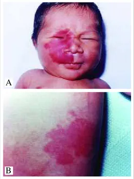
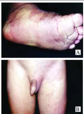
malformação vascular presente ao nascimento e que não apre-senta tendência à involução. Freqüentemente unilateral e seg-mentar, em geral respeita a linha média. Aumenta proporcional-mente ao crescimento da criança, pode estar presente em qual-quer área do corpo, sendo a face e a região cervical os locais mais comuns (Figuras 1A e 1B). As lesões podem ser róseas na infância, mas tendem a se tornar vinhosas com a idade. De iní-cio são totalmente maculares, mas, com a idade, sobretudo após a quarta década, podem apresentar superfície irregular,
espessa-da e nodular.9Em poucas crianças a lesão pode tornar-se mais
clara com a idade, mas a regressão total é excepcional.10A lesão
branqueia levemente à digitopressão, e a cor intensifica-se com o choro da criança. Microscopicamente a mancha em vinho do Porto é composta de capilares dilatados maduros na derme, sem nenhuma evidência de proliferação celular. A etiologia da lesão é desconhecida, e especula-se a existência de fragilidade na parede dos capilares e déficit do número de nervos perivascula-res levando a uma deficiência na neuromodulação do fluxo
vas-cular no local da lesão.11Em dois estudos realizados a mancha
em vinho do Porto foi encontrada em 0,36%12 e 1,2%13dos RN.
Há relatos de casos de mancha em vinho do Porto familiares,14adquiridos15,16e bilaterais e simétricos.17
A terapêutica de escolha é o Pulsed dye Laser. O
tra-tamento deve ser o mais precoce possível, uma vez que crianças mais jovens geralmente requerem menor número de sessões e apresentam resultado terapêutico mais favorá-vel. O resultado também é melhor
nas lesões localizadas na face e no tronco em relação às das extremida-des. A hipertrofia dos tecidos moles não responde ao Laser, e a correção cirúrgica pode ser necessária.6,18-20
A síndrome de Sturge-Weber caracteriza-se pela presença da mancha em vinho do Porto na região do primeiro ramo do nervo trigêmeo, com anomalias vascula-res ipsilaterais na leptomeninge, estando presentes um ou mais dos seguintes sinais ou sintomas:
epi-CAPILLARY VASCULAR MALFORMATIONS Port-Wine Stains
Port-wine stain, misnamed as flat hemangioma, is frequently referred to as nevus flammeus, though this expression is also used as a synonym of salmon patch. As these two lesions have distinct significance and prognoses, the term nevus flammeusmust be dropped. Port-wine stain is a congenital vascular malformation that shows no ten-dency toward involution. It is frequently unilateral and seg-mentary, and usually respects the medial line. It increases in proportion to the child's growth, and may be present on any area of the body. The face and cervical regions are the most common sites (Figures 1A and 1B). The lesions may be pink during infancy, but tend to become wine-colored with age. From the outset port-wine stains are totally macular, but with age and especially after the fourth decade of life, they may show an irregular, thick and nodular surface.9In few children, the lesion may become lighter with age, but total remission is unusual.10The lesion turns slightly pale to when pressed with the fingers. Its color is intensified with a child's tears. Microscopically, port-wine stain consists of dilated and mature capillaries in the dermis without any evidence of cellular proliferation. The etiology of the lesion is not known. There is speculation as to fragility in the cap-illary walls and a deficit in the number of perivascular nerves leading to a neuromodulation deficiency of the vas-cular flow at the lesion site.11 In two studies performed, port-wine stain was found in 0.36%12and 1.2%13of NR.
There are case reports of familial,1 4acquired15,16and bilateral and symmetrical port-wine stains.17
The therapy of choice is Pulsed-Dye Laser. The treatment must take place at the earliest possible time, given that the youngest children usually require a fewer ses-sions and show more favorable therapeutic results. Also, the result is better in localized lesions on the face and trunk in relation to those found on the extremities. Hypertrophy of the soft tissues does not respond to the laser, and surgical correction may be necessary.6,18-20
Sturge-Weber Syndrome is characterized by the presence of port-wine stains on the first branch region of the trigeminal nerve, with ipsilateral vascular anomalies on the leptomeninges. One or more of the following signs or symptoms
Figure 1A: Port-wine stain on the face (area of innervation of the first and second trigeminal branches). 1B: Port-wine stain on the abdominal region. Figura 1A: Mancha em vinho do
Porto na face (área de inervação dos primeiro e segundo ramos do trigêmeo). 1B: Mancha em vinho do Porto na região abdominal.
A
may be present: epilepsy, hemiparesis or hemiplegia, intracranial calcifications, cerebral atrophy and vascular lesions of the ipsilateral carotid in association with glauco-ma. It is worth emphasizing that only 10% of the carriers of port-wine stain appearing in the area innervated by the ophthalmic branch show this syndrome.2
Patients showing port-wine stain on the face and glaucoma without meningeal anomalies, or meningeal angiomas without port-wine stain on the face do not sat-isfy the criteria to be considered carriers of Sturge-Weber syndrome.21 Patients whose vascular stains are distrib-uted only along the maxillary sensory branch and mandible regions do not present a risk of neuro-ocular disease. However, repeated ophthalmologic assessment and computerized tomography of the cranium are indi-cated only for patients with port-wine stain on the oph-thalmic area.2 1
The risk of glaucoma grows when there is joint affliction of the ophthalmic and maxillary branches, which may occur in 45% of patients. In 50% of patients intracranial lesion symptoms emerge in the first year of life but very rarely is there onset after age 20 years. Convulsions, which may occur in 80% of cases, are usu-ally premature, with onset in the first three months of life. Hemiplegia is reported in up to 30% of cases, and mental retardation in 60%. Lesions in the oral mucosa may be present.2 2
Phacomatosis pigmentovascularis is a syndrome combining port-wine stain and other cutaneous lesions, such as epidermal or melanocytic nevus (type 1), dermal melanocytosis with or without anemic nevus (type II), nevus
spiluswith or without anemic nevus (type III), or dermal
melanocytosis and nevus spilus with or without anemic nevus (type IV). The letter A is appended when involvement is merely cutaneous; letter B when systemic involvement is associated (hypoplastic larynx, subglottic stenosis, calcifi-cations in the central nervous system, cerebral atrophy, and scoliosis).6,23,24
Beckwith-Wiedemann syndrome includes a capillary malformation on the central region of the forehead or upper eyebrows. It resembles persistent salmon patch in associa-tion with a somatic and visceral overgrowth, with macroglossia, enlarged kidneys and exomphalos.25
Robert's syndrome is characterized by severe tetraphocomelia, leporine lip and palatine fissure, mental retardation and few possibilities for survival. Medial facial capillary malformation is usually present.26
Salmon patch
Salmon patch was first reported by French physi-cians in 1881 under the term tache sanguine. But it was Unna, the German dermatologist, who first gave it a detailed description in 1884. Various names are used, such as capillary telangiectasis, telangiectasia nevus of the nape of neck, Unna nevus, erythema of the nape of neck,
heman-lepsia, hemiparesia ou hemiplegia, calcificações intracra-nianas, atrofia cerebral e lesões vasculares da coróide ipsi-lateral associadas com glaucoma. Cumpre ressaltar que apenas 10% dos portadores de mancha em vinho do Porto localizada na área inervada pelo ramo oftálmico
apresen-tam a síndrome.2
Os pacientes que apresentam mancha em vinho do Porto na face e glaucoma sem anomalias meníngeas ou angiomas meníngeos sem a mancha em vinho do Porto na face não preenchem critérios para ser considerados
portado-res da síndrome de Sturge-Weber.21Os pacientes cujas
man-chas vasculares se distribuem apenas ao longo das regiões dos ramos sensoriais maxilares e mandibulares não apre-sentam risco de doença neuroocular. Portanto, a avaliação oftalmológica repetida e a tomografia computadorizada do crânio estão indicadas apenas para os pacientes com
man-cha em vinho do Porto na área oftálmica.21
O risco de glaucoma aumenta quando há acometi-mento dos ramos oftálmico e maxilar em conjunto, poden-do ocorrer em 45% poden-dos pacientes. Em 50% poden-dos pacientes os sintomas das lesões intracranianas surgem no primeiro ano de vida e muito raramente se iniciam após os 20 anos. As convulsões, que podem ocorrer em 80% dos casos, são geralmente precoces, com início nos três primeiros meses de vida. Hemiplegia é relatada em até 30% dos casos, e retardo mental em 60%. Lesões na mucosa oral podem estar presentes.22
A facomatose pigmento-vascular é síndrome em que se combinam mancha em vinho do Porto e outra lesão cutânea como nevo epidérmico ou melanocítico (tipo I), melanocitose dérmica com ou sem nevo anêmico (tipo II),
nevus spiluscom ou sem nevo anêmico (tipo III) ou
mela-nocitose dérmica e nevus spiluscom ou sem nevo anêmico
(tipo IV). A letra A é acrescentada quando o envolvimento é apenas cutâneo, e a letra B quando se associa acometi-mento sistêmico (laringe hipoplásica, estenose subglótica, calcificações no sistema nervoso central, atrofia cerebral, escoliose).6,23,24
A síndrome Beckwith-Wiedemann compreende uma malformação capilar na região central da fronte ou pálpe-bras superiores, semelhante a uma mancha salmão persis-tente, associada a um supercrescimento somático e visceral,
com macroglossia, rins aumentados e onfalocele.25
A síndrome de Robert caracteriza-se por tetrafoco-melia grave, lábio leporino e fenda palatina, retardo mental e poucas possibilidades de sobrevivência. Malformação
capilar médio-facial geralmente está presente.26
Mancha salmão
A mancha salmão foi primeiramente relatada por
médicos franceses em 1881 sob o termo tache sanguine,
hemangio-ma da nuca e nuca vinhosa. Popularmente as lesões encon-tradas nas pálpebras e fronte são denominadas beijo do anjo, e as lesões occipitais são consideradas a marca do bico
da cegonha.27Acredita-se que haja um componente
genéti-co, possivelmente autossômico dominante, na etiologia da mancha salmão.
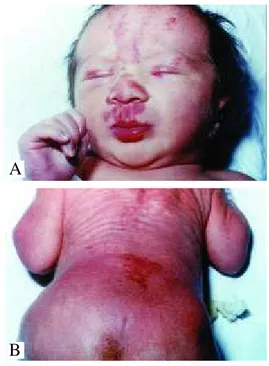
Clinicamente caracteriza-se por lesões planas, róseas ou avermelhadas, muitas vezes com telangiectasias, locali-zadas na região occipital, nuca, glabela, fronte, pálpebras superiores e regiões nasolabiais (Figura 2A). As lesões geralmente sobressaem quando a criança chora e podem desaparecer totalmente quando comprimidas. São presumi-velmente compostas por capilares dérmicos ectásicos, que representam a persistência dos padrões de circulação fetal na pele. Localizam-se geralmente na linha média, exceto as lesões das pálpebras, e devem ser diferenciadas da mancha em vinho do Porto, que tende a ser unilateral e mais vinho-sa. As manchas salmão usualmente estão presentes em mais
de um local do mesmo recém-nascido.27,28
A freqüência relatada na literatura varia de 1,5% a 74%, e as regiões mais afetadas são nuca (37,1%), glabela (19,6%), pálpebras (15,4%), nariz (1,5%) e lábio superior
(0,5%).27 As lesões das pálpebras parecem regredir mais
rapidamente do que as da glabela, e estas de forma mais
rápida do que as localizadas na nuca.27,29 A maioria das
lesões desaparece até os seis anos de idade; as localizadas nas pálpebras e glabela o fazem durante o primeiro ano de vida. A persistência da mancha na região occipital nos
adul-tos é freqüente e ocorre em até 50% dos indivíduos.30
Mancha vascular telangiectásica medial sacral
Como já visto, a mancha em vinho do Porto caracte-riza-se por ser unilateral e persistir inalterada durante a vida, enquanto a mancha salmão habitualmente se localiza na linha média (exceto a localizada
nas pálpebras) e apresenta involu-ção espontânea (exceto a localizada na região occipital, que persite na vida adulta em cerca de 50% dos pacientes).
Máculas angiomatosas localizadas na linha média da região sacral, geralmente em forma de borboleta e que tendem a persistir durante a vida, são vistas em recém-nascidos e não são habi-tualmente classificadas como man-cha em vinho do Porto ou manman-cha salmão (Figura 2B). Essas lesões
gioma of the nape of neck and wine-stained nape of neck. Lesions found on the eyebrows and forehead are commonly known as 'angel's kiss'. The occipital lesions are considered to be the marks of a stork's bite.2 7A genetic component, pos-sibly autosomal dominant, is thought to be involved in the etiology of salmon patch.
It is clinically characterized by flat pink or red-dish lesions that often have telangiectasias localized on the occipital region, neck, glabella, forehead, upper eye-brows and nasolabial regions (Figure 2A). Lesions usu-ally project when the child cries and may disappear totally when compressed. They presumably consist of ecstatic dermal capillaries, which represent persistent fetal circulation patterns in the skin. They are usually localized on the medial line, except with eyebrow lesions. And they must be differentiated from port-wine stain, which tends to be unilateral and more wine-like. Salmon patch is usually present on more than one site in the same newborn.27,28
The frequency reported in the literature varies from 1.5% to 74%. The most affected regions are the nape of the neck (37.1%), glabella (19.6%), eyebrows (15.4%), nose (1.5%) and upper lip (0.5%).27 Eyebrow lesions appear to regress more rapidly than those of the glabella, and the lat-ter more rapidly than those localized on the nape.27,29 Most lesions disappear by age six. Those localized on the eye-brows and glabella disappear during the first year of life. The persistence of the stain on the occipital region in adults is frequent and occurs in up to 50% of individuals.30
Sacral medial telangiectatic vascular stain
As we have already seen, port-wine stain is charac-terized by being unilateral and it persists unaltered during the course of one's lifetime. By contrast, salmon patch is often localized on the medial line (except for the localization on the eyebrows) and shows spontaneous involution (except for the localiza-tion on the occipital region, which persists in adult life for roughly 50% of patients).
Angiomatous spots localized on the medial line of the sacral region, usually in a butterfly shape that tends to persist during one's entire lifetime, can be seen in new-borns. They are not usually classi-fied as port-wine stains or salmon patches (Figure 2B). These lesions
Figure 2A: Salmon patch on the face 2B: Sacral medial telangiectatic vascular stain. Figura 2A: Mancha salmão na
face. 2B: Mancha vascular telangiectásica medial sacral.
A
appear to be associated with bifid spine, spinal dys-raphism, intraspinal masses, imperforated anus, and kid-ney or genital abnormalities as occur with sacral heman-giomas. They show very similar characteristics and pro-gression to salmon patches on the nape. Some authors prefer using the term sacral telangiectatic vascular nevus for these lesions. Others have already included them among salmon patches.3 1
Telangiectasia
Essential telangiectasia (localized or generalized) is a vascular alteration frequently found in women. It is typi-cally localized on the lower limbs. Onset occurs during or after puberty. It may present as irregular fine lines, or as dot-or star-like macules either with dot-or without an anemic halo.5,6 Unilateral nevoid telangiectasia occurs predomi-nantly in women. Onset occurs during puberty and may intensify with pregnancy. A high number of estrogen and progesterone receptors has been demonstrated in the skin areas involved.6
Benign hereditary telangiectasia is a family disorder characterized by the presence of cutaneous and labial telangiectasias. As opposed to Rendu-Osler-Weber syn-drome, it does not show any visceral hemorrhaging.6
Generalized essential telangiectasis is another syn-drome appearing sporadically in adult females.6
Hereditary hemorrhagic telangiectasia (Rendu-Osler-Weber syndrome) is an autosomal dominant disorder manifested during infancy or adolescence with telangiec-tasias on the face, tongue, lips, nose, conjunctiva, fingers, ungual bed, liver, lungs, spleen, pancreas and brain.5 Bleeding in the mucouses (recurrent epistaxis with onset during infancy and aggravation in adulthood) and in the visceras (bleeding in the upper and lower gastrointestinal tract) may cause anemia. Hereditary hemorrhagic telang-iectasia is characterized by genetic heterogeneity with vari-able phenotypes. Two defective genes have already been found (9q33-34 and 12q). Electrodissection, Laser (Nd: YAG, carbon dioxide or argon), and sclerotherapy have been used to stanch the hemorrhaging.2,6
Ataxia/telangiectasia (Louis-Bar syndrome) is a recessive autosomal disorder that is usually fatal for patients up to their twenties. Cerebellar ataxia (cerebellar degeneration with progressive motor degeneration) is associated with the presence of ocular telangiectasias (mainly in the bulbar conjunctiva, close to the corner of the eye). Cutaneous telangiectasia may also occur on the face, throat and dorsal aspect of the hands and feet. Humoral immunodeficiencies (IgA and IgG-2 deficiency) and cellular ones (lymphopenia and reduction of CD4+) in association with repeated respiratory infections are characteristic. Telangiectasia usually emerges at three years of age, and cerebellar ataxia in the second decade of life. Endocrinologic dysfunction (insulin-resistant diabetes, gonadal insufficiency and growth retardation), pre
-parecem não estar associadas com espinha bífida, disrafis-mo espinhal, massas intra-espinhais, ânus imperfurado, anormalidades renais ou genitais como ocorre com os hemangiomas sacrais. Apresentam características e evolu-ção muito semelhantes à mancha salmão da nuca. Há auto-res que preferem a denominação nevo vascular telangiectá-sico sacral para essas lesões; outros já as incluem na
man-cha salmão.31
Telangiectasias
A telangiectasia essencial (localizada ou generaliza-da) é uma alteração vascular freqüente em mulheres, tipica-mente localizada nas extremidades inferiores, e que se ins-tala durante ou após a puberdade. Pode apresentar-se como linhas finas irregulares, máculas puntiformes ou estelares
com ou sem halo anêmico.5,6
A telangiectasia unilateral nevóide ocorre predomi-nantemente em mulheres, iniciando-se na puberdade e podendo intensificar-se com a gravidez. Um número eleva-do de receptores de estrógenos e progesterona tem sieleva-do
demonstrado nas áreas da pele envolvida.6
A telangiectasia benigna hereditária é dermatose familiar caracterizada pela presença de telangiectasias cutâ-neas e labiais, mas, ao contrário da síndrome de
Rendu-Osler-Weber, não apresenta hemorragia visceral.6
A telangiectasia essencial generalizada é outra
sín-drome que aparece esporadicamente em mulheres adultas.6
A telangiectasia hemorrágica hereditária (síndrome de Rendu-Osler-Weber) é doença autossômica dominante que se manifesta na infância ou adolescência com telangiec-tasias na face, língua, lábios, nariz, conjuntiva, dedos, leitos
ungueais, fígado, pulmão, baço, pâncreas e cérebro.5O
san-gramento nas mucosas (epistaxe recorrente com início na infância e agravamento na vida adulta) e nas vísceras (san-gramento do trato gastrointestinal superior e inferior) pode causar anemia. A telangiectasia hemorrágica hereditária se caracteriza por heterogeneidade genética com fenótipos variáveis. Dois genes defeituosos já foram encontrados (9q33-34 e 12q). Eletrodissecação, Laser (Nd:YAG, dióxi-do de carbono ou argônio) e escleroterapia têm sidióxi-do
utiliza-dos para estancar a hemorragia.2,6
cresci-mento), envelhecimento precoce, efélides e perda do tecido subcutâneo. Os portadores da síndrome apresentam risco de câncer de 61 a 184 vezes maior do que a população geral (principalmente linfomas, leucemias e carcinomas), e essas neoplasias representam a principal causa de óbito. O defeito
genético está no cromossoma 11q22-23.2,5,6
Cútis marmórea telangiectásica congênita
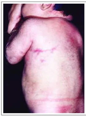
A cútis marmórea telangiectásica congênita (síndro-me de van Lohuizen) é lesão vascular reticulada, de cor azul-violeta, usualmente presente ao nascimento e que, diferente do livedo reticular, está sempre visível, mas pode também acentuar-se com o frio (Figura 3). As lesões cutâ-neas tendem a melhorar espontaneamente, sobretudo nos dois primeiros anos de vida. Podem ser localizadas (89% dos casos) ou mais extensas (11%), mas não há relatos de
formas generalizadas.32A patogênese é desconhecida, mas
uma herança autossômica dominante com penetrância variável ou a presença de um gene letal, mas que sobrevive graças ao fenômeno do mosaicismo, são hipóteses
aventa-das.32Um defeito funcional da inervação simpática vascular
(tônus neurogênico) pode explicar as lesões cutâneas.33
Anomalias associadas incluem assimetria do corpo, outras anomalias vasculares, glaucoma, aplasia cutânea congênita, fenda palatina, retardo mental ou psicomotor, atrofia
cutâ-neas, ulcerações.32,33O Pulsed dye Laser pode ser usado para
tratar as lesões cutâneas residuais.6
A síndrome de Adams-Oliver, de herança autossô-mica dominante, é formada pela cútis marmórea telangiec-tásica congênita associada com múltiplas lesões de aplasia cutânea no couro cabeludo, com ou sem defeito ósseo
sub-jacente, e defeitos nos membros.6
MALFORMAÇÕES VASCULARES LINFÁTICAS
As malformações linfáticas estão presentes ao nascimento em 60% dos casos, tornam-se aparentes até o segundo ano de vida em 90%, em geral não regridem espontanea-mente, e seu volume pode aumentar por hemorragia, acúmulo de líquidos
ou inflamação.5,6
Malformações linfáticas macrocísticas (higroma cístico) caracterizam-se como lesões gran-des, macias, de superfície lisa e translúcida com pele normal ou azu-lada sobrejacente. Localizam-se tipi-camente no triângulo posterior do
mature aging, ephelides and loss of subcutaneous tissue are also observed. Syndrome carriers show a risk of can-cer 61 to 184 times higher than in the general population (mainly lymphomas, leukemias and carcinomas). These neoplasias represent the main cause of death. The genetic defect is found in chromosome 11q22-23.2,5,6
Congenital telangiectatic cutis marmorata
Congenital telangiectatic cutis marmorata (van Lohuizen Syndrome) is a reticulated vascular lesion with a blue-violet color. It is usually present at birth. As opposed to reticular livedo it is always visible and may also be accentuated with cold weather (Figure 3). Cutaneous lesions tend to improve spontaneously, above all in the first two years of life. They may be localized (89% of cases) or more extensive (11%). But there are no reports on general-ized forms.32 Its pathogenesis is not known, but autosomal dominant inheritance with variable penetrance, or the pres-ence of a lethal gene, which survives due to the phenome-non of mosaicism,, are among the hypotheses formulated.32 A functional defect of vascular sympathetic innervation (neurogenic tonus) may explain cutaneous lesions.33 Associated anomalies include body asymmetry, other vas-cular anomalies, glaucoma, congenital cutaneous aplasia, palatine fissure, mental or psychomotor retardation, cuta-neous atrophies, and ulcerations.32,33 The pulsed-eyed laser may be used to treat residual cutaneous lesions.6
Adams-Oliver syndrome, with autosomal dominant inheritance, is formed by congenital telangiectatic cutis marmorata in association with multiple lesions of cuta-neous aplasia on the scalp, either with or without underly-ing bone defects and defects in the limbs.6
LYMPHATIC VASCULAR MALFORMATIONS
Lymphatic malformations are present congenitally in 60% of cases. They become apparent by the second year of life in 90% of patients. Usually, they do no regress spontaneously, and their volume may increase with hemor-rhages, liquid build-up or inflam-mation.5,6
Macrocystic lymphatic mal-formations (cystic hygroma) are characterized as large, soft lesions with a smooth surface and translu-cent with normal skin or blue with overlying skin. They are localized
Figure 3: Congenital telangiectatic cutis marmorata. Figura 3:
Figure 4: Microcystic-type lymphatic vascular malforma-tion (circumscribed
lymphangioma). Figura 4: Malformação
vascular linfática do tipo microcístico (linfangioma circunscrito).
typically in the rear triangle of the throat. Onset may occur in the popliteal space, and retroperitoneal and virilian areas. They are caused by a communication defect between the lymphatic system and the fetal venous which, if it is reestablished later, may result in the regression of hygroma and improvement of peripheral edema. Cystic hygroma must be differentiated from the other cranial-cervical mass-es, such as encephaloceles and cystic teratoma. Studies show an increase in a-phetoprotein in the amniotic liquid during the pre-natal period. This lymphatic malformation may be associated to fetal hydrops, which reduce the child's probability of survival.5,6,34
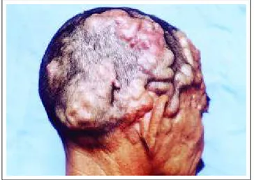
Microcystic lymphatic anomalies (circumscribed lymphangioma) are characterized by the small vessel grouped in plaques, on the skin or in mucouses. They have a translucent content or are slightly hemorrhagic. They pre-dominantly affect the cervical-facial region, axillae, thorax and extremities (Figure 4). There might be hypertrichosis on the site.5,6
Histopathologically, microcystic lymphatic malfor-mations are characterized by fine vessel walls with no blood within. They are localized in the dermis, which may or may not have lymphocytes around. The presence of blood within the vessels may indicate recent hemorrhaging or a combined, usually venous or lymphatic, malformation. With macrocystic lesions, large cisterns are found which do no communicate directly with the lymphatic system. In mixed lymphatic malformations (macro- and microcystic), the deep muscular cisterns communicate with the superficial dermis by means of a complex network of anastomoses.6
If the clinical diagnosis leaves any doubts about the lesion's lymphatic origin, the following techniques may be used: ultrasound to demonstrate anechoic or hypoechoic and homogenic cysts; computerized tomography to reveal hypo-dense cysts; and magnetic resonance to show hypointense cystic spaces on T1. A puncture of the lesion may be carried out directly to show the presence of a clear liquid.6The lym-phatic and vena malformations show patterns similar to magnetic resonance. As opposed to other lesions, no vessel or lobular architecture is demonstrated. The contrast medi-um may be useful, because lymphatic malformations do not intensify with the con-trast. The main role of mag-netic resonance is to deter-mine the extension and infil-tration of the deep tissues and assist in the differential diagnosis with other soft
tis-pescoço, podendo também instalar-se na fossa poplítea, áreas retroperitoneais e virilhas. Ocorrem por um defeito de comunicação entre o sistema linfático e venoso fetal que, se posteriormente reestabelecida, pode resultar na regressão do higroma e melhora do edema periférico. O higroma cís-tico deve ser diferenciado de outras massas craniocervicais, como encefalocele e teratoma cístico. Há estudos
demons-trando aumento da α-fetoproteína no líquido aminiótico no
período pré-natal, e essa malformação linfática pode estar associada à hidropisia fetal, o que diminui a probabilidade
de sobrevivência da criança.5,6,34
Anomalias linfáticas microcísticas (linfangioma cir-cunscrito) caracterizam-se por pequenas vesículas agrupa-das em placas, na pele ou nas mucosas, de conteúdo trans-lúcido ou levemente hemorrágico. Acometem predominan-temente a região cervicofacial, axilas, tórax e extremidades
(Figura 4). Pode haver hipertricose no local.5,6
Histopatologicamente, as malformações linfáticas microcísticas caracterizam-se por vasos de parede fina sem sangue em seu interior, localizados na derme, podendo ou não apresentar linfócitos ao redor. A presença de sangue no interior dos vasos pode indicar uma hemorragia recente ou uma malformação combinada, geralmente venosa e linfáti-ca. Nas lesões macrocísticas há grandes cisternas que não se comunicam diretamente com o sistema linfático. Nas mal-formações linfáticas mistas (macromicrocísticas), as cister-nas musculares profundas comunicam-se com a derme
superficial por meio de uma rede complexa de anastomoses.6
Se o diagnóstico clínico deixar dúvida quanto à ori-gem linfática das lesões, as seguintes técnicas podem ser empregadas: ultra-sonografia demonstrando cistos anecóicos ou hipoecóicos e homogêneos; tomografia computadorizada demonstrando cistos hipodensos; e ressonância magnética demonstrando espaços císticos hipointensos em T1. A punc-tura direta da lesão pode ser realizada e demonstra a
presen-ça de um líquido claro.6As malformações linfáticas e
veno-sas mostram padrões similares à ressonância magnética. Ao contrário de outras lesões, nenhum vaso ou arquitetura lobu-lar é demonstrada. O meio de contraste pode ser útil, pois as malformações linfáticas não se intensificam com o contraste. O principal papel da
ressonân-cia magnética é determinar a extensão e infiltração de teci-dos profunteci-dos e ajudar no diagnóstico diferencial com outros tumores de tecidos moles.35
Lesões que obstruem as vias aéreas respiratórias requerem excisão, aspiração, traqueostomia e sondas para manter a alimentação. Infecções devem ser tratadas com antibióticos. A remoção cirúrgica está indicada quando a lesão interfere com a função, causa problemas estéticos ou se infecta facil-mente. Para as lesões localizadas a excisão completa é pos-sível. Nas formas difusas, geralmente associadas com teci-dos normais, como nervos, uma excisão mais cuidateci-dosa torna-se necessária, geralmente em várias sessões. Como se trata de lesões benignas, não há nenhuma indicação para excisar nervos ou músculos. O tratamento também pode ser tentado com agentes esclerosantes, como bleomicina, OK-432, também denominado picibanil (uma cepa morta do
grupo da bactéria Streptococcus pyogenes), solução de
sul-fato de tetradecil de sódio, com risco potencial de recorrên-cia e/ou infecção.5,7,34,36,37
A síndrome de Turner é definida como uma disge-nesia gonadal devido a cromossoma X ausente ou defeituo-so (46XO). Asdefeituo-socia-se freqüentemente a malformações lin-fáticas congênitas, como vasos linfáticos hipoplásicos, res-ponsáveis pela presença de linfedema nos membros infe-riores ao nascimento, que desaparece em poucos meses ou anos na maioria dos casos. Outras malformações, como higroma cístico, hidropisia fetal e ascite, podem ser detec-tadas a partir do segundo trimestre pela ultra-sonografia. Fenotipicamente caracteriza-se também pela presença de baixa estatura, tórax largo com grande espaço entre os
mamilos, pescoço encurtado com membrana (pterygium
colli), palato ogival, unhas hipoplásicas, orelhas
malforma-das e múltiplos nevos melanocíticos.34
A síndrome de Noonan é fenotipicamente seme-lhante à síndrome de Turner, mas o cariótipo é normal (46XY ou 46XX). Caracteriza-se pela presença de baixa estatura, pescoço curto e largo, hipertelorismo, epicanto, linha dos cabelos baixa e micrognatia. O linfedema, ao contrário do que ocorre na síndrome de Turner, em geral persiste na vida adulta, de forma estacionária ou lenta-mente progressiva. Observa-se também aplasia ou hipo-plasia dos vasos linfáticos e linfagiectasia. O pescoço largo pode ser explicado pela regressão de higromas cís-ticos com formação de canais linfácís-ticos colaterais. Questiona-se uma herança autossômica dominante para
essa síndrome.34
MALFORMAÇÕES VASCULARES VENOSAS
As malformações vasculares venosas apresentam um largo espectro, variando de ectasias cutâneas isoladas até lesões volumosas envolvendo múltiplos tecidos e órgãos. São macias e compressíveis e não apresentam alte-ração na temperatura da pele, frêmitos ou sopros. São fre-qüente e erroneamente denominadas hemangiomas caver-nosos. Malformações venosas puras geralmente apresen-tam coloração azulada na pele ou mucosa suprajacente enquanto as combinadas capilares venosas exibem
tonali-dade que varia do vermelho-escuro ao violáceo.5,6
sue tumors.35
Treating lymphatic malformations is difficult. Lesions that end up obstructing the respiratory tract require excision, aspiration, tracheostomy and sounding to maintain patient feeding. Infections must be treated with antibiotics. Surgical removal is indicated when the lesion interferes with the function, causes aesthetic problems or gets easily infected. For localized lesions, complete exci-sion is possible. In diffuse forms usually associated with normal tissues, such as nerves, more careful excision becomes necessary, usually in several sessions. As for benign lesions, there is no indication whatsoever to excise nerves or muscles. Treatment may also be attempted with sclerosing agents, such as bleomycin, OK-432, also known as picibanil (a dead fungus of the Streptococcus pyogenes bacteria group), a solution of sodium tetradecyl sulphate, with a potential risk of recurrence and/or infection.5,7,34,36,37
Turner's syndrome is defined as a gonadal dysgene-sis due to chromosome X being absent or defective (46XO). It is frequently associated with congenital lymphatic mal-formations, such as hypoplasic lymphatic vessels, which are responsible for the presence of lymphedema in the lower limbs at birth; in most cases these disappear within a few months or years. Other malformations, such as cys-tic hygroma, fetal hydrops and ascites, may be detected from the second trimester on by ultrasound. Phenotypically, it is also characterized by physical features such as short stature, large thorax with a wide space between the nipples, short webbed neck (pterygium colli), ogival palate, hypoplastic nails, malformed ears and mul-tiple melanocytic nevi.34
Noonan's syndrome is phenotypically similar to Turner's syndrome, but the cariotype is normal (46XY or 46XX). It is characterized by physical features such as short stature, a short and thick throat, hypertelorism, epi-canthis, low hair line and micrognathia. As opposed to what occurs in Turner's syndrome, the lymphedema is usu-ally persistent in adult life, either in a stationary or slow-ly progressive form. Aplasia or hypoplasia of the slow- lymphat-ic vessels and lymphangiectasis can also be observed. The thick neck can be explained by the regression of cystic hygromas with collateral formation of lymphatic channels. Questions remain as to whether autosomal dominant inheritance characterizes this syndrome.34
VENOUS VASCULAR MALFORMATIONS
dark-red to violet.5,6
The venous malformations are hemodynamically inactive, with a low flow. Their volume increases when the person is standing or doing physical efforts. They are pres-ent at birth, and worsen progressively during infancy, and, to a lesser degree, during adulthood. Their volume may also increase with pregnancy or trauma.2,38 They do not usually involve only the skin, but also underlying structures, such as muscles and fascia. There is no overgrowth of the limbs, which differs from combined vascular malforma-tions, such as Klippel-Trenaunay syndrome. There may be refinement, demineralization, hypoplasia or lytic changes in the underlying bones in up to 71% of cases.2
The diagnosis is clinical for most cases, but a simple radiography may reveal phleboliths (calcified thrombi) already at age two or three years. These round calcifica-tions are pathognomic of venous vascular lesions. Simple radiography can also be useful to assess bone distortions. Magnetic resonance is the best examination to delimit vas-cular malformation.6
Venous thrombosis is a regular complication, and the thrombi may be palpated at the point of pain. Another possible complication is the development of consumption coagulopathy for stasis in the ecstatic vascular canals. With stasis there is formation of microthrombi and, secondly, consumption coagulation factors. Platelet numbers are moderately low (usually around 100,000/mm3). However these are higher than what is observed in Kasabach-Merritt syndrome. Similar alterations may occur in lymphatic or combined (venous lymphatic) malformations. Treatment is carried out with low-molecular-weight heparin. The possi-bility of consumption coagulopathy must be investigated prior to undertaking any invasive procedures.2,5,39
Venous vascular malformations cannot be eradicat-ed completely. The usual treatment is sclerotherapy, with a local injection of sclerosing solutions, like 95% alcohol or sodium tetradecyl sulfur 1% for small lesions. Surgical resection may be performed after successful obliteration by sclerotherapy. The embolization of arteries sustaining the malformation is counter-indicated since it may provoke tis-sue necrosis. Photocoagulation by laser may obstruct small superficial vessels. Surgical excision is the definitive thera-py, often rendered impossible however by anatomic, esthet-ic and functional limitations.5,40
Familial cerebral venous malformations constitute an autosomal dominant disorder. But the gene carriers may remain free of symptoms. Patients may suffer fainting, cere-bral hemorrhaging, and neurological or cephalic deficits. Magnetic resonance demonstrates venous malformations found on the skin and retina as well. There is probably a genetic heterogeneity, but with a defective gene involved. A defective gene was already found on chromosome 7q 11-22. It was mapped in several families with this syndrome, but was absent in others.2
Cutaneous venous malformations and multiple
As malformações venosas são hemodinamicamente inativas, de baixo fluxo. Apresentam aumento de volume quando a pessoa está de pé ou faz esforços físicos. Estão presentes ao nascimento, progressivamente pioram na infância e, em menor grau, durante a vida adulta. Seu
volu-me também pode auvolu-mentar com gravidez ou trauma.2,38
Geralmente não envolvem apenas a pele, mas também as estruturas subjacentes, como músculo e fáscia. Não há supercrescimento dos membros, diferenciando-se assim das malformações vasculares combinadas, como a síndrome de Klippel-Trenaunay. Pode haver afinamento, desmineraliza-ção, hipoplasia ou alterações líticas nos ossos subjacentes
em até 71% dos casos.2
O diagnóstico é clínico na maioria dos casos, mas uma radiografia simples pode revelar flebólitos (trombos calcificados) já na idade de dois a três anos. Essas calcifica-ções arredondadas são patognomônicas de lesões vascula-res venosas. A radiografia simples pode ser útil também para avaliar distorções ósseas. A ressonância magnética é o
melhor exame para delimitar a malformação vascular.6
A trombose venosa é complicação comum, e os trombos podem ser palpados na área de dor. Outra possível complicação é o desenvolvimento de coagulopatia de con-sumo pela estase nos canais vasculares ectásicos. Com a estase há formação de microtrombos e, secundariamente, consumo de fatores de coagulação. O número de plaquetas
é moderadamente baixo (usualmente 100.000/mm3), porém
superior ao observado na síndrome de Kasabach-Merritt. Alterações semelhantes podem ocorrer nas malformações linfáticas ou combinadas (linfáticas venosas). O tratamento é realizado com heparina de baixo peso molecular. A possi-bilidade de coagulopatia de consumo deve ser pesquisada
antes de qualquer procedimento invasivo.2,5,39
As malformações vasculares venosas geralmente não podem ser completamente erradicadas. O tratamento usual é a escleroterapia, com injeção local de soluções esclerosantes, como álcool a 95% ou sulfato tetradecil de sódio 1% para lesões pequenas. A ressecção cirúrgica pode ser realizada após obliteração conseguida por escleroterapia. A emboliza-ção das artérias que nutrem a malformaemboliza-ção está contra-indi-cada, pois pode provocar necrose do tecido. Fotocoagulação pelo Laser pode obstruir pequenos vasos superficiais. A exci-são cirúrgica é a terapia definitiva, muitas vezes impossível
pelas limitações anatômicas, estéticas e funcionais.5,40
As malformações venosas cerebrais familiares consti-tuem afecção autossômica dominante, mas portadores do gene podem estar livres dos sintomas. Pacientes podem apre-sentar desmaios, hemorragias cerebrais, déficits neurológicos ou cefaléias. A ressonância magnética demonstra as malfor-mações venosas, também encontradas na pele e na retina. Há provavelmente uma heterogeneidade genética, com mais de um gene defeituoso envolvido. Um gene defeituoso já foi encontrado no cromossoma 7q 11-22, mapeado em várias
famílias com essa síndrome, mas ausente em outras.2
fami-liares múltiplas são doenças autossômicas dominantes com-postas por malformações venosas cutâneo-mucosas cujo defeito se encontra no cromossoma 9p. As lesões cutâneas
assemelham-se às da síndrome blue rubber bleb nevus, mas
não há acometimento do trato gastrointestinal.2,6
Na síndrome blue rubber bleb nevus (síndrome de
Bean) as malformações vasculares estão presentes na pele e nas vísceras. As lesões cutâneas caracterizam-se por nódulos azulados, isolados ou agrupados, macios, dolorosos ou não. Há casos em que a cor violácea está ausente e as lesões se
asseme-lham a um mamilo elástico (rubber bleb). As lesões viscerais
acometem principalmente o trato gastrointestinal (esôfago, estômago, intestino delgado e grosso, ânus, mesentério), gerando sangramentos recorrentes, anemia ferropriva e, mais raramente, choque hipovolêmico. A cavidade oral, nasofarin-ge, genitália, bexiga, cérebro, medula espinhal, fígado, baço,
pulmões, ossos e músculos podem também ser acometidos.6
Histologicamente as lesões apresentam uma rede de lagos vasculares, delineados por células endoteliais
achata-das e ausência de células glômicas.6
As lesões vasculares cutâneas podem ser tratadas com escleroterapia, excisão, criocirurgia e Laser Nd:YAG. As lesões viscerais com sangramento podem requerer
foto-coagulação ou ressecção cirúrgica.6
MALFORMAÇÕES VASCULARES ARTERIAIS
Malformações arteriais (atresia, ectasia, aneurisma ou coarctação), malformações arteriovenosas (conglomera-ção difusa ou localizada de artérias e veias com fístulas
vas-culares microscópicas) e fístulas arteriovenosas (shunts
entre braços arteriais a veias vizinhas) são anomalias vascu-lares de alto fluxo caracterizadas pelo aumento da
tempera-tura local, frêmito e sopro.5,38As malformações vasculares
arteriais puras, como os aneurismas, estenoses e ectasias, raramente ocorrem na pele como lesões sintomáticas. Na pele, ao contrário do cérebro, uma fístula arteriovenosa
geralmente é resultado de um trauma.6
MALFORMAÇÕES VASCULARES COMBINADAS
Anomalias vasculares podem ocorrer junto com outros erros morfogênicos de estruturas mesenquimais rela-cionadas, tais como as do tecido ósseo. Muitas dessas alterações são conhecidas como epônimos.
Malformações arteriovenosas (MAV)
As fístulas arteriovenosas apresentam um epicentro
denominado ninho (nidus) que consiste em artérias que
ali-mentam e auali-mentam o volume das veias. Podem estar sentes ao nascimento ou tornar-se evidentes na infância pre-coce. Nunca regridem espontaneamente, e a puberdade ou trauma podem acionar seu crescimento. Clinicamente carac-terizam-se por massa coberta por pele normal ou angiomato-sa, geralmente tensa e brilhante, com aumento do calor,
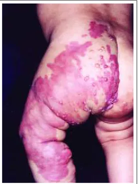
frê-mito e sopro no local. Com a progressão da MAVas veias de
drenagem tornam-se mais evidentes, tortuosas e distendidas
familial mucouses are dominant autosomal diseases con-sisting of cutaneous-mucosa venous malformations whose defect is found in chromosome 9p. The cutaneous lesions resemble blue rubber bleb nevus syndrome lesions, but there is no involvement of the gastrointestinal tract.2,6
In blue rubber bleb nevus (Bean's syndrome), vascu-lar malformations are present on the skin and in the vessels. The cutaneous lesions are characterized by blue nodules, isolated or in groups. They are soft, and can either be painful or not. Cases exist in which the violet color is absent or the lesions resemble an elastic nipple (rubber bleb). Visceral lesions mainly afflict the gastrointestinal tract (esophagus, stomach, small or large intestines, anus, and mesentery). There is usually recurrent bleeding, ferroprive anemia and, more rarely, hypovolemic shock. The oral cav-ity, nasopharynges, genitalia, bladder, brain, spinal medul-la, liver, spleen, lungs, bones and muscles may also be affected.6
Histologically, the lesions show a network of vascu-lar lakes, delineated by flattened endothelial cells and absence of glomic cells.6
Cutaneous vascular lesions may be treated with sclerotherapy, excision, cryosurgery and Nd: YAG laser. Visceral lesions with bleeding might require photocoagula-tion or surgical resecphotocoagula-tion.6
ARTERIAL VASCULAR MALFORMATIONS
Arterial malformations (atresia, ectasia, aneurysm or coarctation), arteriovenous malformations (diffuse or localized conglomeration of arteries and vessels with micro-scopic vascular fistulas) and arteriovenous fistulas (shunts between the arterial branch to neighboring veins) are high-flow vascular anomalies characterized by increased local temperature, thrill and bruit.5,38 Pure arterial vascular mal-formations, such as aneurysm, stenosis and ectasias, rarely occur on the skin as symptomatic lesions. When the skin is involved, as opposed to the brain, an arteriovenous fistula is usually the result of trauma.6
COMBINED VASCULAR MALFORMATIONS
Vascular anomalies may occur jointly with other errors of morphogenesis in related mesenchymal structures, such as those of the bone tissue. Many of these disorders are better known by their eponyms.
Arteriovenous Malformations (AVM)
distended (Figure 5). The hemorrhage is an important com-plication that can put the patient's life at risk.
Doppler ultrasound may be useful as a screening method, because it is a non-invasive examination capable of diagnosing a vascular lesion and differentiating it from a solid tumor mass. In addition, it allows us to distinguish vascular malformations consisting of arterial vessels, such as (high flow)
AVM, from others whose arterial flow is absent (low flow).41 Magnetic resonance better shows lesion extension as well as differentiates the AVMfrom a hemangioma, and venous or lym-phatic malformation.6Invasive examinations of the venous sys-tem (venography) and of the arterial syssys-tem (arteriography) are carried out only by planning and performing the treatment.7
As for prognosis, arteriovenous fistula is the most predictable vascular malformation. It can cause the destruction of local tissues and systemic effects. Partial excision or linking of the feeder artery may set off lesion growth and considerably worsen the prognosis. Alterations of a Kaposi pseudo-sarcoma type may be initiated on the skin at the region affected by the arteriovenous fistula.6
The arteriovenous fistula is not usually treated in its quiescent phase. Premature embolization or surgical treat-ment in these phases are controversial and must only be indicated when it can be performed easily. Linking or prox-imal embolization of the feeder arteries are counter-indi-cated, because the neighboring arteries grow and increase the lesion volume. The treatment must be conducted only by experienced individuals, preferentially in a multidiscipli-nary approach.7 The following therapeutic resources may be employed: transcatheter embolization of the nest, accompanied by pulsed-dye laser for residual skin lesions; superselective arterial embolization of the nest and surgical resection usually performed 24 to 72 hours after arterial embolization.6,42
Wyburn-Mason syndrome
(Bonnet-Dechaune-Blanc syndrome)
Components of this unilateral retinoic arteriove-nous malformation syndrome involve the optic and orbital nerves, ipsilateral arteriovenous malformation of the brain and ipsilateral vascular cuta-neous malformation (usually with increased local tempera-ture and thickness). This cutaneous vascular malfor-mation does not accompany the trigeminal nerve, thereby differentiating it from port-wine stain.6
Brégeat's Syndrome
This does not show a
(Figura 5). A hemorragia é uma complicação importante, podendo colocar em risco a vida do paciente. A
ultra-sono-grafia com Doppler pode ser útil como um método de scree
-ning, pois é exame não invasivo capaz de diagnosticar uma
lesão vascular e diferenciá-la de uma massa tumoral sólida. Além disso, permite distinguir as malformações vasculares
compostas de vasos arteriais, como a MAV(alto fluxo), de
outras cujo fluxo arterial está ausente (baixo fluxo).41A
res-sonância magnética demonstra melhor a extensão da lesão e
também diferencia a MAVde um hemangioma, malformação
venosa ou linfática.6Exames invasivos do sistema venoso
(venografia) e do sistema arterial (arteriografia) são
realiza-dos apenas para o planejamento e realização do tratamento.7
Quanto ao prognóstico, a fístula arteriovenosa é a malformação vascular mais imprevisível, podendo causar destruição dos tecidos locais e efeitos sistêmicos. A excisão parcial ou ligação da artéria nutridora pode acionar o cres-cimento da lesão e piorar em muito o prognóstico. Alterações tipo pseudo-sarcoma de Kaposi podem
instalar-se na pele da região acometida pela fístula arteriovenosa.6
A fístula arteriovenosa usualmente não é tratada em sua fase quiescente. Embolização precoce ou tratamento cirúrgico nessa fase são controversos e só devem ser indica-dos quando facilmente realizáveis. Ligação ou embolização proximal das artérias nutridoras estão contra-indicadas, pois as artérias vizinhas crescem e aumentam o volume da lesão. O tratamento deve ser conduzido apenas por pessoas
expe-rientes, de preferência em caráter multidisciplinar.7 Os
seguintes recursos terapêuticos podem ser empregados:
embolização transcatéter do ninho, acompanhada por Pulsed
dye Laserpara as lesões residuais da pele; embolização
arte-rial superseletiva do ninho e ressecção cirúrgica geralmente
realizada 24 a 72 horas após a embolização arterial.6,42
Síndrome de Wyburn-Mason
(Síndrome de Bonnet-Dechaune-Blanc)
São componentes dessa síndrome malformação arte-riovenosa retiniana unilateral, envolvendo o nervo óptico e a órbita, malformação arteriovenosa do cérebro ipsilateral e malformação cutânea vascular ipsilateral (geralmente com aumento da temperatura
local e espessa). Esta mal-formação vascular cutânea não acompanha o nervo tri-gêmeo, diferenciando-se assim da mancha em vinho do Porto.6
Síndrome de Brégeat
Não apresenta fístula arteriovenosa retiniana, mas apresenta anomalias
vascu-Figure 5: Arteriovenous fistulas, with dilated and tortuous veins. Figura 5: Fístula arteriovenosa,