Protective Effects of Dietary
Supplementation with a Combination of
Nutrients in a Transgenic Mouse Model of
Alzheimer
’
s Disease
Shengyuan Wang4☯, Yu Cu1☯, Chao Wang2, Wei Xie3, Lan Ma3, Jinfeng Zhu3, Yan Zhang3, Rui Dang3, Decai Wang3, Yonghui Wu4*, Qunhong Wu1*
1Department of Social Medicine, Public Health College, Harbin Medical University, Harbin, China, 2Department of Nutrition, YanZhou people's hospital, YanZhou, China,3Department of Nutrition and Food Hygiene, Public Health College, Harbin Medical University, Harbin, China,4Department of Occupational Health, Public Health College, Harbin Medical University, Harbin, China
☯These authors contributed equally to this work.
*[email protected](YHW);[email protected](QHW)
Abstract
Objective
This study investigated the effects of intervention with a combination of nutrients in the amy-loid precursor protein-presenilin (APP-PSN) C57BL/6J double transgenic mouse model of Alzheimer’s disease (AD).
Methods
A total of 72 2-month-old APP-PSN mice were randomly assigned to three groups. The model group (MG) was fed regular, unsupplemented chow, while the low- and high-dose treatment groups (LG and HG, respectively) were given a combination of nutrients that included phosphatidylserine, blueberry extracts, docosahexaenoic acid, and eicosapentae-noic acid as part of their diet. An additional 24 wild-type littermates that were fed unsupple-mented chow served as the negative control group (NG). After 3 and 7 months of treatment, the cognitive performance was assessed with the Morris water maze and the shuttle box escape/avoidance task, and the biochemical parameters and oxidative stress were evaluated in both the blood and brain.
Results
An improvement in antioxidant capacity was observed in the treatment groups relative to the MG at 3 months, while superior behavioral test results were observed in the mice of the HG and NG groups. In the MG, pycnosis was detected in neuronal nuclei, and a loss of neu-rons was observed in the cerebral cortex and the hippocampus. At 7 months, theβ -amy-loid1–42peptide accumulation was significantly elevated in the MG but was markedly lower OPEN ACCESS
Citation:Wang S, Cu Y, Wang C, Xie W, Ma L, Zhu J, et al. (2015) Protective Effects of Dietary Supplementation with a Combination of Nutrients in a Transgenic Mouse Model of Alzheimer’s Disease. PLoS ONE 10(11): e0143135. doi:10.1371/journal. pone.0143135
Editor:Maria Eugenia Saez, CAEBi, SPAIN
Received:March 9, 2015
Accepted:October 31, 2015
Published:November 25, 2015
Copyright:© 2015 Wang et al. This is an open access article distributed under the terms of the
Creative Commons Attribution License, which permits unrestricted use, distribution, and reproduction in any medium, provided the original author and source are credited.
Data Availability Statement:All relevant data are within the paper.
Funding:The study was supported by a grant from the National Natural Science Foundation of China (no. 81273193).
in the mice fed the nutrient combination. The antioxidant capacity and behavioral test scores were also higher in these mice.
Conclusions
Early intervention with a combination of nutrients should be considered as a strategy for pre-venting cognitive decline and other symptoms associated with AD.
Introduction
Alzheimer’s disease (AD) is increasingly becoming a global public health burden with an esti-mated prevalence of over 100 million individuals by 2050 [1] because of the longer lifespan. As the most common form of dementia, AD is a progressive neurodegenerative disorder that is characterized by the presence of extracellular neuritic plaques and intracellular neurofibrillary tangles. The clinical symptoms of AD include memory loss, impaired cognitive function, a decline in the ability to perform day-to-day activities, and changes in personality and behavior [2,3]. Although the precise etiology of AD is not fully known, it likely involves several pro-cesses, such as oxidative stress, abnormal processing ofβ-amyloid, epigenetic mechanisms, low levels of acetylcholine (ACh), mitochondrial dysfunction, and membrane deterioration, among others [4–9].
At present, there are not any effective therapeutic agents for treating AD [10,11]. Further-more, there are significant barriers to treatment, ranging from the physiological (the side effects of currently available drugs) to the economical (high fees for medical services and nursing care) factors [12]. Thus, new approaches for delaying the onset and progression of AD are urgently needed. With this goal in mind, early intervention with a combination of nutrients shows prom-ise. More specifically, studies have reported that a variety of nutrients are beneficial for AD pre-vention, including B vitamins [13], lutein [14], lecithin [15], and omega-3 polyunsaturated fatty acids [16,17], such as docosahexaenoic acid (DHA) and eicoapentaenoic acid (EPA). For instance, DHA can reduce Aβaccumulation as well as participates in neural repair and protec-tion [16], and antioxidants reduce ROS-induced neuronal damage and stabilize membranes [18]. Furthermore, PS can improve cognitive function due to its numerous functions, including the regulation of receptors, enzymes, ion channels, and signaling molecules [19,20]. Recently, the nutritional interventions study in humans suggested that supplementation with the macular carotenoids has the benefits on the patients with AD because of the improvements of visual function [21], while oral supplementation with long-chain polyunsaturated fatty acids or lutein/ zeaxanthin had no statistically significant effect on cognitive function [22].
In this sudy, the aim is to investigate the effect of dietary supplementation with phosphati-dylserine (PS), blueberry extracts, DHA, and EPA on the cognitive function and oxidative stress in an amyloid precursor protein-presenilin (APP-PSN) double transgenic mice. The results demonstrated that the AD symptoms of animals receiving dietary supplementation were significantly diminished and oxidative stress were improved, which suggested that the incorporation of these ingredients into a diet could lead to protect against the cognitive deficits and cellular damage in the onset and the development of AD.
Materials and Methods
Materials
The Morris water maze, SDT-8 platform, and STT-100 shuttle box device were provided by the Cheng Du Taimeng Technology Co., Ltd. (Sichuan, China). The ingredients for the regular chow (corn starch, casein, maltodextrin, sucrose, powdered cellulose, andL-cysteine) were
obtained from the Star Biological Technology Co., Ltd. (Beijing, China). The ingredients for the treatment (selenium; zinc; vitamins E, B12, and B6; folic acid; lutein; PS; DHA; and EPA) were purchased from the Beijing Boxing Biological Technology Co., Ltd. (Beijing, China). Blue-berry extracts (50% procyanidine) were from the Daxinganling Lingonberry Organic Food-stuffs Co., Ltd. (Shanghai, China), and the anti-β-amyloid (Aβ)1–42antibody (ab10148) was
obtained from Abcam (Hong Kong).
Animals
All experiments were performed in strict accordance with the recommendations found in the Guide for the Care and Use of Laboratory Animals of the Institute of Zoology. Furthermore, the study protocol was approved by the Medical Ethics Committee of Harbin Medical Univer-sity. Amyloid precursor protein-presenilin (APP-PSN) C57BL/6J double transgenic mice (n = 72) and their wild-type littermates (n = 24) were purchased from the Model Animal
Fig 1. The possible mechanisms of action of compound nutrients.Ach: acetylcholine; cAMP: Cycilic adenosine monophospate; APP: amyloid precursor protein-presenilin; Aβ: anti-β-amyloid; GSH-PX: glutathione peroxidase; SOD: superoxide dismutase; TChE: total cholinesterase.
Resource Platform of Nanjing University (license number SCXK [Su] 2010–0001). The trans-genic mice express a chimeric mouse/human APP (Mo/HuAPP695swe) and mutant human PS (PS1-dE9) in CNS neurons under the control of independent mouse prion protein promoter elements associated with early onset AD [30,31], and they develop Aβdeposits in the brain by 6 to 7 months of age [32]. Genotypes were confirmed by PCR genotyping using genomic DNA extracted from tail tissue samples, and the process was performed by the Model Animal Resource Platform of Nanjing University.
Diet
The animals (2 months old) were acclimatized for 1 week, and then, the transgenic mice were randomly divided into three groups of 24 animals each, with equal numbers of males and females: the model group (MG) that was fed normal chow and the low and high treatment groups (LG and HG, respectively). Wild-type mice were fed normal chow and served as the negative control group (NG). The animals were maintained in a pathogen-free environment on a 12:12-h light/dark cycle with free access to food and water. The composition of ingredients in the nutrient combination diet is shown inTable 1[33]. The high dose corresponded to the highest recommended intake for humans, while the low dose was half that of the high dose. The feed was prepared as granules, and both the food intake and body weight were recorded weekly for each animal.
Morris water maze
At 3 and 7 months after the start of the treatment regimen, 12 mice were randomly selected from each of the four groups for behavioral testing. Spatial learning and memory were assessed with the Morris water maze [34,35]. The test consisted of 5 days of place learning and 1 day of space exploration in a black circular pool (diameter × height, 120 × 40 cm), with antholeucin (Harbin Sunshine Food Additive Co., Ltd., Harbin, China) added to the water (20°C ± 1°C) to render it opaque and the platform invisible. The pool was divided into four quadrants (I–IV) and was surrounded by black curtains. Landmarks placed inside the enclosure were used by the animal to locate the platform (7 cm diameter and 1.5 cm below the water surface) that was placed in quadrant III. Before the experiment, the animals were placed in a pool without the platform for 60 s to allow adaptation, and then, they were gently released from a starting point
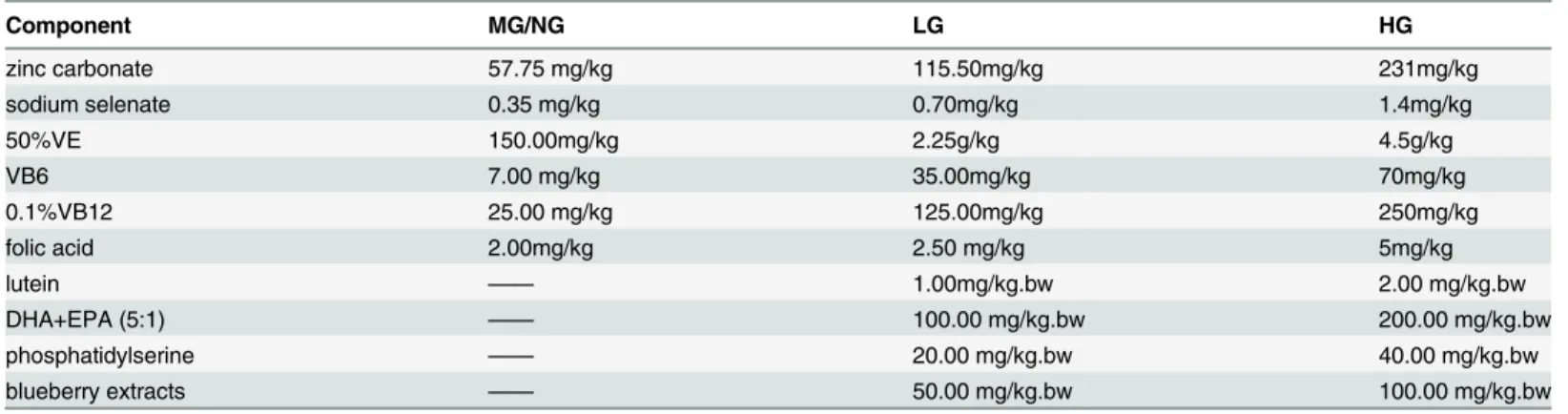
Table 1. Feed formula and amount of intervention dose.
Component MG/NG LG HG
zinc carbonate 57.75 mg/kg 115.50mg/kg 231mg/kg
sodium selenate 0.35 mg/kg 0.70mg/kg 1.4mg/kg
50%VE 150.00mg/kg 2.25g/kg 4.5g/kg
VB6 7.00 mg/kg 35.00mg/kg 70mg/kg
0.1%VB12 25.00 mg/kg 125.00mg/kg 250mg/kg
folic acid 2.00mg/kg 2.50 mg/kg 5mg/kg
lutein —— 1.00mg/kg.bw 2.00 mg/kg.bw
DHA+EPA (5:1) —— 100.00 mg/kg.bw 200.00 mg/kg.bw
phosphatidylserine —— 20.00 mg/kg.bw 40.00 mg/kg.bw
blueberry extracts —— 50.00 mg/kg.bw 100.00 mg/kg.bw
Note: (1) LG-low intervention group, HG- high intervention group, MG- model group, NG- negative control group. (2) Other components of the feed formula were based on AIN-93M. (3) The purity of each constituent was based on AIN-93M.
in one of the quadrants. Mice that found the platform within 60 s were permitted to stay on the platform for 10 s; the others were placed on the platform for 10 s. The place learning task con-sisted of four trial sessions per day for 5 days, and on the sixth day, the platform was removed from the maze, and the mice performed a 60-s probe trial test. A computerized tracking system was used to record and analyze the mice’s movement.
Shutter box escape/avoidance task
The experiment was carried out in shutter boxes consisting of two equal-sized compartments (25 × 25 × 28 cm) connected by an opening (8 × 10 cm) [36]. Diffused illumination was pro-vided by a fluorescent bulb that was placed in the ceiling of the boxes. Mice were acclima-tized inside the shutter box shock area for 5 min and were then given the conditioned stimulus (lights) for 20 s, followed by the unconditioned stimulus (a 24-V, 50 Hz electric shock though the grid floor) for 10 s. One round of the task was complete when the animal crossed to the other compartment, with crossing during the conditioned or unconditioned stimulation being considered as avoidance and escape responses, respectively. The training consisted of a single 40-trial session over the course of 1 day, with the test repeated after 24 h. A camera was placed in the center of the ceiling in each box t so aso record and analyze the mice’s movement.
Biochemical parameters
A total of 12 mice in each group were used for the behavioral studies after 3 and 7 months of treatment. The mice fasted for 12 h; then, they were anesthetized with 10% chloral hydrate, and a blood sample was drawn from the eyeball for biochemical analyses. The brain was removed from six of the animals in each group, frozen in liquid nitrogen, cut into two halves by midsagittal dissection, and then kept at−80°C. The left half was ground using a mortar and
pestle on dry ice and afterwards was homogenized in normal saline (1:9 weight in g:volume in ml). The homogenate was centrifuged at 4°C and 2500 ×gfor 15 min, and the supernatant was
used to measure the levels of superoxide dismutase (SOD), glutathione peroxidase (GSH-Px), malondialdehyde (MDA), total cholinesterase (TChE), and total protein by spectrophotometry (except for MDA) (8500 UV-VIS; Techcomp, Ltd., Shanghai, China) using the appropriate kit (Nanjing Jiancheng Biochemistry Co., Nanjing, China) according to the manufacturer’s proto-cols. The only deviation was a dilution of the supernatant by 10-fold with normal saline for the SOD and protein measurements. The blood was centrifuged at 20°C and 3000 ×gfor 15 min.
The serum was stored at 4°C for a maximum of 3 days before its use in assays for MDA, SOD, acetylcholine (ACh), and TChE.
MDA was assayed with the thiobarbituric acid heating method. To measure the SOD activ-ity, the method employing the reaction of superoxide radicals with hydroxylamine to form a red formazan dye at 550 nm was used. The SOD activity was then measured as the degree of inhibition of this reaction, and the results were given as U/mg or U/ml in the brain and serum, respectively. The GSH-PX activity was measured by the consumption of reduced glutathione in the enzymatic reaction with 5,5'-dithiobis(2-nitrobenzoic acid), which resulted in a yellow solution at 412 nm. TChE hydrolyzes ACh, generating acetic acid and choline, which can react with a thiol chromogenic agent to form the yellow compound 1,3,5-trinitrobenzene at 412 nm. The protein content in the brain was quantified using the Coomassie brilliant blue assay.
Histology and immunohistochemistry
ml of 4% paraformaldehyde in 0.01 M phosphate-buffered saline (pH 7.2–7.4). Their brains were removed and post-fixed in the same solution overnight, and then, they were embedded in paraffin and sectioned at a thickness of 4μm. The resulting sections were placed overnight in
an oven at 68°C, then deparaffinized in three changes of xylol, rehydrated in a graded series of alcohol, and washed with distilled water. They were then immersed in fresh 3% H2O2to
inacti-vate endogenous peroxidase and were again washed with distilled water. For antigen retrieval, the sections were placed in 10 mmol citrate buffer (pH 6.0) for 4 min at 2100°C in a pressure cooker. Afterwards, they were cooled to room temperature over 40 min and then blocked in 5% bovine serum albumin and incubated with a primary antibody against Aβ1–42overnight at 4°C. The sections were incubated with biotinylated anti-mouse IgG secondary antibody for 20 min and then with a diaminobenzidine (DAB) solution using the Vector DAB peroxidase sub-strate kit (Vector Laboratories, Burlingame, CA, USA), followed by counterstaining with meth-ylene blue for 20 s. After clearing in xmeth-ylene, the sections were cover-slipped for examination under a light microscope. Some sections were stained with hematoxylin and eosin (H & E) for histopathological examination [37].
Data analysis
Statistical analyses were performed with SPSS 17.0 for Windows (SPSS Inc., Chicago, IL, USA). Data corresponding to biochemical parameters were presented as mean ± standard error of the mean and were analyzed with the unpaired student’s t test. The results from the Morris water maze and avoidance task were analyzed by analysis of variance (ANOVA) for repeated mea-sures and theχ2test, respectively. P<0.05 was considered statistically significant.
Results
Animal body weight and food intake
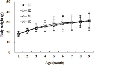
There was no significant difference in the daily food intake across groups (LG, 3.39 ± 0.18 g; HG, 3.34 ± 0.22 g; MG, 3.63 ± 0.26 g; NG, 3.51 ± 0.23 g), and over time, the body weights of all the mice (regardless of group affiliation) were similar up to 9 months of age (Fig 2).
Fig 2. The trend of the average weights of the four groups.
Behavioral tests
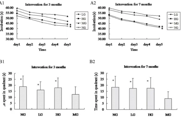
The Morris water maze test took place over 5 days, and the mice were provided different entry points to the pool (Fig 3). The latency until the mice found the platform in each trial became shorter over this period, indicating that the mice were learning to navigate the maze (Fig 4A). At 3 months after the start of treatment, the NG and HG had shorter latencies than the MG (repeated measures ANOVA [38]; P<0.001), but the same was not true for the LG
(P = 0.310). Likewise, at 7 months, only the NG and HG (P<0.001), but not the LG
(P = 0.972), performed better on the test than the MG. There was no statistical difference in the latency between the NG and HG (at 3 months, P = 0.596; at 7 months, P = 0.972). Similarly, the time spent in quadrant III on the sixth day after the removal of the platform was shorter in the NG and HG than in the MG (unpaired student’s t test; P<0.01 for both groups at 3 and 7
months), while the LG did not differ from the MG (at 3 month, P = 0.106 and at 7 months, P = 0.203) (Fig 4B).
In the shutter box escape/avoidance test, the percentage of active or passive avoidances and escape failures during the 40-trial session was recorded (Table 2). Compared to the MG, there was a higher incidence of active avoidance and fewer escape failures in the NG, HG, and LG at 3 and 7 months, (P<0.001 for all groups at both time points). The escape latency was also
lower in the NG and HG than in the MG (unpaired student’s t test; at 3 and 7 months, P<0.01 for both groups).
ACh and TChE levels in serum and brain
The ACh serum level was similar across all groups at 3 months (Table 3); however, at 7 months, a higher level was observed in the HG and LG compared to the MG (P<0.05).
Con-versely, the serum TChE level was reduced in the HG, LG, and NG compared to the MG at both 3 and 7 months (P<0.05).
Fig 3. Trajectories of mice in the Morris water maze tests.
In the brain, ACh was increased in only the HG at 3 months and in the HG, LG, and NG at 7 months (Table 3). In contrast, the TChE levels were lower in the HG, LG, and NG than in the MG at both 3 and 7 months.
Indicators of oxidative stress
The MDA level in the serum at 3 months was lower in both the LG (P<0.05) and HG
(P<0.01) than in the MG (Table 4); moreover, the level in the HG was markedly lower than
Table 2. Results from the two-way (shuttle-box) active avoidance training.
LG*** HG*** MG NG***
Intervention for 3 months
Active avoidances response(%) 45.00 46.67 20.83 44.38
Passive avoidances response(%) 30.42 27.92 40.63 28.96
Escape failures(%) 24.58 25.41 38.54 26.66
Escape latency(s) 6.90±2.29* 5.75±2.58* 9.58±2.85 5.15±1.52*
Intervention for 7 months
Active avoidances response(%) 30.63 45.00 29.38 42.29
Passive avoidances response(%) 53.33 25.63 21.46 32.92
Escape failures(%) 16.04 29.38 49.17 24.79
Escape latency(s) 7.60±2.57* 7.33±2.05* 10.58±3.00 6.92±1.67*
Note:(1) LG-low intervention group, HG- high intervention group, MG- model group, NG- negative control group. (2) The percentage of active avoidances, passive avoidances, and escape failures***P<0.001, compared with MG. (3) Escape latency(s): the average response latency for the 40-trials in the shuttle box training session; n = 12,*P<0.01.
doi:10.1371/journal.pone.0143135.t002
Fig 4. Results from Morris water maze tests.(A) The average incubation period from the first to fifth day for the four groups. After 3 months and 7 months of intervention, the mice in HG and NG spent much less time searching for new (reversal) hidden platform on each reversal training day compared to MG,* P<0.001. (B) The time spent in the third quadrant for HG, LG, and NG was much greater than that of the mice in MG,*P<0.01.
in the LG (P<0.05). In contrast, the SOD level was higher in the NG, HG, and LG compared
to the MG at 3 months. Similar trends were also observed at 7 months.
The MDA, SOD, and GSH-Px levels in the brain tissue are shown inTable 4. Relative to the levels in the MG, the MDA level was lower in the NG, HG, and LG, whereas the SOD and GSH-Px levels were higher. Furthermore, the total protein content in the brain was lower in these three groups than in the MG at 7 months (P<0.05).
Pathologic changes in brains
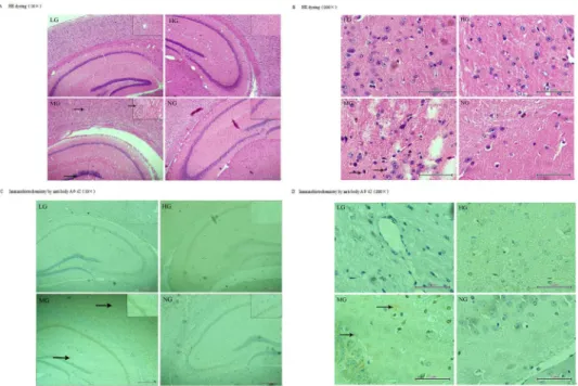
At 9 months old, fewer neurons and pycnotic neuronal nuclei were observed in the cerebral cortex and hippocampus of the transgenic mice but not the wild-type by H & E staining (Fig 5A and 5B). Immunohistochemistry with an antibody against Aβ1–42(Fig 5C and 5D) revealed an increase in the number and volume of amyloid plaques proportional to the age of the mice (data not shown). Additionally, fewer plaques were observed in the cortex and hippocampus of
Table 3. Distribution of Ach and TChE in serum and brains for the four groups (Mean±SD, n = 6/12).
LG HG MG NG
3 m 7 m 3 m 7 m 3 m 7 m 3 m 7 m
In Brain
Ach (μg/mg port) 56.43±12.03 56.80
±17.10*
74.81±9.45** 53.11±12.59** 50.74±11.65 35.08±3.44 70.50
±10.01**
58.28±8.96***
TChE (U/mg port)
0.91±0.29 0.84±0.37 0.83±0.18* 0.73±0.24* 1.14±0.29 0.99±0.15 0.79±0.17* 0.74±0.16*
In serum
Ach (μg/ml) 282.54
±123.42 305.95 ±98.88 321.82 ±120.41 380.65
±115.32*
294.18 ±96.33 269.64 ±108.59 355.53 ±141.71 431.84
±175.96* TChE (U/ml) 47.31±3.51** 59.75±7.56* 46.82±3.08** 53.23±5.54*** 52.60±5.48 66.40±5.55 46.83±3.99** 58.17±6.13**
Note:(1) LG-low intervention group, HG- high intervention group, MG- model group, NG- negative control group. (2)*:P<0.05,**:P<0.01,***P<0.001
compared with MG. (3) in brain, n = 6; in serum, n = 12.
doi:10.1371/journal.pone.0143135.t003
Table 4. Distribution of indicators of oxidative stress of brain and serum for the four groups (Mean±SD, n = 6/12).
LG HG MG NG
3 m 7 m 3 m 7 m 3 m 7 m 3 m 7 m
In Brain
Total Protein (g/l) 0.40±1.13 0.47±0.22* 0.36±0.06* 0.41±0.05*** 0.50±0.10 0.68±0.08 0.34±0.05** 0.39±0.06***
SOD (U/mg port) 3.09±0.39* 2.41±0.36* 3.18±0.64* 2.72±0.42** 2.25±0.76 1.94±0.33 3.08±0.76* 2.47±0.43* MDA (nmol/mg) 2.30±0.66* 2.82±0.92* 2.23±0.95* 2.83±0.89* 3.73±1.15 4.62±1.65 2.06±0.85* 2.74±0.72* GSH-PX (U/mg
port)
26.03±14.90 30.51±9.15* 41.12±5.71*** 31.45 ±1.39***
22.44±4.35 20.55±1.36 41.59±6.49*** 31.10 ±2.03*** In serum
MDA (n mol/ml) 6.89±2.64* 9.15±3.47* 6.71±1.75** 7.36±2.06*** 8.71±1.19 12.04±1.28 6.43±1.68*** 9.41±3.30*
SOD (U/ml) 129.16
±37.28**
123.26
±28.567*
140.17
±30.85***
129.90
±19.80**
96.05
±11.79
86.06
±43.53
139.80
±37.28***
131.91
±25.49**
Note:(1) LG-low intervention group, HG- high intervention group, MG- model group, NG- negative control group. (2)*:P<0.05,**:P<0.01,***P<0.001 compared with MG. (3) in brain, n = 6; in serum, n = 12.
the mice in the LG and HG after 7 months of treatment compared to the MG at the same age (Fig 5D), thus suggesting that early intervention with this nutrient combination can suppress the development of amyloid plaques.
Discussion
In this study, it was demonstrated that APP-PSN mice experience a significant improvement in cognitive function as well as in biochemical and histological indicators of AD after consuming a mixture of nutrients for up to 7 months.
Cognitive dysfunction is one of the major clinical manifestations of AD and is correlated with the loss of hippocampal and cortical synapses [39] that serve as the cellular substrates for spatial learning and memory [35]. In this study, the rate of memory decline was reduced along with the amelioration in cognitive function in mice after dietary intervention, thereby suggesting that this could be a possible strategy for delaying or preventing AD symptoms in humans [40].
During aging, oxidative stress resulting from reactive oxygen species (ROS) production increases even as endogenous antioxidant mechanisms progressively decay. These could be exacerbated in AD. In fact, a growing body of evidence implicates the early involvement of oxi-dative stress in the pathogenesis and progression of AD [41,42]. The attack of lipids, proteins, sugars, and nucleic acids by free radicals leads to the formation of byproducts that can be detected in fluids and tissues and ultimately can serve as markers of oxidative damage [43]. Accordingly, lower levels of the oxidative stress indicators MDA, SOD, and GSH-PX were observed in the mice that were receiving the dietary supplement in this study, which could be due to the antioxidant micronutrients, such as selenium, lutein, zinc, and vitamin E.
Acetylcholinesterase and the cholinergic system, which are involved in learning and mem-ory, may also play a role in AD. TChE belongs to a family ofα/βhydrolases that catalyze the
Fig 5. Immunofluorescence staining with rabbit anti-Aβ1–42 and HE dyeing of the temporal cortex and hippocampus of APP/PSN mice.(1) LG and HG: APP/PSN (Fig 5A) mice with 7 months of intervention; MG: APP/PSN (Fig 5B) mice without intervention and their wild-type littermates (NG). (2) LG and HG:±, MG:
+++, NG:–(Fig 5C and 5D).
hydrolysis of ACh at the cholinergic synapse [44]. Some studies have described a decrease in ACh and an increase in the TChE level in the brain as contributing factors to memory dysfunc-tion in AD [45–47]. The present findings were consistent with these reports. The AD mice con-suming the nutrient mixture had a higher ACh level than those who were fed regular chow, while the TChE level was reduced. Thus, increasing the ACh level in the brain by modulating TChE activity and signaling pathways mediated through cholinergic receptors that promote nonamyloidogenic APP processing and decrease tau phosphorylation are potential strategies for AD therapy [48]. Furthermore, the amyloid cascade hypothesis has been proposed to explain the pathogenesis of AD. The deposition of Aβis the initial pathological event, which then triggers a cascade of events that include the formation of senile plaques, followed by neu-rofibrillary tangles, and this cascade ultimately results in neuronal death and dementia [49,50]. Here, it was found that the Aβdeposition in the treatment groups was decreased compared to the AD model mice who were fed a regular diet. Thus, the protective effects of these nutrients may be direct—by promoting neural repair and inhibiting the formation of amyloid plaques—
or else auxiliary, through antioxidative mechanisms or by lowering homocysteine.
Lastly, there are some limitations to this study. First, it is not clear that the beneficial effects of the tested nutrient combination would translate to humans. Moreover, not all of the compo-nents are commercially available. In the future, preclinical trials will be carried out to assess specific early biomarkers of AD using metabolomics approaches. In conclusion, the findings presented here suggest that a combination of nutrients may synergize to delay the symptoms of AD and that early intervention by dietary supplementation is a viable strategy for AD
management.
Acknowledgments
The study was supported by a grant from the National Natural Science Foundation of China (no. 81273193). No conflicts of interest are declared.
Author Contributions
Conceived and designed the experiments: QHW YHW DCW. Performed the experiments: YC SYW CW YZ. Analyzed the data: SYW YC WX LM. Contributed reagents/materials/analysis tools: JFZ RD. Wrote the paper: SYW YC CW.
References
1. Brookmeyer R, Johnson E, Ziegler-Graham K, Arrighi HM. Forecasting the global burden of Alzhei-mer’s disease. Alzheimers Dement. 2007; 3: 186–191. doi:10.1016/j.jalz.2007.04.381PMID:
19595937
2. Goedert M, Spillantini M G. A century of Alzheimer's disease. Sci. 2006; 314: 777–781.
3. McKeith I, Cummings J. Behavioural changes and psychological symptoms in dementia disorders. Lan-cet Neurol. 2005; 4: 735–742. PMID:16239180
4. Butterfield D A, Swomley A M, Sultana R. Amyloidβ-Peptide (1–42)-Induced Oxidative Stress in Alzhei-mer Disease: Importance in Disease Pathogenesis and Progression. Antioxid Redox Signal. 2013; 19: 823–835. doi:10.1089/ars.2012.5027PMID:23249141
5. Mastroeni D, Grover A, Delvaux E, Whiteside C, Coleman PD, Rogers J. Epigenetic mechanisms in Alzheimer's disease. Neurobiol Aging. 2011; 32: 1161–1180. doi:10.1016/j.neurobiolaging.2010.08. 017PMID:21482442
6. Pimplikar S W, Nixon R A, Robakis N K, Shen J, Tsai LH. Amyloid-independent mechanisms in Alzhei-mer's disease pathogenesis. J Neurosci. 2010; 30: 14946–14954. doi:10.1523/JNEUROSCI.4305-10. 2010PMID:21068297
7. Moreira PI, Carvalho C, Zhu X, Smith MA, Perry G. Mitochondrial dysfunction is a trigger of Alzheimer's disease pathophysiology. Biochim Biophys Acta. 2010; 1802: 2–10. doi:10.1016/j.bbadis.2009.10.006
8. Bignante E A, Heredia F, Morfini G, Lorenzo A. Amyloidβprecursor protein as a molecular target for amyloidβ–induced neuronal degeneration in Alzheimer's disease. Neurobiol Aging. 2013; 34: 2525–
2537. doi:10.1016/j.neurobiolaging.2013.04.021PMID:23714735
9. Querfurth HW, LaFerla FM. Alzheimer’s disease. N. Engl. J.Med. 2010, 362, 329–344. doi:10.1056/ NEJMra0909142PMID:20107219
10. Ji HF, Shen L. Berberine: a potential multipotent natural product to combat Alzheimer’s disease. Mole-cules. 2011; 16: 6732–6740. doi:10.3390/molecules16086732PMID:21829148
11. Hofrichter J, Krohn M, Schumacher T, Lange C, Feistel B, Walbroel B, et al. Reduced Alzheimer's dis-ease pathology by St. John's Wort treatment is independent of hyperforin and facilitated by ABCC1 and microglia activation in mice. Curr Alzheimer Res. 2013; 10: 1057–1069. PMID:24156265
12. Colantuoni E, Surplus G, Hackman A, Arrighi HM, Brookmeyer R. Web-based application to project the burden of Alzheimer's disease. Alzheimers Dement. 2010; 6: 425–428. doi:10.1016/j.jalz.2010.01.014
PMID:20691645
13. Douaud G, Refsum H, de Jager CA, Jacoby R, Nichols TE, Smith SM, et al. Preventing Alzheimer’s dis-ease-related gray matter atrophy by B-vitamin treatment. Proc Natl Acad Sci U S A. 2013; 110: 9523–
9528. doi:10.1073/pnas.1301816110PMID:23690582
14. Johnson E J. A possible role for lutein and zeaxanthin in cognitive function in the elderly. Am J Clin Nutr. 2012; 96: 1161S–1165S. doi:10.3945/ajcn.112.034611PMID:23053547
15. Little A, Levy R, Chuaqui-Kidd P, Hand D. A double-blind, placebo controlled trial of high-dose lecithin in Alzheimer's disease. J Neurol Neurosurg Psychiatry. 1985; 48: 736–742. PMID:3897460
16. Hjorth E, Zhu M, Toro VC, Vedin I, Palmblad J, Cederholm T, et al. Omega-3 Fatty Acids Enhance Phagocytosis of Alzheimer's Disease-Related Amyloid-β42 by Human Microglia and Decrease Inflam-matory Markers. J Alzheimers Dis. 2013; 35: 697–713. doi:10.3233/JAD-130131PMID:23481688
17. Yurko-Mauro K, McCarthy D, Rom D, Nelson EB, Ryan AS, Blackwell A, et al. Beneficial effects of docosahexaenoic acid on cognition in age-related cognitive decline. Alzheimers Dement. 2010; 6: 456–464. doi:10.1016/j.jalz.2010.01.013PMID:20434961
18. Scheltens P, Kamphuis PJ Verhey FR, Olde Rikkert MG, Wurtman RJ, Wilkinson D, et al. Efficacy of a medical food in mild Alzheimer’s disease: A randomized, controlled trial. Alzheimers Dement. 2010; 6: 1–10.e1. doi:10.1016/j.jalz.2009.10.003PMID:20129316
19. Kamphuis PJ, Scheltens P. Can nutrients prevent or delay onset of Alzheimer's disease?. J Alzheimers Dis. 2010; 20: 765–775. doi:10.3233/JAD-2010-091558PMID:20182021
20. Reiserer RS, Harrison FE, Syverud DC, McDonald MP. Impaired spatial learning in the APPSwe+ PSEN1ΔE9 bigenic mouse model of Alzheimer’s disease. Genes Brain Behav. 2007; 6: 54–65. PMID:
17233641
21. Nolan JM, Loskutova E, Howard A, Mulcahy R, Moran R, Stack J, et al. The impact of supplemental macular carotenoids in Alzheimer's disease: a randomized clinical trial. J Alzheimers Dis. J Alzheimers Dis. 2015; 44:1157–1169. doi:10.3233/JAD-142265PMID:25408222
22. Chew EY, Clemons TE, Agrón E, Launer LJ, Grodstein F, Bernstein PS, et al. Effect of Omega-3 Fatty Acids, Lutein/Zeaxanthin, or Other Nutrient Supplementation on Cognitive Function: The AREDS2 Randomized Clinical Trial. JAMA. 2015; 314: 791–801. doi:10.1001/jama.2015.9677PMID:
26305649
23. Jankowsky JL, Fadale DJ, Anderson J, Xu GM, Gonzales V, Jenkins NA, et al. Mutant presenilins spe-cifically elevate the levels of the 42 residue beta-amyloid peptide in vivo: evidence for augmentation of a 42-specific gamma secretase. Hum Mol Genet. 2004; 13: 159–170. PMID:14645205
24. Reiserer RS, Harrison FE, Syverud DC, McDonald MP. Impaired spatial learning in the APPSwe+ PSEN1DeltaE9 bigenic mouse model of Alzheimer’s disease. Genes Brain Behav. 2007; 6: 54–65. PMID:17233641
25. Subash S, Essa MM, Al-Asmi A, Al-Adawi S, Vaishnav R, Guillemin GJ. Effect of dietary supplementa-tion of dates in Alzheimer's disease APPsw/2576 transgenic mice on oxidative stress and antioxidant status. Nutr Neurosci. 2015; 18: 281–288. doi:10.1179/1476830514Y.0000000134PMID:24954036
26. Subash S, Essa MM, Braidy N, Awlad-Thani K, Vaishnav R, Al-Adawi S, et al. Guillemin. Diet rich in date palm fruits improves memory, learning and reduces beta amyloid in transgenic mouse model of Alzheimer's disease. J Ayurveda Integr Med. 2015; 6: 111–120. doi:10.4103/0975-9476.159073
PMID:26167001
28. Garthe A, Kempermann G. An old test for new neurons: refining the Morris water maze to study the functional relevance of adult hippocampal neurogenesis. Front Neurosci. 2013; 7: 63. doi:10.3389/ fnins.2013.00063PMID:23653589
29. Dong Z, Bai Y, Wu X, Li H, Gong B, Howland JG, et al. Hippocampal long-term depression mediates spatial reversal learning in the Morris water maze. Neuropharmacology. 2013; 64: 65–73. doi:10. 1016/j.neuropharm.2012.06.027PMID:22732443
30. Banerjee R, Ghosh AK, Ghosh B, Mondal AC, Banerjee R. Effect of chronic inescapable footshock and antidepressant treatment on BDNF/TrkB levels in rat hippocampus. Research and Reviews: A Journal of Neuroscience. 2012; 2: 12–21.
31. Shi JQ, Shen W, Chen J, Wang BR, Zhong LL, Zhu YW, et al. Anti-TNF-αreduces amyloid plaques and tau phosphorylation and induces CD11c-positive dendritic-like cell in the APP/PS1 transgenic mouse brains. Brain Res. 2011; 1368: 239–247. doi:10.1016/j.brainres.2010.10.053PMID:20971085
32. Qiu H, Jin GQ, Jin RF, Zhao WK. Analysis of variance of repeated data measured by water maze with SPSS. Journal of Chinese Integrative Medicine. 2007; 5: 101–105. PMID:17214947
33. Mitew S, Kirkcaldie MT, Dickson TC, Vickers JC. Altered synapses and gliotransmission in Alzheimer's disease and AD model mice. Neurobiol Aging. 2013; 34: 2341–2351. doi:10.1016/j.neurobiolaging. 2013.04.010PMID:23643146
34. Ansari MA, Scheff SW. Oxidative stress in the progression of Alzheimer disease in the frontal cortex. J Neuropathol Exp Neurol. 2010; 69: 155–167. doi:10.1097/NEN.0b013e3181cb5af4PMID:20084018
35. Reddy VP, Zhu X, Perry G, Smith MA. Oxidative stress in diabetes and Alzheimer's disease. J Alzhei-mers Dis. 2009; 16: 763–774. doi:10.3233/JAD-2009-1013PMID:19387111
36. Mangialasche F, Polidori MC, Monastero R, Ercolani S, Camarda C, Cecchetti R, et al. Biomarkers of oxidative and nitrosative damage in Alzheimer's disease and mild cognitive impairment. Ageing Res Rev. 2009; 8: 285–305. doi:10.1016/j.arr.2009.04.002PMID:19376275
37. Kaladhar D, Anusha N, Rao VV, Surekha C, Meesala S. Regulation of Metabolic Syndromes by means of controlling diseased Ache and Bche with Multitarget Inhibitors through in silico techniques. Journal of Computational Methods in Molecular Design. 2012; 2: 122–129.
38. Naik RS, Hartmann J, Kiewert C, Duysen EG, Lockridge O, Klein J. Effects of rivastigmine and donepe-zil on brain acetylcholine levels in acetylcholinesterase-deficient mice. J Pharm Pharm Sci. 2009; 12: 79–85. PMID:19470293
39. Golde TE, Petrucelli L, Lewis J. Targeting Abeta and tau in Alzheimer's disease, an early interim report. Exp Neurol. 2010; 223: 252–266. doi:10.1016/j.expneurol.2009.07.035PMID:19716367
40. Darreh-Shori T, Soininen H. Effects of cholinesterase inhibitors on the activities and protein levels of cholinesterases in the cerebrospinal fluid of patients with Alzheimer's disease: a review of recent clini-cal studies. Curr Alzheimer Res. 2010; 7: 67–73. PMID:20205672
41. Hu JJ, Li J, Wei Z, Gu HY, Shao BY, Long DW, et al. The PARF Antagonist AChE Inhibitor PMS777 Attenuates LPS-Induced Acute Neuroinflammation. Neuroscience & Medicine. 2011; 2: 355–362. 42. Armstrong RA. The pathogenesis of Alzheimer's disease: a reevaluation of the“amyloid cascade
hypothesis”. Int J Alzheimers Dis. 2011; 2011:630865. doi:10.4061/2011/630865PMID:21331369
43. Lane RM, Kivipelto M, Greig N H. Acetylcholinesterase and its inhibition in Alzheimer disease. Clinical neuropharmacology. 2004; 27: 141–149. PMID:15190239
44. Munoz-Torrero D. Acetylcholinesterase Inhibitors as Disease-Modifying Therapies for Alzheimers Dis-ease. Current medicinal chemistry. 2008; 15: 2433–2455 PMID:18855672
45. Jäger R, Purpura M, Kingsley M. Phospholipids and sports performance. Journal of the International Society of Sports Nutrition. 2007; 4: 5. PMID:17908342
46. Parker AG, Gordon J, Thornton A, Byars A, Lubker J, Bartlett M, et al. The effects of IQPLUS Focus on cognitive function, mood and endocrine response before and following acute exercise. J Int Soc Sports Nutr. 2011; 8: 16. doi:10.1186/1550-2783-8-16PMID:22017963
47. Bhalla V, Arora H, Dhir A, Kumar M. A triphenylene based zinc ensemble as an oxidation inhibitor. Chem. Commun. 2012; 48: 4722–4724.
48. Aliev G, Obrenovich ME, Reddy VP, Shenk JC, Moreira PI, Nunomura A, et al. Antioxidant therapy in Alzheimer’s disease: theory and practice. Mini Rev Med Chem. 2008; 8: 1395–1406. PMID:18991755
49. Tassoni D, Kaur G, Weisinger RS, Sinclair AJ. The role of eicosanoids in the brain. Asia Pacific journal of clinical nutrition. 2008; 17: 220–228. PMID:18296342