The Role of Heparanase in Pulmonary Cell
Recruitment in Response to an Allergic but
Not Non-Allergic Stimulus
Abigail Morris1☯, Bo Wang2☯, Ida Waern3☯, Radhakrishnan Venkatasamy1‡, Clive Page1,
Eric P. Schmidt4‡, Sara Wernersson3‡, Jin-Ping Li2, Domenico Spina1☯‡
*
1Sackler Institute of Pulmonary Pharmacology, Institute of Pharmaceutical Science, King’s College London, London, United Kingdom,2Department of Medical Biochemistry and Microbiology, Uppsala University, Box 582, Uppsala, Sweden,3Department of Anatomy, Physiology and Biochemistry, Swedish University of Agricultural Sciences, Box 7011, Uppsala, Sweden,4Program in Translational Lung Research, University of Colorado School of Medicine, Aurora, CO, United States of America
☯These authors contributed equally to this work. ‡These authors also contributed equally to this work.
Abstract
Heparanase is an endo-β-glucuronidase that specifically cleaves heparan sulfate proteogly-cans in the extracellular matrix. Expression of this enzyme is increased in several pathologi-cal conditions including inflammation. We have investigated the role of heparanase in pulmonary inflammation in the context of allergic and non-allergic pulmonary cell recruit-ment using heparanase knockout (Hpa-/-) mice as a model. Following local delivery of LPS
or zymosan, no significant difference was found in the recruitment of neutrophils to the lung between Hpa-/-and wild type (WT) control. Similarly neutrophil recruitment was not inhibited
in WT mice treated with a heparanase inhibitor. However, in allergic inflammatory models, Hpa-/-mice displayed a significantly reduced eosinophil (but not neutrophil) recruitment to the airways and this was also associated with a reduction in allergen-induced bronchial hyperresponsiveness, indicating that heparanase expression is associated with allergic re-actions. This was further demonstrated by pharmacological treatment with a heparanase in-hibitor in the WT allergic mice. Examination of lung specimens from patients with different severity of chronic obstructive pulmonary disease (COPD) found increased heparanase ex-pression. Thus, it is established that heparanase contributes to allergen-induced eosinophil recruitment to the lung and could provide a novel therapeutic target for the development of anti-inflammatory drugs for the treatment of asthma and other allergic diseases.
Introduction
Heparan sulfate proteoglycans consist of a protein core decorated with heparan sulfate linear polysaccharides. This family includes syndecans, glypicans and secreted forms of extracellular matrix proteoglycans (e.g. perlecan). Heparan sulfate can bind to a range of proteins (e.g.
OPEN ACCESS
Citation:Morris A, Wang B, Waern I, Venkatasamy
R, Page C, Schmidt EP, et al. (2015) The Role of Heparanase in Pulmonary Cell Recruitment in Response to an Allergic but Not Non-Allergic Stimulus. PLoS ONE 10(6): e0127032. doi:10.1371/ journal.pone.0127032
Academic Editor:Bernhard Ryffel, French National
Centre for Scientific Research, FRANCE
Received:January 27, 2015
Accepted:April 11, 2015
Published:June 3, 2015
Copyright:© 2015 Morris et al. This is an open access article distributed under the terms of the Creative Commons Attribution License, which permits unrestricted use, distribution, and reproduction in any medium, provided the original author and source are credited.
Data Availability Statement:All relevant data are
available within the paper, or from Figshare, under the DOI:http://dx.doi.org/10.6084/m9.figshare. 1386646.
Funding:These studies were supported by a
Mammalian heparanase is an enzyme that specifically cleaves heparan sulfate chains. This enzyme was first cloned in 1999 and is the only known mammalian enzyme capable of disas-sembling heparan sulfate [9,10] and is expressed in many inflammatory cells including dendrit-ic cells, T and B cells, platelets, macrophages, neutrophils and mast cells and is impldendrit-icated in mediating diapedesis and recruitment of leukocytes to inflammatory sites [11]. Increased ex-pression of heparanase in various tumours correlates with tumour invasiveness, angiogenesis and poor prospective survival. There is also evidence of increased heparanase expression in in-flammatory bowel disease [12] and in the synovium of rheumatoid arthritis patients [13]. Heparanase overexpression in mice led to an exacerbation of delayed type hypersensitivity (DTH) reaction in the skin [14] and may specifically regulate transmigration of monocytes in this inflammatory response [15]. Few studies have examined the role of this enzyme in regulat-ing pulmonary recruitment of inflammatory cells. This is relevant to an understandregulat-ing of the pathophysiology of inflammatory diseases like asthma and COPD which are characterized by the presence of inflammatory cells like eosinophils and neutrophils. It has been reported that mice deficient in heparanase are protected from sepsis-induced acute lung injury [16] and al-lergic inflammation [17], suggesting that heparan sulfate provides a pro-inflammatory cue. However, elimination of syndecan-1 appears to lead to an exacerbation of the allergic inflam-matory response [7]. Hence, we hypothesize that heparanase could play a role in inflaminflam-matory
cell migration in lung inflammation and used heparanase knockout (Hpa-/-) mice and
pharma-cologically treated mice as two models to investigate the role of heparanase in pulmonary in-flammation and changes in pulmonary lung mechanics.
Methods
Human lung tissue
Lung tissue from human subjects (anonymized) was obtained from the National Jewish Health, Denver USA (normal controls) and from the Lung Tissue Repository Consortium (COPD lungs) [18] and tissue heparanase expression was detected using a previously described method [19].
Lung samples were obtained either from surgical lung biopsy or explants at the time of lung transplantation. Severity of COPD was stratified according to the 2013 Global Initiative for Ob-structive Lung Disease (www.goldcopd.com) guidelines, which ranged from Stage 0 (normal
spirometry with chronic symptoms), Stage I (FEV1/FVC<70% with FEV1>80% predicted),
Stage II (FEV1/FVC<70% with FEV1 50–79% predicted), Stage III (FEV1/FVC<70% with
FEV1 30–69% predicted), and Stage IV (FEV1/FVC<70% with FEV1<30% predicted).
4-μm paraffin-embedded sections using a rabbit polyclonal antibody to human heparanase
(Pro-spec, Ins-26-2, 1:1000). Rabbit IgG (Abcam, 27472, 1:1000) served as an isotype control. We measured intensity of staining using Metamorph (Molecular Devices, Sunnyvale, CA), as previ-ously described [19].
Animal Welfare and Maintenance
Female BALB/c mice (18-20g; Charles River or Harlan, UK) were housed in rooms maintained at a constant temperature (21 ± 2°C) and humidity (55 ± 15%) with a 12 hour light-dark cycle.
Animals had food (SDS, UK) and water availablead libitum. All experiments were performed
under The Scientific Procedures (Animals) Act 1986 and a Home Office project licence (PPL 70/8021) approved by the Animal Welfare and Ethical Review Board at King’s College London. Studies using mice deficient in heparanase [20] were carried out at the Department of Medical Biochemistry and Microbiology, Uppsala University, Uppsala, Sweden and approved by Upp-sala Ethical Committee on Animal Experiments (ethical approval number; C228/12 and C135/ 8). Animals had free access to food and water and were housed with suitable enrichment and bedding with 12h day/night cycle. All surgery was performed under general anaesthesia (King’s College London, urethane; Uppsala University, pentobarbital sodium) and all efforts made to minimize suffering.
Induction of non-allergic airway inflammation
In order to establish lung inflammation, mice were anaesthetised with isoflurane and dosed in-tranasally (i.n.) with either zymosan (4 mg/kg) (Sigma-Aldrich, UK) or LPS (1.25 mg/kg) (from Escherichia Coli, Sigma-Aldrich, UK). Control mice received the same volume (i.n) of sterile saline.
Induction of allergic airway inflammation
For pharmacological studies, allergic airway inflammation was induced in WT BALB/c mice. In these studies, mice (6–8 weeks) were sensitised with ovalbumin (OVA, i.p.) (Chicken
oval-bumin, grade V. Sigma, UK). Each mouse received 30μg of OVA absorbed in 16μL of
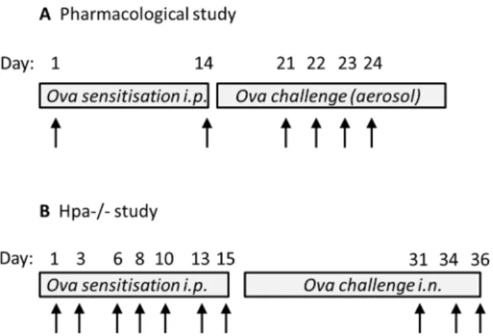
alumini-um hydroxide (Ems, Brazil) in saline to give a final volalumini-ume of 0.4 mL. Sham mice received saline and aluminum hydroxide alone. Control and OVA mice were sensitised on day 1 and boosted on days 7 and 14. On days 21–23 (BAL) or 21–24 (lung function), OVA-sensitised mice were exposed to 3% OVA by aerosol in a small chamber attached to a nebuliser (DeVill-bisS 99; HCE, UK). On each challenge day mice were exposed to OVA for 30 minutes (Fig 1A).
In a separate series of experiments, allergic airway inflammation was induced in Hpa-/-mice
backcrossed for more than 10 generations to the C57BL/6J genetic background [20].
Experi-mental groups were age and sex matched (WT control: 6♀,5♂; WT OVA: 7♀,5♂; Hpa
-/-control: 5♀,2♂: Hpa-/-OVA: 5♀,4♂) and WT littermates were used as controls. In these
ex-periments, mice (10–14 weeks) were immunized i.p. with 10μg of OVA (Sigma-Aldrich) in
100μl of PBS on days 1, 3, 6, 8, 10, 13 and 15. Control mice were non-immunized. On days 31,
34 and 36, mice were anaesthetized with isofluorane (4%) for intranasal administration with
20μg of OVA in 20μL of PBS (Fig 1B).
Pharmacological intervention
Mice were subcutaneously (s.c.) administered with the heparanase inhibitor (Muparfostat,
PI-88; 30–100 mg/kg: Progen Pharmaceuticals, Brisbane Australia) or vehicle either once (q.i.d.)
model. In the case of allergic studies, animals were treated on 3 (eosinophil recruitment) or 4 (lung function) consecutive days.
Assessment of Lung Function
Animals were anaesthetised with urethane (2 g/kg) i.p. 24 hours after the last OVA challenge and lung function was measured using a previously described technique [21]. A tracheotomy was performed and a cannula inserted and sutured in place. Mice were then placed in a plethys-mograph chamber via the cannula connected to a 4-way manifold with one port attached to a differential pressure transducer (± 20 cm H2O: Validyne, UK). Two ports were connected to the inspiratory and expiratory ports of a volume cycled ventilator (CWA Incorporated, USA). Mice were ventilated at 150 breaths/minute with a tidal volume of 0.15–0.20 mL and a positive
expiratory pressure between 3 and 5 cm H2O. Transpulmonary pressure was estimated as the
difference between mouth pressure and box pressure, as the chest wall contributes little to the overall compliance of the respiratory system. Changes in flow were determined with a Fleisch pneumotachograph connected to a side port of the chamber. Flow was measured with a
dif-ferential pressure transducer (± 20 cm H2O: Validyne, UK). The flow was recorded to give a
continuous reading of tidal volume. Breath-by-breath recording of total lung resistance (RL:
cmH20.s/L) was calculated by an online respiratory analyser on a computer (LFR 7; Mumed
Ltd., UK).
Airway responsiveness was assessed by inhaled aerosolised methacholine at a concentration range of 3.125–50 mg/mL. Aerosols of methacholine generated from an ultrasonic nebulizer were administered directly to the lungs via the cannula and a port of the plethysmograph. Aerosolised solutions were delivered to the mouse for 8 seconds, at a rate of 150 breaths/minute with a tidal volume of 0.15–0.2 mL. Total airway resistance was measured prior to, and follow-ing administration of saline and then followfollow-ing administration of methacholine. Upon comple-tion of experiments, animals were humanely killed by cervical dislocacomple-tion. The provocative concentration (PC) of methacholine which caused a 100% increase in post saline RL (RL PC100) and peak response (i.e. maximum percentage change in post saline RL) for each animal was used as a measure of airway sensitivity and reactivity, respectively.
Fig 1. Protocol for allergen sensitization and challenge.Diagrammatic representation of the sensitization and challenge protocols to induce allergic lung inflammation in WT BALBc (A) and Hpa-/-(B) mice. Arrow represents exposure to ovalbumin (OVA). Biological readouts were undertaken 24 h following the last OVA administration. In (A), bronchoalveolar lavage and lung function was measured on day 24 and day 25 respectively. In (B), BAL and serum samples were collected on day 37.
BAL fluid analysis
Mice were humanely killed 24 h after receiving the inflammatory stimulus with an
intraperito-neal (i.p) injection of 0.2 mL 50% urethane (King’s College London) or 40–60 mg/kg
pento-barbital sodium (Uppsala University). Lungs were lavaged via a cannula inserted into the trachea and instilled with 3 x 0.5 mL of sterile saline (King’s College London) or with 2 x 1 mL
of sterile HBSS (Uppsala University). In experiments using Hpa-/-allergic mice, BAL fluid
anal-ysis was performed as previously described [22]. For differential cell counts, cytospin slides
were prepared using 150μL of BAL fluid spun at 1000 x g for 1 minute. Cytospins were allowed
to dry before staining using a Diff-Quick stain. Differential cell counts were performed accord-ing to standard morphological criteria on Diff-Quick stained cytospins (100 cells/sample). Cells were identified as mononuclear cells, neutrophils and eosinophils. Almost all mononucle-ar cells observed were macrophages, but for accuracy and clmononucle-arity of presentation, were analyzed
and analysed as mononuclear cells. To perform total cell counts, 50μL of BAL fluid was mixed
well with 50μL of Türk solution (Merck, Germany) and counts performed using a
haemocyt-ometer. The total number of cells counted was then standardised to the number of total cells x
106/mL. In both instances, the individual counting the cells was blind to treatment
or genotype.
IgE ELISA
IgE anti-OVA ELISA were performed as previously described in allergic WT and Hpa-/-mice
[22]. For analysis of OVA-specific IgE in sera, 96-well microtiter plates (Nunc) were incubated
overnight at 4°C with goat anti-mouse IgE coating antibody (10μg/mL, 100μL) from the
Mouse IgE ELISA Quantification Kit (Bethyl Laboratories). Plates were washed three times
with 200μL washing buffer (50 mM Tris, 0.14 M NaCl, 0.05% Tween-20, pH 8) and then
incu-bated with 200μL blocking buffer (50 mM Tris, 0.14 M NaCl, 1% BSA, pH 8) for 2 h in room
temperature. Mouse sera were diluted 1:3 in blocking buffer and incubated for 2 h (100μL).
Plates were washed and bound anti-OVA antibodies were quantified by first incubating with
biotinylated OVA (2μg/mL, 100μL) for 60 minutes. Plates were then washed five times before
adding strepavidin-peroxidase conjugate (0.05 U, 100μL, Roche Diagnostics) and incubating
for 30 minutes. After five washes, 3,30,5,5’-tetramethylbenzidine liquid substrate (100μL,
Sigma-Aldrich) was added. The reaction was stopped after 30–45 minutes by adding 100μl 2M
H2SO4and plates were read at 450 nm.
In the biotinylation procedure 10 mg OVA (Grade V, Sigma-Aldrich) in 1 mL PBS was mixed with 2.5 mg biotinamidocaproic acid 3-sulpho-N-hydroxy-succinimide ester (Sigma-Al-drich) dissolved in 0.25 mL distilled water. The mixture was stirred for 2 h at room tempera-ture. To remove unreacted biotin the mixture was dialysed against PBS at 4°C and then the biotinylated OVA was stored at 4°C in 0.1% sodium azide.
Statistical Analysis
Data was expressed as median, with interquartile range unless otherwise specified. Cell data was analyzed using non-parametric methods (Kruskal Wallis, and Mann Whitney U test). In some instances, neutrophil or eosinophil recruitment to the lung was expressed as a percentage in terms of the average number of total cells. Analysis of variance with Dunnett’s post hoc test was used to analyze RL PC100 (log transformed) and peak response in RL. Data was analyzed
Results
Non-allergic inflammation in Hpa
-/-mice
Zymosan induced inflammation. The total number of cells in the BAL fluid of saline
in-jected WT mice (median, 25–75 percentile; 0.72 (0.60–0.96) x 106cells/ml, n = 5) was
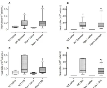
pre-dominantly comprised of mononuclear cells and represented 99% of the total cell population with an absence of neutrophils. There was a significant increase in total number of cells (Fig 2A, p = 0.038) and neutrophils (Fig 2B, p = 0.0016) recruited to the airways following challenge with zymosan. There was no significant difference in the number of total cells (Fig 2A) or
neu-trophils (Fig 2B) between Hpa-/-mice and WT mice indicating that heparanase is not involved
in the recruitment of inflammatory cells to the lung following an acute challenge with zymosan.
Lipopolysaccharide-induced lung inflammation. The total number of cells in the BAL
fluid of saline injected WT mice (median, 25–75 percentile: 0.92 (0.35–1.26) x 106cells/ml,
n = 5) was predominantly comprised of mononuclear cells and represented 99% of the total cell population with an absence of neutrophils. There was a tendency (although not statistically significant) increase in the total number of cells (Fig 2C, p = 0.0652) and a significant increase in neutrophils (Fig 2D, p = 0.0005) recruited to the airways following challenge with LPS. There was no significant difference in the number of total cells (Fig 2C) or neutrophils (Fig 2D) between Hpa-/-mice and WT mice indicating that heparanase is not involved in the re-cruitment of inflammatory cells to the lung following an acute challenge with LPS.
The effect of Muparfostat on non-allergic lung inflammation. The majority of the cells
in the saline treated animals were predominantly comprised of mononuclear cells (>95%),
but in zymosan vehicle treated animals neutrophils comprised 85% of the total cell number.
Fig 2. The effect of genetic modification of heparanase on neutrophil recruitment to the lung.The total number of inflammatory cells (A, C) and neutrophils (B, D) recruited to the lung following acute exposure of wild-type (WT), and Hpa-/-mice to intranasal (i.n.) saline, zymosan (A, B) or LPS (C, D). Data presented as box plots (median, 25–75 percentile) with whiskers representing 5–95% confidence interval. The number of animals represented by box plots in each panel is as follows; Panel A, B: Saline treated group (WT; n = nil♀, 5♂; Hpa-/-, n = 2♀, 1♂) and zymosan treated group (WT; n = 4♀, 4♂; Hpa-/-, n = 3♀, 4♂).
*p<0.05 compared with saline;†No significant difference compared with WT zymosan. Panel C, D: Saline treated group (WT: wild type; n = 2♀, 3♂; Hpa-/-, n = 3♀) and LPS treated group (WT; n = 5♀; Hpa-/-, n = 4♀). *p<0.05 compared with saline;†No significant difference compared with WT LPS. Data obtained from one experiment.
Treatment of mice with the heparanase inhibitor muparfostat once a day (q.d) or twice a day (b.i.d) did not significantly alter the total number of inflammatory cells (Fig 3A) or neutrophils (Fig 3B) infiltrating the lung in response to zymosan.
Similarly, intranasal administration of LPS also caused a significant increase in total cell re-cruitment to the lung due to the infiltration of neutrophils (80% of total cell count). Muparfo-stat did not significantly reduce total number of cells (Fig 3C) or neutrophils (Fig 3D) in response to LPS.
Heparanase expression in human lung tissue
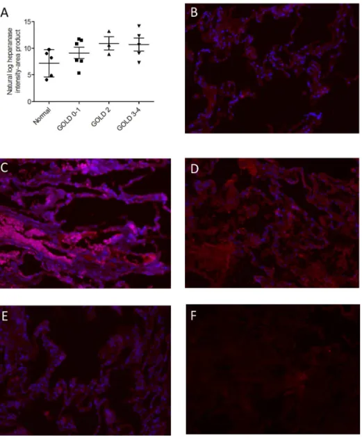
Available clinical data from de-identified healthy and COPD cohorts providing lung tissue are
shown inTable 1. Immunofluorescence revealed that heparanase expression increased with
se-quential increases in GOLD stage (Fig 4, linear trend by ANOVA p = 0.029).
Fig 3. The effect of pharmacological treatment with the heparanase inhibitor Muparfostat on neutrophil recruitment to the lung.The total number of inflammatory cells (A, C) and neutrophils (B, D) recruited to the lung following acute exposure to zymosan (A, B) or LPS (C, D). Wild-type (WT), and Hpa -/-mice received intranasal (i.n.) saline, zymosan (A, B) or LPS (C, D). The number of total cells (A) and neutrophils (B) in bronchoalveolar lavage fluid 24 h after intranasal administration of zymosan in BALB/c mice treated with Muparfostat (30 mg/kg s.c.) dosed 30 minutes before (q.d.) and 6 h after (b.i.d) zymosan exposure, n = 4 per group. Data presented as box plots (median, 25–75 percentile) with whiskers representing 5–95% confidence interval.*p<0.05 compared with saline. Data obtained from one experiment. In other experiments total number of cells (C) and neutrophil (D) recruitment to the lung was measured following intranasal administration of LPS in vehicle and Muparfostat (b.i.d.) treated mice (n = 3–9). Data presented as box plots (median, 25–75 percentile) with whiskers representing 5–95% confidence interval.*p<0.05 compared with saline. Treatment groups were not significantly different from vehicle (symbol not reported). Data was obtained from two independent experiments.
doi:10.1371/journal.pone.0127032.g003
Table 1. Patient demographics of COPD patients.
Group Gender (F/M) Age (mean, (SD)) % FEV1 (pre) % FEV1 (post) Pack years
GOLD (0–1) 2/4 56 (8) 89 (78–100) 94 (85–100) 39 (4–74)
GOLD (2) 1/3 51 (6) 48 (38–59) 54 (46–63) 30 (-3–63)
GOLD (3–4) 2/3 54 (4) 24 (8–41) 46 48 (15–81)
Heparanase plays a role in allergic inflammation
Adult WT mice immunized with OVA and repeatedly exposed to this antigen induced an aller-gic inflammatory response (Fig 5) characterized by an eosinophilic rich cell infiltrate (42% of
total cells, n = 12). The significant increase in total cells (Fig 5A, p<0.05), eosinophils (Fig 5B,
p<0.05) and macrophages (Fig 5C, p<0.05) following OVA challenge in wild type mice was
inhibited in Hpa-/-mice. Neutrophil and lymphocyte recruitment to the airways were not
sig-nificantly increased following antigen challenge, and the absence of heparanase did not affect the migration of these cell types into the airways. Th2 cell activation was indirectly assessed by
Fig 4. Heparanase expression in lung tissue from normal subjects and COPD patients of varying severity.Each point represents the intensity of staining for heparanase expression in normal human lung tissue and in subjects with varying severities of COPD (A). Representative images are shown (B-F reflecting Gold 0–1, Gold 2, Gold 3–4, healthy subject, and isotype control, respectively. Red: heparanase; blue: DAPI (DAPI not performed for isotype control). Image brightness was uniformly adjusted for clarity and all heparanase quantification was performed using unadjusted (raw) images. Magnification was 40 x for all images. The data was analyzed as continuous variables and there was a significant linear trend (post-ANOVA, r-squared = 0.27 and p = 0.029). Horizontal line represents mean and limits represent SEM.
measurement of OVA specific IgE, which was significantly elevated in WT and Hpa-/-OVA sensitized and challenged mice compared with their respective controls (Fig 5).
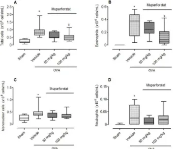
The effect of Muparfostat against OVA induced allergic lung inflammation. Mice
treat-ed with OVA and vehicle showtreat-ed a significant increase in the total number of cells compartreat-ed
to sham animals (Fig 6A, p<0.05). Eosinophils comprised 36% of the total cell infiltrate
Fig 5. Effect of heparanase gene deletion on allergic inflammation induced by ovalbumin (OVA). Box plots showing (A) total cell, (B) eosinophil, (C) macrophages, (D) lymphocytes, (E) neutrophils in bronchoalveolar lavage fluid and (F) OVA specific IgE in serum in control and OVA immunized and challenged mice. Mice were sensitized by repeated i.p. injections with 10μg OVA or were non-sensitized (controls). All mice received three i.n. instillations with 20μg OVA and lavage was undertaken 24 h after the last intranasal administration of OVA. Data expressed as blox plots (median, 25–75 percentile) with whiskers representing 5–95% confidence interval (7–12 animals for cell data and 6–10 for IgE data).*p<0.05 compared with control;†No significant difference compared with Hpa-/-control. Data pooled from four
independent experiments.
doi:10.1371/journal.pone.0127032.g005
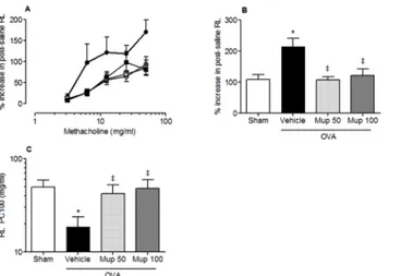
Fig 6. Effect of Muparfostat on allergic inflammation induced by OVA.Box plots showing (A) total cell, (B) eosinophil, (C) mononuclear cell and (D) neutrophil cell number in the lungs of mice following sensitisation and aerosol challenge with OVA. Mice were treated with Muparfostat (50 mg/kg and 100 mg/kg s.c.) 30 minutes before challenge. Mice were challenged with aerosolised 3% OVA for three consecutive days and lavage undertaken 24 h after the last challenge. Data expressed as blox plots with whiskers representing 5–95% confidence interval (n = 9–15).*p<0.05 compared with sham and‡p<0.05 compared with vehicle. Data obtained from four independent experiments.
(Fig 6B, p<0.05). In addition, there was also a small and significant increase in the number of
neutrophils and mononuclear cells in the OVA vehicle treated group compared to sham (Fig 6C and 6D). The heparanase inhibitor Muparfostat at the highest dose evaluated caused a sig-nificant inhibition of eosinophil recruitment to the airways (Fig 6B).
The effect of Muparfostat against OVA induced allergen-induced bronchial
hyperre-sponsiveness. Methacholine caused a dose-dependent increase in post-saline resistance (RL)
(Fig 7A). The percentage increase in post-saline resistance (%RL) of the lungs (i.e. peak
re-sponse) to increasing concentrations of methacholine was significantly greater in OVA vehicle treated animals compared with sham sensitised animals (mean ± s.e.m; 107.6 ± 16.8, n = 10
versus 213.8 ± 26.4, respectively, n = 11 p<0.05;Fig 7B). In animals treated with muparfostat
50 mg/kg and 100 mg/kg there was a significant decrease in the peak response (107 ± 10.1,
n = 8, p<0.01 and 121 ± 21.7, n = 11 p<0.05 respectively for dose). Similarly, airway
sensitiv-ity to methacholine (RL PC100, mg/mL) was reduced by approximately 3 fold in OVA vehicle treated animals compared with sham sensitised animals (mean ± s.e.m; 18.5 ± 5.2, n = 10
ver-sus 49.7 ± 9.5 respectively, n = 10, p<0.05;Fig 7C) indicating an increase in airway sensitivity
to methacholine. This increase in airway sensitivity was significantly inhibited in OVA
sensi-tized and challenged mice, treated with Muparfostat (50 mg/kg or 100 mg/kg;Fig 7C). The RL
PC100 (mg/mL) was significantly increased compared with vehicle treated sensitized animals
(42 ± 10.2, n = 8 and 47.8 ± 11.5 n = 11, p<0.05 respectively for dose).
Discussion
Inflammatory cell migration to the lung has a number of similarities with tumour cell metasta-sis, for example, secreting heparanase to degrade ECM structure. The results demonstrated that heparanase plays a greater role in regulating the pulmonary cell recruitment of eosinophils following an allergic, compared with neutrophils following a non-allergic, stimulus.
A previous study has reported that the constitutive cell population within the airways of mice deficient in heparanase was similar to the number of cells in naïve mice [23], an observation that was also observed in our study. It has been suggested that the deletion of the
Fig 7. Effect of Muparfostat on OVA induced changes in lung function.Graphs showing (A) % increase in total lung resistance versus methacholine dose in sham immunized (), OVA sensitized in vehicle-treated
(●) or OVA-immunized mice treated with Muparfostat 50 mg/kg (Mup,□) or Muparfostat 100 mg/kg (Mup,■) s. c. before being challenged with aerosolised 3% OVA for 4 consecutive days, (B) peak percentage increase in post saline resistance, (C) RLprovocative concentration (PC)100 for methacholine. Each point or columns
represent mean±SEM n = 8–11, p<0.05 versus sham*or vehicle‡. Data obtained from four independent experiments.
heparanase gene may lead to a compensatory increase in MMP’s primarily MMP-2, MMP-14 and MMP-9, which are known to degrade the ECM [24], although this is not a consistent find-ing [17]. Whether compensation by MMPs in the lung could explain why mice deficient in heparanase were able to recruit neutrophils in response to zymosan or LPS remains to be established.
We also used a pharmacological approach to inhibit heparanase activity in WT mice using the heparanase inhibitor, Muparfostat derived from yeast. This phosphosulfomannan has dem-onstrated efficacy against several cancers in phase I and II clinical trials, either administered alone or in combination with other therapy [25,26]. Muparfostat inhibits heparanase with an IC50 of 7.9 nM [27] and has previously been shown to suppress experimental lung metastases in a B16 melanoma model [28]. However, Muparfostat, in a dose range similar to those re-ported in this melanoma model did not significantly reduce the number of neutrophils infil-trating to the lung in response to zymosan or LPS in our study.
The current findings do not support the notion that heparanase is important in the acute in-nate inflammatory response in response to airway injury, although we have recently reported that recombinant heparanase is able to promote neutrophil recruitment into tissues [29]. It has also been reported that heparanase inhibition prevents sepsis-induced mortality and acute lung injury (ALI) in mice [16]. This apparent discrepancy may reflect pathophysiologic differences between direct (i.e. intranasal) and indirect (i.e. systemic-induced) lung injury, as suggested by previously-observed differences in patients with direct (pneumonia) and indirect (sepsis, pan-creatitis) lung injury [16,30]. Alternatively, heparanase might be important during the early phase of the inflammatory response, as leukocyte attachment was measured 6 h after acute lung injury [16] whilst in our studies we measured leukocyte recruitment at a 24 h time point.
It has recently been shown that heparanase is involved in the chronic inflammatory re-sponse in ulcerative colitis [31] and chronic inflammation is known to have a profound effect on tumour progression [32]. Consequently, heparanase is a potential target for therapeutic in-tervention in chronic inflammatory disease states. We investigated the expression of hepara-nase in subjects with different severities of COPD and found an association of heparahepara-nase expression in lung tissue and COPD severity. This would be consistent with evidence of in-creased expression of this enzyme in other chronic inflammatory conditions including protei-nuric disease [33], chronic haemodialysis [34], rheumatoid arthritis [13], Crohn’s disease and ulcerative colitis but not in infection colitis [12]. We cannot draw any conclusion concerning cause and effect, but our data highlight a further avenue for research regarding the role of heparanase in COPD.
Whilst mice deficient in heparanase have been shown to have no detectable difference in sponse to a DTH reaction [24], although there was a reduction in the number of monocytes
re-cruited to the inflammatory site [15], a pharmacological approach using thein vivo
administration of anti-heparanase siRNA or an inhibitor of heparanase enzymatic activity ef-fectively abolished the DTH inflammatory response in the skin of mice [14]. Hence, we investi-gated the role of heparanase in allergic inflammation in the lung using an allergic model of murine lung inflammation which is characterized by a Th2 phenotype [35]. Mice sensitized and then exposed to aerosolized OVA demonstrated a significant increase in inflammatory cell influx, predominantly composed of eosinophils which was accompanied with changes in pul-monary lung mechanics, as evident by an increase in airways responsiveness to methacholine.
We have demonstrated significantly lower number of eosinophils in the BAL fluid of Hpa
cells [42]. Other cell types involved in the allergic inflammatory response in the airways includ-ing dendritic cells [17,43], T cells [44–50] and vascular endothelium [16,50,51] also express heparanase to potentially regulate the migration of leukocytes to sites of inflammation. Selec-tive ablation of heparanase in individual cell types would be required to investigate the role of heparanase expressing cells in the allergic inflammatory response and is beyond the scope of this study.
Conclusions
We have demonstrated a role for heparanase in acute allergic inflammation, in the lung and our findings highlight a potential role for this enzyme in cell recruitment to the airways and that targeting heparanase may be beneficial in the treatment of allergic pulmonary diseases.
Acknowledgments
We acknowledge Prof. Israel Vlodavsky (Technion, Israel) for his generous support for
provid-ing the Hpa-/-mice. Abigail Morris was supported by a Capacity Building Award in Integrative
Mammalian Biology funded by the BBSRC, BPS, HEFCE, MRC and SFC. Grants from Swedish Research Council (to JPL and SW) and Konsul Th C Bergh Foundation also contributed to as-pects of this research.
Author Contributions
Conceived and designed the experiments: DS SW JPL. Performed the experiments: AM RV BW IW. Analyzed the data: DS SW AM. Contributed reagents/materials/analysis tools: JPL EPS. Wrote the paper: DS SW JPL CP. Provided heparanase knockout mice: JPL. Analyzed heparanase expression in human airway tissue: EPS.
References
1. Vlodavsky I, Beckhove P, Lerner I, Pisano C, Meirovitz A, Ilan N et al. (2012) Significance of hepara-nase in cancer and inflammation. Cancer Microenviron 5: 115–132. doi:10.1007/s12307-011-0082-7
PMID:21811836
2. Bishop JR, Schuksz M, Esko JD (2007) Heparan sulphate proteoglycans fine-tune mammalian physiol-ogy. Nature 446: 1030–1037. PMID:17460664
3. Forsberg E, Kjellen L (2001) Heparan sulfate: lessons from knockout mice. J Clin Invest 108: 175–180. PMID:11457868
5. Alexopoulou AN, Multhaupt HA, Couchman JR (2007) Syndecans in wound healing, inflammation and vascular biology. Int J Biochem Cell Biol 39: 505–528. PMID:17097330
6. Maeda T, Desouky J, Friedl A (2006) Syndecan-1 expression by stromal fibroblasts promotes breast carcinoma growth in vivo and stimulates tumor angiogenesis. Oncogene 25: 1408–1412. PMID:
16247452
7. Xu J, Park PW, Kheradmand F, Corry DB (2005) Endogenous attenuation of allergic lung inflammation by syndecan-1. J Immunol 174: 5758–5765. PMID:15843578
8. Chan SC, Leung VO, Ip MS, Shum DK (2009) Shed syndecan-1 restricts neutrophil elastase from alpha1-antitrypsin in neutrophilic airway inflammation. Am J Respir Cell Mol Biol 41: 620–628. doi:10. 1165/rcmb.2008-0185OCPMID:19251947
9. Vlodavsky I, Friedmann Y, Elkin M, Aingorn H, Atzmon R, Ishai-Michaeli R et al. (1999) Mammalian heparanase: gene cloning, expression and function in tumor progression and metastasis. NatMed 5: 793–802. PMID:10395325
10. Gandhi NS, Freeman C, Parish CR, Mancera RL (2012) Computational analyses of the catalytic and heparin-binding sites and their interactions with glycosaminoglycans in glycoside hydrolase family 79 endo-beta-D-glucuronidase (heparanase). Glycobiology 22: 35–55. doi:10.1093/glycob/cwr095
PMID:21746763
11. Vaday GG, Lider O (2000) Extracellular matrix moieties, cytokines, and enzymes: dynamic effects on immune cell behavior and inflammation. J Leukoc Biol 67: 149–159. PMID:10670574
12. Waterman M, Ben-Izhak O, Eliakim R, Groisman G, Vlodavsky I, Ilan N (2007) Heparanase upregula-tion by colonic epithelium in inflammatory bowel disease. ModPathol 20: 8–14.
13. Li RW, Freeman C, Yu D, Hindmarsh EJ, Tymms KE, Parish CR et al. (2008) Dramatic regulation of heparanase activity and angiogenesis gene expression in synovium from patients with rheumatoid ar-thritis. Arthritis & Rheumatism 58: 1590–1600.
14. Edovitsky E, Lerner I, Zcharia E, Peretz T, Vlodavsky I, Elkin M (2006) Role of endothelial heparanase in delayed-type hypersensitivity. Blood 107: 3609–3616. PMID:16384929
15. Stoler-Barak L, Petrovich E, Aychek T, Gurevich I, Tal O, Hatzav M et al. (2015) Heparanase of murine effector lymphocytes and neutrophils is not required for their diapedesis into sites of inflammation. FASEB J Jan 29. pii: fj.14-265447. [Epub ahead of print].
16. Schmidt EP, Yang Y, Janssen WJ, Gandjeva A, Perez MJ, Barthel L et al. (2012) The pulmonary endo-thelial glycocalyx regulates neutrophil adhesion and lung injury during experimental sepsis. Nat Med 18: 1217–1223. doi:10.1038/nm.2843PMID:22820644
17. Poon IK, Goodall KJ, Phipps S, Chow JD, Pagler EB, Andrews DM et al. (2014) Mice deficient in hepar-anase exhibit impaired dendritic cell migration and reduced airway inflammation. Eur J Immunol 44: 1016–1130. doi:10.1002/eji.201343645PMID:24532362
18. Yoshida T, Mett I, Bhunia AK, Bowman J, Perez M, Zhang L et al. (2010) Rtp801, a suppressor of mTOR signaling, is an essential mediator of cigarette smoke-induced pulmonary injury and emphyse-ma. Nat Med 16: 767–773. doi:10.1038/nm.2157PMID:20473305
19. Lygizos MI, Yang Y, Altmann CJ, Okamura K, Hernando AA, Perez MJ et al. (2013) Heparanase medi-ates renal dysfunction during early sepsis in mice. Physiol Rep 1: e00153. doi:10.1002/phy2.153
PMID:24400155
20. Zcharia E, Jia J, Zhang X, Baraz L, Lindahl U, Peretz T et al. (2009) Newly generated heparanase knock-out mice unravel co-regulation of heparanase and matrix metalloproteinases. PLoS ONE 4: e5181. doi:10.1371/journal.pone.0005181PMID:19360105
21. Riffo-Vasquez Y, Ligeiro de Oliveira AP, Page CP, Spina D, Tavares-de-Lima W (2007) Role of sex hormones in allergic inflammation in mice. ClinExpAllergy 37: 459–470.
22. Waern I, Jonasson S, Hjoberg J, Bucht A, Abrink M, Pejler G et al. (2009) Mouse mast cell protease 4 is the major chymase in murine airways and has a protective role in allergic airway inflammation. J Immu-nol 183: 6369–6376. doi:10.4049/jimmunol.0900180PMID:19841188
23. Waern I, Jia J, Pejler G, Zcharia E, Vlodavsky I, Li J-P et al. (2010) Accumulation of Ym1 and formation of intracellular crystalline bodies in alveolar macrophages lacking heparanase. Molecular Immunology 47: 1467–1475. doi:10.1016/j.molimm.2010.02.004PMID:20226534
24. Zcharia E, Jia J, Zhang X, Baraz L, Lindahl U, Peretz T et al. (2009) Newly Generated Heparanase Knock-Out Mice Unravel Co-Regulation of Heparanase and Matrix Metalloproteinases. PLoS ONE 4: e5181. doi:10.1371/journal.pone.0005181PMID:19360105
539452PMID:24509853
31. Lerner I, Hermano E, Zcharia E, Rodkin D, Bulvik R, Doviner V et al. (2011) Heparanase powers a chronic inflammatory circuit that promotes colitis-associated tumorigenesis in mice. J Clin Invest 121: 1709–1721. doi:10.1172/JCI43792PMID:21490396
32. Coussens LM, Werb Z (2002) Inflammation and cancer. Nature 420: 860–867. PMID:12490959
33. Garsen M, Rops AL, Rabelink TJ, Berden JH, van der Vlag J (2014) The role of heparanase and the en-dothelial glycocalyx in the development of proteinuria. Nephrol Dial Transplant 29: 49–55. doi:10. 1093/ndt/gft410PMID:24166469
34. Cohen-Mazor M, Sela S, Mazor R, Ilan N, Vlodavsky I, Rops AL et al. (2008) Are primed polymorphonu-clear leukocytes contributors to the high heparanase levels in hemodialysis patients? Am J Physiol Heart Circ Physiol 294: H651–658. PMID:18032524
35. Lloyd CM, Hessel EM (2010) Functions of T cells in asthma: more than just T(H)2 cells. Nat Rev Immu-nol 10: 838–848. doi:10.1038/nri2870PMID:21060320
36. Ewart SL, Kuperman D, Schadt E, Tankersley C, Grupe A, Shubitowski DM et al. (2000) Quantitative trait loci controlling allergen-induced airway hyperresponsiveness in inbred mice. AmJRespirCell Mol-Biol 23: 537–545. PMID:11017920
37. Pitchford SC, Riffo-Vasquez Y, Sousa A, Momi S, Gresele P, Spina D et al. (2004) Platelets are neces-sary for airway wall remodeling in a murine model of chronic allergic inflammation. Blood 103: 639– 647. PMID:14504080
38. Pitchford SC, Momi S, Giannini S, Casali L, Spina D, Page CP et al. (2005) Platelet P-selectin is re-quired for pulmonary eosinophil and lymphocyte recruitment in a murine model of allergic inflammation. Blood 105: 2074–2081. PMID:15528309
39. Pitchford SC, Momi S, Baglioni S, Casali L, Giannini S, Rossi R et al. (2008) Allergen Induces the Mi-gration of Platelets to Lung Tissue in Allergic Asthma. American Journal of Respiratory and Critical Care Medicine 177: 604–612. PMID:18096710
40. Ludwig RJ, Schultz JE, Boehncke WH, Podda M, Tandi C, Krombach F et al. (2004) Activated, not rest-ing, platelets increase leukocyte rolling in murine skin utilizing a distinct set of adhesion molecules. J In-vest Dermatol 122: 830–836. PMID:15086572
41. Hoogewerf AJ, Leone JW, Reardon IM, Howe WJ, Asa D, Heinrikson RL et al. (1995) CXC chemokines connective tissue activating peptide-III and neutrophil activating peptide-2 are heparin/heparan sulfate-degrading enzymes. J Biol Chem 270: 3268–3277. PMID:7852412
42. Zetser A (2006) Heparanase Induces Vascular Endothelial Growth Factor Expression: Correlation with p38 Phosphorylation Levels and Src Activation. Cancer Research 66: 1455–1463. PMID:16452201
43. Benhamron S, Nechushtan H, Verbovetski I, Krispin A, bboud-Jarrous G, Zcharia E et al. (2006) Trans-location of active heparanase to cell surface regulates degradation of extracellular matrix heparan sul-fate upon transmigration of mature monocyte-derived dendritic cells. J Immunol 176: 6417–6424. PMID:16709798
44. Yahalom J, Fibach E, Bar-Tana R, Fuks Z, Vlodavsky I (1988) Differentiating human leukemia cells ex-press heparanase that degrades heparan sulfate in subendothelial extracellular matrix. LeukRes 12: 711–717. PMID:2973542
46. Lider O, Mekori YA, Miller T, Bar-Tana R, Vlodavsky I, Baharav E et al. (1990) Inhibition of T lympho-cyte heparanase by heparin prevents T cell migration and T cell-mediated immunity. Eur J Immunol 20: 493–499. PMID:2318247
47. Vlodavsky I, Eldor A, Haimovitz-Friedman A, Matzner Y, Ishai-Michaeli R, Lider O et al. (1992) Expres-sion of heparanase by platelets and circulating cells of the immune system: possible involvement in dia-pedesis and extravasation. Invasion Metastasis 12: 112–127. PMID:1399400
48. Gilat D, Hershkoviz R, Goldkorn I, Cahalon L, Korner G, Vlodavsky I et al. (1995) Molecular behavior adapts to context: heparanase functions as an extracellular matrix-degrading enzyme or as a T cell ad-hesion molecule, depending on the local pH. J Exp Med 181: 1929–1934. PMID:7722469
49. Goldshmidt O, Zcharia E, Abramovitch R, Metzger S, Aingorn H, Friedmann Y et al. (2002) Cell surface expression and secretion of heparanase markedly promote tumor angiogenesis and metastasis. Proc Natl Acad Sci U S A 99: 10031–10036. PMID:12097647
50. Sotnikov I, Hershkoviz R, Grabovsky V, Ilan N, Cahalon L, Vlodavsky I et al. (2004) Enzymatically qui-escent heparanase augments T cell interactions with VCAM-1 and extracellular matrix components under versatile dynamic contexts. J Immunol 172: 5185–5193. PMID:15100255