Albanian j. agric. sci. 2013;12 (2): 159-162 Agricultural University of Tirana
Correspondence: Elenica Dimço, Affiliation; Agricultural University of Tirana, AlbaniaEmail: [email protected] (Accepted for publication 19 february 2013)
ISSN: 2218-2020, © Agricultural University of Tirana
RESEARCH ARTICLE
(Open Access)
Effect of pregnancy in hematological profile of dogs
ELENICA DIMÇO*, JETMIRA ABESHI, ERINDA LIKA, GERTA DHAMO
Agricultural University of Tirana, Albania
Abstract
Pregnant impact on hematological parameters in the dogs was award in this study. Blood was collected in the cephalic vein of 32 female animals, clinically healthy, in different physiological condition. Hematological parameters that studied were RBC, WBC and platelets count, Hgb concentration, HCT, differential counting of leukocytes and erythrocyte indicators. The obtained results from the analysis showed that pregnancy can affect the values of hematological parameters. The number of erythrocytes, the level of hematocrit and hemoglobin concentration in pregnant animals resulted lower (P <0.05) than in non-pregnant animals. No volatility resulted in the values of MCV, MCH and MCHC. In the pregnant animals group was found the increase of the circulating leucocytes number, of relative value of neutrophils and lymphocytes decrease. The pregnancy condition should be taken in consideration during the results interpretation of laboratory analysis.
Keywords: RBC, WBC, hematocrit, hemoglobin, pregnant, dog.
1. Introduction
Clinical problems and their monitoring and treatment effectiveness necessarily require laboratory examinations. The analyze and correct interpretation of the laboratory tests results provide adequate information for assessing of conditions of breeding, feeding, stress conditions, non-specific resistance, etc., [18]. They also serve to monitor specific therapeutic protocols and to determine prognosis. At the same time, they are the basis of experimental and scientific research. However, the basis of these parameters must be stabilized before the data can be interpreted and applied.
Blood parameters vary in different physiological stages, as well as in pathological conditions [5]. Pregnancy is one of the physiological conditions that lead significant changes in hematological and biochemical parameters of all animal species. During pregnancy some blood components are subject to significant changes, confusing the correct interpretation of blood parameters in diseases or disorders that may occur during this period [2]. In order to prevent metabolic disorders or subclinical disease during the pregnancy, it is necessary to define the physiological fluctuations of hematological and biochemical parameters of blood in healthy bitches and carried out systematic monitoring of these parameters in different stages of pregnancy [17].
2. Material and methods:
To study the influence of pregnancy in blood parameters of dogs, 32 healthy bitches were divided
into two groups. The first group was represented by 16 non-pregnant bitches and the second group of 16 bitches that were in the last stage of pregnancy. The females of the second group had similar body weight and age with them of the first group.
The samples were collected from September 2010 to February 2012. Clinically healthy status was based on these criteria: no visible signs of illness were observed, while temperature, pulse and respiratory rate were normal. All of the dogs were clinically examined and none of these dogs receive any treatment before. To determine the condition of pregnancy was based on the date of copulation and on manual methods to control it.
th M a [ I b δ tr R t o 1 r d c h hrough the May-Grunwa and reading 11]. For stat Inc program. by the above
δ, minimum ruthfulness o Results were
est, t-test and
Param Non-pregn Pregnan Reference i norma We foun of MCV, MC 1, the averag reduced from dogs to 5.8 count was de hemoglobin 5.4 5.6 5.8 6.0 6.2 6.4 6.6 0 5 10 15 20 F smears prep ald-Giemsa by immersi tistical proce .16.0. For di analysis it w and maximu of change av e evaluated s
d P < 0.05 w
meters nant animals t animals intervals for al dogs S
Table 2. Cha
Hematolog tD non-preg
Fig
nd no signifi CH and MCH ge number o m 6.4 x 106
x 106/µl in ecreased from
level from nonpreg M
1 2 3
samp
Fluctuation of Hg
pared with f laboratory ion objective essing of data
ifferent para was determin
um values o verages two statistically u were consider
Table 1. Va
RBC (x1 6.4±0
5.8±0
5.5-8
Statistical diffe
anging the ave
gical paramete gnant/ pregna
gure. 1. Diagr
icant fluctuat HC. As it is f red blood 6
/µl in non-n pregnon-nanon-nt a m 3.4 x 10⁵/µ
15.2 g/dl t gnant Mean values o
4 5 6 7
ples number
b concentration
Dimço et al
fresh blood, staining me e was perfor a was used S ameters meas ned the avera of fluctuation populations using the Stu red significan
alues of hemat
106/μl) PLT .76a 3
0.6a 2
8.5
ference betwee
erages (tD) of
ers
ant 2
ams of the flu
tion in the v reflected in cells (RBC) -pregnant fe animals, plat µl to 2.8 x 10 to 13.9 g/dl
pregnant f RBC
8 9 10 11
nonpreg pregnan l
160
after ethod rmed SPSS sured age ± n, the (tD). udent nt. flu sec par and ani tological paramT (x105/μl) .4±0.97b 2.8±1.06b
2-5
en the average
hematologica
RBC PT
2.151* 2.09 P <0:05 *
uctuation of th
alues table ) was emale telets 05/µl, l and hem fou in exp hem inc the 38 40 42 44 46 gnant nt 1 1 1 1 1 W BC v a lue s 3. Resul From th uctuation of cond mon rameters suc d Hgb valu imals, than th
meters (mean±
HCT (%) 45.4±3.6c 41±4.9c
37-55
e values of gro
al indices acco
TL HCT
92* 2.371*
he values betw
matocrit from und by some
The decre female anim plained by t modilution a creased plasm e erythrocyte 8 0 2 4 6 nonpre M 0 2 4 6 8 0 2 4 6 8
1 2 3
Fluctuation of
lts and discu
e results blood values th of pr ch as numbe ues were lo
he values of
±sd) in bitche
Hgb (g/dl) 15.2±1.4d 13.9±1.6d
12-18
oups a,b,c,d P<0
ording to phys
Hgb * 2.126*
ween the two g
m 45.4% to 4 e authors [4, 8 ease of RBC,
mals in the the fact that and a slight a ma volume.
es life in pr gnant
ean values of
4 5 6 7
samples number
WBC values
ussion:
obtained w s in female regnancy. er of RBC a
wer (P<0.05 f non-pregnan es MCV (fl) 69.9±4.6 70.7±4.5 60-77 0:05 siological statu MCV M -0.374 0 groups 41%. Simila 8, 10 and 12 PLT, HCT a ir last stage
in pregnant anemia as a In addition, regnant anim
pregnant HCT
7 8 9 10
r nonpreg pregnan were observ animals, in Hematologi and PLT, H 5) in pregn nt ones. MCHC (%) 33.5±1.9 33.9±1.5 32-36 us MCHC 0.000
ar findings w ].
and Hgb valu e pregnancy t animals oc consequence a shortening mals is anot
Effect of pregnancy in hematological profile of dogs
cause of the reduction of red blood cells number [5, 16]. While, other authors [7, 8, 9] report increased levels of these indicators in pregnant cattle.
According to statistical processing of the data the number of circulating leucocytes in pregnant animals was high. The number of white blood cells in the average value was increased from 8.4 x10³ to 10.2 x10³/μL blood, while neutrophils were increased and lymphocytes were decreased. This change is comparable to that reported by Coles (1986). According to some authors, it is expected a physiological increase of leucocytes and mostly
granulocytes neutrophil in female animals in the last stage of pregnancy [9]. This increase can be attributed to a number of reasons, such as the secretion of estrogen, a higher level of cortisol in plasma, or maternal immune system response to fetal allograph [2]. The increase of granulocytes neutrophil during the recent weeks of pregnancy may reflect the numerous neutrophils aggregation in placental blood vessels, close to the degenerative and necrotic cells [14]. On the contrary, some authors [5] have observed the reduction of leucocytes number in pregnant animals because of immunosuppression in young bitch.
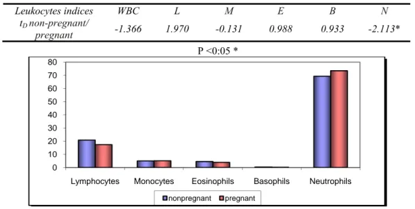
Table 3. Values of leukocytes parameters (mean ± sd) in female dogs
Leukocytes parameters WBC (x103/μl)
Leukograma (%)
Lymphocytes Monocytes Eosinophils Basophils Neutrophils Non-pregnant 8.4±2.2 20.8±4.2 5.0±1.3 4.5±1.2 0.4±0.5 69.3±3.1a Pregnant 10.2±3.8 17.4±4.1 5.1±1.9 3.9±1.8 0.2±0.4 73.5±5.8a Reference intervals for dogs 6-17 12-30 3-9 2-10 0-0.5 60-75
Statistical difference between the average values of two groups aP<0.05
Table 4. Changing the averages (tD) indices according to physiological status
Leukocytes indices WBC L M E B N
tD non-pregnant/
pregnant -1.366 1.970 -0.131 0.988 0.933 -2.113* P <0:05 *
161
0 10 20 30 40 50 60 70 80
Lymphocytes Monocytes Eosinophils Basophils Neutrophils
nonpregnant pregnant
Figure 2. Diagram of differential leucocytes count in both groups of animals
Female animals in the last month of pregnancy showed different neutrophils/lymphocytes ratio, compared with non-pregnant group, because the relative values of lymphocytes decrease while neutrophils increase at this stage of pregnancy (Figure 2). In fact, the absolute number of lymphocytes remains constant, but while the number of white blood cells increases, the percentage (relative value) of lymphocytes decreases. On the other hand, the reduction of lymphocyte values during pregnancy can be attributed to the physiological stress leading to lymphopenia [3, 8]. However, this finding is different of that reported by some authors [4], who observed increased lymphocytes during the last period of pregnancy.
In conclusion, statistical tests for the difference of average values of the parameters of red blood cells between the pregnant and non-pregnant animals showed significant difference (P <0.05) only for RBC, HCT and Hgb. Statistical tests for the change of average values of white blood cells were not significant, excepted of the relative number of neutrophils.
Dimço et al
162
4. References:
1. Alexander RR, Griffiths JM: Basic biochemical methods; 1993: 187-189.
2. Awodu OA, Enosolease ME, Ubaru AG, Famodu AA : Leukocyte count in Pregnant Nigerian woman with Sickle cell train.African Journal of
Reproductive Health, 2002, 6 (3): 112-116.
3. Balikci E, Yildiz A: Haematological parameters in single and twin pregnancies of sheep. Indian
Veterinary Journal, 2005, 82 (7): 721-723.
4. Bozdogan O, Baysal AI: The effect of age, sex, pregnancy and housing system on some blood parameters of ARMY sheep. Turkish Journal of
Veterinary and Animal Sciences, 2003, 27:
521-524.
5. Chaudhari SUR, Mshelia, GD: Evaluation of the Hematologic Values of Bitches in Northern Nigeria for the Staging of Pregnancy. Pakistan
Journal of Biological Sciences, 2006, 9 (2):
310-312.
6. Coles EH: Veterinary Clinical Pathology. 4th Edition: WB Saunders Co. Philadelphia. 1986.
7. El-Sherif, MMA, Assad F: Changes in some blood constituents of Barki ewes during pregnancy and lactation under semi arid conditions. Small Ruminant Research, 2001, 40: 269-277.
8. Feldman BR, Zinkl JG, Jain NC: Schalm's Veterinary Hematology. 5th Edition: Lippincott, Williams & Wilkins, Baltimore, Maryland, 2000.
9. Getnet AM, Abebe W: The influence of late pregnancy and excitement on blood parameters of Issa type dromedaries in eastern Ethiopia. Israel Journal of Veterinary Medicine. 2005, 60(4): 34-36.
10. Kimberly ET, Casal ML, O'Donnell PA, Haskins ME: Effects of pregnancy on complete blood cell counts and serum biochemical profiles in
dogs. Theriogenology, 2006, 66: 663-687.
11. Latimer KS: Leukocytes in Health and Disease.
Textbook of Veterinary Internal Medicine 4th ed. 1995, 2:1892 - 1929.
12. Mbassa GK, Poulsen JSD: Influence of pregnancy, lactation and environment of hematological profiles in Danish landrace Dairy goats of different parity. Comparative
biochemistry and Physiology, 1991, 100(2):
403-412.
13. Meyer DJ, Harvey JW: Veterinary Laboratory Medicine: Interpretation and Diagnosis. WB. Saunders Co., Philadelphia. 1998.
14. Peck T, Arias F: Hematologic Changes Associated with pregnancy. Clin Obstet Gynecol 1979, 22:785-798.
15. Rizzi TE, Meinkoth JH, Clinkenbeard KD:
Schalm's Veterinary Haematology. 6th Edition: Weiss DJ, Wardrop DJ, 2010:799-810.
16. Singh R, Singh SPS, Setia MS: Distribution of Trace Elements in blood, plasma and erythrocyte during different stages of gestation in Buffalo. Buffalo Journal, 1991, 1: 77-85.
17. Stojević Z, Piršljin J, Milinković-To S, Zdelar-Tuk M, Ljubic BB: Activities of AST, ALT and GGT in clinically Healthy Dairy cows during lactation and dry period. Veterinarski Arhiv, 2005, 75 (1): 67-73.