137
UNUSUAL BEHAVIOR OF GROWING POLLEN TUBES IN THE OVARY OF PLUM CULTURE (PRUNUS DOMESTICA L.)
MILENA ĐORĐEVIĆ1, R. CEROVIĆ1, D. NIKOLIĆ2 and SANJA RADIČEVIĆ1
1Fruit Research Institute, 32000 Čačak, Serbia
2Faculty of Agriculture, University of Belgrade, 11080 Belgrade, Serbia
Abstract – Unusual behavior of growing pollen tubes in different combinations of pollination was observed in the ovary of the plum (Prunus domestica L.) cv ‘Čačanska Lepotica’. It primarily refers to several issues, i.e. the curling up of pollen tubes within the micropyle, the growth of two pollen tubes into the nucellus of an ovule, the occurrence of a bundle above the nucellar cap and fluorescence of the part of the embryo sac containing the egg apparatus. Upon the growth of pollen tubes into the nucellus of the ovule, subsequently penetrating pollen tubes form a bundle either above the micropyle entrance or above the nucellus. Branching and bending of pollen tubes by 180º upon their growth into the micropyle was also observed.
Key words: Plum, ovary, pollen tube growth, fluorescent microscopy
UDC 634.22:631.52
INTRODUCTION
The interaction between the pollen and the pistil within the scope of the stigma and the style, in both compatible and incompatible variants of pollination and fertilization in the Prunus genus has generally been given attention in literature. On the other hand, the data on the possible interaction between the pollen and the pistil in the ovary are sparse. Some earlier reports on the occurrence of wandering, branching and curling up of pollen tubes in the ovary are brought into connection with incompatibility in this part of the pistil (Seavey and Bawa, 1986; Sage et al., 1994). Herrero and Hormaza (1996) put forward that the ovary plays an important role in the directed growth of pollen tubes in compatible combinations. Untypical behavior of growing pollen tubes into the ovary was also observed in cases of compatible pollination (Herrero, 2000). It is related to a phase of the development of female structures which cannot provide further growth of pollen tubes when they reach certain parts of the ovary.
In the majority of the Prunus genus, pollen tubes in the ovary grow along the entire length of the placenta, obturator, micropyle, nucellus
reaching eventually the embryo sac through one of the synergid cells. The growth of the pollen tubes is unsteady, varying from a rapid to a slow rate of growth through the female sporophyte. Pollen tube growth into the ovary is also characterized by some specific changes in several structures directly related to the pollen tubes. These changes are visible in the placenta, specifically on the obturator, and in the ovule, i.e. in the egsostoma (the entrance on the outer integument layer), micropyle (the entrance on the inner integument layer), nucellus and in the megagametophyte.
layer occurs along with the accumulation of callose which progressively covers the entire obturator. The chaotic growth of pollen tubes in this region is indicative of the chemotrophic activity of certain ovule parts rather than its nutritive function.
The objective of this paper was to investigate the occurrence of the unusual behavior of growing pollen tubes in the ovary of the plum (Prunus domestica L.) cv ‘Čačanska Lepotica’ under three pollination variants: open-pollination, cross-pollination and self-cross-pollination. Atypical behavior of the pollen tubes’ growth was noticed both prior to and upon penetration of the pollen tube into the nucellus of the ovule.
MATERIAL AND METHODS
The trial (2004–2005) was set up in a plum orchard planted with the plum cv ‘Čačanska Lepotica’ at a site of the Fruit Research Institute, Čačak. The orchard was established in 1997 with one-year old virus-free plants, planting density being 4 x 1 m. The cultivar was grafted on Prunus cerasifera L. rootstock.
Plum cv ‘Čačanska Lepotica’ was developed at the Fruit Research Institute Čačak. It derived from the cross of ‘Wangenheims Frühzwetsche’ x ‘Požegača’. It is partly fertilising to self-fertilising. The plum cv ‘Čačanska Najbolja’, which resulted from the same cross (‘Wangenheims Frühzwetsche’ x ‘Požegača’), was used as its pollinator.
The trial, set up under field conditions, included three pollination variants – open-pollination, cross-pollination (pollen of plum cv ‘Čačanska Najbolja’) and self-pollination (its own pollen). Branches were collected in the phenophase of the late balloon stage and flowers were emasculated by removing the perianths and anthers. Branches bearing emasculated flowers were isolated with pergament bags to prevent uncontrolled pollination. In the period of full bloom (85% flowers open), the emasculated flowers were manually pollinated with earlier prepared
pollen of the plum cv ‘Čačanska Najbolja’ (cross-pollination) and its own pollen (self-(cross-pollination). The branches were once more isolated with the bags. Upon pollination, the pollinated pistils were
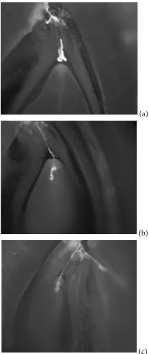
(a)
(b)
(c)
subjected to triple successive fixation – 72 h, 144 h, 240 h in FPA fixative (70% ethanol, propionic acid and formaldehyde, 90 : 5 : 5percentage by volume). Concurrently with the flower fixation under cross- and self-pollination, randomly chosen flowers of cv ‘Čačanska Lepotica’ were subjected to fixation for the purpose of studying pollen tube growth in the ovary under conditions of open pollination. A total of 30 pistils were analyzed within all fixation terms and under all pollination variants.
Aniline blue staining was used for the study of pollen tube growth in the ovary (Preil, 1970; Kho and Baër, 1971). The ovaries were separated along the suture. For better monitoring of the penetration of pollen tubes into the micropyle and nucellus of the ovule, the ovule was cut longitudinal-tangentially (Cerović, 1994). Pollen tubes were UV monitoredon a OLYMPUS BX61 microscope.
The rate of the specific growth of pollen tubes was determined collectively, i.e. identified over the entire period of fixation.
RESULTS
Investigation on the pollen tube growth into the ovary of plum cv ‘Čačanska Lepotica’ revealed the occurrence of those characterized by untypical growth patterns.
Before the penetration of pollen tubes into the nucellus of the ovule, they were characterized by more or less pronounced branching before micropyle, whereby they curled up above the nucellar cap (Fig. 1a), the pollen tube tip bending upon penetration of the micropyle. The concurrent growth of two pollen tubes into the nucellus of the ovule was also observed whereby one of them grew thicker and displayed pronounced fluorescence. The penetration of two pollen tubes is sometimes accompanied by the fluorescence of the entire embryo sac.
Intensive fluorescence of a part of the embryo sac containing the egg apparatus was observed after the penetration of the pollen tube into the nucellus
of the ovule. In addition, the pollen tube was observed to fill up the embryo sac, forming a bundle (Fig. 1b). The occurrence of fluorescence of the entire embryo sac was also detected (Fig. 1c). Under the conditions of self- and open-pollination, insignificant growth of pollen tubes between the nucellus and integument was also discovered. Under the conditions of cross-pollination, in two ovaries, pollen tubes formed a bundle in the embryo sac. In the first year of study, on the sixth day upon pollination, the intensive fluorescence of the ovary tissue and the embryo sac was detected. On this occasion, a large number of pollen tubes penetrating the micropyle and forming bundle above the nucellar cap was also observed.
Over both years of the study, as well as in all three variants of pollination, we observed the unusual behavior of the growing pollen tubes, which refers to several issues, i.e. the curling up of pollen tubes in the micropyle, the growth of two pollen tubes into the nucellus of the ovule and the occurrence of a bundle above the nucellar cap and fluorescence of the upper part the embryo sac containing the egg apparatus. The frequency of the phenomenon described in all the combinations of pollination was approximately the same in both years, and it amounted to 8%. The growth rate ranged from 6.7% in the open-pollination variant, 7.2% in the cross-pollination variant and 7.9% in the open-pollination variant.
Upon growth of the pollen tubes into the nucellus of the ovule, the penetrating pollen tubes formed a bundle either above the micropyle entrance or above the nucellus (Fig. 2a and 2b). The branching and bending of the pollen tubes by 180º upon growth into the micropyle was also evidenced. The specific growth of subsequently penetrating pollen tubes was particularly detected in the cross-pollination variant (9.3%).
DISCUSSION
growth deviates from the common behavior characteristic of the final sequences of the progamic phase of fertilization. According to Herrero (2003), the unusual or untypical behavior of growing pollen tubes occurring in the ovary is classified as follows: a) wandering pollen tubes that change their direction of growth, b) branching pollen tubes, c) pollen tubes that grow through the chalazogamic region (the occurrence of chalazogamy).
The key issues related to the unusual behavior of growing pollen tubes refer to the influence of some components of female sporophyte and gametophyte on the directions of growth of pollen tubes in the ovary. Some changes in several structures in immediate interaction with pollen tubes have been observed. These changes are visible
in the placenta, specifically on the obturator, whereas in the ovule these changes are detected in egsostoma, micropyle, nucellus and the very megagametophyte.
In the obturator area, a more or less pronounced branching of pollen tubes was observed, specifically in cases when a large number of pollen tubes penetrated the ovary. Pollen tubes mostly formed a bundle above and at the egsostome entrance, at the micropyle entrance and above the nucellus cap. In several cases, a thickening of pollen tube tips and the bending of pollen tube tips by 180º were discovered upon the growth into the micropyle and its outward movement.
The obturator covers the ovule entrance and links the style base and the ovule, whereas the cell secretion of the obturator provides passage to the pollen tubes. There is no consensus on the nature of secretion of the obturator cells and its influence on pollen tubes that subsequently arrive in the area. In the Nicotiana species the occurrence of arabinogalactan in the style intercellulars is observed as well as on the secretional placenta (Gane et al., 1995). In maize (Zea mays L.), the occurrence of papillar hairs covering the ovule entrance was detected. After the pollen tubes had passed, these hairs lost their turgor which prevents subsequently arriving pollen tubes from further growing (Heslop–Harison and Heslop–Harison, 1985).
Investigating the growth of pollen tubes into the ovary of peach (Prunus persica var. vulgaris L.), Herrero and Arbeloa (1989) report that, upon passing the obturator zone, pollen tubes terminate their growth in the area of egsostoma. Investigations into the final stages in pollen tube growth in the sour cherry (Prunus cerasus L.) ovary infer the occurrence of the specific nature of growth of pollen tubes in the region of obturator (Cerović, 1994). The same author also suggests that the obturator in the sour cherry is not as much of a limiting factor in the further growth of pollen tubes in the region, adding that some other structures play a much more essential role in controlling the (a)
(b)
direction of the final phases of pollen tube growth. The penetration of pollen tubes is evidenced in the ovules demonstrating the signs of fluorescence, which is indicative of the occurrence of secretion in the ovules that show signs of degeneration which directs the growth of pollen tubes (Cerović, 1996).
The secretion of the cells building the micropyle as well as the presence of starch grains in the cells was evidenced in the majority of species. Cerović et al. (1999) report that the highest concentration of large starch grains in the ovule of sour cherry is observed in the integument cells making up the micropyle. Similarly, it was discovered that after the growth of pollen tubes through the micropyle, a very positive PAS-tainted reaction of the micropilary area is evidenced. The origin of the secretion, along with its linkage with starch grains, still remains unrevealed. Some authors suggest that it originates from the cells of micropyle area, whereas others assume that it is the result of the synergides.
However, the observed secretion of the micropyle cells in a large number of different types of angiosperms, even in the ovules of the gymnosperms, may be indicative of a convergent evolutionary process where the ovules play a principal function in the controlled growth of pollen tubes towards the ovule. It remains an open question whether or not there is, a biochemical influence of this secretion after the growth of a single pollen tube through the micropyle on subsequent pollen tubes, and if so what kind. It is assumed that the growth of a single pollen tube into the micropyle is followed by the termination of the secretion, which results in the chaotic behavior of subsequently penetrating pollen tubes.
Synergides play a most important role in the control of the final stages of pollen tube growth as well as in gamete fusion and migration of spermatic cells (Russell, 1996; Punwani and Drews, 2008). If the sinergides degenerate before growth of pollen tubes into the synergides, the embryo sac is not capable of attracting the pollen tubes (Hegashiyama et al., 2001).
This paper shows that sometimes more than three pollen tubes grow through the micropyle of the ovule. If the secretion of the micropyle cell one way or another has an impact on the growth of the pollen tubes, the results of the study infer that it is by no means a limiting factor of the penetration of a multitude of pollen tubes. Under such circum-stances, it is indicative of the growth of a pollen tube into the nucellus of the ovule, whereas the other pollen tubes most commonly form a bundle above the nucellar cap. The examples that describe the unusual behavior of pollen tubes just before penetration into the nucellus of the primary ovule, could affect the number of fertilized ovules, the percentage of finally germinated fruits and the yield
of the plum cv ‘Čačanska Lepotica’.
Acknowledgement – This work was supported by the
Ministry of Science of the Republic of Serbia.
REFERENCES
Arbeloa, A., and M. Herrero (1987). The significance of the obturator in the control of pollen tube entry into the ovary in peach (Prunus persica). Ann. Bot. 60, 681-685.
Cerović, R. (1994). Histocitološki aspekti dinamike oplodnje kod višnje (Prunus cerasus L.). Doktorska disertacija, Univerzitet u Beogradu, Biološki fakultet.
Cerović, R. (1996). Unusual behaviour of growing pollen tubes in the ovary of sour cherry. Acta Hort. 423, 171-176.
Cerović, R., Vujić, R., and N. Mićić (1999). Localization of polysaccharides in the ovary of sour cherry.
Gartenbauwissenschaft, 64, 40-46.
Gane, A. M., Clarke, A. E., and A. Bacic (1995): Localisation and expression of arabinogalactan-proteins in the ovaries of Nicotiana alata Link and Otto. Sex. Plant Reprod.8, 278-282.
Hegashiyama, T., Yabe, S., Sasake, N., Nishimura, Y., Miyagishima, S., Kuroiwa, H., and T. Kurowia (2001). Pollen tube attraction by synergid cell. Science, 293, 1480-1483.
Herrero, M., and A. Arbeloa (1989). Influence of the pistil on pollen tube kinetics in peach (Prunus persica). Amer. J. Bot. 76, 1441-1447.
Herrero, M. (2000). Changes in the ovary related to pollen tube guidance. Ann. Bot. 85, 79-85.
Herrero, M. (2003). Male and female synchrony and the regulation of mating in flowering plants. Phil. Trans. R. Soc. Lond. B. 358, 1019-1024.
Heslop-Harison, J., and Y. Heslop-Harison (1985). Surfaces and secretions in the pollen-stigma interaction: a brief review. Jour. of Cell Sci. 2, 287-300.
Kho Y. O., and J. Baër (1971). Fluorescence microscopy in botanical research. Zeiss Inform. 76, 54-57.
Preil, W. (1970). Observing of pollen tube in pistil and ovarian tissue by means of fluorescence microscopy. Zeiss Inform. 75, 24-25.
Punwani J. A., and G. N. Drews (2008): Development and function of the synergid cell. Sex. Plant Reprod. 21, 7-15.
Rusell, S. D. (1996). Attraction and transport of male gametes for fertilization. Sex. Plant Reprod. 9, 337-342.
Sage, T. L., Bertin, R. I., and E. G. Williams (1994). Ovarian and other late-action self-incompatibility systems, In:
Genetic control of self-incompatibility and reproductive development in flowering plants (Eds. E. G. Williams, A. E. Clarke, and R. B. Knox), 116-140. Kluwer Academic Publishers, the Netherlands.
Seavey, S. R., and K. S. Bawa (1986). Late-action self-incompatibility in angiosperms. The Botanical Review, 52, 195-215.
СПЕЦИФИЧАН РАСТ ПОЛЕНОВИХ ЦЕВЧИЦА
У ПЛОДНИКУ ШЉИВЕ (PRUNUS DOMESTICA L.)
МИЛЕНА ЂОРЂЕВИЋ1, Р. ЦЕРОВИЋ1, Д. НИКОЛИЋ2 и САЊА РАДИЧЕВИЋ1
1Институт за воћарство, 32000 Чачак, Србија
2Пољопривредни факултет, Универзитет у Београду, 11080 Београд, Србија
У раду су приказани двогодишњи резултати испитивања специфичног раста поленових
цев-чица у плоднику шљиве (Prunus domestica L.)
сорте ‘Чачанска лепотица’, у варијантама сло-бодног, страно- и самоопрашивања. Специфи-чан раст поленових цевчица утврђен је пре и на-кон продора поленове цевчице у нуцелус семе-ног заметка. У обе године истраживања, у све три варијанте опрашивања, констатован је спе-цифичан раст поленових цевчица, који се од-носи на клупко поленових цевчица у микропи-ли, продор две поленове цевчице у нуцелус се-меног заметка и клупко над нуцеларном капом, са флуоресценцијом дела ембрионове кесице у