(Annals of the Brazilian Academy of Sciences) ISSN 0001-3765
www.scielo.br/aabc
Ultrastructural features of
Mimulus aurantiacus
(Scrophulariaceae)
pollen tubes
in vivo
NURAN EK˙ICI˙1, FERUZAN DANE2 and GÖKSEL OLGUN2
1Department of Science Education, Faculty of Education, Trakya University, Ismail Hakki Tonguc Yerleskesi
22030 Aysekadin, Edirne, Turkey
2Department of Biology, Faculty of Science and Arts, Trakya University, Gullapoglu Yerleskesi, 22030, Edirne, Turkey
Manuscript received on September 2, 2007; accepted for publication on May 26, 2008; presented bySERGIOVERJOVSKI-ALMEIDA
ABSTRACT
The aim of this study is to give information on ultrastructure ofin vivopollen tubes ofMimulus aurantiacuswhich were collected from the Botanical Garden of the University of California at Berkeley. Materials were prepared according to electron microscopy methods and examined under Zeiss electron microscope. Four zones were examined in the pollen tubes ofMimulus aurantiacus.Apical zone: Mitochondria, smooth endoplasmic reticulum, rough endoplasmic reticulum, dictyosomes and secretory vesicles were observed. Subapical zone: This area contained abundant rough endoplasmic reticulum and occasionally some smooth endoplasmic reticulum. The polysomes, mitochondria, proplas-tids that contain starch, small vacuoles and a few lipid bodies were detected. Nuclear zone: Both generative and vegetative cell nuclei lie in this zone. The vegetative cell nucleus was large and long. Rough endoplasmic reticulum, mitochondria, ribosomes, dictyosomes, and amyloplasts that are rich of starch were observed.Vacuolation and plug formation zone: Cytoplasm of the tubes was full of large vacuoles. Few organelles such as mitochondria, dictyosome and rough endoplasmic reticulum were detected along their periphery.
Key words:in vivo,Mimulus, pollen tube, ultrastructure.
INTRODUCTION
Pollen, the male gametophyte of higher plants, is a bio-logical system playing a central role in sexual plant reproduction (Cresti et al. 1992, Moscatelli and Cresti 2001). Interactions between pollen and the stigma sur-face initiate pollen germination, which involves an asym-metric extrusion of the pollen cytoplasm through a ger-mination pore to initiate the outgrowth of a pollen tube. Various studies have been done about pollen tubes mostly concerning molecular (Graaf et al. 2005), cyto-chemical (Georgieva 1987), biocyto-chemical (Zonia et al. 2002) physiological;in vitro(Dane et al. 2004),in vivo
(Unal 1986), semivivo (Bergamini Mulcahy and
Mul-Correspondence to: Nuran Ek˙ıc˙ı E-mail: nuranekici@yahoo.com
cahy 1985) andin situ(Dane 2000) aspects and pollen-pistil interactions (Ferrari et al. 1985). The influences of various external factors; such as some chemicals (Kan-dasamy and Kristen 1987, Kim et al. 2003, Röderer and Reis 1988, Sawidisand and Reiss 1995) heat shock (Kandasamy and Kristen 1989), caffeine (Lancelle et al. 1997) and also pistil (Herrero and Arbeloa 1989) on pollen tube growth have been investigated. But ultra-structural studies about pollen tubes are very rare. Initial studies have been done inPetunia(Solanaceae) (Sassen
1964), (Cresti and Van Went 1976) andLilium longiflo-rum(Liliaceae) (Rosen et al. 1964). Few reports have appeared in 1980s, concerning the fine structure of the pollen tubes inLycopersicum peruvianum(Solanaceae) (Cresti et al. 1977), (Cresti et al. 1980),Prunus avium
30 NURAN EK˙IC˙I, FERUZAN DANE and GÖKSEL OLGUN
Prunussp. (Rosaceae) (Ciampolini et al. 1982), Nico-tiana alata(Solanaceae) (Lancelle et al. 1987). In re-cent years, ultrastructural studies about pollen tubes have continued withHosta ventricosa(Liliaceae) (Shi-Yi et al. 1992),Lilium longiflorum(Liliaceae) (Pierson et al. 1990), (Lancelle and Hepler 1992),Nicotiana tabacum
(Solanaceae) (Yu and Russell 1993, Rutten 1993), As-clepias exaltata (Asclepiadaceae) (Sage and Williams 1995),Arabidopsis thaliana(Brassicaceae) (Lennon et
al. 1998), and Conospermum species (Magnoliaceae) (Stone et al. 2004).
Those studies were generally on the ultrastructure of in vitropollen tubes except the studies withPrunus avium(Cresti et al. 1979), (Uwate and Lin 1980). It is too hard to perform and to follow the development ofin vivopollen tubes, so these studies are very important and restricted. Therefore, the number of those studies should be increased.
Some electron microscope observations on pollen grains have been done in some members of subtribe
Castilleiinae(Jensen et al. 1974) and in the genus Mi-mulus of subtribeGratioleae Scrophulariaceae (Argue 1980). Olgun has done embryological researches on
Digitalis sp. (Scrophulariaceae) with light microscope (Olgun 1979), (Yakar and Olgun 1984). She continued her studies with TEM such as ultrastructures of embryo sac (Olgun and Jensen 1987) and endothelium (Dane et al. 2007) in Penstemon gentianoideswhich belongs to the same family. This study can be regarded as the follow-up of those studies.
The ultrastructure of in vivo pollen tubes of
Mimulus aurantiacus(Scrophulariaceae) which is natu-rally grown in North America, and possible similarities related to the ultrastructure ofin vitroandin vivopollen tubes in other angiosperms (Moscatelli and Cresti 2001) were emphasized in this study.
Ultrastructural studies were generally realized with the families Liliaceae, Solonaceae, Rosaceae and in re-cent years with some members of Asclepiadeceae, and Magnoliaceae. However, no studies have been found on ultrastuctural level with the pollen tubes of Scrophula-riaceae family members neitherin vivonorin vitro. This will be the first ultrastructural study on in vivo pollen tubes ofMimulus aurantiacusand also in Scrophularia-ceae family.
MATERIALS AND METHODS
Mimulus aurantiacus (Scrophulariaceae) flowers were
collected from the Botanical Garden of the University of California at Berkeley. Gynoecia were removed from flowers and they were fixed in 4% glutaraldehyde in 0.1M cacodylate buffer pH 6.8, for 2 hours. Then, the pollinated pistils were washed several times in buffer, fixed overnight with 2% buffered OsO4; dehydration was applied with gradually increasing aceton-propylenoxide series with staining in 70% acetone containing 1% uranyl nitrate overnight. The material was embedded in Spurr’s medium. 0.5-1 m thick semi-thin sections were stained with methylene blue (1%) using 0.02 M NaOH to con-trol pH and observed by light microscopy. Thin sections were cut on a Porter-Blum ultramicrotome with a glass or diamond knife. Sections were stained on grids with lead citrate (Reynolds 1963) for one minute and observed by a Zeiss EM 9A electron microscope.
RESULTS
In this study, four zones were examinedin vivopollen tubes ofMimulus aurantiacusas similar in the previous studies.
APICALZONE
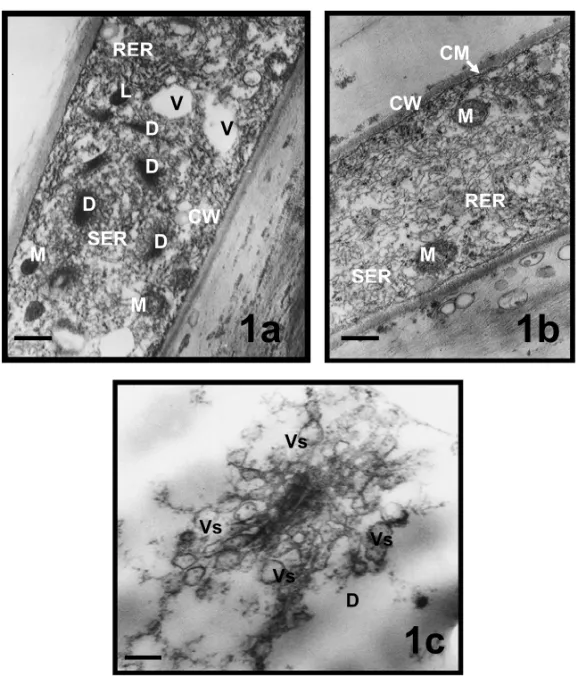
The cytoplasm of that zone was rich of organelles. Many electron dense dictyosomes and spherical mitochondria were visible. Small vacuoles, abundant rough endoplas-mic reticulum (RER), occasionally some smooth endo-plasmic reticulum (SER) and electron dense lipid bod-ies were also seen (Fig. 1a). A well-developed RER (Fig. 1b) and dictyosomes were observed. Many vesi-cles budded out from the active dictyosomes were seen, in various sizes around cis and trans regions of dictyo-somes, in the apical zone (Fig. 1c). Cell wall and cell membrane were also clearly visible in the electron micrographs of apical zone (Figs. 1a, b).
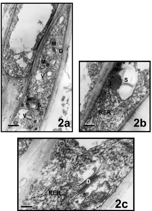
SUBAPICALZONE
Fig. 1 – Apical zones of pollen tubes inMimulus aurantiacus. a, b, views from apical zone; c, well-developed dictyosome in the apical zone. a, bar = 1µm; b, bar = 0,5µm; c, bar = 150 nm. (CM, cell membrane; CW, cell wall; D, dictyosome; L, lipid; M, mitochondria; RER, rough endoplasmic reticulum; SER, smooth endoplasmic reticulum; Vs, vesicule).
as polysomes. Well-developed active dictyosomes and budding vesicles were also found in the subapical zone (Fig. 2c).
NUCLEARZONE
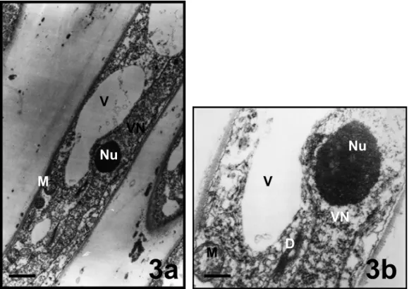
Vegetative and generative nuclei were located in this zone (Figs. 3, 4a, b). The vegetative cell nucleus was large and long. It contained one nucleolus and several weakly-stained chromatin aggregations. It was rich of euchromatin material. Vacuoles in different sizes, mito-chondria and dictyosomes were observed around vege-tative nucleus (Figs. 3a, b). There were 2, 3 amyloplasts
containing starch grains and spherical shaped mitochon-dria around generative nucleus (Figs. 4a, b). Electron dense lipid bodies were seen near the amyloplast full of starch grains (Fig. 4b).
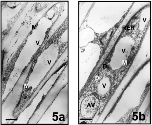
VACUOLATION ANDPLUGFORMATIONZONE
32 NURAN EK˙IC˙I, FERUZAN DANE and GÖKSEL OLGUN
Fig. 2 – Subapical zones of pollen tubes inMimulus aurantiacus. a, bar = 1,5µm; b, bar = 0,5µm; c, bar = 0,2µm; (D, dictyosome; L, lipid; M, mitochondria; RER, rough endoplasmic reticulum; S, starch; V, vacuol).
DISCUSSION
The term “zonation” was introduced to describe the functional cytoplasmic distribution in the pollen tubes of Lycopersicum perivuanum (Cresti et al. 1977); in which four different areas were identified: (i) the apical zone, rich in SVs (Secretory Vesicles); (ii) the subapical area, containing organelles; (iii) the nuclear zone, com-prising the MGU (Male Germ Unite); and (iv) the zone of vacuolization and callose plug formation (Moscatelli and Cresti 2001), (Bhojwani and Soh 2001). The pres-ence of four zones was detected in the in vivo grow-ing pollen tubes ofMimulus aurantiacusas reported by
Cresti et al. (1977), Ciampolini et al. (1982) in theirin vitroand by Cresti et al. (1979, 1980), Uwate and Lin’s (1980)in vivostudies. In general, the constitution of or-ganelles in various zonesin vitroandin vivoare quite similar as also seen inLycopersicum(Cresti et al. 1977), (Cresti et al. 1980), inMalus(Speranza et al. 1982), in
Prunus avium(Cresti et al. 1979), in cherry (Ciampolini et al. 1982), and inConospermum(Stone et al. 2004).
Fig. 3 – Nuclear zones (vegetative) of pollen tubes inMimulus aurantiacus. a, bar = 1µm; b, bar = 100 nm. (D, dictyosome; M, mitochondria; Nu, nucleolus; V, vacuol VN, vegetative nucleus).
34 NURAN EK˙IC˙I, FERUZAN DANE and GÖKSEL OLGUN
Fig. 5 – Vacuolation and plug formation zones of pollen tubes in Mimulus aurantiacus. a,b bar = 1,5µm; (AV, autophagic vacuole; D, dictyosome; M, mitochondria; RER, rough endoplasmic reticulum; V, vacuol).
which concentrates Golgi-derived SVs at the tip (Heslop-Harrison and Heslop-Harrison 1989) where they fuse with the plasma membrane. This process of exo-cytosis provides wall polysaccharides and new plasma membrane for tube elongation (Moscatelli and Cresti 2001). Apical and subapical zones are active zones be-cause they are rich of organelles such as: mitochondria, well-developed dictyosomes and lipid bodies. We also observed areas which would be designated as smooth and rough endoplasmic reticulum zones (Uwate and Lin 1980). Our micrographs showed small vesicles arising from the well-developed dictyosome cisternae. Besides playing a major role in the carbohydrate metabolism, dictyosomes have functioned as sorting and dispatching station for the ER products (Dupree and Sherrier 1998). According to Uwate and Lin (1980), these vesicles have been arisen from smooth endoplasmic reticulum cister-nae, and rough endoplasmic reticulum circumscription of vacuoles. Lipid bodies were visible in various zones in our study. This was also mentioned by Uwate and Lin (1980).
Since pollen tubes go into the stigma and stylus, they need glycoproteins and glycolipids to build pollen tube
wall. These molecules are synthesized in these well-developed dictyosomes. Amiloplasts detected in vivo
growing pollen tubes ofMimulus auriaticusare less than
in vitroones ofPrunus avium(Ciampolini et al. 1982).
In vivogrowing pollen tubes can absorb nutrients from the stylar tissue surrounding them. However, this may also occurin vitrostudies due to high concentration of sucrose used in culture mediums.
According to Cresti and Van Went (1976), there are two ways of callose deposition inPetunia pollen tubes growing in the style. The first one is callose deposi-tion outside the plasma membrane; the second one is callose deposition within the cytoplasm as distinct cal-lose grains, leading to the formation of calcal-lose plugs. Both pollen tube elongation and organelle distribution must be highly coordinated, so that pollen tube growth makes the oldest part of the vegetative cytoplasm move forward, and it becomes isolated by callose plug forma-tion. The mechanism of growth in pollen tubes as well as in other cell types, requires the integrity of the se-cretory system, namely ER and Golgi apparatus. The secretory system involves the secretion of protein and polysaccharide components. In this study, the elements of RER, SER and Golgi apparatus dispersed through the pollen tube ofMimulus aurantiacus.
In conclusion, the ultrastructure of zonation in pol-len tubes ofMimulus aurantiacus in vivohad similarities with the ultrastructure ofin vivopollen tubes inPrunus avium(Cresti et al.1979, Uwate and Lin 1980). In addi-tion, it was seen that callose deposition occurred within the cytoplasm of pollen tubes inMimulus aurantiacus.
RESUMO
O objetivo deste estudo é informar sobre a ultraestrutura de tubos de pólen deMimulus aurantiacus in vivocoletados no “Botanical Garden” da Universidade da Califórnia em Berke-ley. O material foi preparado de acordo com os métodos de microscopia eletrônica e examinado em microscópio eletrônico Zeiss. Quatro zonas dos tubos de pólen deMimulus auran-tiacusforam examinadas. Zona apical: foram observados mitocôndrias, retículo endoplasmático liso; retículo endoplas-mático rugoso, dictiossomos e vesículas secretoras.Zona sub-apical: esta área continha retículo endoplasmático rugoso em abundância e, ocasionalmente, algum retículo endoplasmático liso. Foram detectados polissomos, mitocôndrias, proplas-tídeos que contêm amido, pequenos vacúolos e alguns corpos lipídicos. Zona nuclear: nesta área, existem tanto núcleos de células geradoras como vegetativas. O núcleo de célula vegetativa é grande e longo. Foram observados retículo endo-plasmático rugoso, mitocôndria, ribossomos, dictiossomos e amiloplastos ricos em amido. Zona de vacuolização e de formação de “plug”: o citoplasma dos tubos estava cheio de grandes vacúolos. Algumas organelas como mitocôndria,
dictiossomo e retículo endoplasmático rugoso foram detectadas em toda a periferia desta área.
Palavras-chave: in vivo,Mimulus, tubo de pólen, ultraestru-tura.
REFERENCES
ARGUE CL. 1980. Pollen morphology in genus Mimulus
(Scrophulariaceae) and its taxonomic significance. Amer J Bot 67: 68–87.
BERGAMINIMULCAHYGANDMULCAHYDL. 1985. Ovar-ian influence on pollen tube growth, as indicated by the
semivivotechnique. Amer J Bot 72: 1078–1080. BHOJWANISSANDSOHWY. 2001. Current trends in the
embryology of angiosperms. Kluwer Academic Publish-ers: Dordrecht, The Netherlands, p. 33–65.
CIAMPOLINIF, CRESTIMANDKAPILRN. 1982. Germi-nation of cherry pollen grainsin vitro. An Ultrastructural Study. Pyhtomorphology 32: 364–373.
CRESTI MAND VAN WENT JL. 1976. Callose deposition and plug formation inPetuniapollen tubesin situ. Planta 133: 35–40.
CRESTI M, PACINI E, CIAMPOLINI F ANDSARFATTI G. 1977. Germination and early tube developmentin vitroof
Lycopersicum peruvianumpollen: ultrastructural features. Planta 136: 239–247.
CRESTI M, CIAMPOLINI F, PACINI E, SARFATTI G AND DONINIB. 1979. Ultrastructural features ofPrunus avium
L. pollen tubesin vivo. I. The compatible pollen tube. Caryologia 32: 433–440.
CRESTIM, CIAMPOLINIFANDSARFATTIG. 1980. Ultra-structural investigations on Lycopersicum peruvianum
pollen activation and pollen tube organization after self-and cross-pollination. Planta 150: 211–217.
CRESTIM, BLACKMORESAND VANWENTJL. 1992. Atlas of sexual plant reproduction in flowering plants, Springer-Verlag, Berlin. 75 p.
DANEF. 2000. In situgermination of pollen tetrads in Peri-ploca greaceaeL. (Periplocaceae). Turk J Biol Tubitak 24: 337–343.
DANE F, OLGUNGANDDALGICO. 2004. In vitropollen germination of some plant species in basic culture medium. J Cell Mol Biol (Halic Univ. Press, Turkey) 3: 71–76. DANE F, OLGUNGANDEK˙IC˙IN. 2007. Ultrastructure of
36 NURAN EK˙IC˙I, FERUZAN DANE and GÖKSEL OLGUN
DUPREEPANDSHERRIERDJ. 1998. The plant Golgi appa-ratus. Biochem Biophys Acta 1404: 259–270.
FERRARITE, BESTV, MORETA, COMSTOCKP, MUHAM-MAD AANDWALLACEDH. 1985. Intercellular adhe-sions in the pollen-stigma system: Pollen capture, grain binding, and tube attachments. Amer J Bot 72: 1466– 1474.
GEORGIEVAID. 1987. Cytochemical investigation of pollen and pollen tubes afterγ-irradiation: II. Effect of the irra-diation on quinine formation. Phytomorphology 37(2-3): 159–163.
GRAAF BHJ, CHEUNG AY, ANDREYEVA T, LEVASSEUR K, KIELISZEXSKIMANDWUH. 2005. Rab11 GTPase-regulated membrane trafficking is crucial for tip-focused pollen tube growth in tobacco. Plant Cell 17: 2564–2579. HERREROMANDARBELOAA. 1989. Influence of the pistil on pollen tube kinetics in peach (Prunus persica) Amer J Bot 76: 1441–1447.
HESLOP-HARRISON J AND HESLOP-HARRISON Y. 1989. Actomyosin and movement in the angiosperm pollen tube: An interpretation of some recent results. Sex Plant Reprod 2: 199–207.
JENSENWA, ASHTONMANDHECKARDLR. 1974. Ultra-structural studies of the pollen of subtribeCastilleiinae, family Scrophulariaceae. Bot Gaz 135: 210–218. KANDASAMYMKANDKRISTENU. 1987.
Pentachlorophe-nol affects mitochondria and induces formation of golgi apparatus-endoplasmic reticulum hybrids in tobacco pol-len tubes. Protoplasma 141(2-3): 112–120.
KANDASAMY MKANDKRISTENU. 1989. Ultrastructural responses of tobacco pollen tubes to heat shock. Proto-plasma 153(1-2): 104–110.
KIMS, MOLLETJC, DONGJ, ZHANGK, PARKSYAND LORD EM. 2003. Chemocyanin, a small basic protein from the lily stigma, induces pollen tube chemotropism. Plant Biol 100: 16125–16130.
LANCELLE SAANDHEPLER PK. 1992. Ultrastructure of freeze-substituted pollen tubes of Lilium longiflorum. Protoplasma 167(3-4): 215–230.
LANCELLESA, CRESTIMANDHEPLERPK. 1987. Ultra-structure of the cytoskeleton in freeze-substituted pollen tubes ofNicotiana alata. Protoplasma 140(2-3): 141–150. LANCELLESA, CRESTIMANDHEPLERPK. 1997. Growth inhibition and recovery in freeze-substitutedLilium longi-florumpollen tubes: structural effects of caffeine. Proto-plasma 196(1-2): 21–33.
LENNONKA, ROYS, HEPLERPKANDLORDEM. 1998. The structure of the transmitting tissue of Arabidopsis thaliana(L.) and the path of pollen tube growth. Sex Plant Reprod 11: 49–59.
MOSCATELLIAANDCRESTIM. 2001. Pollen germination and pollen tube growth. In: BOJWANISSANDSOHWY (Eds.), Current trends in the embryology of angiosperms, Kluwer Academic Publishers: Dordrecht, The Nether-lands, p. 33–65.
OLGUNG. 1979. Comparative investigations on the karyol-ogy and embryolkaryol-ogy (Embryo sac and endosperm devel-opment) ofDigitalis viridifloraLindl. andDigitalis lutea
L. Istanbul Univ. J Sci Fac B Series 44: 1–22.
OLGUNGANDJENSENJA. 1987. The ultrastructure of the egg apparatus and central cell ofPenstemon gentianoides
Poir. Before fertilization (Scrophulariaceae). Istanbul Univ Sci Fac J Series B 52: 35–45.
PIERSON ES, LICHTSCHEIDLIL ANDDERKSEN J. 1990. Structure and behaviour of organelles in living pollen tubes ofLilium longiflorum. J Exp Bot 41: 1461–1468. REYNOLDSES. 1963. The use of lead citrate of high pH as
an electron-opaque stain in electron microscopy. J Cell Biol 17: 208–212.
ROSEN WG, GAWLICK SR, DASHEK WV AND SIEGES-MUND KA. 1964. Fine structure and cytochemistry of
Liliumpollen tubes. Amer J Bot 51: 61–74.
RÖDERERGANDREIS HD. 1988. Different effects of in-organic and triethyl lead on growth and ultrastructure of lily pollen tubes. Protoplasma 144(2-3): 101–109. RUTTENALM. 1993. Organelle distribution and cytoskeleton
in the cytoplasm of tobacco pollen tube. Ph.D. Thesis. Chap. 6. University of Nijmegen. The Netherlands. SAGETLANDWILLIAMSEG. 1995. Structure,
ultrastruc-ture, and histochemistry of the pollen tube pathway in the milkweed Asclepias exaltata L. Sex Plant Reprod 8(5): 257–265.
SASSENMMA. 1964. Fine structure ofPetuniapollen grain and pollen tube. Acta Bot Neerl 13: 174–181.
SAWIDISAND T AND REISS HD. 1995. Effects of heavy metals on pollen tube growth and ultrastructure. Proto-plasma 185(3-4): 113–122.
SHI-YIH, CHUN-GUILANDCHENGZ. 1992. Ultrastruc-ture of microfilaments in pollen and pollen tubes ofHosta ventricosa(=H. coerulea). Zhíwùxué Bào 34: 8–14. SPERANZAA, CALZONIGL, CRESTIMANDCIAMPOLINI
germi-nation and ultrastructure of apple pollen. Env Exp Bot 22: 339–347.
STONELM, SEATONKA, KUOJANDMCCOMBJA. 2004. Fast Pollen Tube Growth inConospermumSpecies. Ann Bot 93: 369–378.
UNALM. 1986. A comperative cytological study on compat-ible and in-compatcompat-ible pollen tubes ofPetunia hybrida. Istanbul Univ Sci Fac J Series B 51:1–12.
UWATE WJ AND LIN J. 1980. Cytological zonation of
Prunus aviumL. pollen tubesin vivo. J Ultrastruct Res 71: 173–184.
YAKARNANDOLGUNG. 1984. Comparative investigations on the megasporogenesis and embryo sac development of
Digitalis divisianaHeyw. andDigitalis ambigua. Murr J Nat Sci 8(3): 1–22.
YUHSANDRUSSELLSD. 1993. Three-dimensional ultra-structure of generative cell mitosis in the pollen tube of
Nicotiana tabacum. Eur J Cell Biol 61: 338–348. ZONIAL, CORDEIROS, TUPYJANDFEIJOJA. 2002.