ii
THAÍS RIBEIRO SANTIAGO
A DEEP ANALYSIS OF THE GENETIC STRUCTURE OF
Ralstonia solanacearum IN BRAZIL REVEALS NOT MUCH SEX
IN THE POPULATION!
Thesis presented to the Universidade Federal de Viçosa as part of the requirements for the degree of Doctor
Scientiae in Plant Pathology.
VIÇOSA
iv
THAÍS RIBEIRO SANTIAGO
A DEEP ANALYSIS OF THE GENETIC STRUCTURE OF
Ralstonia solanacearum IN BRAZIL REVEALS NOT MUCH SEX
IN THE POPULATION!
Thesis presented to the Universidade Federal de Viçosa as part of the requirements for the degree of Doctor
Scientiae in Plant Pathology.
v
To my mother Neide, my father and my sister Thássia, for all their love, sacrifice
vi
ACKNOWLEDGMENTS
Thanks are due to the Universidade Federal de Viçosa, especially to the Plant Pathology Department and to Foundation for Research Assistance of Minas Gerais State (FAPEMIG) for granting a scholarship.
Thanks are also due to CNPq for providing financial support to the research.
Thanks to my parents Joaquim and Neide, my sister Thássia and my
boyfriend Geraldo for their immense and unconditional support.
I am indebted to Professor Eduardo Seiti Gomide Mizubuti for his valuable advice, guidance, patience, friendship and opportunity.
Thanks are due to Professor José Rogério de Oliveira for his friendship and contributions to my professional education.
Thanks to Dr. Carlos Alberto Lopes for his contributions and suggestions for improving this work.
I am grateful to all my colleagues in the Graduate Program in Plant Pathology at UFV, especially my friends Robson, Saulo, Lahyre, Jaime, Miller, Sara, Jonas, Carlos, Alessandro, Maria, Adriana and Silvia. I am also grateful to the following members of the undergraduate programs at UFV: Sara, Lillian, Leonardo, Claudiney, Renato, Pollyany, Raquel, Luciano and Luana for their help and encouragement during this time.
Thanks to my friends Arshan, Fizza, Muhamad, Tânia and Kênia for their friendship and collaboration in my sandwich program.
Thanks are due to Professor Gustavo Caetano-Anolles for all his collaboration and guidance.
I am grateful to the Sara Moreira for her friendship and collaboration during this period.
vii BIOGRAPHY
THAIS RIBEIRO SANTIAGO, daughter of Joaquim Santiago Sobrinho and Maria Rizoneide Ribeiro Rodrigues, was born in Brasiléia, AC, Brazil on November 10th, 1986.
In January of 2009, she graduated with a degree in Agronomy at Universidade Federal de Viçosa, Viçosa, MG.
In July 2010, she obtained her Master Scientiae degree in Plant Pathology from Universidade Federal de Viçosa, Viçosa, MG.
viii CONTENT
RESUMO ... ix
ABSTRACT ... xi
GENERAL INTRODUCTION ... 1
CHAPTER 1 - GENETIC STRUCTURE OF Ralstonia solanacearum CAUSING BACTERIAL WILT IN BRAZIL ... 4
1. Introduction ... 5
2. Material and methods ... 7
3. Results ... 12
4. Discussion ... 16
5. Conclusions ... 22
6. Acknowledgments ... 23
7. References ... 23
CHAPTER 2 - PARAMETRIZATION OF EVOLUTIONARY MECHANISMS THAT AFFECT THE POPULATION OF Ralstonia solanacearum IN BRAZIL USING GENE GENEALOGIES AND THE COALESCENT APPROACH ... 41
1. Introduction ... 41
2. Material and methods ... 43
3. Results ... 47
4. Discussion ... 49
5. Conclusion ... 54
6. Acknowledgements ... 54
7. References ... 54
ix RESUMO
SANTIAGO, Thaís Ribeiro, D.Sc., Universidade Federal de Viçosa, agosto de 2014. Uma análise profunda da estrutura genética de Ralstonia solanacearum no Brasil revela não muito sexo na população! Orientador: Eduardo Seiti Gomide Mizubuti. Coorientadores: Carlos Alberto Lopes e José Rogério de Oliveira.
x
xi ABSTRACT
SANTIAGO, Thaís Ribeiro, D.Sc., Universidade Federal de Viçosa, August, 2014. A deep analysis of the genetic structure of Ralstonia solanacearum in Brazil reveals not much sex in the population!Adviser: Eduardo Seiti Gomide Mizubuti. Co-advisers: Carlos Alberto Lopes and José Rogério Oliveira.
xii
1
GENERAL INTRODUCTION
Bacterial wilt caused by Ralstonia solanacearum is one of the most destructive diseases in various crops cultivated in the tropics. The inoculum can be dispersed by infested soil, irrigation water and tubers (Caruso et al., 2005; Denny, 2006). Several species of plants, distributed in more than 50 botanical families, are reported as hosts (Denny, 2006). Among the economically important crops, those belonging to the Solanaceae family are relevant, such as
tomatoes, potatoes, peppers and eggplant (Hayward, 1994).
In an attempt to study the variability, different classification methods commonly used in bacteria species have been adopted. The main divisions are biovar, phylotype and sequevar (Buddenhagen et al., 1964; Fegan; Prior, 2005). Methods based on genome analysis, analysis of repeated sequences and sequencing of genomic regions are useful to elucidate the relationships within the species R. solanacearum (Strange, 2005). Although there is information on the genetic variability of R. solanacearum in Brazil and its geographical distribution, these studies were conducted for a reduced number of hosts and sampling areas (Costa et al., 2007; Pinheiro et al., 2011; Rodrigues et al., 2012; Santana et al., 2012). The knowledge about the pathogen population structure is useful for the understanding and development of control strategies and to guide breeding programs that aim to develop cultivars with durable resistance (Milgroom and Fry, 1997).
Despite several efforts, epidemics of bacterial wilt remain responsible for the low productivity in several regions. Under favorable temperatures, the appearance of wilt occurs initially in upper leaves and after a few days in all plant (Hayward, 1991; Akiew and Trevorrow, 1994). The difficulty of developing bacterial wilt control strategies have been caused by the lack of knowledge of ecology, variability and about the evolutionary mechanisms that affect the pathogen population (Strange, 2005). The limited knowledge of the variability in
2
The objectives of this study were to: (1) characterize the isolates of R.
solanacearum by biovar, phylotype and sequevar, (2) determine the genetic
structure of pathogen population by geographic region and by host and (3) parameterize the evolutionary mechanisms affecting the population of R.
solanacearum in Brazil.
References
Akiew, E. B. and Trevorrow, P. R. 1994.Management of bacterial wilt of tobacco.Pages 179-198.in Bacterial Wilt: The disease and its causative agent,
Pseudomonas solanacearum. A. C. Hayward and G. L. Hartman, eds. CAB
International, Wallingford, UK.
Buddenhagem, I.; Sequeira, L. and Kelman, A. 1962.Designation of races in
Pseudomonas solanacearum (Abstr.).Phytopathology 52: 726.
Casuro, P.; Palomo, J. L. Bertolini, E., Alvarez, B., Lopez, M. M., Biosca, E. G. 2005. Seasonal variation of Ralstonia solanacearum biovar 2 populations in a Spanish river: recovery of stressed cells at low temperatures. Applied and Environmental Microbiology 71: 140-148.
Costa, S. B., Ferreira, M. A. S. V. and Lopes, C. A. 2007. Diversidade patogênica e molecular de Ralstonia solanacearum da região amazônica brasileira. Fitopatologia Brasileira 32:285-294.
Denny, T. P. 2006. Plant pathogenic Ralstonia species.Pages 573-644 in Plant-associated Bacteria. S. S. Gnanamanickan. The Nertherlands, Springer Publishing
Fegan, M. and Prior, P. 2005. How complex is the “Ralstonia solanacearum species complex”? Pages 449-461 in: Bacterial wilt disease and the Ralstonia
solanacearum species complex. C. Allen, P. Prior, and A. C. Hayward.
American Phytopathological Society Press, St. Paul, MN.
Hayward, A. C. 1991. Biology and epidemiology of bacterial wilt caused by
Pseudomonas solanacearum. Annual Review Phytopathology 29: 65-87.
Hayward, A.C. 1994. The hosts of Pseudomonas solanacearum.Pages 9-24 in Bacterial Wilt: The disease and its causative agent, Pseudomonas
solanacearum.A. C. Hayward and G. L. Hartman, eds. CAB International,
3
Lopes, C. A. 1994. Situação da murcha bacteriana no Brasil. Pages 13-16 in Taller sobre enfermedades bacterianas de la papa. Embrapa CNPH CIP, Brasília, DF.
Milgroom, M. G. and Fry, W. E. 1997.Contribution of population genetics to plant disease epidemiology and management.Advances in Botanical Research 24:1-30.
Pinheiro, C. R., Amorim, J. A. E., Diniz, L. E. C., Silva, A. M. F., Talamini, V., Souza Júnior, M. T. 2011. Diversidade genética de isolados de Ralstonia
solanacearum e caracterização molecular quanto a filotipos e sequevares.
Pesquisa Agropecuária Brasileira46:593-602.
Rodrigues, L. M. R., Destéfano, S. A. L., Silva, M. J., Costa, G. G. L. and Maringoni, A. C. 2012. Characterization of Ralstonia solanacearum from Brazil using molecular methods and pathogenity tests.Journal Plant Pathology 94: 505-516.
Santana, B.G., Lopes, C. A., Alvarez, E., Barreto, C. C., Allen, C. and Quirino, B. F. 2012. Diversity of Brazilian biovar 2 strains of Ralstonia solanacearum. Journal of General Plant Pathology 78: 190-200
4 CHAPTER 1
GENETIC STRUCTURE OF Ralstonia solanacearum CAUSING BACTERIAL WILT IN BRAZIL
T. R. Santiago1, C. A. Lopes2, G. Caetano-Anollés3, and E. S. G. Mizubuti1,4
Abstract: Understanding the genetic variability of populations of the plant pathogenic β-proteobacterium Ralstonia solanacearum (Rs)is key to the implementation of control measures, mainly breeding for disease resistance. In order to study the genetic structure of Rs in Brazil, 301 isolateswerecollectedfrom nine plant species and from all major geographic regions. Isolates were characterized according to biovar, phylotype, and sequevar to determine the amount and distributionof geneticvariability. Isolates were classified into biovars 1 (52%), 2 (32%) and 3 (16%) and phylotypes I (48 isolates) and II (253 isolates) with two subclusters, IIA and IIB. Phylotype II was found to be widespread in Brazil, whereas Phylotype I isolates were found in the Central, North and Northeast regions, the warmest areas. Based on the endoglucanase (egl)gene sequences, seven known sequevars (1, 4, 18, 27, 28, 41 and 50) and four new sequevars (54, 55, 56, 57) were identified. Large clonal fraction and high number of haplotypes (282)were observed inBrazil. Three groups were identified: group 1 made of phylotype I isolates; and two other groups comprised of phylotypes I and II isolates but split according to geographic regions; group 2 made of isolates collected in the North/Northeast and group 3 from the Central/Southeast/South regions. Absence of genetic separation of the South-Southeast-Central and North-Northeast regions supports the occurrence of gene flow at different latitudes probably due to long distance dissemination of planting materials or rivers. There was evidence of recombination in the population. Among the isolates collected in the South-Southeast-Central regions those from tomato were genetically distinctfrom the potato isolates. Thereishighgeneticvariabilityin theBrazilian population ofR.
solanacearum. Thismakesbacterialwiltcontrolusinghostresistance
achallengingtask.
Key words: Brown rot,population genetics, molecular epidemiology, variability, diversity.
1
Departamento de Fitopatologia, Universidade Federal de Viçosa, Viçosa, Minas Gerais, Brazil. 2
Centro Nacional de Pesquisa de Hortaliças, Gama, Distrito Federal, Brazil. 3
Department of Crop Sciences, University of Illinois, Urbana, Illinois, United States. 4
5 1. Introduction
Ralstonia solanacearum [(Smith, 1896) Yabuuchi et al. 1995],
previously known as Pseudomonas solanacearum and Burkholderia
solanacearum, is the causal agent of bacterial wilt in many plant species (Yao &
Allen, 2006). Despite its wide distribution, epidemics are more intense in the tropical regions of the world (Hayward, 1994). There are recordsof occurrenceof bacterial wilt inplantsfrom more than50botanical families, includingmonocots and dicots (Elphinstone, 2005; He et al., 1983). Economic lossesare difficult to
quantify, but are high. While an evaluation of direct yield lossesispossible,estimatingindirect losses commonly associated with bacterial wiltis not straight forward. Specifically, the occurrenceof bacterial wiltin certain areasmay hamper the cultivation ofsusceptiblehosts, which imposes severerestrictions on the economic viabilityof land use. Manycrops of economic importance such as potato (Solanum tuberosum), tomato (S. lycopersicum), eggplant (S. melongena), banana (Musa spp.) and peanut (Arachis
hypogea)are affected by the pathogen. In addition, recent reports show that the
range ofplantsinfected by R.solanacearumis increasing, including in Brazil (Wilson et al., 2005; Malavolta, 2008).
Ralstoniasolanacearumhas highphenotypicand genotypic variability and
strains have been isolated from virgin jungle soils in Asia and the Americas (Hayward, 1991). Currently, R. solanacearum is considered a species complex and there is variation according to host range, geographic distribution, pathogenicity and physiological properties (Denny, 2006). The origin of the species complex is believed to predate the geological separation of the continents (Hayward, 1991).
Before thegenomic revolution, the characterization ofraces was used toseparate thestrains ofR.solanacearum in relation to the range of hosts(Buddenhagem et al., 1962), whereas biovarswere defined asphysiologicalgroups that vary according to the abilityto usecarbohydrates(He
6
endoglucanase (egl) genes (Fegan &Prior, 2005). Four phylotypes that make use of multiplex PCR and various sequevars derived from egl gene sequences variations were identified. Nowadays, this classification is the most accepted and is used in a number of studies of R. solanacearum strains from different regions, including the Americas.
Despite their considerable phylogenetic diversity, R. solanacearum strains are unified by their common pathology. All cause bacterial wilt, which is characterized by bacterial colonization of the xylem vessels to very high cell
densities (109- 1010 CFU/mL xylem fluid), vascular browning, stunting, wilting, and often rapid death (Kelman, 1956; Buddenhagen et al., 1964; Peeters et al., 2013). The bacterium is dispersed by soil, water and infected propagation material. Strains infect plants most commonly through the roots, but sometimes through insect wounds (Hayward, 1991).
Bacterial wilt is difficult to control because the bacteria survive for years in infested soils and weed hosts. Breeding for host resistance is the best management strategy, but is complicated by the high genetic variability of the pathogen population. For example, tomatoes resistant to R. solanacearum in one region can be susceptible in another (Boshou, 2005). The lackof knowledge aboutthe variabilityof the pathogen, its magnitude and geographic distribution, make difficult the development and the strategic use of resistant varieties inseveralareas, particularly in Brazil.Thestrategic useof resistant cultivarsdepends on basic informationrelatingthe environmental conditions, the characteristics of the hostand genetic variabilityofthe pathogen population, which is often subject to changes. Therefore, successfulbreeding programsrequire knowledge of theevolutionary biologyof the pathogenand the mechanismsthat influencetheir pathogenicity(Strange, 2005). Since the variability of the pathogendirectly affects themanagement of the disease, epidemiological studies are also important.
While knowledge of the local pathogen population is required for
successful breeding and integrated pest management programs, there is scarce information about the genetic variability of the Brazilian populations of R.
solanacearum. It has been proposed that R. solanacearum which was putatively
7
Thus, the Brazilian population is very old and has had the opportunity to co-evolve with a high diversity of plant species throughout millions of years, but with few commercially grown hosts that were introduced and have been more intensively cultivated in the past 200 years. In one study, isolates from tomato and a few other hosts sampled in the Amazon region were found to be highly polymorphic and were classified into five groups that had no correlation with host range, biovar, ecosystem and sampling location of the pathogen (Costa et al. 2007). Similar results were observed for isolates from different hosts and
different regions of the country (Rodrigues et al., 2012). However, the sampling limitations of both studies (only 22 isolates) restrict the scope of inferences. In another study, 53 R. solanacearum strains mainly from potato collected in seven Brazilian states were characterized using phylotype and sequevar (Santana et al., 2012). There was high genetic variation, but information on the amount and distribution of genetic variation in populations of R. solanacearum associated to other host species remains largely unknown. In this study, we are interested in answering the following questions: Which biovar, phylotype and sequevar are found in Brazil? Is the genetic diversity high? Is there spatial genetic differentiation or any population structure according to host? Is there evidence of recombination in the population of R. solanacearum in Brazil?
2. Material and methods
2.1. Bacterial strains and growth conditions
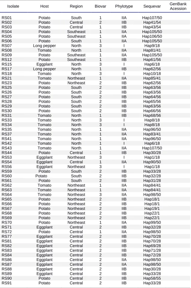
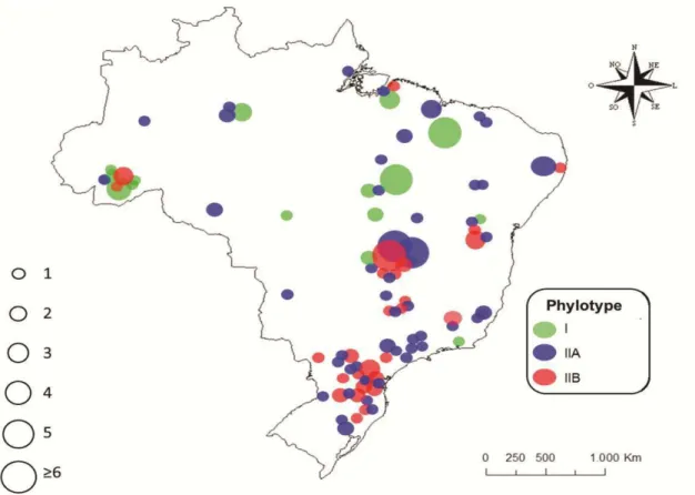
Bacterial strains used in this work are listed in Table 1 and Fig. 1. The isolates are deposited in the culture collection maintained by EMBRAPA Hortaliças (Brasília, Brazil) and Universidade Federal de Viçosa. A total of 301 isolates were analyzed. Isolates were obtained from different host plants, including Solanumlycopersicum (137 isolates), S. tuberosum (98), Capsicum spp. (4), Piperhispidinervum (20), S. melongena (19), Eucalyptus sp. (15), Musa spp. (2), S. gilo (3), and Geranium sp. (3), collected from 1987 to 2012, from
8
Kelman agar medium (Kelman, 1954) incubated for 48h at 28°C and maintained in cryogenic storage tubes containing sterilized water.
2.2. DNA extraction and identification of bacterial strains
Bacteria were streaked on Kelman solid medium without tetrazolium chloride and incubated at 28°C for 48 h. Bacterial growth from a single, isolated, and typical colony was transferred to another plate and incubated in the same way. Two mL of saline solution (0.01%) was added to plates with developed
colonies that were scraped so as to prepare a cell suspension. The entire suspension was then pelleted by centrifugation and total genomic DNA was extracted using the Wizard® Genomic DNA Purification Kit (Promega) following the manufacturer`s instructions. The quality of DNA was checked by gel electrophoresis. DNA was quantified with a NanoDrop 2000 spectrophotometer (Thermo Fisher Scientific) and adjusted to 25 ηg.
2.3. Species specific primers
The species-specific primer pair 759/760 designed to identify R.
solanacearum was used in all isolates. PCR conditions were as described by
Opina et al. (1997) in a total volume of 25 µL. Amplification was performed in a MJ PTC-100 thermocycler. PCR products were analyzed by eletrophoresis in 1% (wt/vol) agarose gels with 0.001 µg/mL GelRed (Biotium), and photographed under UV light. Fragments were compared with a 100 base pair (bp) DNA ladder. Control reactions without DNA were made to validate the results. A positive identification was based on the presence of a 282 bp amplicon.
2.4. Biovar characterization
Biovarswere determined using the physiological tests developed by Hayward (1964). Culture mediacontaining 1%filter-sterilized maltose, lactose,
9
to yellow indicated carbohydrate utilization by bacteria and was regarded as positive. No differentiation of biovar 2A and 2B was tested.
2.5. DNA typing
The phylotypes were identified using multiplex PCR (Fegan & Prior, 2005). Four direct primers were used, Nmult 21:1F, Nmult 21:2F, Nmult 22:Inf, and Nmult 23:AF, and only one primer in the reverse direction, Nmult 22:RR. These phylotype-specific primers generate different products for each phylotype
allowing strain differentiation (Fegan & Prior, 2005). Identification of phylotypes I, II, III and IV were based on the amplification of the expected 144 bp, 372 bp, 91 bp and 213 bp fragments, respectively. PCR amplifications were done in 25 l reaction volumes containing 1X PCR buffer, 0.2 mM of each dNTP, 1.5 mM MgCl2, 0.24 l of each phylotype-specific primer (Nmult:21:1F, Nmult:21:2F, Nmult:22:InF), 0.4 l Nmult22:RR, 0.72 µL Nmult 23:AF, 2 U of Taq DNA polymerase and 25 ηg of DNA template. Thermocycling conditions included an initial melting phase at 96o C for 5 min followed by 30 cycles of 94o C/15s, 59o C/30s, and 72o C/30s, and a final hold of 72o C for 10 min. All multiplex PCR amplifications were performed in a MJ PTC-100 thermocycler. For each PCR amplification, 5 l PCR products were subjected to electrophoresis in 1.0% (wt/vol) agarose gels with 0.001 µg/mL GelRed (Biotium) and photographed under UV light. Theamplifiedfragment sizeswere estimatedby comparisonwith a 1kb markerladder when visualized in theultraviolet transilluminator. In each experiment, reference strains for the different phylotypes were used for comparison: GMI1000, UW443, UW386, K60, IPO1609, NCPPB3987, UW129, ANT307, CFBP3059, CFBP734, NCPPB1018, CFBP734, CRM39, R28, UW162, T1UY and Ralstonia syzygii.
2.6. Sequevar characterization
The genetic diversity and phylogenetic relationships of strains were
characterized by comparative analysis of the partial nucleotide sequences of the egl gene. PCR amplification of a 750 bp region of egl was performed using
10
The solution was vortexed and quickly centrifuged. The thermocycling protocol included an initial denaturation step at 96°C for 1 min, an amplification phase made of three steps repeated 30 times (a denaturation step at 95°C for 1 min; an annealing step at around 70°C for 2 min, an exte nsion step at 72°C for 2 min) and a final extension step at 72°C for 10 min in the MJ PTC-100 thermocycler. PCR products were purified using Illustra GFX PCR DNA and the Gel Band Purification (GE Healthcare) kits and sequenced by Macrogen Services (Kumchun-ku, Seoul, Korea) using Endo-F and Endo-R primers
(Poussier et al., 2000). The nucleotide sequences were edited with Sequencher 4.6 (Gene Codes Corp., Ann Arbor, Michigan) and aligned using Clustal W in MEGA 5.0 (Tamura et al., 2011) and were deposited in the GenBank database. Two sets of aligned sequences were studied. The first set, comprised sequence alignments of all isolates obtained in the present study and representative isolates downloaded from GenBank, so as to include at leastonesequence ofeachsequevar of the R. solanacearum species complex. Phylogenies were reconstructed using three methods. The neighbor-joining was used with the evolutionary model of Jukes & Cantor (1969) and 1000 bootstrap resampling in MEGA 5.0 (Tamura et al., 2011). We also performed maximum likelihood (ML) in RAxML (Stamatakis, 2006). This analysis was done with the GTR+G model of evolution, based on the Akaike Information Criterion, running 1000 pseudo replicates. Bayesian phylogenetic analysis (MB) used MrBayes v. 3.2.2 (Ronquist & Huelsenbeck, 2003). The HKY+I nucleotide substitution model was selected using MrModeltest v. 2.2 (Nylander, 2004). The Markov Chain Monte Carlo (MCMC) analysis was conducted with four chains starting from a random tree topology and lasted 10 million generations. MB trees were saved at every 100 generations, resulting in 100.000 saved trees. A burn-in of 25% was applied and posterior probabilities (PP’s) were calculated.
The second set was constructed after analyzing the first one. From each cluster of similar sequevars, one representative sequence was chosen for
maximum parsimony (MP) analysis which was carried out using heuristic searches with 1000 random-addition sequence replicates and tree bisection reconnection (TBR) branch swapping in PAUP* (4.0 beta 10) (Swofford, 2002). Pairwise distance values were calculated using p-distance in MEGA. We
11
sequevar based on egl gene. Representative sequences of all phylotypes were used in the analyses. The ancestral and most divergent phylotype IV was used as outgroup (Wicker et al., 2012).
2.7. Genomic fingerprinting and genetic analysis
Aliquots of genomic DNA were used as templates to generate repetitive element PCR genomic fingerprints with the BOX A1R primer (Louws et al., 1994). PCR products were separated by electrophoresis on 2% agarose gels at
80 V, stained with GelRed (Biotium) and photographed under UV light. Digital images of the stained agarose gels were stored and the banding pattern was scored as presence or absence with the BioNumerics 4.5 software package (Applied Maths, Sint-Martens-Latem, Belgium).
The data set was analyzed to investigate potential subdivision of the population in relation to hosts and geographic regions. Additionally, clonality, genotypic diversity and genotypic differentiation were also quantified. Clonality of the population and subpopulations was estimated based on the frequency of clones in the population adjusting for the differences in sample size and the number of genotypes (Zhang et al.,2002). Genotypic diversity was assessed using the Stoddard and Taylor’s G index (Stoddard & Taylor, 1988) using vegan and vegetarian packages of the R program (R Development Core Team 2011). Bootstrap standard error of the estimates was obtained with 1000 replicates. The index was scaled to allow for comparisons among different sample sizes. Rarefied genotypic richness estimates were calculated to study the proportion of genotypes in a population (Grünwald et al.,2003). The percentage of polymorphism and Nei’s gene diversity (Nei, 1978) in each population was defined using Popgene 1.32 software (Francis & Yang, 2000).
Genetic differentiation between populations was estimated based on Θ
values calculated with Multilocus 1.3 software (Agapow & Burt, 2001) using 1000 randomizations of the data set to generate the null distribution for region,
12
Gis the number of haplotypes within a population and N is the total number of
isolates.
Populations were grouped using the unweighted pair group method with arithmetic mean (UPGMA) of Nei’s unbiased genetic distances and used to construct a phylogenetic tree with PopGen 1.32 (Yeh & Boyle, 1997). The SEQBOOT, GENDIST, CONSENSE and NEIGHBOR programs of PHYLIP 3.6 were used to calculate the bootstrap values for branches of the trees from 100 replicates (Felsenstein, 1993). Genotype relationships were studied using Nei's
genetic distance matrices, UPGMA trees, and 1000 bootstrap replications generated with PAUP*. Analysis of molecular variance (AMOVA) based on Euclidean distances was used to test hypotheses regarding the partitioning of variability for R. solanacearum in order to assess the hierarchical population
structure by host. All tomato isolates of the South, Southeast and Central regions were pooled and compared with the potato isolates distributed in the same region. The AMOVA was conducted in Arlequin 2.0 (Excoffier et al., 2005).
3. Results
3.1. Species-specific PCR confirmation of R. solanacearum species complex
The expected 281 bp amplicon was detected in all the 301 isolates under test, including the reference isolates. All isolates belong to the R.
solanacearum species complex (Table 1).
3.2. Biovar classification of R. solanacearum
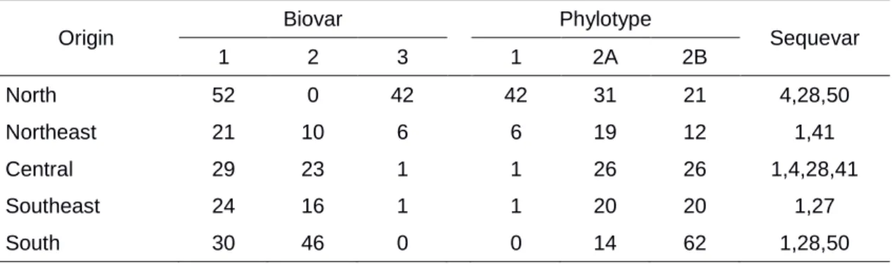
Biovar classification revealed bias in the geographic and host distribution of R. solanacearum. More than half of the isolates (52%) were assigned to biovar 1, while 32% and 16% of the isolates corresponded to
13
eggplant. Isolates of biovar 3 also occurred in high frequency in the northern parts of the country, and few isolates in the Central and Southeast regions. No biovar 3 isolates were found in the South region. Biovar 3 was isolated from tomato, eggplant, bell pepper and capsicum. Most isolates (38) were found associated with tomato.
In the South, 39% and 61% of the isolates were of biovar 1 and biovar 2, respectively. In the Southeast region, 59%, 40% and 1% of the isolates were of biovars 1, 2 and 3, respectively. In Central Brazil: 55% were biovar 1, 43%
biovar 2 and 2% biovar 3; in the Northeast: 57% biovar 1, 27% biovar 2 and 16% biovar 3; and in the North: 55% biovar 1 and 45% biovar 3. Biovar 3 was not found in the South region, but was highly correlated (100%) with phylotype I (see below). No isolates were assigned to biovars 4 or 5 (Table 2).
3.3. Phylotype classification and distribution
Phylotype-specific multiplex PCR analyses amplified an 114 bp fragment, typical of phylotype I, in 48 isolates and a 372 bp fragment, typical of phylotype II, in 253 isolates. Isolates belonging to phylotype I were found in different locations in at least three different states of the North and Northeast regions, as well as in the Distrito Federal, in the Central region and in Rio de Janeiro state, in the Southeast region. Phylotype I strains were obtained from tomato, eggplant, capsicum and bell pepper (Table 2). Phylotype II is widely distributed in the country and isolates were associated with tomato, potato, eggplant, bell pepper, eucalyptus, capsicum, geranium, banana and scarlet eggplant. In the South and Southeast regions, all isolates were classified as phylotype II. Isolates in the North, Northeast and Central regions belong to phylotypes I and II. Phylotype III and IV were not found.
3.4. Sequevar diversity
Partial sequencing of the endoglucanase (egl) gene showed restricted sequevar diversity. PCR amplification of a 750 bp region of the egl gene, which
14
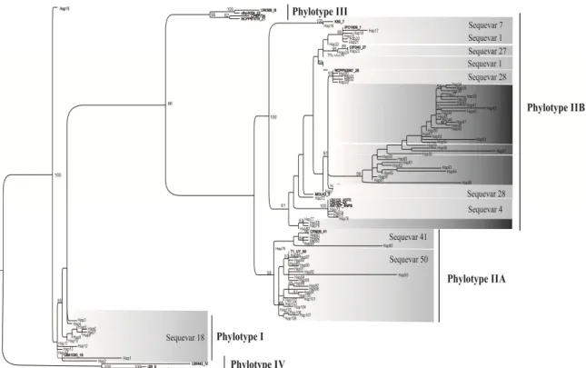
phylogenetic position, which was entirely consistent with their phylotype determination on the basis of multiplex PCR. Phylogenetic analysis differentiated Brazilian isolates into phylotypes I, IIA and IIB. The first reconstructed tree included 301 Brazilian sequences and 52 reference sequences of different sequevars of isolates collected in different countries. In total, 108 variants were found. The variants were recorded as varying from 1 to 108.
Phylotype I was assigned to 48 Brazilian isolates and included 15
variants. All phylotype I isolates (48) were sequevar 18. Phylotype II isolates were divided into two subclusters: IIA and IIB, with 28 and 65 variants, respectively. The phylotype IIA subcluster was composed of isolates that infect different hosts, mainly tomato and eucalyptus, and encompasses sequevars 41 (16 isolates) and 50 (95 isolates). Subcluster IIB was comprised of 142 isolates and sequevars 1, 4, 27 and 28. Subcluster IIB was predominantly made of isolates from the South region and was more commonly associated with potato, eggplant, geranium, banana and bell pepper. The phylotype IIB isolates were split into two groups. One group was made of isolates from bell pepper, tomato, potato, eggplant and eucalyptus; while the other group consisted solely ofisolates frompotato of the South, Southeast and Central regions.
Sequevars 4, 28 and 41 were detected in isolates from different hosts and distinct regions. Sequevar 41 is reported in Brazil for the first time. Sequevar 50 was found in tomato isolates from all regions and sequevar 1 was found in the South, Southeast, Central and Northeast regions in potato isolates. Isolates of banana were sequevar 4. One isolate collected from eucalyptus was characterized as sequevar 27. Phylogenetic analysis showed that four new sequevars were found in phylotype IIB.
3.5. BOX-PCR cluster analysis
The BOX-PCR fingerprints of the 301 isolates produced 35 bands that
15
the same haplotype were sampled from different regions. The most frequent genotype was observed three times in each state (data not shown).
Similar high levels of genotypic diversity were detected when analyses were partitioned by region or host (Table 3). The highest genotypic diversity was detected in the North region, followed by the South, Central, Southeast and Northeast. Genotypic diversity was highest in tomato and potato isolates, at least 5 times higher than in isolates from eggplant, eucalyptus and bell pepper. Since there was a limited number of isolates from capsicum, geranium, banana
and scarlet eggplant, we did not estimate the genotypic diversity among isolates from these hosts. The genotypic richness in the Southeast and South regions was higher than in other regions. The tomato subpopulation had the highest genotypic richness while the bell pepper had the lowest. The highest gene diversity values were estimated for the North (HE = 0.39, 92.9% polymorphic loci) and Northeast (HE = 0.37, 91.4% polymorphic loci) regions and in the long pepper (HE= 0.43, 100% polymorphic loci), eggplant (HE= 0.42, 100% polymorphic loci) and tomato (HE= 0.39, 94.6% polymorphic loci) subpopulations. The potato subpopulation from the South region had the lowest gene diversity. Relatively low values of HE were estimated for phylotype I isolates (HE = 0.21, 55.7% polymorphic loci) when compared with isolates from other phylotypes (data not shown).
In most tests the random mating hypothesis could not be rejected, thus there is evidence of recombination in the population of R. solanacearum. In all populations, except the eucalyptus population (IA= 0.28), the null hypothesis of
recombination could not be rejected.
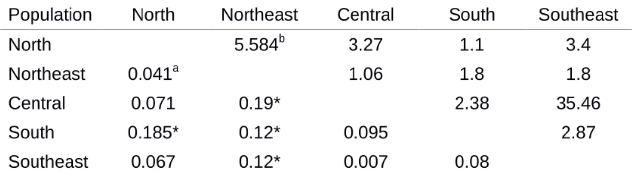
The population was moderately structured according to geographic region and host. The FST value between North and South populations was 0.19
and those between the Northeast populations and all other regions varied from 0.12 to 0.19. Apparently, the Northeast population differs from other populations and the populations from the Central, Southeast and South regions were more similar among each other since FST values varied from 0.007 to 0.095. The
16
population structuring according to host, especially between tomato and potato subpopulations. Molecular variance was mainly partitioned according to among (55.6%), followed by a similar variation level within subpopulations (44.4%) (Table 5).
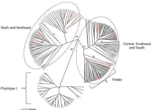
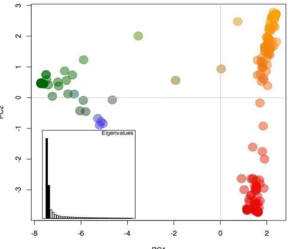
Genotypes were sorted into three major and well-supported groups (Fig. 3). The first group included isolates of the Central, Southeast and South regions mainly those of phylotype IIA. The second group was comprised mainly by isolates from the North and Northeast regions and of phylotype IIA. The third
group was comprised of phylotype I isolates. Haplotypes of phylotype IIB, mainly from potato in the South, Southeast and Central regions, were assigned to a separate cluster. Few isolates from eggplant and tomato were also clustered, though with little bootstrap support, into the latter group.
Gene flow seems to take place between the Southeast and Central regions, mainly among Minas Gerais, São Paulo, Goiás and Distrito Federal (Fig. 3). Evidence for the occurrence of migration was also detected between all regions of the country (Fig. 3). The UPGMA tree revealed distinct groups such as those formed by isolates of phylotype I and II, and phylotype II isolates from the South, Southeast and Central regions from populations of the Northeast and North regions.
4. Discussion
Brazil, a putative ancient center of diversity of R. solanacearum, has an extensive territory, approximately 8.5 million squared-kilometers, in which different ecosystems and a highly diverse community of plants can be found. This allows for the occurrence of different genoytpes of plant-associated organisms, including bacteria. Plants infected with R. solanacearum were detected in latitudes in Brazil as extreme as 00o02’20” N to 29o17’29S. Consequently, a diverse population of R. solanacearum was expected to be found in Brazil. High gene and genotypic diversity were estimated and two phylotypes of R. solanacearum, I and II, were detected. The population is
17
Mutation, recombination and gene flow seem to be the major evolutionary mechanisms that contribute to shape the R. solanacearum population.
4.1. Origin and natural history of the pathogen in South America
Worldwide, the different phylotypes of R. solanacearum are relatively distributed according to geographic region: phylotype I isolates most likely originated in Asia, phylotype II in America, phylotype III in Africa and phylotype IV in Indonesia, nevertheless more than one phylotype is commonly reported in
a country due to effective dispersion processes (Horita et al., 2010; Sanchez-Perez et al., 2008; Xue et al., 2011; Ramsubhag et al., 2012). Most Brazilian isolates of R. solanacearum (84%) belong to phylotype II, although phylotype I isolates were also detected. The reasons for the absence of isolates of phylotypes III and IV in Brazil cannot be completely ascertained at the moment. Probably, phylotype III isolates have never been introduced in Brazil due to no transmission by true seeds. On the other hand, the insect-transmitted nature of phylotype IV isolates can impair its distribution due to the lack of vector/host in other countries (Remenant et al., 2011).
The occurrence of phylotype I isolates, which are originated in Asia, in different Brazilian regions can be attributed to human-mediated processes. We speculate that one possible pathway was through contaminated seeds or seedlings of black pepper brought to Brazil by Japanese immigrants in the early 20th Century. Black pepper is native to India and the crop was introduced by Japanese immigrants. It has been documented that 20 seedlings were brought from Singapore in 1933, but only two plants survived the trip (Yamauchi, 1992). The plants promptly adapted to warm regions in Brazil and within a 20 year-period the cultivated area quickly expanded (Yamauchi, 1992). Between 1940-1950, Japanese immigrants established new colonies in the Amazon region (Northern Brazil), Pernambuco state (Northeast), Minas Gerais, São Paulo and Rio de Janeiro states (Southeast), and in Brasília and Mato Grosso do Sul state
18
(Rodrigues et al., 2012; Silveira et al., 1998; Coelho Netto et al., 2004; Garcia et al., 2013).
Phylotype I isolates were identified as belonging to biovar 3 and phylogenetically associated with sequevar 18, which is also found in Guatemala (Sanchez Perez et al., 2008), French Guiana (Milling et al., 2009) and Trinidad (Ramsubhag et al., 2012), countries located from the equator up to 15o N. In Brazil, phylotype I isolates predominantly occurred in regions around the equator down to 15o S. Isolates of biovar 3 were more frequently associated with Capsicum species, as compared to tomatoes, as previously reported
(Coelho Neto et al., 2004). These are the two most important solanaceous hosts affected by this variant in the northern and northeast Brazil. There are also interesting indication that breeding for resistance to biovar 3 is easier for tomatoes and more difficult to Capsicum (Lopes et al., 1993; Lopes & Boiteux, 2004). We speculate that biovar 3/ phylotype I isolates are well adapted to regions with higher temperatures.
The predominance of the American-originated phylotype II isolates in Brazil was expected (Wicker et al., 2012). However, we detected variation in the geographic distribution and host range of phylotype II isolates in the country. Phylotype IIA isolates were present in all regions and were obtained from different hosts, but most came from infected tomato plants cultivated in lowland areas. Isolates of phylotype IIB were pathogenic to potato and tomato, but most came from infected potato plants cultivated in highland areas (altitude ranging from 800m to 1600m). Additionally, phylotype and biovar were associated, as previously recorded (Hayward, 1991; Fegan & Prior, 2005).
Brazilian phylotype II isolates were phylogenetically diverse and included the known sequevars 1, 4, 27, 28, 41 and 51. In addition, four other sequences that did not cluster with any previously known sequevars were also found in phylotype IIB. There was predominant correlation with potato in the South, Southeast and Central regions. Curiously, sequevar 4 causes banana
19
sequevars 1, 4, 28 and 41 were found in several regions. Isolates collected in eucalyptus and potato were classified as sequevar 41, as previously described (Fonseca et al., 2013). The present study is the first formal report of sequevar 50 in Brazil. Using the proposed criterion of Fegan & Prior (2005) and Poussier et al. (2006) we classified four new sequevars in the phylotype IIB: sequevars 53, 54, 55 and 56.
4.2. Processes underlying the structure of the Brazilian population of R. solanacearum
Using BOX-PCR it was possible to detect three groups of R.
solanacearum in Brazil: one formed by phylotype I isolates, a second group
formed by isolates predominantly from the South, Southeast and Central regions, and a third group formed by isolates from the North and Northeast regions. We hypothesize that the spatial distribution of the population of R.
solanacearum is largely affected by ecological differences between regions.
The subpopulation of the Northeast region can be clearly differentiated from those of all other geographical regions, but the North. The difference between the Northeast and the non-North subpopulations can be explained by environmental properties such as temperature, humidity, altitude and absence of some host species (Tang, 2009).Further investigation on the influence of climate on R. solanacearum populationdiversity is required.
There is evidence of spatial population subdivision of Brazilian phylotype IIA between the North and South regions. Absence of regional structure in the Central, Southeast and South regions and North and Northeast regions suggested no isolation by distance and/or occurrence of gene flow between these large spatial groups. Previous studies used repetitive DNA elements to define groups for R. solanacearum.The population from Trinidad was found to be highly polymorphic and showed high gene diversity using BOX-PCR (Cruz et al., 2012; Ramsubhag et al., 2012). Contrary to what we found,
20
(Ramsubhag et al., 2012). The decrease in the community similarity with geographic distance is a universal biogeographic pattern observed for all domains of life, mainly organisms with limited dispersal (Martiny et al., 2011). When analyzing the genetic structure at a smaller geographic scale such as the South, Southeast and Central regions and between North and Northeast regions we detected weak population structure. Using BOX-PCR analysis, regional differentiation was also detected when comparing populations of R.
solanacearum in the Philippines (Ivey et al., 2007). In the Amazon region, no
correlation with host and geographic origin was found (Coelho Neto et al., 2004). Additional studies using rep-PCR to characterize the Brazilian isolates may provide higher resolution data that allow the assessment of the relationship of genotypes to location and aggressiveness. This knowledge can be useful for breeding for resistance and better inspection of propagative materials.
Transportation of infected propagative materials as well as natural dispersal by rivers may have contributed to distribute variants of the pathogen across regions and influenced the genetic structure of the population. It was previously suggested that the occurrence of biovar 2 in potato fields in the Northeast and Central regions of Brazil may be associated with transport of latently infected tubers from colder southern regions to warmer northern regions (Santana et al., 2012). We detected biovar 2 potato isolates in the Central and Northeast regions of Brazil and haplotype 72 found in the South was detected in the Northeast regions (in Paraná and Bahia states, respectively). Similarly, haplotype 111 was found in the South and in Central regions (in Paraná and Goiás states, respectively). The lower genetic diversity among potato isolates than among tomato isolates also provides some support to the role played by transportation of infected or infested seed-tubers. The wide distribution can also be associated with fruit dissemination, but despite the fact that R. solanacearum can be spread by infected tomato fruits, this does not seem to occur very often (Sanchez Perez et al., 2008). In Europe and in the United States, R.
solanacearum was detected in river water and the origins of introduction in
21
solanacearum over short and long distances (Elphinstone et al.,1998). Biovar
characterization also helped to understand the route of migration of R.
solanacearum into Brazil.
The Brazilian population of R. solanacearum has higher genetic diversity than other organisms thought to reproduce predominantly asexually and with limited dissemination. In all regions and host subpopulations estimates of gene and genotypic diversity were high. High gene diversity provides indirect evidence for the occurrence of recombination (Maynard Smith, 1971).
Furthermore, when isolates from all geographic regions were tested for linkage disequilibrium, there was strong evidence of occurrence of random mating and consequently that recombination is involved in bacterial diversity. A number of studies revealed the importance of recombination in R. solanacearum (Guidot et
al., 2009; Coupat-Goutaland et al., 2011; Ramsubhag et al., 2012; Wicker et al., 2012). In a previous study, the multilocus sequence characterization of a world collection of R. solanacearum suggested that recombination is an active evolutionary mechanism that shapes population diversity (Wicker et al., 2012). Identification of recombination events provides insights into epidemiology and disease management and from a biological point of view has consequences for microbial diversification and adaptive evolution. For instance, recombination could facilitate adaptation through the acquisition of new metabolic capabilities, promoting speciation or diversification (Jain et al., 2003). A more thorough investigation using robust methods to identify recombination needs to be conducted with the Brazilian isolates of R. solanacearum.
Genetic divergence of potato and tomato occurs between populations that are geographically in close proximity. This process can lead to a pathogen adaptation to host plants (Stukenbrock & McDonald, 2008). The populations of
R. solanacearum associated with potato and tomato in the Central, Southeast
and South regions of Brazil are genetically different, but the highest fraction of molecular variation was attributed to be due to differences within host. Genetic
variation is key to evolution. Selection and gene acquisition can be involved with host preference or differential aggressiveness. Host specialization changes may be caused by positively selected genetic variation, though no study on selection has been conducted to assess the influence of the host in the populations of R.
22
influenced by horizontal gene transfer (de la Cruz & Davies, 2000). A single gene can be responsible by the change in host range with type III protein secretion gene product (avrA and popP1) (Poueymiro et al., 2009). The influence of the host in the rhizosphere microbiome and its role in shaping pathogen populations is discussed by Berendsen et al. (2012). A complex study is necessary and other pathogenesis-related genes need to be studied to properly address speciation issues in the population of R. solanacearum in Brazil.
The striking genetic diversity of the Brazilian population of R. solanacearum raises important issues related to both the applied and basic
scenarios. From the applied perspective, the high genetic variability of the population is one of the reasons for the limited success of several management strategies aimed at controlling bacterial wilt in several hosts. It can be anticipated that this constitutes a major challenge to the control of bacterial wilt, particularly for the strategies associated with biological control and the deployment of resistant varieties. Additionally, the high variability associated with the positive selection exerted by the host can contribute to host adaptation and emergence of new ecotypes. The findings of the present study prompt the need for greater caution during the commercialization and use of propagation materials and the quality of irrigation water in clean areas. From the evolutionary biology perspective, the high genetic variability supports its categorization as an ancient population and the claim of South America as a center of origin of the pathogen: The high frequency of genotypes, high genotypic diversity, high gene polymorphism, the occurrence of bacterial wilt in native host plants, and the occurrence of the pathogen in virgin areas, including the Cerrado and the Amazon, suggest that R. solanacearum is native to these areas (Takatsu & Lopes, 1997). A more thorough investigation is being conducted to assess the likelihood of South America, Brazil in particular, be one of the centers of origin of R. solanacearum.
5. Conclusions
Our study represents the first analysis of the genetic structure of the highly diverse Brazilian R. solanacearum populationsthat is capable of
23
estimated genetic diversity by geographic distance, but were restricted to a limited number of isolates (Rodrigues et al., 2012; Cruz et al., 2012). Moreover, the fingerprinting methods that were used have been described as being of low resolution and incapable of reconstructing significant patterns of descent. In contrast, we used methods based on the scanning of entire genomes that are more powerful and appropriate for the comparative analysis of population diversity (Rademaker & De Bruijn, 1997), especially of microbial populations that are clonal. The spatial separation and high variability we detected in the
populations we examined provided important information that could explain why a plant variety is only resistant in a given geographic region and susceptible to other bacterial populations. Our results explain why breeding programs for durable resistance appear more efficient at regional level and provide a basis to study environmental and historical constraints on the diversity and evolution of the pathogen. There is however much to learn about microbial diversification and pathogenesis. In future studies, we will use multi-locus sequence typing to study the influence of recombination, explore isolate variability in crop fields, the influence of the environment, and reconstruction of patterns of descent in genealogical analyses of Brazilian isolates of phylotype I and II. In addition, we will identify genomic regions and biological tests that can explain the wide range of plant hosts used by the pathogen.
6. Acknowledgments
This research was supported by CNPq grant 476940/2011-7. We gratefully acknowledge Maurício Rossato (Universidade de Brasília) and Saulo Oliveira (EMBRAPA Mandioca e Fruticultura) for technical support. We thank FAPEMIG for providing the scholarship to TRS and Caitilyn Allen, for providing the phylotype standards.
7. References
Agapow PM, Burt A, 2001. Indices of multilocus linkage disequilibrium.Molecular Ecology Notes1, 101-2.
24
Bertolla F, Gijsegem FV, Nesme X, Simonet P, 1997. Conditions for natural transformation of Ralstonia solanacearum.Applied Environmental Microbiology63, 4965-68.
Buddenhagem I, Sequeira L, Kelman A, 1962. Designation of races in
Pseudomonas solanacearum.Phytopathology52, 726.
Coelho Netto RA, Pereira BG, Noda H, Boher B, 2004. Murcha bacteriana no estado do Amazonas, Brasil. Fitopatologia Brasileira 29, 21-7.
Costa SB, Ferreira MASV, Lopes CA, 2007. Diversidade patogênica e molecular de Ralstonia solanacearum da região amazônica brasileira.
Fitopatologia Brasileira32, 285-94.
Coupat-Goutaland B, Bernillon D, Guidot A, Prior P, Nesme X, Bertolla F, 2011.
Ralstonia solanacearum virulence increased following large interstrain gene
transfers by natural transformation. Molecular Plant-Microbe Interactions24, 497-505.
Cruz L, Eloy M, Quirino F, Oliveira H, Tenreiro R, 2012. Molecular epidemiology of Ralstonia solanacearum strains from plants and environmental source in Portugal. European Journal Plant Pathology133, 687-706.
de la Cruz F, Davies J, 2000. Horizontal gene transfer and the origin of species: lessons from bacteria. Trends in Microbiology 8, 128–33.
Denny TP, 2006.Plant pathogenic Ralstonia specie. In: Gnanamanickam SS, ed. Plant-associated Bacteria. Dordrecht, The Netherlands: Springer Publishing, 573-644.
Elphinstone JG, Stanford HM, Stead DE, 1998.Detection of Ralstonia
solanacearum in potato tubers, Solanum dulcamara and associated irrigation
water. In: Prior P, Allen C, Elphinstone JG, eds. Bacterial Wilt Disease:
Molecular and Ecological Aspects. Springer-Verlag, Berlin, 133-9.
Elphinstone JG, 2005. The current bacterial wilt situation: a global overview. In: C. Allen C, Prior P, Hayward AC, eds. Bacterial Wilt Disease and the Ralstonia
solanacearum Specie Complex. St. Paul, MN: American Phytopathological
Society Press, 9-28.
Excoffier L, Laval G, Schneider S, 2005. Arlequin (version 3.0): an integrated software package for population genetics data analysis. Evolutionary
Bioinformatics Online1, 47–50.
Fegan M, Prior P, 2005. How complex is the “Ralstonia solanacearum species complex?” In: C. Allen C, Prior P, Hayward AC, eds. Bacterial Wilt Disease and
the Ralstonia solanacearum Specie Complex. St. Paul, MN: American
Phytopathological Society Press, 449-61.
25
Francis CY, Yang RC, 2000. Popgene version 1.32. [http//www. http://www.ualberta.ca/~fyeh/popgene.html]. Acessed 17 September 2014.
Garcia AL, Lima WG, Souza EB, Michereff SJ, Mariano RLR, 2013. Characterization of Ralstonia solanacearum causing bacterial wilt in bell pepper in the state of Pernambuco, Brazil. Journal of Plant Pathology95, 237-45.
Guidot A, Prior P, Schoenfeld J, Carrère S, Genin S, Boucher C, 2007. Genomic structure and phylogeny of the plant pathogen Ralstonia
solanacearum inferred from gene distribution analysis. Journal of
Bacteriology189, 377-87.
Guidot A, Elbaz M, Carrère S, Siri MI, Pianzzola MJ, Prior P, Boucher C, 2009. Specific genes from the potato brown rot strain of Ralstonia solanacearum and their potential use for strain detection. Phytopathology99, 1105-12.
Hayward AC, 1964.Characteristics of Pseudomonas solanacearum.Journal
Applied Microbiology27, 265-77.
Hayward AC, 1991.Biology and epidemiology of bacterial wilt caused by
Pseudomonas solanacearum.Annual Reviews Phytopathology.29, 65-87.
Hayward AC, 1994.The hosts of Pseudomonas solanacearum. In: Hayward AC, Hartman GL, eds. Bacterial Wilt: The disease and its causative agent,
Pseudomonas solanacearum. Wallingford, UK: CAB International, 9-24.
He LY, Sequeira L, Kelman A, 1983.Characteristic of strains of Pseudomonas
solanacearum from China. Plant Disease67,1357-61.
Hong JC, Momol, M.T., Jones, J. B., Ji, P., Olson, S. M., Allen, C., Perez, A., Pradhanag, P. and Guven, K. 2008. Detection of Ralstonia solanacearum in irrigation ponds and aquatic weeds associated with ponds in North Florida.
Plant Disease 92,1674-82.
Peeters N, Guidot A, Vailleau F, Valls M, 2013. Ralstonia solanacearum, a widespread bacterial plant pathogen in the post-genomic era.Molecular Plant
Pathology 14, 651-62.
Horita M, Suga Y, Ooshiro A, Tsurchiya, K, 2010. Analysis of genetic and biological characters of Japanese potato strains of Ralstonia solanacearum.
Journal of General Plant Pathology 76, 196-207.
Jain R, Rivera MC, Moore JE, Lake JA, 2003. Horizontal gene transfer accelerates genome innovation and evolution. Molecular Biology Evolution20, 1598-1602.
Jukes TH, Cantor CR, 1969. Evolution of protein molecules. In: Munro HN, ed.
26
Kelman A, 1954. The relationship of pathogenicity of Pseudomonas
solanacearum to colony appearance on a tetrazolium
medium.Phytopathology44, 693-5.
Lambert CD, 2002. Agricultural bioterrorism protection act of 2002: Possession, use, and transfer of biological agents and toxins; interim and final rule (7 CFR Part 331). Fed Regist67, 76908-38.
Lopes CA, Boiteux LS, 2004. Biovar-specific and broad-spectrum sources of resistance to bacterial wilt (Ralstonia solanacearum) in capsicum.Crop Breeding
and Applied Biotechnology 4, 350-5.
López MM, Biosca EG, 2005. Potato bacterial wilt management new prospects for an old problem. In: C. Allen C, Prior P, Hayward AC, eds. Bacterial Wilt
Disease and the Ralstonia solanacearum Specie Complex. St. Paul, MN:
American Phytopathological Society Press, 205-24.
Louws FJ, Fulbright DW, Stephens CT, de Bruijn FJ, 1994. Specific genomic fingerprints of phytopathogenic Xanthomonas and Pseudomonas pathovars and strains generated with repetitive sequences and PCR. Applied and
Environmental Microbiology 60, 2286-95.
Maynard Smith J, 1971. What use is sex? Journal of Theoretical Biology30,319-35.
Milling A, Meng F, Denny TP, Allen C, 2009. Interactions with hosts at cool temperatures, not cold tolerance, explain the unique epidemiology of Ralstonia
solanacearum race 3 biovar 2. Phytopathology99, 1127-34.
Nei M, 1978. Estimation of average heterozygosity and genetic distance from a small number of individuals.Genetics89, 583-90.
Nylander JAA, 2004. MrModeltest v2. Program distributed by the author. Evolutionary Biology Centre, Uppsala University.
Opina N, Tavner F, Holloway G, Wang J-F, Li T-H, Maghirang R, Fegan M, Hayward AC, Krishnapillai V, Hong WF, Holloway BW, Timmis JN, 1997. A novel method for development of species and strain-specific DNA probes and PCR primers for identifying Burkholderia solanacearum (formerly Pseudomonas
solanacearum).Asia Pacific Journal of Molecular Biology and Biotechnology5,
19–33.
Pinheiro CR, Amorim JAE, Diniz LEC, Silva AMF, Talamini V, Souza Júnior MT, 2011. Diversidade genética de isolados de Ralstonia solanacearume caracterização molecular quanto a filotipos e sequevares. Pesquisa
Agropecuaria Brasileira46, 593-602.
Poussier S, Prior P, Luisetti J, Hayward C, Fegan M, 2000. Partial sequencing of the hrpB and endoglucanase genes confirms and expands the known diversity within the Ralstonia solanacearum species complex. Systematic and
27
Rademaker JLW, de Brujin FJ, 1997. Characterization and classification of microbes by rep-PCR genomic fingerprinting and computer assisted pattern analysis. In: Caetano-Anolles G, Gresshoff, eds. DNA Markers: Protocols,
Applications and Overviews. New York, NY: John Wiley and Sons Inc, 151-71.
Ramsubhag A, Lawrence D, Cassie D, Fraser R, Umaharan P, Prior P, Wicker E, 2012. Wide genetic diversity of Ralstonia solanacearum strains affecting tomato in Trinidad, West Indies. Plant Pathology61, 844-57.
Rodrigues LMR, Destéfano SAL, Silva MJ, Costa GGL, Maringoni AC. 2012. Characterization of Ralstonia solanacearum from Brazil using molecular methods and pathogenity tests.Journal of Plant Pathology94, 505-16.
Ronquist F, Huelsenbeck JP, 2003. MrBayes 3: Bayesian phylogenetic inference under mixed models. Bioinformatics19, 1572-74.
Sanchez Perez A, Meija L, Fegan M, Allen C, 2008. Diversity and distribution of
Ralstonia solanacearum strains in Guatemala and rare occurrence of tomato
fruit infection. Plant Pathology57, 320-31.
Santana BG, Lopes CA, Alvarez E, Barreto CC, Allen C, Quirino BF, 2012. Diversity of Brazilian biovar 2 strains of Ralstonia solanacearum. Journal of General Plant Pathology 78, 190-200.
Silveira, EB, Gomes AMA, Michereff SJ, Mariano RLR, 1998. Variability of
Ralstonia solanacearum populations causing wilt on tomato in agreste of
Pernambuco, Brazil.Bacterial Wilt Newsletter15, 8-10.
Smith JJ, Offord LC, Holderness M, Saddler GS, 1995. Genetic diversity of
Burkholderia solanacearum (synonym Pseudomonas solanacearum) race 3 in
Kenya. Applied and Environmental Microbiology61, 4263-8.
Stamatakis A, 2006. RAxML-VI-HPC: maximum likelihood-based phylogenetic analyses with thousands of taxa and mixed models. Bioinformatics22, 2688-90.
Stoddart JA, Taylor JF, 1988. Genotypic diversity: estimation and prediction in samples. Genetics118, 705-11.
Strange RN, Scott PR, 2005. Plant Disease: A Threat to Global Food Security.
Annual Review of Phytopathology43, 83-116.
Stukenbrock EH, McDonald BA, 2008.The origins of plant pathogens in agro-ecosystems.Annual Review Phytopathology46, 75-100.
Swofford DL, 2002. Paup*: Phylogenetic analysis using parsimony (*and other methods). Sinauer Associates. Sunderland, MA
Tamura K, Dudley J, Nei M, Kumar S, 2007. MEGA4: Molecular Evolutionary Genetics Analysis (MEGA) software version 4.0. Molecular Biology and
28
Yabuuchi E, Kosako Y, Yano I, Hotta H, Nishiuchi Y, 1995. Transfer of two
Burkholderia and an Alcaligenes species to Ralstonia gen. nov.: Proposal of Ralstonia picketii (Ralston, Palleroni and Doudoroff 1973) comb. nov. Ralstonia solanacearum (Smith 1896) comb.nov. andRalstonia eutropha (Davis 1969)
comb. nov. Microbiology and Immunology 39, 897-904.
Yamauchi A, 1992. Uma epopéia moderna: 80 anos da imigração japonesa no Brasil. São Paulo, SP: Hucitec.
Yao J, Allen C, 2006. Chemotaxis is required for virulence and competitive fitness in the bacterial wilt pathogen Ralstonia solanacearum. Journal of
Bacteriology 188, 3697-708.
Yeh FC, Boyle TJB, 1997. Population genetic analysis of co-dominant and dominant markers and quantitative traits.Belgian Journal of Botany 129, 157.
Wicker E, Grassart L, Coranson-Beaudu R, Mian D, Guilbaud C, Fegan M, Prior, 2007. Ralstonia solanacearum strain from Martinique (French West Indies) exhibiting a new pathogenic potential. Applied and Environmental
Microbiology 73, 6790-801.
Wicker E, Lefeuvre P, de Cambiaire JC, Lemaire C, Poussier S, Prior P, 2012. Contrasting recombination patterns and demographic histories of the plant pathogen Ralstonia solanacearum inferred from MLSA. The ISME Journal6, 961-74.
Wilson DJ, Falush D, McVean G, 2005.Germs, genomes and genealogies.Trends in Ecology and Evolution 20, 39-45.
Xue QY, Yin YN, Yang W, Heuer H, Prior P, Guo JH, Smalla K, 2011. Genetic diversity of Ralstonia solanacearum strains from China assessed by PCR-based fingerprints to unravel host plant- and site-dependent distribution patterns. FEMS Microbiology Ecology75, 507-19.
Figure 1.Map of B sam
29
FIGURES AND TABLES
Brazil showing the origin isolates and biova ampled in different regions of the country.
30
Bootstrap values >50% derived of 1000 replicates are reported at the nodes. The scale bar shows genetic distance; the bar represents 11 nucleotide differences.
31
Bootstrap support values above 50% are shown. Migrations between the groups are indicated with red branches.