PRISCILA VENDRAMINI SILVA
GENE EXPRESSION IN SOWS WITH HIGH AND LOW
OVULATION RATE, HIGH AND LOW BIRTH WEIGHT
LITTER AND THE EFFECT OF SOW CATABOLIC STATUS ON
EMBRYONIC GENE EXPRESSION
Thesis presented to the Genetics and Breeding Graduate Program of the Universidade Federal de Viçosa, in partial fulfillment of the requirements for degree of Doctor Scientiae.
VIÇOSA
Ficha catalográfica preparada pela Seção de Catalogação e Classificação da Biblioteca Central da UFV
T
Silva, Priscila Vendramini, 1982-
S586g Gene expression in sows with high and low ovulation rate, 2012 high and low birth weight litter and the effect of sow catabolic
status on embryonic gene expression / Priscila Vendramini Silva. – Viçosa, MG, 2012.
x, 117f. : il. ; (algumas color.) ; 29cm.
Inclui apêndices.
Orientador: Simone Eliza Facioni Guimarães
Tese (doutorado) - Universidade Federal de Viçosa. Inclui bibliografia.
1. Suíno - Melhoramento genético. 2. Regulação de
expressão gênica. 3. Suíno - Reprodução. 4. Reação em cadeia de polimerase. 5. Ovulação. 6. Suíno - Embrião.
I. Universidade Federal de Viçosa. Departamento de Zootecnia. Programa de Pós-Graduação em Genética e Melhoramento. II. Título.
PRISCILA VENDRAMINI SILVA
GENE EXPRESSION IN SOWS WITH HIGH AND LOW
OVULATION RATE, HIGH AND LOW BIRTH WEIGHT
LITTER AND THE EFFECT OF SOW CATABOLIC STATUS ON
EMBRYONIC GENE EXPRESSION
Thesis presented to the Genetics and Breeding Graduate Program of the Universidade Federal de Viçosa, in partial fulfillment of the requirements for degree of Doctor Scientiae.
APROVADA: September 11, 2012.
___________________________ ___________________________ Prof. Fabyano Fonseca e Silva Prof. Paulo Sávio Lopes (Co-adviser) (Co-adviser)
____________________________ ___________________________ Prof. José Domingos Guimarães Dr. George R. Foxcroft
ACKNOWLEDGEMENTS
I would like to thank Universidade Federal de Viçosa and the Genetics and Breeding Program, for providing me the opportunity to conclude my degree and post-graduate education. I am also grateful to CNPq for the financial support for those years and opportunity to stay one year abroad working on complementary research projects at the AGPU Lab in the Agriculture/Forestry Centre at the University of Alberta.
I would like to thank my advisor, Prof. Simone Guimarães for her guidance and support throughout my academic life, her confidence on my work and her encouragement that has been shaped my scientific endeavors.
I am also very grateful to Dr. George Foxcroft for his invaluable support and guidance that were essential for enriching my knowledge and improving the quality of my PhD studies. I would also like to thank my co-advisors Professor Paulo Sávio and Professor Fabyano for their encouragement and advices, and to Professor JD for his help and suggestions in the experiments.
Many thanks to Department of Agricultural, Food and Nutritional Science, the Natural Sciences and Engineering Research Council (NSERC) and EmbryoGENE NSERC Strategic Research Network and members of their research group that kindly receive me and letting me take part of this great project. Particularly, I would like to thank Gina Oliver for the orientation in the lab activities and her valuable suggestions and discussions, Jennifer Patterson for coordinating the animal work, Joan Turchinsky for her technical support and Stephen Tsoi for his help and suggestions during microarray analysis. Also, to Jamie Wilkinson, Tracy Gartner, François Paradis and specially, Walter Dixon and Michael Dyck, thank you for your help and support.
Em uma gaiola dourada um pintassilgo vivia Cantava numa toada, tão cheia de nostalgia Que parecia seu canto, um ai dorido e queixoso
Era um suspiro, era um pranto Pelo seu ninho saudoso. Talvez buscasse o coitado,
Neste cantar esquecer a vida livre do prado Desde a alvorada nascer.
Uma bondosa menina, cheia de amor e piedade, À ave gentil, pequenina
Depressa deu liberdade. Com a alma então à vibrar, Numa expressão de alegria,
De quem vai ver o seu lar que a longo tempo não via, O passarinho galante, batendo as asas voou, E num gorjeio triunfante, no espaço, além recuou!
(Inah Glanzmann Vendramini, 92 anos)
À minha querida avó Inah
Biography
Priscila Vendramini Silva, daughter of Décio Evandro da Silva and Livia Vendramini, born on april 5th, 1982 in São Paulo, SP, Brazil. She began her studies at the Universidade Federal de Viçosa in 2002 obtaining her degree in Biochemistry in 2006.
In same year of 2006 she started her Master on in Genetics and Breeding Program at the same Institution under supervision of Prof Simone Guimarães. She finished her master in 2008, entitled Gene expression in pig ovary during the estrous cycle.
Summary
ABSTRACT...VII
RESUMO ...IX
GENERAL INTRODUCTION... 1
References ...5
CHAPTER1... 11
Abstract ... 11
Resumo ... 12
Introduction... 13
Materials and Methods ... 14
Results ... 18
Discussion ... 20
Conclusions ... 25
Acknowledgements ... 26
References ... 33
CHAPTER2... 41
Abstract ... 41
Resumo ... 42
Introduction... 43
Materials and methods... 44
Results and Discussion... 47
Conclusions ... 51
Acknowledgements ... 52
References ... 58
CHAPTER3... 64
Abstract ... 64
Resumo ... 65
Introduction... 66
Materials and methods... 67
Results ... 72
Discussion ... 76
Conclusions ... 84
Acknowledgements ... 84
References ... 103
General Conclusions... 114
References ... 116
ABSTRACT
SILVA, Priscila Vendramini; D.S., Universidade Federal de Viçosa, September, 2012. Gene expression in sows with high and low ovulation rate, high and low birth weight litter and the effect of sow catabolic status on embryonic gene expression. Adviser: Simone Eliza Facioni Guimarães. Co-Advisers: Paulo Sávio Lopes e Fabyano Fonseca e Silva.
angiogenesis-related genes investigated were higher expressed in CL tissue in the low group. Among intrafollicular growth factors, only IGFR1 and BMPR2 were differently expressed in GC and denuded oocytes, respectively. Findings from the present study suggest that differences in CL vascularity and function, as well as in follicle development, may be in part, driving differences between-litter variation in birth weight in contemporary sows. Finally, microarray data from the third experiment revealed different pattern of distribution of biological functions across treatments for the commercial sows. Validation by QPCR showed a differential expression for CYR61 and MYOF, specifically for the female sex, in the pair-wise contrast FHvsNC and CLvsNC, respectively (P≤0.05). Additional contrasts in female sex were marginally significant (P≤0.10) for MYOF, BCSL1, CYR61, RAD21 and SOD1, but no difference was found for ETFA, ACDSB, TFPI2 and TNFRSF21. Furthermore, skip sows showed higher total corpus luteum weight, total corpus luteum average and litter growth average compared to “first heat” sows. These results suggest that the differential expression observed in female embryos may be an adaptive response to the intrauterine conditions, which may mediate epigenetic programming in the offspring. This study reinforces and provides additional insights into the role of nutrition and maternal metabolic state in determining the dynamics of early embryonic development and embryo quality in pigs.
RESUMO
SILVA, Priscila Vendramini; D.S., Universidade Federal de Viçosa, Setembro, 2012.
Expressão gênica em porcas de alta de baixa taxa de ovulação, alto e baixo peso médio de leitegada ao nascimento e efeito do estado catabólico na expressão gênica de embriões. Orientadora: Simone Eliza Facioni Guimarães. Co-orientadores: Paulo Sávio Lopes e Fabyano Fonseca e Silva.
General Introduction
The pig is one of the most economically important domesticated animals and the major meat consumed worldwide according to the United States Department of Agriculture (http://www.fas.usda.gov/dlp/circular/2012/livestock_0412.pdf). The worldwide demand for a protein source of high quality has promoted an increase in pig production in developing countries (McManus et al. 2010). For instance, Brazilian swine production is experiencing a change of position in international scenario in recent years, and emerging as one of majors exporters of pork according to Abipecs (Associação Brasileira da Indústria Produtora e Exportadora de Carne Suína, 2010).
Traditionally, pig breeding programs have focused on the genetic improvement of production traits, such as growth rate, lean meat percentage and feed efficiency. However, as these traits have reached desirable values, more attention has been given to the genetic improvement of reproductive traits (Lopes et al. 2001). Among the reproductive traits, the litter size and pre-weaning mortality of piglets are important components in reducing costs, resulting in an increase in the number of piglets weaned per sow per year and therefore, greater economic return for producers (Aherne, 1994). More recently, birth litter weight and weaning weight has been associated with improved postweaning performance and therefore included in swine genetic programs (Bergstrom et al. 2009, Schinckel et al. 2010).
Genetic regulation of litter size is complex and affected by multiple components such as ovulation rate, fertilization rate and embryonic/fetal survival (Distl, 2007). In pigs, fertilization rate is the less significant component, generally superior to 95% (Soede et al. 1992). Therefore, litter size is mainly determined by the balance between ovulation rate and embryonic and/or fetal losses. According Foxcroft et al. (1997, 2006) the pattern of prenatal losses in commercial sows maybe changing due to ongoing selection for ovulation rate, associated with increased rate of embryonic survival in the pre-implantation period. As a consequence, a large proportion of prenatal loss has been attributed to the post-implantation period, which is primarily limited by uterine capacity, defined as the number of conceptuses that can be maintained by the uterus until term (Christenson et al. 1987). Further losses may occur during later gestation due to the effects of intrauterine crowding that impair placental development (Almeida et al. 2000, Vonnahme et al. 2002; Town et al. 2004). In addition, the nutritional status of the lactating sow may also increase embryonic loss (Foxcroft et al. 1997).
formation, changes in morphology and expansion (elongation) of the extra-embryonic and primordial placental tissue (Blomberg et al. 2010). These events are finely regulated by thousands of genes related with important biological process, such as compaction/cavitation, metabolism, transcription/translation, DNA methylation and histone modification, oxidative stress, response to or production of growth factors, cytokine signalling, cell cycle regulation and apoptosis (Ruddock-D’Cruz et al. 2007). Equally important for conceptus implantation and survival is a synchrony between the embryo and the uterine environment (Geisert and Schmitt, 2001), or maternal ability to respond appropriately to embryo derived signals (Hansen, 2002).
Classical methods of selection have allowed only slow genetic progress for litter size due to its low heritability, 0.10 to 0.15 (Avalos and Smith, 1987, See et al. 1993, Spotter and Distl, 2006). Major advances in genetic improvement of reproductive characteristics were obtained through the association of molecular genetics with quantitative genetics. The identification of candidate genes underlying, or associated with, phenotypic trait through quantitative trait loci (QTLs) has allowed identification of candidate genes. Several candidate genes with an important role in reproductive physiology can be cited, such as the estrogen receptor (ESR) (Rothschild et al. 1996), binding protein 4 retinol (RBP4) (Ollivier et al. 1997; Rothschild et al., 2000), prolactin receptor (PRLR) (Vincent et al., 1998), epidermal growth factor (EGF) and follicle stimulating hormone β (fSHb) (Linville et al., 2001). However, the use of marker assisted selection is limited by the numbers of isolated and/or known genes of interest available for inclusion in genetic programmes, considering the whole genome. More recently, efforts to sequence the pig genome have resulted in the release of the first draft of the swine genome sequence with an overall depth of 4X coverage (Sscrofa 9) available in September 2009 (Archibald et al. 2010). The second draft (Sscrofa10.2) has been released in
the current year (http://www.ncbi.nlm.nih.gov.ilsprod.lib.neu.edu/Traces/wgs/?&val=AEMK&size=50&page
Overall, the present study aimed to clarify the biological question regarding the genetic control of intrafollicular mechanisms responsible for normal preovulatory follicle development in the pig. The first chapter provided a new insight into genetic diversity of the local Brazilian Piau breed and a commercial line, specifically in regard to recruitment, selection and the establishment of the preovulatory follicle population. The Piau breed was introduced by Portuguese settlers in the 16th century and is composed for Iberian breeds with influences of Dutch and African breeds (Vianna, 1985). These breeds have acquired adaptive traits in specific local environments over time, and are considered as (fat-type), yielding larger amount of fat and lower amount of carcass in comparison to commercial line (Benevenuto Júnior, 2001). Since 1998, a nucleus of conservation of this breed has been allocated at Pig Breeding Farm at Universidade Federal de Viçosa. Several studies have been conducted by our group since then, such as QTL mapping using divergent crosses (Guimarães and Lopes, 2000; Pires et al., 2008; Paixão et al., 2008; Silva et al., 2011), association studies between expression of candidate genes and intramuscular content (Serão et al., 2011), phylogeny and genetic diversity (Schierholt et al. 2008, Sollero et al. 2008, Souza et al. 2009) and transcriptional profiling during skeletal muscle development (Sollero, 2010). Divergences between Piau and commercial line is also extended for reproductive traits, such as ovulation rate, total born, total born alive and birth weight litter (data presented in the first chapter). Therefore, comparative studies using Piau and commercial line may contribute to the identification of genes related to ovulation rate, oocyte quality, and consequently embryonic development and survival, in the pig. This may be more evident considering that local breeds may have allelic variants no longer found in European lineages; continuously selected for commercial purposes (Vianna et al., 1956). Moreover, local breeds may represent an important genetic and ecological component of biodiversity (Hall and Bradley, 1995).
already of considerable economic interest, since they have been associated with reduced growth potential and poor carcass quality (Foxcroft et al. 2007; 2009). In the second chapter the biological origins of low birth weight litters in commercial line of mature sows were used to investigate the role of these intrafollicular factors in creating the differences in corpus luteum vascularity and function, as well as follicle and oocyte quality. It is also becoming increasingly apparent that nutritional or other environmental stimuli, acting both in utero and during critical periods of relative plasticity beyond birth, may exert detrimental effects on cellular structure and function and, consequently, on the postnatal growth and risk of developing chronic disease in adulthood (Jones et al. 2009). Lactational and nutritional management strategies are likely to influence the ovarian function through gonadotrophin-mediated effects and/or through direct effects of nutrients and metabolic hormones (Prunier and Quesnel, 2000, Soede et al. 2009). In the third chapter a refined feed restriction model known to negatively affect litter weaning weight and embryonic development of the next litter was used to investigate how the previous catabolic state of lactating primiparous sows affects embryonic gene expression using microarray analysis.
Microarray technologies have been widely applied in the past decade and broaden the number of applications from gene screening and target identification to emerging approaches such as developmental biology, disease classification, pathways studies, drug discovery and toxicology (Russo et al. 2003). The purpose of a microarray is to detect the presence and abundance of labelled nucleic acids in a biological sample, which will hybridise to the DNA on the array via Watson–Crick duplex formation, and which can be detected via the labelling. In the majority of microarray experiments, the labelled nucleic acids are derived from the mRNA of a sample or tissue, and so the microarray measures gene expression. The power of a microarray is that there may be many thousands of different DNA molecules bonded to an array, and so it is possible to measure the expression of many thousands of genes simultaneously (Stekel, 2003). However, a cautionary interpretation of differentially expressed data is needed to have a comprehensive picture of the events that occurs during early embryonic development, implantation and maternal-embryo communication (Almiñana and Fazeli, 2012). This knowledge may contribute to an overall understanding of molecular interactions as key drivers for understanding complex cellular processes and the respective biological functions (Dreher et al. 2012).
References
ABIPECS: ASSOCIAÇÃO BRASILEIRA DA INDÚSTRIA PRODUTORA E EXPORTADORA DE CARNE SUÍNA. Estatistica. Produção brasileira de carne suína - 2004 a 2011 < http://www.abipecs.org.br/uploads/relatorios/mercado interno/producao/producao_2011.pdf.> 20 de Mar. 2012
Aherne, F.X. (1994). Litter size and sow productivity. Proceedings of the 1994 Banff Pork Seminar. Advances in Pork Production. Vol. 5, 113-132.
Almeida, F. R. C. L., Kirkwood, R. N., Aherne, F. X., Foxcroft, G. R. (2000). Consequences of different patterns of feed intake during the estrous cycle in gilts on subsequent fertility. J. Anim. Sci. 78, 1556–1563.
Almiñana, C., Fazeli, A. (2012). Exploring the application of high-throughput genomics technologies in the field of maternal-embryo communication. Theriogenology 77(4), 717–37.
Archibald, A. L., Bolund, L., Churcher, C., Fredholm, M., Groenen, M. A. M., Harlizius, B., Lee, K. T., et al. (2010). Pig genome sequence--analysis and publication strategy. BMC genomics 11, 438
Avalos, E., Smith, C. (1987). Genetic improvement of litter size in pigs. Animal Production
44, 153-163.
Benevenuto Júnior, A. A. (2001). Avaliação de Rendimento de Carcaça e de Qualidade da Ca-rne de Suínos Comerciais, de Raça Nativa e Cruzados. 93f. Dissertação (Mestrado). Universidade Federal de Viçosa, MG, Brasil.
Bergstrom, J. R., Potter, M. L., Tokach, M. D., Henry, S. C., Nelssen, J. L., Goodband, R. D., DeRouchey, J. M. (2009). Effects of piglet birth weight and litter size on the
preweaning growth performance of pigs on a commercial farm. In: Swine Day, 1, 1–7. Manhattan, KS, November 19, 2009.
Christenson, R. K., Leymaster, K.A., Young, L.D. (1987). Justification of unilateral hysterectomy-ovariectomy as a model to evaluate uterine capacity in swine. J. Anim. Sci. 65, 738-744.
Dom Anim. 42, Suppl 2:10-6.
Dreher, F., Kamburov, A. (2012). Construction of a pig physical interactome using sequence homology and a comprehensive reference human interactome. Evolutionary
Bioinformatic 8, 119-126.
Foxcroft, G.R. (1997). Mechanisms mediating nutritional effects on embryonic survival in pigs. Journal of Reproduction and Fertility Supplement . 52, 47-61.
Foxcroft, G., Bee, G., Dixon, W., Hahn, M., Harding, J., Patterson, J., Putman, T., Sarmento, S., Smit, M., Tse, W. Y., and Town, S. (2007). Consequences of selection for litter size on piglet development. In: Paradigms of Pig Science, J. Wiseman, M. A. Varley, S, McOrist and B. Kemp, Eds., Nottingham University Press, Nottingham. 207-229. Foxcroft, G. R., Dixon, W. T., Dyck, M. K., Novak, S., Harding, J. C. S., and Almeida, F. C.
R. L. (2009). Prenatal programming of postnatal development in the pig. In ‘Control of Pig Reproduction VIII’. (Eds H. Rodriguez-Martinez, J. L. Vallet and A. J. Ziecik.).
213–232. (Nottingham University Press: Nottingham.).
Foxcroft, G.R., Dixon, W.T., Novak, S., Putman, C.T., Town, S.C., Vinsky, M.D.(2006). The biological basis for prenatal programming of postnatal performance in pigs. J Anim Sci.
84, E105-E112.
Foxcroft, G. R., and Town, S. (2004). Prenatal programming of postnatal performance - the unseen cause of variance. Proceedings of the 2004 Banff Pork Seminar. Advances in Pork Production. 15, 269-279.
Foxcroft, G. R. (2012). Reproduction in farm animals in an era of rapid genetic change: will genetic change outpace our knowledge of physiology? Reprod Dom Anim. 47 (Suppl. 4), 313–319.
Geisert, R. D., Schmitt, R. A. M. (2000). Early embryonic survival in the pig : Can it be improved ? J Anim Sci. 80, E54-E65.
Guimarães, S. E. F., Lopes, P. S. (2000) Use of native genetic resources as a tool for the genomic mapping in swine. In: Rare Breeds International Symposium, Brasília, DF, Brazil.
Ecology and Evolution. 10, 267–270.
Hansen, P.J. (2002). Embryonic mortality in cattle from embryo’s perspective. J. Anim. Sci.
80(2), E33-E34.
Han, J. D. (2008). Understanding biological functions through molecular networks. Cell Res.
18(2), 224–37.
Ideker, T., Sharan, R. (2008). Protein networks in disease. Genome Res. 18(4), 644–52. Jones, R. H., Ozanne, S. E. (2009). Fetal programming of glucose-insulin metabolism. Mol
Cell Endocrinol. 297(1–2), 4–9.
Linville, R. C., Pomp, D., Johnson, R. K., Rothschild, M. F. (2001). Candidate gene analysis for loci affecting litter size and ovulation rate in swine. . J. Anim. Sci. 79, 60–67.
Lopes, P. S., Freitas, R. T. F., Ferreira, A. S. (2001). Melhoramento de suínos. UFV. 39. (Caderno Didático, 37).
Mcmanus, C., Paiva, S. R., Vanessa, A., Silva, R., Sayori, L., Louvandini, H., Patrícia, G., et al. (2010). Phenotypic Characterization of Naturalized Swine Breeds in Brazil, Uruguay and Colombia. Braz. arch. biol. technol. 53(3), 583–591.
Ollivier, L., Messer, L. A., Rothschild, M. F., Legault, C. (1997). The use of selection experiments for detecting quantitative trait loci. Genetical Research. 69, 227–232. Paixão, D. M., Silva Filho, M. I., Pereira, M. S., Lopes, M. S., Barbosa, L., Souza, K. R. S., et
al. (2008). Detection of quantitative trait loci on chromosomes 16, 17 and 18 for
carcass, internal organs and meat quality traits is pigs. Genetics and Molecular Biology.
31, 898‐901.
Pires, A. V., Lopes, P. S., Guimarães, C. T., Guimarães, S. E. F. (2007). Mapping
0quantitative trait loci for performance traits on pig chromosome 6 (SSC6). Archivos
Latinoamericanos de Producción Animal 15, 25‐32.
Pope, W. F. (1994). Embryonic mortality in swine. In: R. D. Geisert andM. T. Zavy (ed.) Embryonic Mortality in Domestic Species 53–78. CRC Press, Boca Raton, FL.
Prunier, A., Quesnel, H. (2000). Influence of the nutritional status on ovarian development in female pigs. Anim Reprod Sci. 60-61, 185-197.
Rothschild, M. F., Jacobson, C., Vaske, D. A., Tuggle, C. K., Wang, L., Short, T., Erchardt, G., Sasaki, S., Vincent, A., McLaren, D. G., Southwood, O., Van der Steen, H., Mileham, A., and Plastow, G. (1996). The estrogen receptor loco is associated with a major gene influencing litter size in pigs. Proc. Nat. Acad. Sci. 93, 201.
Rothschild, M. F., Messer, L. A., Day, A., Wales, R., Short, T. (2000). O. Southwood, and G. Plastow. Investigation of the retinol binding protein (RBP4) gene as a candidate gene for increased litter size in pigs. Mammalian Genome 11, 75.
Ruddock-D'Cruz, N.T., Hall, V.J., Tecirlioglu, R.T., French, A.J. (2007). Gene expression analysis of single preimplantation bovine embryos and the consequence for developmental potential. Soc Reprod Fertil Suppl.64, 341-63.
Russo, G., Zegar, C., & Giordano, A. (2003). Advantages and limitations of microarray technology in human cancer. Oncogene 22(42), 6497–507.
Schierholt, A.S., Fonseca, I., Silva, P.V., Paiva, S.R., Chaves, L.C.S., Lopes, P.S., Faria, D.A., & Guimarães, S.E.F.. (2008). Análise filogenética do gene da miogenina. (Phylogenetic analysis of the myogenin gene). Arquivo Brasileiro de Medicina Veterinária e Zootecnia 60(1), 156-162.
Serão, N. V., Veroneze, R., Ribeiro, A. M., Verardo, L. L., Braccini, N. J., Gasparino, E., Campos, C. F., Lopes, P. S., Guimarães, S. E. (2011) Candidate gene expression and intramuscular fat content in pigs. J. Anim. Breed. Genet. (1), 28-34.
See, M. T., Mabry, J. W., & Bertrand, J. K. (1993). Restricted maximum likelihood estimation of variance components from field data for number of pigs born alive. . J. Anim. Sci. 71(11), 2905–9.
Silva, K. M., Bastiaansen, J. W., Knol, E. F., Merks, J. W., Lopes, P. S., Guimarães, S. E., Van Arendonk, J. A. (2011). Meta-analysis of results from quantitative trait loci mapping studies on pig chromosome 4. Anim Genet. 42(3), 280-92.
Silva, K. M., Guimarães, S. E. F., Lopes, P. S., Nascimento, C. S., Lopes, M. S., Weller, M. M. D. C. A. (2009). Mapeamento de Locos de Características Quantitativas para Desempenho no Cromossomo 4 de suínos. Revista Brasileira de Zootecnia / Brazilian
Journal of Animal Science 38, 474‐479.
Soede, N. M., Kemp, B., and Noordhusizen, J. P. T. M. (1992). The duration of ovulation in pigs, studied by transrectal ultrasonography, is not related to early embryonic diversity. Theriogenology 38, 653–666.
Soede NM, Hazeleger W, Gerritsen R, Langendijk P, Kemp B. (2009): Ovarian responses to lactation management strategies. In: Rodriguez-Martinez H, Vallet JL, Ziecik AJ (eds), Control of Pig Reproduction VIII. Nottingham Univer- sity Press, Nottingham, UK. Soc Reprod Fertil Suppl. 66, 117–186.
Sollero, B. P., Guimarães, S. E. F., Rilington, V. D., Tempelman R. J., Raney N. E., Steibel J. P., Guimarães J. D., Lopes P. S., Lopes M. S., Ernst C. W. (2010). Transcriptional profiling during fetal skeletal muscle development of Piau and Yorkshire-Landrace crossbred pigs. Animal Genetics 42(6), 600-612.
Spotter, A., Distl, O. (2006) Genetic approaches to the improvement of fertility traits in the pig. The Veterinary Journal. 172, 234–247.
Stekel, D. (2003). Microarray Bioinformatics. 1ª ed. Cambrige: Cambrige University Press. Town, S. C., Putman, C. T., Turchinsky, N. J., Dixon, W. T., and Foxcroft, G. R. (2004).
Number of conceptuses in utero affects porcine fetal muscle development. Reproduction 128, 443–454.
Vianna, A. T. (1985). Os suínos. (14th ed.). São Paulo, Brasil: Nobel.
Vincent, A., Wang, L., Tuggle, C. K., Rothschild, M. F. (1998). Linkage and physical mapping of prolactin to porcine chromosome 7. Anim. Genet. 29(1), 27.
Vonnahme, K. A., Wilson, M. E., Foxcroft, G. R. (2002). Ford, S.P. Impacts on conceptus survival in a commercial swine herd. J. Anim. Sci. 80, 553–559.
CHAPTER 1
(Article 1)
Follicular dynamics and gene expression in granulosa cells, corpus luteum and oocyte in
gilts from breeds with low and high ovulation rates
P. V.SilvaA, S. E. F. GuimarãesA,E, J. D GuimarãesB, Nascimento, C. SA, P.S LopesA, J.B. SiqueiraA, Amorim, L. SB, F. Fonseca e SilvaC and G.R. FoxcroftD
A
Departamento de Zootecnia, Universidade Federal de Vicosa, Vicosa, MG, Brasil
B
Departamento de Veterinária, Universidade Federal de Vicosa, Vicosa, MG, Brasil
C
Departamento de Estatística, Universidade Federal de Vicosa, Vicosa, MG, Brasil
D
Swine Reproduction-Development Program, Department of Agricultural, Food and Nutritional Science, University of Alberta, Edmonton, AB, T6G 2P5, Canada.
E
Corresponding author. Email: [email protected] Abstract
Follicular dynamics and the expression of candidate genes using real-time PCR were compared during the estrous cycle of pig breeds with high (commercial-line: n=24) and low (local Brazilian Piau: n=21) ovulation rates and prolificacy. Gilts were slaughtered on days 0, 4, 10, and 18 of the estrous cycle and visible ovarian follicles were classified by follicular diameter. Recovered cumulus-oocyte complexes were classified as normal or atretic and frozen in liquid nitrogen until RNA extraction. Low ovulation rates/prolificacy in Piau gilts was associated with a different pattern of follicle development, with lower numbers of small follicles at day 18, fewer large follicles at days 0 and 18 (P≤0.05) and a higher proportion of atretic follicles at days 0 and 18 (P≤0.05). Compared to commercial-line gilts, less prolific Piau gilts showed higher expression of apoptotic genes during luteolysis (CASP3 and FASL, P≤0.05), decreased expression of TGFBR2 and BAX mRNA in CL (P≤0.05), higher expression of apoptotic genes (FAS, BCL2 and CASP8, P≤0.05) in GC, and a greater abundance (P≤0.05) of genes controlling oocyte secreted factors (GDF9, BMP15 and BMP6), suggesting underlying mechanisms controlling differences in follicular development, ovulation rate and inherent prolificacy in this pig breed.
Resumo
A dinâmica folicular e o perfil de expressão de genes candidatos obtido por meio do PCR em tempo real foram comparados durante o ciclo estral em linhagens de alta (linhagem comercial: n=24) e de baixa (raça local Brasileira Piau: n=21) taxa de ovulação e prolificidade. As porcas foram abatidas nos dias 0, 4, 10 e 18 do ciclo estral e folículos ovarianos visíveis foram classificados de acordo com o diâmetro folicular. Células do cumulus foram coletadas e classificadas como normal ou atrésicas, e em seguida foram congeladas em nitrogênio líquido, até o momento de extração de RNA. A baixa taxa de ovulação/prolificidade da raça Piau foi associada com diferente padrão de desenvolvimento folicular, caracterizado como pequeno número de folículos no dia 18, poucos folículos grandes no dia 0 e 18 (P≤0,05) e maior proporção de folículos atrésicos no dia 0 e 18 (P≤0,05). Comparadas com as da linhagem comercial, as porcas menos prolíficas da raça Piau apresentaram maior expressão de genes apoptópticos durante a luteólise (CASP3 e FASL, P≤0,05), decréscimo da expressão do TGFBR2 e BAX mRNA no corpo lúteo (P≤0,05), maior expressão de genes apoptóticos (FAS, BCL2 e CASP8, P≤0,05) nas células da granulosa, e maior abundância (P≤0,05) de genes que controlam a secreção de fatores de crescimento pelos oócitos (GDF9, BMP15 e BMP6), sugerindo que mecanismos intrínsecos controlam diferenças no desenvolvimento folicular, taxa de ovulação e prolificidade inerentes a raça Piau.
Introduction
As in sheep, differences in ovulation rate in different breeds of pigs are presumably linked to differences in intra-ovarian regulatory mechanisms and overall prolificacy. For example, according to Manabe et al. (2004), the higher ovulation rate in the Meishan sow is related to differences in both follicular recruitment and atresia. The same group reported that a lower ovulation rate and a smaller number of pigs born in local Hungarian Mangalica sows, in comparison with other European breeds, was also related to differences in follicle recruitment and atresia (Rátky et al. 2005). It was also postulated that follicles of Meishan sows provide a “better” environment for oocyte maturation than follicles of Large-White hybrid sows, which may also contribute to the prolificacy of Meishan females (Bazer et al. 1988a; Haley and Lee, 1993, Hunter and Picton, 1995). Similarly, different patterns of follicle and oocyte development may exist between prolific commercial breeds and the less prolific Brazilian Piau breed, and comparative studies using these breeds may contribute to the identification of genes related to ovulation rate, oocyte quality, and consequently embryonic development and survival, in the pig. Preliminary studies comparing different traits in contemporary commercial sows with sows from the less prolific Brazilian Piau breed (Peixoto et al. 2006) also revealed differences in ovulation rate (OR) (15.5 ± 1.9 vs 11.1 ± 2.4, P≤0.05), number of total pigs born (TB) (14.3 ± 3.7 vs 9.3 ± 2.7, P≤0.05) and pigs born alive (BA) (12.7 ± 3.1, 7.9 ± 2.6, P≤0.05) (unpublished data). As the Piau breed has never been subjected to intensive genetic selection, it may carry allelic variants that are no longer found in highly selected and more prolific commercial lines.
oocyte and consequently embryonic development and survival (Hunter et al. 2004; Hunter and Paradis, 2009). The bi-directional communication between oocyte and somatic cells is also considered to be essential for follicular and theca cell development, and for oocyte maturation (Singh et al. 1993; Gilchrist et al. 2004). However, once having initiated development, the majority of follicles become atretic before ovulating due to apoptosis of GC, irrespective of the stage of follicular development (Tilly, 1996; Guthrie, 2005). The fate of follicles during follicular development is, therefore, determined by the balance between pro-apoptotic and survival molecules. The molecules and the processes in which they are involved may be summarized as follows: (i) atresia: B/linfoma-2 cell family members (Bcl2), tumor necrosis factor (TNF) and caspases, (ii) follicle selection: Bcl2, Bax, FSH, inhibin, Fas ligand (FASL), caspases, and (iii) luteolysis: Fas/FasL, caspase-3 (CASP3), Bax, BMP ligands and receptors (Hussein, 2005). However, the precise role of these proteins and genes in regulating follicle selection and apoptosis in the pig has not been completely elucidated (Inoue et al. 2011). Since ovulation rate and oocyte quality are important determinants of reproductive efficiency, understanding the regulation of follicular growth leading to ovulation is crucial (Webb et al. 2007).
As there are no gene expression data available to explain the lower prolificacy of the Brazilian Piau breed, the aims of the current study were: (1) to investigate and compare the estrous cycle length and the dynamics of follicle growth between the less prolific Piau and commercial-line sows; (2) to elucidate the expression pattern of candidate genes during the estrous cycle using quantitative real-time PCR in both genetic lines, as a means of better understanding the mechanisms controlling ovarian follicular development in the pig.
Materials and Methods
Animals
breed, except day 18, with n=6 Piau gilts. Selected time frame covers the initial phase of follicle development (day 0 to 4), which is characterized as being gonadotrophin-independent and during which initial recruitment and growth of primordial follicles and preantral follicle relies mostly on local ovarian factors (Foxcroft et al. 1994). Day 10 to 18 of the cycle corresponding to the final selection phase when the preovulatory follicle population has been established (Grant et al. 1989, Hunter and Wiesak, 1990). The animals were fed to appetite with a recommended diet twice daily and had free access to water.
Tissue collection
RNA isolation and cDNA synthesis
Immediately after classification of COCs, GC from follicles with the same classification from each animal were washed twice in 1X PBS by centrifugation at 5000 x g for 6 min at 4°C, stored in RLT Buffer provided by RNeasy Mini Kit (Qiagen, Valencia, CA) and frozen at -20°C until RNA extraction. The GC samples were thawed at 36°C for 5 min and extracted according manufacturer’s instructions. RNA isolation from luteal tissue was performed with 30 mg of grounded tissue using the same kit cited above. Oocyte samples were thawed on ice and total RNA isolation was performed using RNeasy Micro Kit (Qiagen, Valencia, CA) according manufacturer’s instructions. All samples were DNase treated using the on-columm DNase digestion with the RNase free DNase Set (Qiagen, Valencia, CA) and the RNA was eluted in 30 µL for GC and corpora lutea, and 10 µL for oocytes. The samples were quantified using the NanoVue Plus spectrophotometer (GE Healthcare Bio-Scinces, Psicataway, NJ, USA) and RNA integrity was verified using Agilent 2100 Bioanalyser Nano Kit (Agilent Technologies, USA). Oocyte, GC and CL total RNA was reverse transcribed with ProtoScript M-MuLV First Strand cDNA Synthesis Kit (New England Biolabs, Ipswich, MA, USA) according to manufacturer’s instructions using 5 µM of oligo dT. cDNA synthesis was performed using 1 µg of GC and CL total RNA and with 6 µL of the oocyte total RNA. The cDNA was then stored at – 20 ºC until analysis by quantitative real-time PCR.
Quantitative real-time PCR
Quantitative real-time PCR was performed using SyBr Green GoTaq qPCR Master Mix (Promega, Madison, WI, USA) according to the manufacturer's instructions. The primer oligonucleotides used for the reactions were designed using “PrimerQuest” software (Integrated DNA Technologies, Inc., Coralville, IA) from swine sequences available in GenBank (htpp://www.ncbi.nlm.nih.gov). In the present study, GAPDH was used as a reference gene against which all gene expression was normalized. The list of primer sequences and expected PCR product lengths are shown in Table 1. Reactions were performed in duplicate in 96-well optical reaction plates sealed with optical adhesive film using 12.5 µl of 2X SyBr Green GoTaq qPCR Master Mix. Prior to quantification by quantitative real-time PCR, the amplification efficiency and optimal primer concentration was determined for each gene using serial dilution of cDNA from each cell type. The PCR efficiencies for all primers pairs were obtained using the formula E =10−1/slope, where E is
amplified separately using the ABI Prism 7300 Sequence Detection System (Applied Biosystems, Foster City, CA) by the amplification program: 40 cycles of 30 s of melting at 95ºC followed by 30 s of annealing and extension at 60 ºC. After the 40 amplification cycles, all samples were subjected to a melt curve analysis in which they were heated at 1 ºC/30 s increments from 60 ºC to 94 ºC to validate the absence of non-specific products.
Statistical analysis
The Chi-square test of independence was used to evaluate the distribution of follicles within different follicle size classes and the sample size deviation was corrected using the Fisher exact test. Statistical tests were performed using the PROC FREQ from SAS statistical package (SAS Institute, Cary, NC, USA), with a significance level of 5% and confidence intervals for the independent proportions were then constructed by the PROP.TEST from the statistical R software system (R Development Core Team, 2008). The number of CL and data on the inter-estrous interval were analyzed using SAS PROC Univariate to verify differences in ovulation rate and estrous cycle length between genetic groups.
The expression for each gene was calculated using the ΔCt method (target gene Ct –
GAPDH gene Ct) for all individual samples, where Ct reflects the PCR cycle number at which
Results
Dynamics of follicle development
Compared to commercial-line gilts, the less prolific Piau gilts tended to have a shorter estrous cycle length (19.4 ± 1.71 vs 20.0 ± 1.66 d, respectively; P=0.068) and a different pattern of follicle development. As well as having a lower OR (15.5 ± 1.9 vs 11.1 ± 2.4 in commercial-line vs Piau, respectively; P≤0.05).
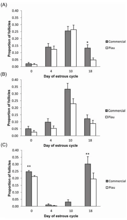
The preovulatory population of follicles for both Piau and commercial gilts reached 6 to 10 mm in diameter. The distribution of both small (P≤0.05) and large (P≤0.01) ovarian follicles during the estrous cycle differed between commercial and Piau gilts throughout the estrous cycle, as illustrated in supplementary Table 1 (Appendix 1). The confidence interval for the difference between proportions allowed the identification of days of the estrous cycle on which the follicular number from Piau and commercial-line were significantly different (Figure 1). The pattern of follicle development in Piau breed was characterized as a lower number of small follicles at day 18, and large follicles at day 0, reflecting their expected lower-ovulation rate compared to the commercial-line.
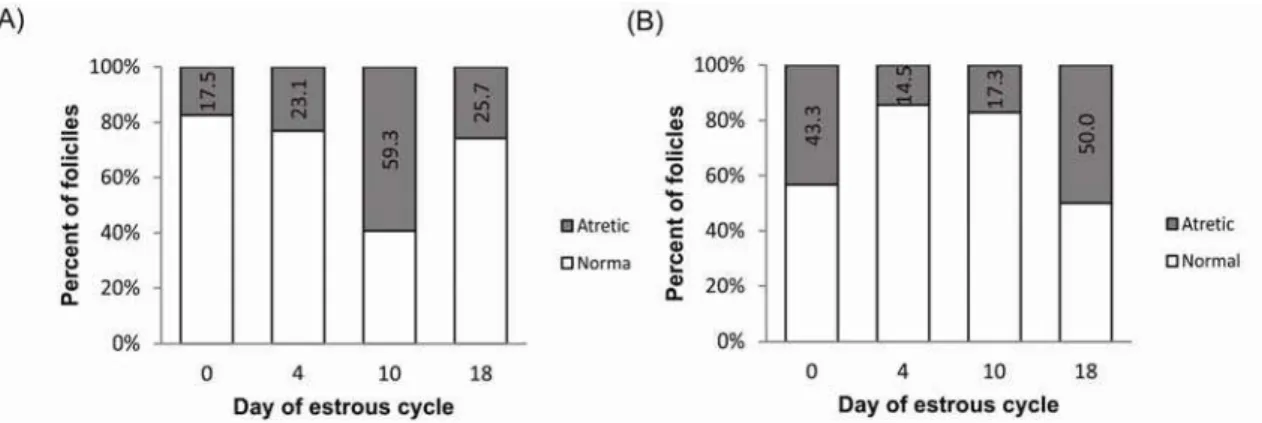
The proportion of normal and atretic follicles during the estrous cycle for all size follicles in commercial and Piau breed is illustrated in Figure 2. The pattern of atresia seems to be different between breeds, with a higher atresia rate in day 10 for commercial and day 18 for Piau breed. In addition to the decline in the number of small and medium sized follicles observed between days 10 and 18 of the estrous cycle for both groups, the incidence of atresia in the total follicle population increased from 17.3% before selection (Day 10) to 50% before estrous (Day 18) in the Piau breed compared to an decrease in atresia from 59.3% to 25.7% in the commercial-line gilts.
Corpus luteum mRNA expression profiles
system, FASL was higher expressed in Piau breed at day 18 (P≤0.01). However, no differences in expression were observed for FAS and the cascade initiator caspase-8 (CASP8). CASP3 mRNA abundance, which is one of the downstream components of CASP8 was higher in Piau breed on day 0 and 4 (P≤0.05 and P≤0.01, respectively). Similar results were observed for TGFBR2, with higher expression in commercial-line gilts on day 0 and day 4 (P≤0.0001 and P≤0.05, respectively) than in the Piau breed.
Granulosa cell mRNA expression profiles
Figure 4 illustrates mRNA expression between the commercial and Piau breed for components of the death receptor apoptotic pathway, with the exception of FASL, which could not be detected in the GC with the current primer set, and TGFBR2. In large normal follicles, TGFBR2 mRNA abundance was higher in the commercial-line on day 0; however, no significant difference was observed in large atretic follicles. FAS mRNA expression in large atretic follicles was higher in Piau than commercial gilts (P≤0.05) on day 0, while no significant difference was observed in large normal follicles. At day 0, medium normal follicles showed higher BCL2 mRNA abundance in GC in the Piau breed (P≤0.01) than in commercial-line. CASP8 mRNA expression was higher in Piau breed for medium normal follicle category at day 18 of the estrous cycle. CASP3 mRNA expression was higher in commercial-line for medium atretic follicles at day 4. In small normal follicles from day 4 CASP3 mRNA expression was higher in commercial-line while CASP8 mRNA expression was higher in the Piau breed. Additional comparisons between follicle size/health status were limited by the number of experimental units at specific days of estrous cycle.
Oocyte mRNA expression profiles
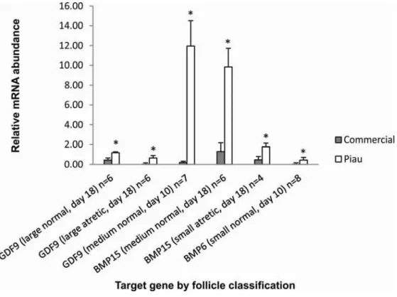
GDF9 mRNA abundance were observed in either normal or atretic follicles on the analyzed days. BMP15 mRNA expression was higher at day 18 in the Piau breed in medium normal and small atretic follicles (P≤0.05) than in the commercial-line. Only one contrast was significant for BMP6 mRNA abundance, which showed higher expression in the Piau breed for small normal follicles at day 10 (P≤0.05).
Discussion
The key ovarian phenotypic traits of the less prolific Piau breed were a lower OR, tendencies for a shorter estrous cycle than in the more prolific commercial-line gilts. A shorter estrous cycle has also been observed in the hyperprolific Meishan breed when compared to nonprolific breeds (Bazer et al. 1988b). It has been postulated that the longer behavioral estrus in Meishan breed would result in recovery of follicles relatively earlier to ovulation; as a result, Meishan sows maintained a large number of follicles during the follicular phase, which keeps constant with no reduction in the proliferating pool, contributing for their high prolificacy (Bazer et al. 1988b). Despite showing a tendency for shorter estrous cycle as observed in Meishan, the Piau breed represents the other extreme of prolificacy. Besides the lack of information about the precise time course of events during the estrous cycle in Piau breed, findings from the present study indicate decrease in the number of small follicles (≤ 3mm diameter) in early follicular phase (Day 18) as illustrated in Figure 1, panel A. The reduction in the number of small follicles along with higher rate of atresia at Day 18 (see Figure 2) suggests a block of follicle replacement in the proliferating pool, an opposite situation that occurs in Meishan, which may be the primary determinants of low prolificacy in this breed.
2004).
It is possible that the different pattern of follicle growth observed in the present study is caused by different timing of the LH surge between breeds, linked to differences in the estrous cycle length between Piau and commercial gilts, as comparative studies reported that behavioral estrous in the hyper-prolific Meishan pig occurred earlier, relative to the LH surge and ovulation, than in Large-White sows (Hunter et al. 1993).
The follicular dynamics between breeds were further corroborated by gene expression analysis using quantitative real-time PCR. Genes of TGF-β superfamily, such as BMPs, GDF9 and their receptors are known modulators of mammalian folliculogenesis, and were selected for study. The expression of BMP6, BMP15 and GDF9 transcripts have recently been reported in porcine oocytes from preovulatory follicles, as well as their cell receptors (Paradis et al. 2009; Sun et al. 2010). However, less information is available about the mechanisms underlying their physiological role in follicular development between species, or differences in their expression in divergent breeds of pigs. Therefore, a comparative study using the less prolific Piau breed and more prolific contemporary commercial-line gilts provided the opportunity to identify genes related to ovulation rate and oocyte quality, and consequently embryonic development and survival in these two breeds.
maturation, as previous studies reported that the Meishan preovulatory follicle is in a more advanced state of maturation than that of the Large-White (Hunter et al., 1993, Faillace and Hunter, 1994, Xu et al., 1998).
BMP6 mRNA expression was also detected in porcine oocytes in this study, corroborating previous findings (Zhu et al. 2008, Paradis et al. 2009). The differential expression of BMP6 mRNA from Piau oocytes may play an important role in FSH-dependent follicle development and in the regulation of luteinization, again affecting the difference in ovulation rate between breeds. It has been suggested in rats that BMP6 mRNA derived from GC is lost during selection of the dominant follicle and that BMP6 mRNA is strongly expressed in GC during atresia (Erickson and Shimasaki, 2003). Moreover, investigation into the mechanism of action found that BMP6, unlike BMP15 and GDF9, does not have proliferative properties on rat GC and is able to suppress FSH-induced progesterone production (Otsuka et al. 2001). Therefore, the results from the present study suggest breed differences in the BMP6, BMP15 and GDF9 mRNA expression profile, and indicate that a larger number of follicles can escape from atresia during early folliculogenesis in the Piau breed, resulting in the lower incidence of atresia at days 4 and 10 compared to commercial-line gilts. These results initially seem contradictory, since the Piau breed is the less prolific. However, oocyte secreted factors may be involved in the recruitment process, leading to differences in oocyte quality (Gilchrist et al. 2008) and the number of tertiary and atretic follicles between lines, as suggested by Manabe et al. (2004). Also associated with their lower prolificacy, differences in oocyte-expressed factors may be contributing to the ability of Piau to use smaller follicles for selection and ovulation, as described for the Meishan breed (Miller et al. 1998).
2001). However, in contrast, Inoue et al. (2006) reported that FAS mRNA expression increased in GC from atretic compared to healthy follicles.
We also detected FAS and FASL mRNA expression in pig luteal tissue as reported in murine and human studies (Kondo et al. 1996; Sakamaki et al. 1997). Interestingly, in the current study no differences were observed for the FAS transcript in CL between breed groups, while FAS mRNA expression in GC was significantly higher in large atretic follicles from Piau gilts than from commercial females at day 0. According to Manabe et al. (2004), differences in the initiation of GC apoptosis between species indicate local mechanisms of regulation, mainly the apoptotic stimuli induction mechanism. In this context, the current study indicates that apoptosis signaling may be differently activated in atretic follicles between distinct breeds. Therefore, it can be suggested that the follicle in the Piau breed provides a different environment for follicle apoptosis than in commercial-line follicles, which may contribute to the lower prolificacy of Piau breed. Many reports have shown that apoptosis occurs during luteolysis, and it is also established that it plays a role in the CL regression at the end of the estrous cycle (Rueda et al., 1997). In the present study the amount of transcript that encodes the membrane protein Fas were similar in CL between breeds, while its ligand FASL mRNA was higher expressed in Piau CL, which may result in a more effective apoptosis signaling towards luteolysis in this breed.
regulation of the CL lifespan by controlling the rate of apoptosis and potentially underlies differences in estrous cycle length and cyclicity between the breeds. However, it is important to emphasize that these events may also be regulated at the post-translational level and modulated by interactions with other molecules such as p53 tumor suppressor protein (Miyashita et al. 1994). In addition, the abundance of transcripts is not always associated with differences in protein secretion (Griffin et al. 2002) and studies at proteomic level are also required.
Many findings indicate that Bcl2 family proteins modulate apoptosis of GC in mammals, where BCL2 over-expression is related with reduced follicular atresia and increased litter size (Hsu et al. 1996; Choi et a., 2004). In the current study BAX mRNA abundance in GC was similar between breeds during estrous cycle, while BCL2 mRNA abundance of medium-sized healthy follicles was higher in the Piau breed compared to commercial-line gilts at day 0, again suggesting decreased apoptosis in Piau GC during early follicular development. CASP3 has been reported as an essential molecule for the apoptosis of GC. Many studies in GC have described changes in caspase-3 protein expression and activity associated with the progression of atresia in ovarian follicles (Boone and Tsang 1998; Berardinelli et al. 2004). In the current study CASP3 mRNA abundance was significant higher in GC from medium-sized atretic and small-sized healthy follicles at day 4 in commercial-line gilts than in the Piau breed. However, little other information is available about CASP3 mRNA expression pattern in different breeds of pigs during the estrous cycle. During the early luteal phase in the pig, a new group of medium-sized follicles starts to growth between days 3and 8 (Guthrie et al. 1995). Although the functional life-span of these follicles is unknown, it is likely that within a day or two after ovulation the medium-sized growing follicles will be eventually deleted by atresia, as described in cattle (Sirois and Fortune, 1988). Therefore, the data on CASP3 mRNA expression in medium- and small-sized follicles in commercial-line gilts suggest that this breed may be more susceptible to atresia than the Piau breed at day 4, driving the different pattern of follicular development observed between breeds. These findings also agreed well with the higher incidence of atresia (23.1% vs 14.5%) at day 4 in commercial-line gilts compared to the Piau gilts in the total follicle population, as shown in Figure 2.
Tsang, 1998) and in sheep (Rueda el al. 1999) CL. Moreover, abundant expression of CASP3 in the human CL was reported and considered to be important for luteal regression (Krajewska et al. 1997). However, little is known regarding the expression and role of caspases in the pig CL. In the current study CASP3 mRNA was expressed in luteal cells throughout estrous cycle and its abundance was higher in the Piau breed at day 0 and day 4. Apoptotic cell death during luteolysis is important to maintain estrous cyclicity (Manabe et al. 2004). However, premature disruption of normal CL function could result in reduction of reproductive efficiency due to irregular estrous cycles and loss of pregnancy (Rueda et al. 1997). In this context, the present results indicate that apoptosis mediated by CASP3 may be differently regulated between the breeds studied and may reflect differences in the estrous cycle length and the time of luteal regression. Recent findings have addressed roles for TGFβ in the follicle maturation and luteinization processes in the pig (Paradis et al. 2009, Sriperumbudur et al. 2010). In the current study, TGFBR2 mRNA expression was decreased in Piau CL at day 0 and day 4 compared to the commercial-line, indicating that the regression process may differ between breeds. In addition, the TGFBR2 mRNA expression in this study was higher in GC from large follicles of the commercial-line gilts at day 0 and may mediate the differences in follicular dynamics leading to differences in ovulation rate and oocyte quality in gilts from the different breeds. This finding is further supported by Sriperumbudur et al. (2010), who suggested that TGFBR2 may have roles in mediating the luteinization process in post-ovulatory porcine follicles.
Conclusions
protein modification, activity or location (Zeng et al. 2004). Additional techniques like RNA interference (RNAi) could be used in future experiments to down-regulate the expression of specific genes in vitro culture systems. For example, RNAi has been used investigate the potential role of growth factors in mediating oocyte regulation of cumulus cells expansion (Gui and Joyce, 2005) and to confirm the pro-activity of apoptotic genes in granulosa cells during atresia (Sai et al. 2012). Apoptosis in the CL, mediated by differences in FASL mRNA, CASP3 mRNA and by decreased expression of TGFBR2 mRNA verified in Piau gilts compared to commercial-line at day 0 and 4 of estrous cycle, may also reflect the tendency for a shorter estrous cycle length and faster luteal regression in the Piau breed. Finally, the higher expression of oocyte secreted factors (GDF9, BMP15 and BMP6) in Piau oocytes may play a role in inhibition of the luteinization process and also affect follicle development and induce the lower ovulation rate that is a key component of the reduced prolificacy of this breed.
Taken together, our results support the hypothesis that differential expression of genes and/or gene pathways controlling follicle growth mediate the different pattern of follicle development observed between the breed studied. This may affect not only ovulation rate but also oocyte and embryo quality.
Acknowledgements
Table 1. List of primer sequences and quantitative real-time PCR quality control data for pig candidate gene expression analysis
1) Gene symbol: glyceraldehyde-3-phosphate dehydrogenase (GAPDH), growth differentiation factor 9 (GDF9), bone morphogenetic protein 15 (BMP15),
bone morphogenetic protein 6 (BMP6), Fas ligand (FASL), Fas protein (FAS), B/linfoma-2 cell (BCL2), BAX protein (BAX), transforming growth factor β
receptor 2 (TGFBR2), caspase-8 (CASP8), caspase-3 (CASP3). 2) Accession number at Genbank ((htpp://www.ncbi.nlm.nih.gov). 3) Cell type which
amplicons are generated: granulosa cell (GC), corpus luteum (CL). 4) Coefficient of determination (R2). Gene Symbol1 Accession number2 Cell type3
Primer Oligonucleotide sequence (5’→3)’ Product size
(bp)
R2(4) Slope of calibration curve Efficiency (%) GAPDH AF017079 GC CL Oocyte Forward Reverse GCAAAGTGGACATTGTCGCCATCA
AGCTTCCCATTCTCAGCCTTGACT 124
0.99 0.99 0.99 -3.33 -3.32 -3.21 2.00 1.99 2.05
GDF9 NM001001909 Oocyte Forward Reverse
TGGTGCAGAACATCATCCACGAGA
GGCTCAATGGCCAACACACTCAAA 100 0.99 -3.32 2.00
BMP15 NM001005155 Oocyte Forward Reverse
AAGCTTGGACGGAGATGGATGTCA
GAAGGCAGTGTCCAGGGATGAAA 162 0.99 -3.53 1.92
BMP6 EU693015 Oocyte Forward Reverse
GGCGGTGACGGCTGCAGAAT
CACACGACGCGGGTGTCCAA 150 0.98 -3.42 1.96
FASL NM213806 CL Forward Reverse
AGGCCTGTGTCTCCTTGTGATGTT
TTTGGCTGGCAGACTCTCTGAGTT 125 0.99 -3.30 2.00
FAS NM213839 GC
CL
Forward Reverse
AGGTGATGATGCCCAAGTGACTGA
AGTCAGCATGTTTCCGTTTGCCAG 149
0.99 0.99 -3.53 -3.55 1.92 1.91
BCL2 NM214285 GC CL
Forward Reverse
TACGGAAACAATGCAGCAGCTGAG
TGGTCATTTCCGACTGAAGAGCGA 123
0.99 0.99 -3.41 -3.55 1.96 1.91
BAX NM138761 GC
CL
Forward Reverse
TTTCTGACGGCAACTTCAACTGGG
TGTCCAGCCCATGATGGTTCTGAT 122
0.99 0.99 -3.73 -3.28 1.85 2.01
TGFBR2 EF396957 GC CL
Forward Reverse
TGAGTCCTTCAAGCAGACGGATGT
TGGAACCAAAGGGTGGCTCATAGT 134
0.99 0.99 -3.33 -3.76 2.00 1.85
CASP8 NM001031779 GC CL
Forward Reverse
TGCCTCCGGTTACAACTACATCCT
AACTTGAGGGAAGCCAGGTCATCA 112
0.99 0.99 -3.31 -3.76 2.01 1.85
CASP3 NM214131 GC CL
Forward Reverse
ATGCTGCAAATCTCAGGGAGACCT
CACCATGGCTTAGAAGCACGCAAA 159
Figure 1. Confidence intervals for the proportion of (A) small, (B) medium and (C) large follicles
during the estrous cycle in commercial and Piau gilts. Asterisks indicate significant differences
Figure 2. Percentage of normal and atretic follicles during the estrous cycle for all follicle sizes
Figure 3. Comparison of mRNA abundance in corpus luteum from commercial and Piau genetic
group (A) BCL2, (B) BAX, (C) FAS, (D) FASL, (E) TGFBR2, (F) CASP3, (G) CASP8 throughout
estrous cycle. Relative mRNA abundance is expressed as lsmeans of 2-ΔCt ± S.E.M. Asterisks
Figure 4. Comparison of mRNA abundance in granulosa cells from commercial and Piau
genetic group for target genes by follicle classification on particular days of the estrous cycle
and number of animals. Relative mRNA abundance is expressed as lsmeans of 2-ΔCt ± S.E.M.
Figure 5. Comparison of mRNA abundance in denuded oocytes recovered from commercial
and Piau genetic group for target genes by follicle classification on particular days of the estrous
cycle and number of animals. Relative mRNA abundance is expressed as lsmeans of 2-ΔCt ±
References
Bazer, F.W., Thatcher, W.W., Martinat-Botte, F., and Terqui, M. (1988a). Conceptus development in Large White and prolific Chinese Meishan pigs. J. Reprod. Fertil.
84, 37.
Bazer, F. W., Thatcher, W. W., Martinat-Botte, F., & Terqui, M. (1988b). Sexual maturation and morphological development of the reproductive tract in large white and prolific Chinese Meishan pigs. Journal of reproduction and fertility,
83(2), 723-8.
Berardinelli, P., Russo, V., Martelli, A., Nardinocchi, D., Di Giacinto, O., Barboni, B., and Mattioli, M. (2004). Colocalization of DNA fragmentation and caspase-3 activation during atresia in pig antral follicles. Anat. Histol. Embryol. 33, 23-27. Bonnet, A., Lê, C.K., Sancristobal, M., Benne, F., Robert-Granié, C., Law-So, G.,
Fabre, S., Besse, P., De Billy, E., Quesnel, H., Hatey, F., Tosser-Klopp, G. (2008). In vivo gene expression in granulosa cells during pig terminal follicular development. Reproduction 136, 211-224.
Boone, D.L., and Tsang, B.K. (1998). Caspase-3 in the rat ovary: localization and possible role in follicular atresia and luteal regression. Biology of Reproduction
58, 1533-1539.
Choi, D., Hwang, S., Lee, E., Yoon, S., Yoon, B.K., and Bae, D. (2004). Expression of mitochondria-dependent apoptosis genes (p53, Bax, and Bcl-2) in rat granulosa cells during follicular development. J. Soc. Gynecol. Investig. 11, 311-317.
Clark, J.R., Eday, T.N., First, N.L., Chapman, A.B., Casida, L.E. (1973). The effects of four genetic groups and two feed levels of feeding on ovulation rate and follicular development in pubertal gilts. J Anim Sci. 36,1164-1169.
Dharma, S.J., Kelkar, R.L., Nandedkar, T.D. (2003). Fas and Fas ligand protein and mRNA in normal and atretic mouse ovarian follicles. Reproduction 126, 783-789. Erickson, G.F., and Shimasaki, S. (2003). The spatiotemporal expression pattern of the
bone morphogenetic protein family in rat ovary cell types during the estrous cycle. Reprod Biol Endocrinol. 1(9), 1-20.
Faillace, L.S., and Hunter, M.G. (1994). Follicle development and oocyte maturation during the immediate preovulatory period in Meishan and White hybrid gilts. J. Reprod. Fertil. 101, 571–576.
Feary, E.S., Juengel, J.L., Smith, P., French, M.C., O’connell, A.R., Lawrence, S.B., Galloway, S.M., Davis, G.H., Mcnatty, K.P. (2007). Patterns of expression of messenger RNAs encoding GDF9, BMP15, TGFBR1, BMPR1B, and BMPR2 during follicular development and characterization of ovarian follicular populations in ewes carrying the Woodlands FecX2W mutation. Biology of Reproduction 77, 990-998.
Foxcroft, G.R., and M. G. Hunter. (1985). Basic physiology of follicular maturation in the pig. J. Reprod. Fertil.Suppl. 33,1.
Foxcroft, G.R., Cosgrove, J., Ding, J., Hofacker, S., and Wiesak, T. (1994). Reproductive function: Current Concepts. In: Cole DJA, Wiseman J, Varley M (eds), Principles of Pig Science, Nothingham University Press, pp. 225-252. Gilchrist, R.B., Ritter, L.J., Armstrong, D.T. (2004). Oocyte–somatic cell interactions
during follicle development in mammals. Animal Reproduction Science 82-83, 431-446.
Gilchrist, R.B., Lane, M., and Thompson, J.G. (2008). Oocyte-secreted factors: regulators of cumulus cell function and oocyte quality. Human Reproduction Update 14 (2), 159–177.
Glister, C., Kemp, C.F., Knight, P.G. (2004). Bone morphogenetic protein (BMP) ligands and receptors in bovine ovarian follicle cells: actions of BMP-4,-6 and -7 on granulosa cells and differential modulation of Smad-1 phosphorylation by follistatin. Reproduction 127, 239-54.
Goodman, S.B., Kugu, K., Chen, S.H, Preutthipan, S., Tilly, K.I., Tilly, J.L., Dharmarajan, A.M. (1998). Estradiol-mediated suppression of apoptosis in the rabbit corpus luteum is associated with a shift in expression of bcl-2 family members favoring cellular survival. Biol Reprod. 59, 820-827.
Grant, S.A., Hunter, M.G., and Foxcroft, G.R. (1989). Morphological and biochemical characteristics during ovarian follicular development in the pig. Journal of Reproduction and Fertility 86, 171-183.
Griffin, T.J., Gygi, S.P., Ideker, T., Rist, B., Eng, J., Hood, L., Aebersold, R. (2002). Complementary profiling of gene expression at the transcriptome and proteome levels in Saccharomyces cerevisiae. Mol. Cell Proteomics 1, 323-333.
Gui, L.M and Joyce, I.M. (2005). RNA interference evidence that growth differentiation factor-9 mediates oocyte regulation of cumulus expansion in mice. Biology of reproduction, 72(1), 195–9.
Guthrie, H.D., Grimes, R.W., Cooper, B.S., Hammond, J.M. (1995). Follicular atresia in pigs: Measurement and physiology. J Anim Sci. 73, 2834-2844.
Guthrie, H.D. (2005). The follicular phase in pigs: follicle populations, circulating hormones, follicle factors and oocytes. J Anim Sci. 83, 79-89.
Haley, C.S., and Lee, G.J. (1993). Genetic basis of prolificacy in Meishan pigs. Journal of Reproduction and Fertility Supplement 48, 247–259.
Hsu, S.Y., Lai, R.J., Finegold, M., and Hsueh, A.J. (1996). Targeted overexpression of Bcl-2 in ovaries of transgenic mice leads to decreased follicle apoptosis, enhanced folliculogenesis, and increased germ cell tumorigenesis. Endocrinology 137, 4837-43.
heterogeneity in pigs. J Reprod Fertil Suppl. 40, 163-77.
Hunter, M.G., Biggs, C., Foxcroft, G.R., McNeilly, A.S., Tilton, J.E. (1993). Comparisons of endocrinology and behavioural events during the periovulatory period in Meishan and Large-White hybrid gilts. J. Reprod. Fertil. 97, 475–480. Hunter, M.G., and Picton, H.M. (1995). Effect of hCG administration at the onset of
oestrus on early embryo survival and development in Meishan gilts. Animal Reproduction Science 38(3), 231-238
Hunter, M.G. (2000). Oocyte maturation and ovum quality in pigs. Rev Reprod. 5(2), 122-30.
Hunter, M.G., Robinson, R.S., Mann, G.E., Webb, R. (2004). Endocrine and paracrine control of follicular development and ovulation rate in farm species. Anim Reprod Sci. 82,461-77.
Hunter, M.G., and Paradis, F. (2009). Intrafollicular regulatory mechanisms in the porcine ovary. Soc Reprod Fertil Suppl. 66,149-64.
Hussein, M.R. (2005). Apoptosis in the ovary: molecular mechanisms. Human Reproduction and Embryology 11,162-178.
Inoue, N., Maeda, A., Matsuda-Minehata, F., Fukuta, K., Manabe, N. (2006). Expression and localization of Fas ligand and Fas during atresia in porcine ovarian follicles. J Reprod Dev. 52, 723–730.
Inoue N, Matsuda F, Goto Y, Manabe N. (2011). Role of cell-death ligand-receptor system of granulosa cells in selective follicular atresia in porcine ovary. J Reprod Dev. 57(2), 169-75.
Juengel, J.L., Garverick, H.A., Johnson, A.L., Youngquist, R.S., and SMITH, M.F. (1993). Apoptosis during luteal regression in cattle. Endocrinology 132 (1), 249-254.
Knox, R.V. (2005). Recruitment and selection of ovarian follicles for determination of ovulation rate in the pig. Domestic Animal Endocrinology 29(2), 385-397.
Kondo, H., Maruo, T., Peng, X., Mochizuki, M. (1996). Immunological evidence for the expression of the Fas antigen in the infant and adult human ovary during follicular regression and atresia. J Clin Endocrinol Metab. 81, 2702-2710.
Krajewska, M., Wang, H.G., Krajewski, S., Zapata, J.M., Shabaik, A., Gascoyne, R., Reed, J.C. (1997). Immunohistochemical analysis of in vivo patterns of expression of CPP32 (Caspase-3), a cell death protease. Cancer Res. 57, 1605-1613.
Kutuzov, M.A., Alexandra, A.V., Voyno-Yasenetskaya, T.A. (2007). Regulation of apoptosis signal-regulating kinase 1 degradation by Galpha13 The FASEB Journal
21, 3728-36.
Liu, H.K., Kuo, T.W., Yang, H.S., Chenb, L.R., LI, S. S-L., Huanga, W.H. (2008). Differential gene expression of bone morphogenetic protein 15 and growth differentiation factor 9 during in vitro maturation of porcine oocytes and early embryos. Animal Reproduction Science 103, 312–322.
Livak, K.J., and Schmittgen, T.D. (2001). Analysis of relative gene expression data using real-time quantitative PCR and the 2DDCt method. Methods 25, 402–408. Manabe, N., Goto, Y., Matsuda-Minehata, F., Inoue, N., Maeda, A., Sakamaki, K.,
Miyano, T. (2004). Regulation Mechanism of Selective Atresia in Porcine Follicles: Regulation of Granulosa Cell Apoptosis during Atresia. J. Reprod. Dev.
50, 493-514.
Matsuda-Minehata, F., Maeda, A., Cheng, Y., Sai, T., Gonda, H., Goto, Y., Manabe, N. (2008). Regulation of granulosa cell apoptosis by death ligand–receptor signaling. Animal Science Journal 79 (1), 1–10.
Miller, A.T., Picton, H.M., Craigon, J., Hunter, M.G. (1998). Follicle dynamics and aromatase activity in high-ovulating Meishan sows and in Large-White hybrid contemporaries. Biology of Reproduction 58, 1372-1378.
Miyashita, T., Krajewski, S., Krajewska, M., Wang, H. G., Lin, H. K., Liebermann, D., Hoffman, A.B. and Reed, J.C. (1994). Tumor suppressor p53 is a regulator of bcl-2 and bax gene expression in vitro and in vivo. Oncogene 9, 1799-1805.
Oltvai, Z.N., Milliman, C.L., Korsmeyer, S.J. (1993). Bcl-2 heterodimerizes in vivo with a conserved homolog, Bax, that accelerates programmed cell death. Cell 74, 609–619.
Otsuka, F., Moore, R.K and Shimasaki, S. (2001). Biological function and cellular mechanism of bone morphogenetic protein-6 in the ovary. Journal of Biological Chemistry 276, 32889-32895.
Paradis, F., Novak, S., Murdoch, G.K., Dyck, M.K., Dixon, W.T., Foxcroft, G.R. (2009). Temporal regulation of BMP2, BMP6, BMP15, GDF9, BMPR1A, BMPR1B, BMPR2 and TGFBR1 mRNA expression in the oocyte, granulosa and theca cells of developing preovulatory follicles in the pig. Reproduction 138, 115-129.
Peixoto, J.O., Guimaraes, S.E.F., Lopes, P.S., Soares, M.A.M., Pires, A.V., Silva, M.V., Torres, R.A., Silva, M.A.E. (2006) Associations of leptin gene polymorphisms with production traits in pigs. Journal of Animal Breeding and Genetics 123, 378-383.
Porter, D.A., Harman, R.M., Cowan, R.G., Quirk, S.M. (2001). Relationship of Fas ligand expression and atresia during bovine follicle development. Reproduction
121, 561–566.
Pretheeban, T., Balendran, A., Gordon, M.B., Rajamahendran, R. (2010). mRNA of luteal genes associated with progesterone synthesis, maintenance, and apoptosis in dairy heifers and lactating dairy cows. Animal Reproduction Science 121, 218-224.