JSCS–4147 Review
REVIEW
The role of EPR spectroscopy in studies of the oxidative status of
biological systems and the antioxidative properties
of various compounds
IVAN SPASOJEVIĆ1, MILOŠ MOJOVIĆ2, ALEKSANDAR IGNJATOVIĆ2 and GORAN BAČIĆ2*
1Institute for Multidisciplinary Research, Kneza Višeslava 1, University of Belgrade, 11000
Belgrade and 2Faculty of Physical Chemistry, University of Belgrade,
Studentski trg 12–16, 11000 Belgrade, Serbia (Received 15 October, revised 28 December 2010)
Abstract: In this era of intense study of free radicals and antioxidants, electron paramagnetic resonance (EPR) is arguably the best-suited technique for such research, particularly when considering biochemical and biological systems. No attempt was made to cover all the topics of EPR application but instead at-tention was restricted to two areas that are both novel and received less atten-tion in previous reviews. In the first secatten-tion, the applicaatten-tion of EPR in asses-sing the oxidative status of various biological systems, uasses-sing endogenous sta-bile paramagnetic species, such as the ascorbyl radical, semiquinone, melanin, and oxidized pigments, is addressed. The second section covers the use of EPR in the emerging field of antioxidant development, using EPR spin-trapping and spin-probing techniques. In both sections, in addition to giving an overview of the available literature, examples (mostly from the authors’ recent work) are also presented in sufficient detail to illustrate how to explore the full potential of EPR. This review aims at encouraging biologists, chemists and pharmaco-logists interested in the redox metabolism of living systems, free radical che-mistry or antioxidative properties of new drugs and natural products to take ad-vantage of this technique for their investigations.
Keywords: EPR spectroscopy; oxidative status; antioxidants; spin-probes; spin- -traps.
CONTENT 1. INTRODUCTION
2. EVALUATION OF OXIDATIVE STATUS 2.1. EPR in comparison to other methods
2.2. EPR spectroscopy of the ascorbyl radical 2.3. EPR spectroscopy of the tocopheroxyl radical 2.4. EPR spectroscopy of melanin
2.5. The oxidative status of plants
3. EVALUATION OF ANTIOXIDATIVE ACTIVITY
3.1. EPR spectroscopy – a technique of choice for investigating the antioxidative pro-perties of compounds, extracts and foods
3.2. Applications of EPR spin-trapping in antioxidant research 3.3. Applications of EPR spin-probing in antioxidant research 3.4. Evaluation of antioxidative activity with EPR spin-probing 3.5. Evaluation of the antioxidative capacity against lipid peroxidation 4. CONCLUSIONS
1. INTRODUCTION
The delicate balance between the advantageous and detrimental effects of free radicals is clearly an important aspect of life. Although reactive oxygen spe-cies (ROS) have been labelled as “villains” for a long time,1,2 they also have been found to regulate the activity of a number of enzymes via oxidation in
re-ceptor-mediated signalling pathways, a process known as redox signalling.3–8 This signalling is involved in the regulation of vascular tone, oxygen tension, ac-tivity of the immune system, growth in plants, and some other physiological pro-cesses.9 On the other hand, uncontrolled generation of radicals is highly related to many pathophysiological events, such as neurodegenerative diseases (Alzhei-mer’s disease, amyotrophic lateral sclerosis, Down’s syndrome, etc),10–13
malig-nancy,14 diabetes mellitus,15 sepsis16 and atherosclerosis,17 and also seems to play an important role in the aging process.1,2,18,19 Hence, a certain level of dation performed by free radicals is mandatory in biosystems, but increased oxi-dation may jeopardize normal functioning and lead to pathophysiological con-ditions. Therefore, knowledge of the relative level of oxidation in a biosystem, known as oxidative status, clearly represents an imperative in studies of the me-chanisms of (patho)physiological processes.20
A broad spectrum of techniques has been applied in redox research. Most of them, however, determine only the total antioxidative status or antioxidative capacity and do not provide details on specific reactive species. On the other hand, those which are able to provide more specific data require the use of te-dious laboratory procedures or suffer from low sensitivity or artefacts. Electron Paramagnetic Resonance (EPR) spectroscopy stands out from other methods be-cause of its unique ability to detect either short or long lived radicals with high specificity and sensitivity (e.g., EPR spin-trapping detection of the superoxide
radical is 40 times more sensitive than spectrophotometric analysis with cyto-chrome c 26,27). EPR is also capable of directly detecting a number of specific
markers of the oxidative status, such as the ascorbyl radical,28,29 the tocopherol radical,30,31 melanin,32,33 semiquinone,34 and plant pigments,35,36 in a variety of biosystems – human and animal tissues and fluids, whole insects, plant tissues and others. In an antioxidant investigation, EPR can also be applied to determine the capacity of the selected compound, extract or food to remove specific re-active species, such as superoxide (O2•–), hydroxyl radical (•OH), organic radi-cals, lipid peroxides, or to sequester iron.37–42 In addition, EPR is, in principle, a non-destructive technique, which is a clear advantage over chemical procedures when dealing with biological systems. This paper represents an overview of re-cent applications of EPR spectroscopy in determining the oxidative status of bio-systems and antioxidative properties of compounds, extracts, and foods. The full capacity of a variety of EPR techniques in redox studies is yet to be explored; hence, the aim was to encourage scientists to apply EPR in their studies and to develop new EPR techniques. As a matter of convenience, most of the examples and illustrations are from a series of the authors’ recent studies, with the aim of presenting the main principles of the application of EPR in redox research.
2. EVALUATION OF OXIDATIVE STATUS
2.1. EPR in comparison to other methods
A variety of spectrophotometric assays is available for investigating the oxi-dative status of biochemical systems. Widely applied is the ABTS (2,2-azino- -bis(3-ethylbenz thiazoline-6-sulphonic acid) assay, in which ABTS is added to a system, oxidized by horseradish peroxidase/H2O2 to the radical cation ABTS+ which absorbs light at 660, 734 and 820 nm. Results obtained using the ABTS assay are expressed as equivalents of ascorbic acid, indicating the total antioxi-dant activity of some tissue homogenate, cells (e.g., cultured cells) or liquids
(e.g., serum, CSF, etc.).43,44 Increased total antioxidant activity is usually related
oxidative status. R–SH groups are present in glutathione (GSH), a very important component of the AOS, as well as in proteins.46 A total amount of R–SH groups less than normal usually means that some GSH has been oxidized to GSSG via
ROS reduction. In addition, it could be a consequence of an increased production of reactive nitrogen species (RNS), which leads to the formation of R–SNO groups.47 On the other hand, an increased level of the total amount of R–SH groups indicates a decrease in the production of superoxide, which leads to a de-crease of peroxynitrite production and R–SH nitosylation.48
The ROS/RNS-system and the AOS are just two intertwined components present within the global physiological mechanisms of homeostasis.49 Changes in the oxidative status modify the AOS activity, its composition and structure, striving to provide the best possible protection and preservation of cellular ho-meostasis.33 Therefore, the activity of a battery of AOS enzymes (catalase, GSH peroxidase and reductase, MnSOD, and CuZnSOD) represent excellent marker of the oxidative status and may provide information on the specific mechanisms of oxidative processes.29,50 For example, CuZnSOD is inactivated by H2O2;51 hence a decrease in its activity in some biosystem indicates the development of H2O2-mediated oxidative stress.
Confocal fluorescent microscopy represents a powerful tool for redox studies on cell cultures and ex vivo tissues. With the increased interest in this area of
research, a variety of labels have become available that can be used to investigate the oxidative status of cells under different conditions. For example, the intensity of fluorescence of cells stained with MitoTracker Orange is affected by the intracellular level of hydrogen peroxide (H2O2) and the oxidative status.25,52 Some other dyes, the fluorescence of which is dependent on oxidation, such as carboxy-H2-DCFDA and others, can also be used.53
as-corbate, tocopherol, melanin, or plant pigment system P700, can be modified by ROS to stabile organic radicals with a very long lifetime, allowing their direct detection by EPR spectroscopy.58 Due to the non-destructive nature of EPR, this can be realised without any interference with biochemical processes, which is not the case with any other method.59 By detecting and discriminating these para-magnetic molecules, the level of which represents a marker of oxidative status, EPR provides essential information on redox mechanisms in biosystems. How-ever, in spite of the unique capabilities of EPR, it should be emphasized that, in our experience, the most complete insight into redox processes can be achieved by complementing EPR with other methods, such as analysis of the AOS or SH groups (see the next section).
The detection of different paramagnetic biomolecules requires the explana-tion of some technical details. Usually their signals can be detected by means of a conventional X-band EPR spectrometer operating at a resonant frequency of around 9.5 GHz. The measurements are generally performed at physiological or room temperature, but in some cases lower temperatures (liquid N2) are required to obtain a better S/N ratio, which requires minor technical adjustments. When dealing with oxygen consuming systems or oxygen sensitive processes, it is es-sential to control the gas environment in the sample. This can be easily achieved by placing samples in gas-permeable Teflon tubes or holders and flowing the required gas mixture over the sample.60 In a such manner, a constant level of O2 can be supplied to the system,56,60 or if the radical is sensitive to oxygen (e.g., the tocopheroxyl radical, •TO), to deprive it by using pure N2 or Ar.54 An im-portant component in analyzing any EPR spectrum is to perform spectral simu-lations of each detected signal, to identify species and to determine their signal intensities (in our studies, Brukers’ WINEPR SimFonia was used but there are many other software available).28,60–62 The following sections illustrate different applications of EPR spectroscopy in detecting the most common stabile paramag-netic species in biosystems.
2.2. EPR spectroscopy of the ascorbyl radical
(Cu2+) + Asc– → Fe2+ (Cu1+) + •Asc, ii) in cooperation with H2O2: Fe2+(Cu1+)⋅⋅⋅Asc + H2O2→ Fe3+(Cu2+) + •Asc + OH– + H2O or iii) via •OH, generated in a metal involving the Fenton system: •OH + Asc–→•Asc + OH–.65– 67 Obviously, the level of •Asc can indicate how much redox active metals there are in a system,65,66 but the level of •Asc also depends on the production of free radicals in reactions unrelated to the presence of transition metals and these should be discriminated. It is suggested that the influence of catalytically active metals could be emphasized by the addition of H2O2, which should promote reactions ii and iii depending on the level of the catalytically active metals. On
the other hand, it should be stressed that commercial buffers inevitably contain metal impurities which may represent a source of artefacts in biochemical studies by increasing the basic level of •Asc.65 This potential problem can be eliminated by the addition of strong chelating agents, such as DTPA, which sequester transition metals and diminish their oxidative capacity.67
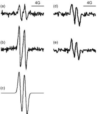
Fig. 1. Left: EPR spectra of the as-corbyl radical (•Asc). EPR detects the single unpaired electron of •Asc, which has a spin S = ½. However, the electron interacts with surround-ing nuclei that also possess spin (here 1H with a nuclear spin of I = = ½), which leads to hyperfine split-ting; hence, two lines emerge in the spectrum. An example of •Asc in hu-man plasma: a) untreated serum;28 b) serum treated with 0.5 mM peroxy-nitrite;64 c) spectral simulation of •Asc EPR signal proving that the sig-nal from serum emerges from this radical, enabling quantification of the signal intensity. Characteristic EPR spectra of •Asc in amnion fluid of d) normal and e) thrombophilic pregnancies29 (showing no differen-ces between the two samples).
then, •Asc has been detected by EPR in a variety of biological samples: plas-ma,68,69 cerebrospinal fluid (CSF),69 skin,70,71 extracellular fluid,72 plant leaves73 and the midgut fluid of insects.74 Increase of the steady-state concen-tration of •Asc was reported in several conditions related to pro-oxidative chan-ges, such as ischemia/reperfusion, sepsis,75 brain injuries,76 paraquat poisoning, iron overload, gastric cancerogenesis,77 pre-eclampsia,78 and others. Recently, EPR spectroscopy of •Asc was employed to investigate the oxidative status of amnion fluid (Figs. 1d and 1e). The similar level of •Asc in the amnion fluid of control and thrombophilic subjects indicates that the disturbed oxidative status of the placenta which was observed by AOS assays,29 was not provoked by an in-creased generation of reactive species in amnion fluid,29 thus enabling a potential mechanisms of thrombophilia to be suggested. There are many other interesting examples. Menditto et al.79 used EPR spectroscopy of •Asc to study the
associ-ation between the oxidative status of seminal fluid and the iron and copper con-tent. A very innovative and elegant experimental design was developed by Shar-ma et al.,80 enabling in vivo EPR measurements of the level of •Asc, as a real-
-time quantitative marker of changes of the oxidative status in dog myocardium during ischemia/reperfusion.
The absolute •Asc concentration which can be detected is as low as ≈5 nM by measuring the intensity of the EPR signal using spectral simulation (Fig. 1) and calibration. However, the basal •Asc level in tissues and fluids can vary significantly between subjects or populations,64,68 and is highly dependent on the level of ascorbate in the system.72 For instance, due to different diets and life styles, the basal level of ascorbate and, consequently, ascorbyl radical in the plasma of the Brazilian population (65 nM)64 is different from that of the Eu-ropean one (100 nM).68 Galleano et al.81 developed an approach that could over-come this potential obstacle in oxidative studies. By combining EPR measure-ments of the •Asc concentration and HPLC analysis for the total ascorbate level, they were able to calculate the ascorbyl radical/ascorbate (•Asc/Asc) ratio, which represents an indicator of oxidative stress independent of individual and popu-lation variations in the ascorbate level. This represents an example of the effec-tiveness of EPR in combination with other techniques, as emphasized before.
2.3. EPR spectroscopy of the tocopheroxyl radical
signal of •TO emerges (Fig. 2b) only after the virtual disappearance of the as-corbyl radical.31 Therefore, the EPR spectroscopy of the tocopheroxyl radical could be used in investigations of oxidative status, emphasizing an occurrence of intense oxidative stress capable of depletion of the antioxidative capacity of the ascorbate in the system.
Fig. 2. Characteristic seven-line EPR signal of •TO. a) Generated by purging the suspension with 0.1 mM tocopherol and 100 mM SDS with NO gas;86 b) in hu-man plasma exposed to O2•– generated by the hypoxan-thine/xanthine oxidase system.31 The interactions which provoke hyperfine splitting resulting in a complex seven-line signal and the spectral parameters were described in detail by Matsuo et al.87
The detection of •TO in biosystems ex vivo is complicated by the fact that •TO can react with oxygen to produce TO and superoxide (an EPR silent spe-cies), which can occur during sample collection and storage, as well as during EPR measurements, hence even EPR measurements have to be performed in an inert atmosphere (N2). For this reason, most of the EPR studies of •TO have been performed on model systems in vitro. Naužil et al.30 applied EPR spectroscopy
of •TO to study the inhibitory effects of α-tocopherol against radical-initiated oxidation of low‐density lipoproteins (LDL) lipids. Zhou et al.88 used a similar
approach to investigate the regeneration of α-tocopherol by green tea polypheno-lics in phospholipid micelle exposed to oxidative stress. The metabolism of TO can also be explored using the more stabile and hydrosoluble vitamin E deri-vative – Trolox (6-hydroxy-2,5,7,8-tetramethylchroman-2-carboxylic acid). For example, Opländer et al.89 applied EPR to investigate the effects of the Trolox
against UV-A induced cell death of human skin fibroblasts. It was determined that the production of the Trolox radical is increased in the presence of nitrite, indicating that the EPR spectroscopy of •TO could be useful for studying not only ROS, but also for nitrogen reactive species.
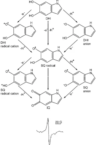
2.4. EPR spectroscopy of melanin
which can be detected by EPR. These properties make the intensity of the EPR signal of melanin an excellent marker signifying the oxidative status of specific melanin-containing systems, such as mammalian skin92 and eyes,93,94 some plants,95 fungi,96,97 insects,33,98 and others. Nevertheless, it should be stressed that although EPR spectroscopy can distinguish between different types of me-lanins, it is virtually impossible to connect the recorded spectrum to a specific eumelanin structure by any spectral analysis.
Fig. 3. Redox chemistry (a) and characteristic EPR signal of eumelanin (b). The key mono-meric building blocks of eumelanin are: DHI (5,6-dihydroxyindole), SQ (semiquinone) and IQ (5,6-indolequinone). Spin–spin interactions, fast electron transfer reactions and anisotropy
related to low mobility of paramagnetic species inside the melanin macromolecules make only one broad line in the melanin EPR spectrum emerge.
Due to the complex chemistry of neuromelanin in the brain, EPR measurements do not provide a straightforward evaluation of the oxidative status, but studies on neuromelanin provide a valuable insight into the mechanisms of neurodege-neration in PD.99–101 The EPR signal of melanin has been used to study the pro-oxidative effects of UV irradiation in skin and eyes. Wood et al.32 used the
melanin signal as an indicator of the oxidative status of melanin-containing skin cells from a genetically melanoma-susceptible cross of Xiphophorus fish (a
mo-del system for human melanoma research) exposed to UV irradiation of different wavelengths and intensities. A strong correlation between the intensity of the melanin signal and melanoma induction was observed, indicating that pro-oxi-dative changes represent an important causative step in melanoma develop-ment.32 Pertinent to this, some attempts have been made to develop an in vivo EPR technique in the diagnostics of melanoma in humans. In a study performed by Vahidi et al.,102 the EPR spectra of melanin in frog skin were recorded in situ
using a surface EPR coil and 2D imaging, and the intensity of the melanin signal was observed to depend on the level of oxygenation, which is one of the para-meters defining the oxidative status. It was proposed that 2D EPR spectroscopy could potentially be useful in early melanoma detection. Seagle et al.93 employed
EPR of melanin to study the effects of UV radiation on human retinal pigment epithelium with different coloured pigments and to compare the observed chan-ges to the modifications in synthetic melanin. Samples were placed in an EPR “flat cell” and irradiated inside the cavity. As expected, the melanin signal from the cells increased with the power of irradiation, but not in two distinguishable portions as observed for synthetic melanin, indicating that in vivo radical
photo-generation is different from photophoto-generation in a chemical system. This shows that chemical systems with synthetic melanin do not represent an adequate model of the structure and metabolism of melanin in biosystems.
Melanin represents an important constituent of the oxidative metabolism of insects. Barbehenn et al.,98 applied EPR spectroscopy of melanin to investigate
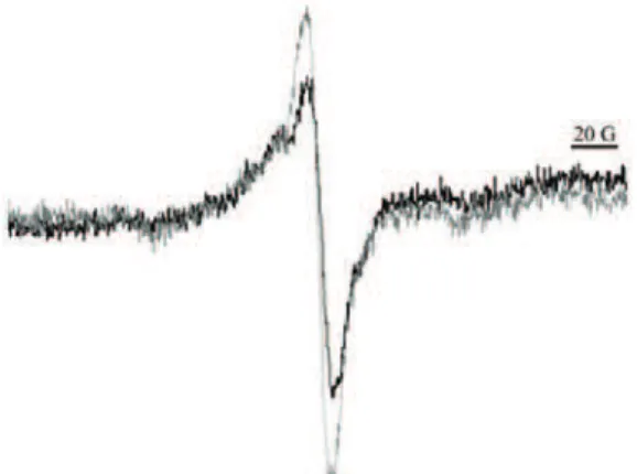
the pro-oxidative defence of plants against herbivorous insects. It was determined that melanin-like reactive substances are formed in the midgut fluid of insects, from the tannic acid present in leaves. In this way, plants disturb the oxidative status of the digestive system of insects and indirectly protect themselves.103,104 On the other hand, some insects generate melanin to use its ROS scavenging ability in the regulation of their oxidative status. For example, it was recently shown using biochemical assays and EPR of melanin that exposure of the Eu-ropean corn borer (Ostrinia nubilalis) to low temperatures leads to an
Fig. 4. EPR spectra of melanin in larvae maintained at: a) 5, b) –3 and c) –16 °C. The downward triangle (▼) marks the characteristic eumelanin-related EPR signal. The signal of pheomelanin (circle) can be observed on the left side of the eumelanin signal in panel c). It can be observed that the underlying signal of pheomelanin slightly shifts the eumela-nin signal. The measurements were performed at liquid N2 temperature in order to obtain a higher S/N ratio than at room temperature.33
Thus, in this case, insects use melanin to protect their cells from the oxi-dative burst involved in the adaptation process.
The presented examples illustrate the vast potential of EPR in studying va-rious organic radicals,105 which should be further utilized in redox studies of biosystems. For example, EPR is able to detect redox-active amino acid radicals: tyrosyl, tryptophanyl, glycyl, and histidyl radical.106 The EPR of these stabile ra-dicals was previously employed to study enzyme activity and protein dama-ge,57,107 but, potentially, it may also find applications in investigations of the oxidative status.108
2.5. The oxidative status of plants
Although previously unappreciated, the application of EPR spectroscopy is becoming more and more important in plant studies. It was previously illustrated that the EPR spin-trapping technique with advanced spin-traps (e.g. DEPMPO) is
processes, Shulaev and Oliver27 concluded that, at present, measurements of oxidative stress in plants are limited and there are no truly non-invasive methods. Although they considered the EPR spin-trapping technique, the application of EPR spectroscopy of stabile organic radicals in plants was not taken into account. Since it enables the acquisition of signals of paramagnetic species in plant cells, parts or even whole plants without any interference with the metabolism, EPR spectroscopy could be the missing ‘truly non-invasive’ technique in plant re-search. It should be emphasized that the measurements can be performed under selected temperature, atmosphere, and light regimes, which can be varied inside the EPR spectrometer cavity during the course of the experiment.
As in animal tissues and fluids, the ascorbyl radical can be used to determine the oxidative status of plants. Malanga et al.110 used the endogenous signal of the
ascorbyl radical in an algal culture (Chlorella vulgaris) and intact soybean leaves
to study UV-provoked oxidative stress. Puntarulo et al.111 applied EPR
measu-rements of •Asc to investigate the effects of NO on the oxidative status of soy-bean chloroplasts, proposing an antioxidative role of NO based on the decreased level of •Asc after treatment with NO.
The photosystem I (PSI), which contains an EPR active specie – oxidized pigment – P700+, could also be used as a marker of the oxidative status.35,36 The increased generation of reactive species leads to photoinhibition of the PSI and a related decrease in the P700+ level.112 Thus, in contrast to previous illustrations, a decrease in the EPR signal signifies pro-oxidative changes of the oxidative sta-tus. This approach was employed to investigate the oxidative stress in intact pea leaves exposed to chilling conditions.36 The light inducible EPR signal, which was recognized to reflect the oxidized form of the PSI pigment P700 (P700+), is shown in Fig. 5. The level of P700+ was lower in plants exposed to cold, show-ing that the chillshow-ing conditions led to pro-oxidative changes of the status in leaves. A similar non-invasive approach could be used to investigate the oxida-tive status of plants exposed to some other stressors.
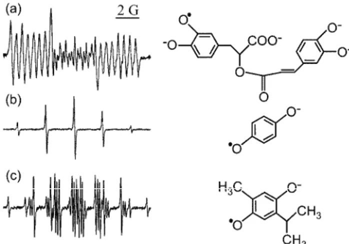
To study paramagnetic species not present in sufficient concentrations in plants to enable non-invasive detection, plant isolates or extracts could be used in EPR studies. For example, the EPR signal of quinhydrone was identified in iso-lated cell walls from pea roots, which could not be detected in the whole plant parts. The intensity of the EPR signal of quinhydrone was highly correlated with the concentration of H2O2 supplemented to the cell wall and with •OH pro-duction evaluated by the EPR spin-trapping technique, showing itself to be a useful marker of oxidative status.34 Pedersen113–115 published several compre-hensive studies on alcohol plant extracts showing that hundreds of species from the families Lamiaceae and Gesneriaceae contain quinone related paramagnetic species easily detectable by EPR, and potentially valuable as markers of oxi-dative status. Examples of EPR signals of such species in extracts of Peltodon radicans, Salvia hispanica and Monarda clinopodia are illustrated in Fig. 6.
Fig. 6. The EPR semiquinone spectra and chemical structures of a) rosmarinic acid from Peltodon radicans, b) hydroquinone from Salvia hispanica and c) thymohydroquinone from
Monarda clinopodia all obtained from crude alcoholic leaf extracts. The signals were identified using the EPR spectra of the corresponding chemicals.115
Spectral simulations should be used to compare the levels of paramagnetic spe-cies in different samples, and to determine whether the signal of the sample is composed of EPR spectra of more than one paramagnetic species.
3. EVALUATION OF ANTIOXIDATIVE ACTIVITY
3.1. EPR spectroscopy – a technique of choice for investigating the antioxidative properties of compounds, extracts and foods
Overproduction or inefficient removal of reactive species by scavenging enzymes, as well as an increase of the level of catalytically active metals (e.g.,
iron or copper) related to the generation of the notorious •OH radical, have been shown to lead to damage of biomolecules and cellular membranes in a process known as oxidative stress,9,116 which has been proposed to be a hallmark of a variety of pathophysiological conditions.9,10–17,29,54 Under such circumstances, the supplement of antioxidants aimed at re-balancing the disturbed oxidative sta-tus could be a very beneficial component of a treatment or a diet.19,20,24,25 To establish whether some food or a compound could be useful in health problems related to oxidative stress, it is necessary to establish their antioxidative proper-ties. A broad range of methods available for the evaluation of the so-called “total antioxidative capacity” of some compounds, plant extracts or foodstuffs were reviewed previously.20,117,118 These assays (such as ABTS, ORAC, TRAP, and DPPH assays118) are easy to perform; hence, they are frequently employed in studies pointing to an antioxidative capacity of various foods or plant extracts. However, the majority of studies were performed in chemical systems not taking into account the specific properties of target biosystems and metabolic processes that can occur in vivo. Although such studies are a prerequisite for further
investi-gations of the antioxidant effectiveness of a certain compound, several other points should be taken into consideration before it should be recommended as a poten-tial cure: task #1: to determine the activity against specific radicals; #2: to de-termine the distribution of active compounds in both principal environments pre-sent in biosystems – hydrophilic and lipophilic; #3: to determine whether the an-tioxidant acts against radicals in cells, extracellular milieu, or both; #4: to take into account metabolic changes of the investigated compound(s) depending on the route of administration; #5: to determine which specific compound(s) present in the metabolized extract or food is (are) active against free radicals; #6: to de-termine whether the active compounds could somehow overcome the refractory response. In the following sections, it will be illustrated that the majority of these tasks can be performed by EPR or by combination of EPR with other methods in a carefully planned experimental setup.
is to generate a specific radical by a selected chemical system and to quantify the inhibiting effects of a compound or extract against radical production (#1). On the other hand, EPR spin-probing can be employed to determine the distribution of potential antioxidants in a hydrophilic, lipophilic, or extracellular environment (#2 and #3). The application of spin-probing in antioxidant research is based on the measurement of the ability of studied compound(s) to reduce synthetic long lived radicals (spin-probes) to EPR-silent hydroxylamines. The spin-probing ap-proach in the study of antioxidants has a long history and has been extensively documented in the literature; in comparison, spin-traps represent an emerging field in EPR spectroscopy.
Tasks #4 and #5 also include the EPR approach, but require specific pro-cessing or analysis of the studied systems prior to EPR measurements. For examp-le, antioxidants are usually applied orally, therefore an in vitro digestion model
system119 or a more simple methanolysis39 can be used to process potential anti-oxidants to compounds that are absorbed in vivo (#4). After establishing the
anti-oxidative properties (#1–#3) and metabolic modifications (#4) of some food or extract, analysis of its chemical composition, using HPLC, GC/MS and other me-thods, should be preformed, to reveal compound(s) that could be “responsible” for antioxidative activity. Selected compounds can be then separately tested for antioxidative activity using EPR techniques (#5).
It should be stressed that even if an investigated compound or extract ap-pears to be an excellent antioxidant on the account of these five points, it might not be effective in vivo if it is unable to overcome the refractory response (#6).18–20
In order to resolve this, the protective effects of a potential antioxidant in cell cultures and experimental animals exposed to oxidative stress should be deter-mined. Fluorescent microscopy with redox sensitive dyes has shown itself to be very useful in studies of antioxidative actions in cell cultures, due to its high sen-sitivity and ability to detect sub-cellular changes related to oxidative stress. On the other hand, a non-destructive in vivo EPR technique is the only available
me-thod to follow up the oxidative status of animals, with the ability to determine the oxidative stress in specific organs.
As an ideal approach covering all six points is rarely seen in one study, it was decided in this review to separately present applications of spin-trapping and spin-probing techniques, indicating each phase of antioxidant research.
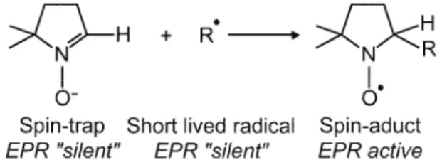
3.2. Applications of EPR spin-trapping in antioxidant research
forming stabile paramagnetic species (spin adducts), which are readily detected by EPR spectroscopy (Fig. 7). Each reactive species that has been trapped shows its own specific signature EPR spectrum. The advantage of such an approach over other methods that measure the total antioxidative capacity of the compound lies in the ability of EPR to distinguish antioxidative activity against different free radicals, even when simultaneously present in the system. This can be per-formed using spectral simulations, which enable the identification of each radical and the quantification of the signal intensity. In addition, recently a number of various hydrophilic and lipophilic spin-traps have evolved120 enabling antioxi-dative measurements in any selected medium.
Fig. 7. The basic principles of the EPR spin-trapping technique.
For antioxidative studies, in vitro chemical systems are used to generate a
specific free radical (e.g., the xanthine/xanthine oxidase system or SOTS1 (di-(4-
-carboxybenzyl)hyponitrite) for O2•– or the Fenton system and Haber–Weiss-like reaction for •OH). The spin-trap is added to a selected system prior to the start of the reaction, and after a specific incubation time, the EPR signal of the spin ad-duct is recorded and the intensity of the specific trapped radical determined.56 The application of an antioxidant to the system (before the initiation of the free radical production) should lead to a decreased generation of the spin adducts, due to radical scavenging or interactions with the reactants of the generating system, which is detected by the lower intensity of the EPR signal of a given radical as compared to the control antioxidant-free system.37–42 The antioxidative activity (AA) is then calculated by comparing the signal intensities obtained in the control
setup and in the system with the antioxidant, using the simple equation:
AA = (I0 – Ix)/I0
where I0 and Ix are the intensities of the EPR spectra obtained in the control and the samples with the antioxidant, respectively. The determined antioxidative ac-tivity can then be compared to the AA of some antioxidant intrinsically present in
metabolism, such as ascorbate or tocopherol.39 This method enables a compari-son of the AA of the investigated compound with the antioxidative properties of
other previously studied compounds. An alternative approach is to determine the
interpolation from the linear regression analysis of several AA values obtained for
different concentrations of the compound or extract. Although attractive for com-parative analysis of data obtained in different studies, this approach suffers from a disadvantage as the EC50 value depends on the concentration of reactants used
in the radical generating system, which may differ from study to study. There-fore, EC50 can be used for comparison of the AA only when they were obtained
in studies using an identical experimental setup.129,130
Antioxidative activity against O2•– can be determined using the xanthi-ne/xanthine oxidase (X/XO) reaction, as an “O2•– generating” system which is also present in biological systems. EPR spin-trapping with the X/XO system was used in a number of studies to determine AA (O2•–) of β-carotene,131 vitamin E,131 glutathione,124 aminoguanidine,123 lazaroids,132 various tea extracts,133 fullerenes42 and others. In antioxidant studies, the spin-trap DMPO (5,5-dime-thyl-L-pyrroline-N-oxide) was usually applied, because of its wide availability
and low price. However, the DMPO adduct with the O2•– radical (DMPO/OOH) has a short lifetime and it is spontaneously transformed into DMPO/OH (the ad-duct of the •OH radical),56 which may result in unrealistically high values of AA (O2•–). Hence, the application of alternative spin-traps, such as DEPMPO (5-(di-ethoxyphosphoryl)-5-methyl-1-pyrroline-N-oxide) is strongly recommended since
the DEPMPO/OOH adduct undergoes transformation at a much lower rate than DMPO/OOH56 or BMPO (5-tert-butoxycarbonyl-5-methyl-1-pyrroline N-oxide), the O2•– adduct of which does not undergo transformation at all. However, DEPMPO is the spin-trap of choice when the identification of different radicals is necessary.134
As an example of the scheme outlined in the previous paragraphs, the anti-oxidative properties of extracts of chestnut (Castanea sativa L.), and fructose and
its phosphorylated forms were examined in two recent studies.25,37,39 EPR, with DEPMPO and X/XO and the Fenton system, was applied to investigate the anti-oxidative properties of extracts of chestnut (C. sativa L.) leaves, catkin, and spiny
burs in an aqueous medium in comparison to the AA of ascorbate (#1 and #2),
while spin-probing with lipophilic probes was used to determine the antioxidative activity in membranes (#2; see the following section). The EPR signals of DEPMPO/OOH in the X/XO system without and with the catkin extract are shown in Figs. 8a and 8b, respectively. Interestingly, the extracts did not show significant antioxidative activity against •OH. Based on the data present in the li-terature that chestnut extracts are predominantly composed of tannins, which are not absorbed as such but are metabolized by intestinal flora, methanolysis of the extracts was performed to simulate the degradation of tannins in human intestines (#4). In order to determine which compound(s) in the extracts may be responsible for the observed high AA values, chemical analysis of the methanolysates was
compounds, ellagic and valoneic acids were recognized previously for their anti-oxidative and anticancer properties.135,136 Hence, it was proposed that the high AA (O2•–) of chestnut extracts is most likely based on the antioxidative pro-perties of these two acids and similar compounds that were detected, such as fla-vogallonic acid. Tasks #3 and #6 were not covered in this study and will be the subject of future research. However, it was shown by others that the derivatives of tannins are present in human plasma137 and have long persistency in the body upon dietary uptake,138 indicating that they may be able to overcome the refract-tory response (#6).
Fig. 8. The characteristic eight-line EPR signals of the DEPMPO/OOH adduct generated in: a) the X/XO system (X 1.6 mM; XO 1.6 IU mL-1); b) X/XO with catkin extract (0.2 mg mL-1).
Gray – spectral simulations of the corresponding DEPMPO/OOH signals. Catkin extract AA(O2•–) = 0.65±0.02; AA(O2•–) for the same concentration of ascorbate (0.2 mg mL-1) was 0.85±0.04.39 Characteristic 8-line EPR spectra of the DEPMPO/OH adduct in: c) the Fenton
reaction (Fe2+ 0.3 mM; H
2O2 1.2 mM); d) Fenton reaction + 3 mM F16BP, AA(•OH) = = 0.91±0.01. Gray – spectral simulation of the DEPMPO/OH signal.37 Characteristic
EPR signals of the DEPMPO/OH adduct in the metal-free Haber–Weiss-like •OH-generating system (KO
It should be noted that in studies of specific compounds, some of the points in the present scheme could be found in the literature. For example, the antioxi-dative properties of fructose and its phosphorylated forms (fructose 1,6-bisphos-phate, F16BP; fructose 1-phoshate, F1P; and fructose 6-phos1,6-bisphos-phate, F6P), the bio-distribution and metabolism were extensively studied previously, were investi-gated.25 Their antioxidative activity against •OH was determined using the Fen-ton reaction (Fe2+ + H2O2→ Fe3+ + OH– + •OH), as the “•OH generating sys-tem”, which represents a constituent of various pathophysiological processes. EPR spin-trapping with the Fenton reaction is the most frequently used method for studying antioxidative properties. It was applied to determine the AA of
mono-saccharides,37 fullerenes,42 polysaccharides,128 vitamins,139 extracts of various plants,140,141 seeds,142 mushrooms,129,130 teas,133 spices143 and others. It was shown that F16BP represents a very efficient antioxidant (#1; Figs. 8c and 8d), which may be useful as an infusion sugar for the treatment of pathophysiological conditions related to oxidative stress.37 It is known that charged F16BP is pre-ferentially located in the hydrophilic medium (#2), that it is transported into the cells (#3) and that it is not metabolized outside the cell (#4), so all these points in addition to #5 were not the subject of our study. However, if not able to over-come the refractory response and to protect cells from oxidative stress (#6), the application of F16BP in treatment could be futile.144 Hence, EPR investigations were complemented by the study of intracellular antioxidative properties of F16BP in a cultured astroglial cell exposed to H2O2-mediated oxidative stress using confocal fluorescent microscopy and fluorescent markers of oxidative stress, which showed that F16BP is indeed able to overcome the refractory response and protect the cells by diminishing oxidative stress. Further research was conducted to resolve the mechanisms of the antioxidative effects of F16BP. In principle, antioxidative actions against •OH generation in the Fenton system can occur via two mechanisms: direct •OH scavenging and sequestration of a transition metal (iron, copper, manganese). In order to establish the mechanisms of antioxidative actions against •OH production, the AA(•OH) of a studied compound should be measured in two “•OH generating” systems: i) the Fenton reaction, which con-tains metal (Fe2+ + H2O2 → Fe3+ + •OH + OH–), and ii) the Haber–Weiss-like reaction, which is a metal-free system (O2•– + H2O2→ •OH + OH– + O2). The difference between AA(•OH) obtained in the first and the second system
repre-sents a measure of the metal sequestration of a certain agent. To the best of our knowledge, we were the first to apply this approach in the study of antioxidative activity of F16BP and some other monosaccharides.37 F16BP showed signifi-cantly higher AA(•OH) in the Fenton system (Figs. 8c and 8d), when compared to
sequestration and •OH scavenging, whereby the first mechanism is predomi-nant.37
The ability of any compound to sequester metals is a very important feature as it enables an antioxidant to prevent progression of Fenton chemistry, which is a more efficient strategy for stopping or slowing down oxidative stress, than attempting to scavenge already produced highly reactive •OH. For example, dif-ferent neurodegenerative conditions, such as Parkinson’s disease, Alzheimer’s disease, are most likely related to the misbalanced metabolism of redox active metals and with consequential propagation of Fenton chemistry.145,146 Contem-porary attempts to treat these conditions are primarily focused on compounds that are efficient in the sequestration of metals.146 The potential of the EPR approach presented here can be used to screen various compounds for their potential ap-plicability in the chelation therapy of neurodegenerative diseases and to further examine their in vivo effects on experimental models.
The presented examples are aimed at illustrating the principles of the ap-plication of EPR spin-trapping in antioxidant research and were focused on the biologically most important free radicals (O2•– and •OH). However, it should be stressed that antioxidative activity against various other radicals can be deter-mined using the corresponding generating system and EPR spin-trapping. In an EPR spin-trapping study of the antioxidative activity of chitosan gallate, Pasan-phan and co-workers128 used UV irradiation of AAPH (2,2’-azobis(2-amidino-propane) dihydrochloride) as the generating system of carbon-centred radicals. Schafer et al.131 applied the Photofrin/light/Fe2+ system to provoke the
genera-tion of lipid radicals in HL-60 cells, and the antioxidative effects of β-carotene, vitamin E and NO against lipid radicals were evaluated using the EPR spin-trap-ping method. Finally, a recent paper of Šentjurc et al.134 on the antioxidative
ca-pacity of leaf extract of the evergreen plant, Sempervivum tectorum, superbly
il-lustrates the outstanding possibilities of EPR spin-trapping in antioxidant re-search. UV irradiation of the liposomal system was used to generate •OH, O2•–, and carbon-centred radicals simultaneously, simulating a real biological setup. Using EPR with the spin-trap DEPMPO and spectral simulations, the authors were able to identify specific radicals, quantify their production and determine the antioxidative activities of the extract against each of these three radicals (Fig. 9).
3.3. Applications of EPR spin-probing in antioxidant research
Nitroxyl radicals (or nitroxides) are N,N-disubstituted >•NO radicals that are
widely used as spin-probes (or spin labels) in various systems, primarily because of the relatively high chemical stability of the nitroxide moiety (up to 30 min in vivo) which enables their detection not only by EPR spectroscopy, but also by
pro-cesses in biochemical and biological systems. The applications of spin-probes go beyond redox research, since their EPR spectra can depict their mobility and different characteristics of their environment (viscosity, pH, pO2, temperature,
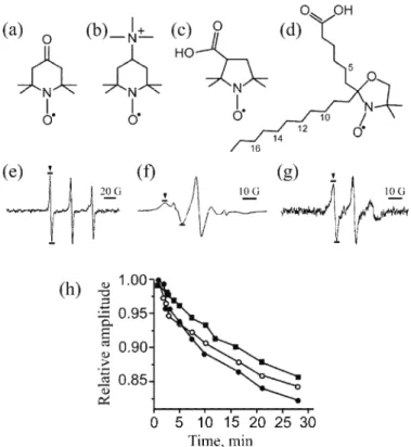
etc.).127,147,148 The three types of rings that are most commonly used for
nitro-xide spin-probes: piperidine (e.g., Tempone, Cat1), pyrrolidine (e.g., PCA) and
doxyl (doxyl stearates, e.g,. 7-DS) are shown in Fig. 10.127
Fig. 9. EPR spectra and spectral simulations of the DEPMPO adducts obtained in liposomes after 2 h irradiation with UV light (λ = 365 nm). a) Control antioxidant-free system; b) computer simulations of the spectra of DEPMPO/OH, DEPMPO/OOH, and carbon-centred radical adduct; c) liposomes + S. tec-torum (12.5 % v/v) after 2 h irradiation with UV light. AA(•OH) = 0.88; AA(•O
2–) = 0.95; AA(C-centred) = = 0.18.134
The stability of nitroxides is primarily based on steric blocking via bulky
groups (usually methyl) on the adjacent ring carbons, but is not absolute since they can be reduced to EPR-silent hydroxylamines in reactions with various antioxidants. The great assortment of available nitroxides, which can be more or less stabile (e.g., pyrrolidines are more stabile than piperidines), hydrophilic
(tempone) or lipophilic (doxyl stearates), charged (Cat1) or neutral,127 enable va-rious applications of the EPR spin-probing technique in redox research. The most frequent approach is to add a spin-probe to some biological system (ex vivo or in vivo) and to follow the decrease of the pertinent signal over time, in order to
eva-luate the intrinsic oxidative status of the system.54,127 Here, the application of EPR spin-probing in the measurement of the antioxidative capacity of a specific compound, extract or food will be illustrated. The basic principle is to combine a spin-probe with a potential antioxidant in vitro and to evaluate the total capacity
of the studied compound to reduce a spin probe, which could be specifically positioned in an aqueous solution, membranes of liposomes or cells, or in the ex-tracellular space (tasks #2 and #3). It should be noted that this approach is not very specific, since spin-probes only represent models of biological free radicals.
3.4. Evaluation of antioxidative activity with EPR spin-probing
grown potato plants exposed to jasmonic acid, in order to evaluate antioxidative and metabolic effects. Kocherginsky and co-workers150 used EPR monitored reduction of tempo and tempone to show that the “reducing power” of beer de-creases with increasing temperature and period of storage. EPR and tempone were also applied in the measurements of the antioxidative activity of various plant extracts.134,151,152 This approach was also utilised in several studies in which the antioxidative activity of chocolate,41 wild garlic (Allium ursinum L.) volatile oil,38 and the reducing power of plant plasma membranes were inves-tigated.153
Fig. 10. Chemical structure of spin-probes: a) tempone (4-oxo-2,2,6,6-tetramethylpiperidine- -1-oxyl); b) Cat1 (4-(trimethylammonio)-2,2,6,6-tetramethylpiperidine-1-oxyl); c) PCA (3-carboxy-2,2,5,5-tetramethylpyrrolidine-1-oxyl); d) 7-DS (2-(5-carboxypentyl)-2-undecyl-
-4,4-dimethyloxazolidine-3-oxyl). The numbers mark the position of the doxyl group on the fatty acid chain in other frequently used doxyl stearates – 5-DS, 10-DS, 12-DS, 14-DS, and 16-DS. Characteristic EPR spectra of tempone (e) in solution; and 7-DS (f) and 12-DS (g)
intercalated into liposomes; h) relative amplitude (compared to the amplitude at the start of incubation) of the EPR signal of tempone (■), 7-DS (○) and 12-DS (●) in the presence of
The charged spin-probe Cat1 cannot pass the membrane of cells. Hence, it can be used to study the antioxidative activity of some compounds or extracts in the extracellular medium. Hochkirch et al.154 used EPR with Cat1 to measure the antioxidative capacity of extracellular solutions in human skin biopsies exposed to UV irradiation. By evaluating the decrease in the EPR signal, they showed that UV light diminishes the activity of antioxidants in the extracellular milieu. Mehl-horn155 developed an assay for determining the concentration of ascorbate in plasma and hemolysates, based on following the rate of EPR signal disappea-rance, provoked by ascorbate-mediated reduction of Cat1.64
EPR with doxyl stearates is used to explore whether a compound or extract component(s) acts as an antioxidant inside cellular membranes. Doxyl stearates inserted in a membrane orient their hydrophilic carboxyl group toward the outer aqueous phase of the lipid bilayer and the fatty acid chain extends toward the core of the membrane. Since nitroxide groups could be placed at different po-sitions on the fatty acid chain, the antioxidative activity at different depths of the membrane could be established by measuring and comparing the kinetics of reduction of specific doxyl stearates. EPR measurements of the rates of reduction of 5-, 7-, 10-, 12-, and 16-DS were applied to evaluate the antioxidative activity of ascorbate in the membrane of unilamellar liposomes, showing that ascorbate does not occupy a specific position in the membrane and that the primary site of antioxidative activity of ascorbate is in the external medium.156 In a similar stu-dy, Schreier-Mucillo et al.157 showed that ascorbate is transported through the
membrane by diffusion, which explains the similar antioxidative activities at dif-ferent depths of the membrane. May and co-workers158 used 5-DS and 16-DS to study the antioxidative activity of ascorbate 6-palmitate (A6P) in the membrane of erythrocytes. A6P reduced 5-DS more efficiently than 16-DS, indicating that the ascorbyl group of A6P is located superficially, but with access to the hydro-phobic membrane interior. Takahashi et al.159 studied the intra-membrane
anti-oxidative activity of tocopherols by measuring the reduction of 5-, 7-, 10-, 12-, and 16-DS. It was demonstrated that tocopherols show a higher antioxidative activity closer to the membrane surface, than deep in the lipid region of the bi-layer membrane.
Measurements of the reduction of tempone and two doxyl stearates (7- and 12-DS) incorporated into liposomes were combined, to study the antioxidative properties of wild garlic (Allium ursinum L.) volatile oil (AUVO) (Fig. 10).38
than one antioxidative compound active in both media. Such complex kinetics may be deconvoluted into components in order to evaluate the number of active components in the system, as was shown on plant plasma membranes.153
3.5. Evaluation of the antioxidative capacity against lipid peroxidation
The ability of antioxidants to remove lipid peroxidation can be assessed by using a specific combination of spin-probing and radical-generating systems. The microenvironment of a spin-probe has a significant impact on its EPR spectrum; thus, specific probes could be used to evaluate membrane fluidity and some other important physiological parameters.127 Although interesting in itself, EPR spin-probing measurements of fluidity could be used in antioxidant research based on the fact that the fluidity of membranes is dependent on lipid peroxidation.160 The basic principle of this indirect approach is to provoke lipid peroxidation by ex-posing membranes to free radicals generated by the Fenton reaction or some other system. The fluidity of the membrane is measured prior and after the ad-dition of a potential antioxidant. If the antioxidant is effective against lipid pe-roxidation, its introduction into the system should remove lipid radicals and com-pensate the decrease of membrane fluidity related to peroxidation.
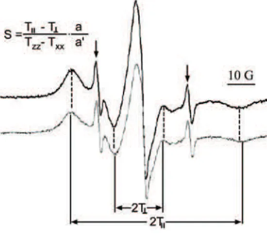
This approach was applied in several studies using liposomes or erythrocytes exposed to the Fenton system as a model of a cellular membrane exposed to oxi-dative stress, before and after the addition of plant extracts39,122 or chocolate.41 Doxyl stearates in a membrane environment have restricted motion which results in a broadening of their spectra, a feature that could be used to measure the order parameter S (Fig. 11), which is reciprocally proportional to membrane
fluidi-ty.127 Peroxidation leads to a decrease in fluidity, so an antioxidant active inside a membrane should be able to enable normal fluidity of the membrane to be re-gained. For example, the S value of an erythrocyte membrane labelled with 7-DS
was around 0.752 in normal cells and ≈0.776 for erythrocytes the membrane of
Fig. 11. EPR spectra of erythrocyte mem-brane labelled with 7-DS. Dark trace – untreated cells; pale trace – cells exposed to the Fenton system. S: order parameter. 2TII: outer hyperfine splitting. 2T┴: inner hyper-fine splitting; a: isotropic hyperfine coupl-ing constant in crystal (a = 1/3(Txx + Tyy + Tzz)); a’: isotropic hyperfine coupling
con-stant in membrane (a’ = 1/3(TII + 2T┴)). Txx, Tyy and Tzz: hyperfine constants (for
7-DS, they were taken to be Txx = Tyy = 6.1 G, Tzz = 32.4 G161). The two narrow lines
which was subjected to lipid peroxidation via the Fenton reaction. The
subse-quent application of the extracts of chestnut catkin reverted the order parameter to 0.754, showing that some lipophilic compound(s) in the extract possess the capacity to remove lipid peroxides in biomembranes.
4. CONCLUSIONS
EPR spectroscopy has played a vital role in redox research and its appli-cations are still growing. Herein two approaches that have not hitherto received full attention were addressed. In the first section, it was demonstrated that endo-genously present stabile radicals could be used for measuring the oxidative sta-tus. Although such an approach is less versatile than the application of EPR spin-traps and spin-probes, its advantage lies in the measurement of the oxidative status of biological systems without any interference with metabolic processes. Secondly, the manners in which different EPR spin-trapping and spin-probing techniques can be used to establish the efficacy of various antioxidants to remove physiologically relevant free radicals and sequestrate metal ions, and thus protect cells from oxidative damage, were presented. The intention was to encourage fel-low colleagues interested in redox research to complement the methods used in their studies with some of the EPR techniques outlined in this review and to en-hance knowledge further in this exciting area.
Acknowledgements. This work was supported by grant 41005 from the Ministry of Science and Technological Development of the Republic of Serbia. We are grateful to Pro-fessor Zlatko Giba from the Institute for Biological Research, Belgrade, Serbia, for helpful discussions.
Ј
Х Х
ХЈ
Ј 1, Ј 2, 2 2
1Institut za multidisciplinarna istra`ivawa, Univerzitet u Beogradu, Kneza Vi{eslava 1,
11000 Beograd i 2Fakultet za fizi~ku hemiju, Univerzitet u Beogradu,
Studentski trg 12–16, 11000 Beograd
,
(EPR) ,
. ,
EPR- ,
. EPR- ђ
-, ,
-, , .
- . ,
( )
- .
,
-.
( 15. , 28. 2010)
REFERENCES 1. D. Harman, J. Gerontol.11 (1956) 298
2. D. Harman, Proc. Natl. Acad. Sci. USA78 (1981) 7124
3. C. K. Mittal, F. Murad, Proc. Natl. Acad. Sci.USA 74 (1977) 4360
4. K. M. Crawford, C. S. Patt, P. J. Lad, J. Biol. Chem. 251 (1976) 7304
5. M. W. Radomski, R. M. J. Palmer, S. Moncada, Br. J. Pharmacol. 92 (1987) 639
6. S. Roth, W. Dröge, Cell Immunol.108 (1987) 417
7. R. Schreck, P. A. Baeuerle, Trends Cell. Biol. 1 (1991) 39
8. S. G. Rhee, Science312 (2006) 1882
9. W. Dröge, Physiol. Rev.82 (2002) 47
10. K. J. Barnham, C. L. Masters, A. I. Bush, Nature Rev.3 (2004) 205
11. M. R. Cookson, P. J. Shaw, Brain Pathol. 9 (1999) 165
12. Y. Luo, G. S. Roth, Antioxid. Redox Signal. 2 (2000) 449
13. J. B. De Haan, E. J. Wolvetang, F. Christiano, R. C. Iannello, C. Bladier, M. J. Kelner, I. Kola, Adv. Pharmacol. 38 (1997) 379
14. D. Dreher, A. F. Junod, Eur. J. Cancer 32 (1996) 30
15. L. W. Oberley, Free Radical Biol. Med. 5 (1988) 113
16. J. Bullen, E. Griffiths, H. Rogers, G. Ward, Microbes Infect.2 (2000) 409
17. R. W. Alexander, Hypertension 25 (1995) 155
18. N. Lane, Oxygen: The Molecule that made the World, Oxford University Press, Oxford, 2002
19. N. Lane, J. Theor. Biol.225 (2003) 531
20. B. Halliwell, Free Radical Biol. Med. 46 (2009) 531
21. M. Levine, C. Conry-Cantilena, Y. Wang, R. W. Welch, P. W. Washko, K. R. Dhariwal, J. B. Park, A. Lazarev, J. F. Graumlich, J. King, L. R. Cantilena, Proc. Natl. Acad. Sci. USA93 (1996) 3704
22. E. Herrera, C. Barbas, J. Physiol. Biochem.57 (2001) 43
23. S. Parthasarathy, N. Khan-Merchant, M. Penumetcha, B. V. Khan, N. Santanam, Curr. Atheroscler. Rep.3 (2001) 392
24. J. M. C. Gutteridge, B. Halliwell, Ann. NY Acad. Sci.899 (2000) 136
25. I. Spasojević, A. Bajić, K. Jovanović, M. Spasić, P. Andjus, Carbohyd. Res. 344 (2009)
1676
26. V. Roubaud, S. Sankarapandi, P. Kuppusamy, P. Tordo, J. L. Zweier, Anal. Biochem.247
(1997) 404
27. V. Shulaev, D. J. Oliver, Plant Physiol.141 (2006) 367
28. G. R. Buettner, B. A. Jurkiewicz, Free Radical Biol. Med. 14 (1993) 49
29. J. Bogdanović Pristov, I. Spasojević, Ž. Miković, V. Mandić, N. Cerović, M. Spasić, Oxi. Med. Cellular Longevity2 (2009) 1
30. J. Neužil, P. K. Witting, R. Stocker, Proc. Natl. Acad. Sci. USA94 (1997) 7885
32. R. S. Wood, M. Berwick, R. D. Ley, R. B. Walter, R. B. Setlow, G. S. Timmins, Proc. Natl. Acad. Sci. USA103 (2006) 4111
33. D. Kojić, I. Spasojević, M. Mojović, D. Blagojević, M. R. Worland, G. Grubor-Lajšić, M. B. Spasić, Eur. J. Entomol. 106 (2009) 451
34. B. Kukavica, M. Mojović, Ž. Vučinić, V. Maksimović, U. Takahama, S. Veljović
Jovanović, PlantCell Physiol. 50 (2009) 304
35. A. G. Ivanov, R. M. Morgan, G. R. Gray, M. Y. Velitchova, N. P. A. Huner, FEBS Lett. 430 (1998) 288
36. J. Bogdanović, M. Mojović, N. Milosavić, A. Mitrović, Ž. Vučinić, I. Spasojević, Eur. Biophys. J.37 (2008) 1241
37. I. Spasojević, M. Mojović, D. Blagojević, S. Spasić, D. Jones, A. Nikolić-Kokić, M. Spasić, Carbohyd. Res. 344 (2009) 80
38. D. Gođevac, L. Vujisić, M. Mojović, A. Ignjatović, I. Spasojević, V. Vajs, Food Chem. 107 (2008) 1692
39. J. Živković, Z. Zeković, I. Mujić, D. Gođevac, M. Mojović, A. Mujić, I. Spasojević, Food Biophys. 4 (2009) 126
40. Z. Oreščanin-Dušić, S. Milovanović, A. Nikolić-Kokić, R. Radojčić, I. Spasojević, M. Spasić, Redox Rep. 14 (2009) 48
41. J. Simonović, A. Ignjatović, I. Spasojević, M. Daković, M. Mojović, in Proceedings of the 9th international conference on fundamental and applied aspects of physical chemistry, Belgrade, Serbia, 2008, p. 391
42. M. Lens, Lj. Medenica, U. Citernesi, Biotechnol. Appl. Biochem.51 (2008) 135
43. A. Cano, J. Hernández-Ruiz, F. Garcia-Cánovas, M. Acosta, M. B. Arnao, Phytochem. Anal.9 (1998) 196
44. R. Re, N. Pellegrini, A. Proteggente, A. Pannala, M. Yang, C Rice-Evans, Free Radical Biol. Med.26 (1999) 1231
45. G. L. Ellman, Arch. Biochem. Biophys. 82 (1959) 70
46. D. J. Townsend, Mol. Intervent.7 (2007) 313
47. M. S. B. Paget, M. J. Buttner, Annu. Rev. Genet.37 (2003) 91
48. A. Nikolić Kokić, Z. Stević, S. Stojanović, P. D. Blagojević, D. R. Jones, S. Pavlović, V. Niketić, S. Apostolski, M. B. Spasić, Redox Rep.10 (2005) 265
49. D. Blagojević, Cryo. Lett.28 (2007) 137
50. M. Slavić, A. Appiah, A. Nikolić-Kokić, R. Radojičić, D. R. Jones, M. B. Spasić, Acta Physiol. Hung. 93 (2006) 335
51. D. C. Salo, R. E. Pacifici, S. W. Lin, C. Giulivi, K. J. A. Davies, J. Biol. Chem. 265
(1990) 11919
52. J. F. Buckman, H. Hernández, G. J. Kress, T. V. Votyakova, S. Pal, I. J. Reynolds, J. Neurosci. Methods104 (2001) 165
53. M. Karbowski, C. Kurono, M. Wozniak, M. Ostrowski, M. Teranishi, T. Soji, T. Wakabayashi, Biochim. Biophys. Acta1449 (1999) 25
54. S. R. Eaton, G. R. Eaton, L. J. Berliner, Biomedical EPR, Part A: Free radicals, metals, medicine and physiology, Kluwer Academic/Plenum, New York, 2005
55. H. Karoui, N. Hogg, C. Fréjaville, P. Tordo, B. Kalyanaraman, J. Biol. Chem.271 (1996)
6000
56. G. Bačić, I. Spasojevic, B. Šećerov, M. Mojović, Spectrochim. Acta A 69 (2008) 1354
58. D. Armstrong, Methods in Molecular Biology, Vol. 108, Free Radical and Antioxidant Protocols, Humana Press Inc., Totowa, NJ, USA, 1999
59. B. Halliwell, M. Whiteman, Br. J. Pharmacol. 142 (2004) 231
60. G. Bačić, M. Mojović, Ann. N. Y. Acad. Sci. 1048 (2005) 230
61. M. Mojović, I. Spasojević, G. Bačić, J. Chem. Inf. Model. 25 (2005) 1716
62. H. Zhao, J. Joseph, H. Zhang, H. Karoui, B. Kalayanaraman, Free Radical Biol. Med.31
(2001) 599
63. G. R. Buettner, Arch. Biochem. Biophys.300 (1993) 535
64. J. Vasquez-Vivar, A. M. Santos, V. B. C. Junqueira, A. Ohara, Biochem. J.314 (1996)
869
65. G. R. Buettner, J. Biochem. Biophys. Meth.16 (1988) 20
66. G. R. Buettner, Free Radical Res. Commun.10 (1990) 5
67. G. R. Buettner, B. Jurkiewicz, Radiat. Res. 145 (1996) 532
68. M.Minetti, T. Forte, M. Soriani, V. Quaresima, A. Menditoo, M. Ferrari, Biochem. J. 282
(1992) 459
69. K. Nakagawa, H. Kanno, Y. Miura, Anal. Biochem.254 (1997) 31
70. G. R. Buettner, A. G. Motten, R. D. Hall C. F. Chignell, Photochem. Photobiol. 46
(1987) 161
71. B. A. Jurkiewicz, G. R. Buettner, Photochem. Photobiol. 59 (1994) 1
72. Q. Chen, M. G. Espey, A. Y. Sun, J.-H. Lee, M. C. Krishna, E. Shacter, P. L. Choyke, C. Pooput, K. L. Kirk, G. R. Buettner, M. Levine, Proc. Natl. Acad. Sci. USA 104 (2007)
8749
73. U. Heber, C. Miyake, J. Mano, C. Ohno, K. Asada, Plant Cell Physiol. 37 (1996) 1066
74. R. V. Barbehenn, U. Poopat, B. Spencer, Insect Biochem. Mol. Biol. 33 (2003) 125
75. H. F. Galley, M. J. Davies, N. R. Webster, Crit. Care Med. 24 (1996) 1649
76. Y. Matsuo, T. Kihara, M. Ikeda, M. Ninomiya, H. Onodera, K. Kogure, J. Cereb. Blood Flow Metabol. 15 (1995) 941
77. I. M. Drake, M. J. Davies, N. P. Mapstone, M. F. Dixon, C. J. Schorah, K. L. White, D. M. Chalmers, A. T. Axon, Carcinogenesis 17 (1996) 559
78. C. A. Hubel, V. E. Kagan, E. R. Kisin, M. K. McLaughlin, J. M. Roberts, Free Radical Biol. Med. 23 (1997) 597
79. A. Menditto, D. Pietraforte, M. Minetti, Hum. Reprod.12 (1997) 1699
80. M. K. Sharma, G. R. Buettner, K. Spencer, R. E. Kerber, Circ. Res.74 (1994) 650
81. M. Galleano, L. Aimo, S. Puntarulo, Toxicol. Lett.133 (2002) 193
82. B. B. Frei, B. N. Ames, in Vitamin E in Health and Diseases, L. Packer, J. Fuchs, Eds., Marcel Dekker, New York, 1993, 131
83. E. G. Janzen, A. L. Wilcox, V. Manoharan, J. Org. Chem.58 (1993) 3597
84. H. Botti, M. Trujillo, C. Batthyany, H. Rubbo, G. Ferrer-Sueta, R. Radi, Chem. Res. Toxicol. 17 (2004) 1377
85. V. E. Kagan, N. V. Gorbunov, in Free Radical and Antioxidant Protocols, D. Armstrong, Ed., Humana Press Inc., Totowa, NJ, USA, 1999, p. 277
86. M. Matsuo, S. Matsumoto, T. Ozawa, Org. Magn. Reson.21 (1984) 261
87. J. E. Packer, T. F. Slater, R. L. Willson, Nature 278 (1979) 737
88. B. Zhou, L. M. Wu, L. Yang, Z. L. Liu, Free Radical Biol. Med.38 (2005) 78
90. P. Meredith, B. J. Powell, J. Riesz, S. P. Nighswander-Rempel, M. R. Pederson, E. G. Moore, Soft Matter2 (2006) 37
91. K. Reszka, K. Jimbow, in Oxidative stress in dermatology, J. Fuchs, L. Packer, Eds., Marcel Dekker Inc., New York, 1993, 287
92. B. Collins, T. O. Poehler, W. A. Bryden, Photochem. Photobiol. 62 (1995) 557
93. B.-L. L. Seagle, K. A. Rezai, Y. Kobori, E. M. Gasyna, K. A. Rezaei, J. R. Norris, Proc. Natl. Acad. Sci. USA 102 (2005) 8978
94. M. Rozanowska, A. Bober, J. M. Burke, T. Sarna, Photochem. Photobiol. 65 (1997) 472
95. A. El-Obeida, S. Al-Harbia, N. Al-Jomaha, A. Hassib, Phytomedicine13 (2006) 324
96. E. Buszman, B. Pilawa, M. Zdybel, S. Wilczyński, A. Gondzik, T. Witoszyńska, T. Wilczok, Sci. Total Environ.363 (2006) 195
97. M. Matuszczyk, E. Buszman, B. Pilawa, T. Witoszyńska, T. Wilczok, Chem. Phys. Lett. 394 (2004) 366
98. R. V. Barbehenn, U. Poopat, B. Spencer, Insect Biochem. Mol. Biol.33 (2003) 125
99. W. S. Enochs, T. Sarna, L. Zecca, P. A. Riley, H. M. Swartz, J. Neural. Transm. Par-kinson’s Dis. Dementia Sect.7 (1994) 83
100. L. Zecca, H. M. Swartz, J. Neural. Transm. Parkinson’s Dis. Dementia Sect. 5 (1993) 203
101. D. Sulzer, J. Bogulavsky, K. E. Larsen, G. Behr, E. Karatekin, M. H. Kleinman, N. Turro, D. Krantz, R. H. Edwards, L. A. Greene, L. Zecca, Proc. Natl. Acad. Sci. USA79 (2000)
11869
102. N. Vahidi, G. Bačić, H. M. Swartz, in Proceedings of 8th annual meeting of society of magnetic resonance in medicine, Amsterdam, Holland, 1989, p. 121
103. C. B. Summers, G. W. Felton, Insect Biochem. Mol. Biol.24 (1994) 943
104. H. M. Appel, L. W. Maines, J. Insect Physiol.41 (1995) 241
105. F. Gerson, W. Huber, Electron Spin Resonance Spectroscopy of Organic Radicals, Wiley-VCH Verlag, Weinheim, 2003
106. S. Un, Magn. Reson. Chem. 43 (2005) S229
107. D. A. Svistunenko, C. E. Cooper, Biophys. J.87 (2004) 582
108. B. Alvarez, V. Demicheli, R. Durán, M. Trujillo, C. Cerveñansky, B. A. Freeman, R. Radi, Free Radical Biol. Med.37 (2004) 813
109. M. Mojović, M. Vuletić, G. G. Bačić, Ž. Vučinić, J. Exp. Bot.55 (2004) 2523
110. G. Malanga, R. G. Kozak, S. Puntarulo, Plant Sci.141 (1999) 129
111. S. Puntarulo, S. Jasid, M. Simontacchi, Plant Signal. Behav.2 (2007) 96
112. I. Terashima, S. Funayama, K. Sonoike, Planta 193 (1994) 300
113. J. A. Pedersen, Phytochemistry17 (1978) 775
114. L. P. Kvist, J. A. Pedersen, Biochem. Syst. Ecol.14 (1986) 385
115. A. J. Pedersen, Biochem. Syst. Ecol.28 (2000) 229
116. E. Crimi, L. J. Ignarro, C. Napoli, Free Radical Res.41 (2007) 1364
117. G. Bartosz, Adv. Clin. Chem.37 (2003) 219
118. B. Halliwell, J. M. C. Gutteridge, Free radicals in biology and medicine, Clarendon, Oxford, 2007
119. A.-M. Aura, In vitro digestion models for dietary phenolic compounds, Espoo VTT Publications, Helsinki, 2005
120. J.-L. Clément, P. Tordo, Electron Paramag. Reson.20 (2007) 29
121. M. Polovka, J. Food Nutr. Res.45 (2006) 1