Gene in Twins with Hereditary Spastic Paraplegia and
Mycobacterial Disease
Xiao-Fei Kong1, Aziz Bousfiha2, Abdelfettah Rouissi3, Yuval Itan1, Avinash Abhyankar1, Vanessa Bryant1, Satoshi Okada1, Fatima Ailal2, Jacinta Bustamante4,5,6, Jean-Laurent Casanova1,4,7., Jennifer Hirst8.,
Ste´phanie Boisson-Dupuis1,4*.
1St. Giles Laboratory of Human Genetics of Infectious Diseases, Rockefeller Branch, The Rockefeller University, New York, New York, United States of America,2Clinical Immunology Unit, Department of Pediatrics, King Hassan II University, Ibn-Rochd Hospital, Casablanca, Morocco,3Pe´diatre du Secteur Libe´ral, neurologie pe´diatre, Casablanca, Morocco,4Laboratory of Human Genetics of Infectious Diseases, Necker Branch, U980, Institut National de la Sante´ et de la Recherche Me´dicale (INSERM), Paris, France,5University Paris Descartes, Paris Cite´ Sorbonne, Necker Medical School, Paris, France,6Study Center for Primary Immunodeficiencies, Assistance Publique-Hoˆpitaux de Paris (AP-HP), Necker Hospital, Paris, France,7Pediatric Immunology-Hematology Unit, Necker Hospital, Paris, France,8Cambridge Institute for Medical Research, University of Cambridge, England, UK
Abstract
We report identical twins with intellectual disability, progressive spastic paraplegia and short stature, born to a consanguineous family. Intriguingly, both children presented with lymphadenitis caused by the live Bacillus Calmette-Gue´rin (BCG) vaccine. Two syndromes – hereditary spastic paraplegia (HSP) and mycobacterial disease – thus occurred simultaneously. Whole-exome sequencing (WES) revealed a homozygous nonsense mutation (p.R1105X) of theAP4E1gene, which was confirmed by Sanger sequencing. The p.R1105X mutation has no effect onAP4E1mRNA levels, but results in lower levels of AP-4e protein and of the other components of the AP-4 complex, as shown by western blotting, immunoprecipitation and immunofluorescence. Thus, the C-terminal part of the AP-4esubunit plays an important role in maintaining the integrity of the AP-4 complex. No abnormalities of the IL-12/IFN-caxis or oxidative burst pathways were identified. In conclusion, we identified twins with autosomal recessive AP-4 deficiency associated with HSP and mycobacterial disease, suggesting that AP-4 may play important role in the neurological and immunological systems.
Citation:Kong X-F, Bousfiha A, Rouissi A, Itan Y, Abhyankar A, et al. (2013) A Novel Homozygous p.R1105X Mutation of theAP4E1Gene in Twins with Hereditary Spastic Paraplegia and Mycobacterial Disease. PLoS ONE 8(3): e58286. doi:10.1371/journal.pone.0058286
Editor:Gisela Nogales-Gadea, University Hospital Vall d’Hebron, Spain
ReceivedOctober 12, 2012;AcceptedFebruary 1, 2013;PublishedMarch 5, 2013
Copyright:ß2013 Kong et al. This is an open-access article distributed under the terms of the Creative Commons Attribution License, which permits unrestricted use, distribution, and reproduction in any medium, provided the original author and source are credited.
Funding:XFK was supported by a Stony Wold-Herbert Fund, Choh-Hao Li Memorial Fund Scholar award and the Shanghai Educational Development Foundation, YI is supported by the AXA Research Fund, and VB is supported by the Stony Wold-Herbert Fund. The Laboratory of Human Genetics of Infectious Diseases is supported in part by grants from the St. Giles Foundation, the Jeffrey Modell Foundation, The Rockefeller University Center for Clinical and Translational Science grant number 8UL1TR000043 from the National Center for Research Resources and the National Center for Advancing Sciences (NCATS), National Institutes of Health, the National Institute of Allergy and Infectious Diseases (grant numbers 5R01AI089970-02 and 5R37AI095983-02), The Rockefeller University, and the European Research Council (ERC). JLC has a research grant from the Jeffrey Modell Foundation in collaboration with Talecris BioTherapeutics. The funders had no role in study design, data collection and analysis, decision to publish, or preparation of the manuscript.
Competing Interests:YI is supported by the AXA Research Fund. JLC has a research grant from the Jeffrey Modell Foundation in collaboration with Talecris BioTherapeutics. There are no patents, products in development or marketed products to declare. This does not alter the authors’ adherence to all the PLOS ONE policies on sharing data and materials.
* E-mail: [email protected]
.These authors contributed equally to this work.
Introduction
Hereditary spastic paraplegia (HSP) constitutes a large, genet-ically diverse group of inherited neurologic disorders characterized by progressive spasticity and weakness of the lower limbs [1]. HSP is uncommon, but not rare, with a prevalence of,3–9/100,000 in most populations [1,2]. Inheritance may be X-linked recessive, autosomal recessive or dominant, and age at onset varies widely, from early childhood to adulthood [1,3]. HSP has historically been classified as ‘pure’ or ‘complicated’ on the basis of the absence (pure) or presence (complicated) of associated clinical features, such as distal amyotrophy, cognitive dysfunction, retinopathy, cataracts, ataxia, thin corpus callosum, peripheral neuropathy and deafness [1,2]. However, HSP is increasingly being classified
subunit. The N-terminal domains of the large adaptins, together with themandssubunits, form the AP ‘core’, and the C-terminal ‘appendage’ domains of the large subunits form binding platforms for the recruitment of additional coat proteins. The five AP complexes have different subcellular distributions and mediate different transport steps [9].
Susceptibility to infection with weakly virulent mycobacteria, including BCG and nontuberculous mycobacteria (or environ-mental mycobacteria), may be seen as one of the symptoms in patients with primary or acquired immune deficiencies, including chronic granulomatous disease [12] and classical severe combined immune deficiency [13], immunocompromised patients with HIV infection [14] or cystic fibrosis patients [15]. However, the infectious phenotype of these patients is not restricted to mycobacteria. By contrast, the syndrome of Mendelian suscepti-bility to mycobacterial disease (MSMD#OMIM209950) is a rare congenital disorder characterized by clinical disease caused by
weakly virulent mycobacteria, such as BCG vaccines or environ-mental mycobacteria, in otherwise healthy children [16,17]. Since the identification of the first genetic etiology of this syndrome in 1996, nine morbid genes (IFNGR1,IFNGR2,IRF8,STAT1,CYBB, NEMO, IL12B, IL12RB1 and ISG15) and 17 different genetic etiologies of MSMD have been discovered. All these defects impair the production of, or the response to IFN-c either directly or indirectly [17,18,19,20,21,22,23,24,25]. The search for new morbid genes by whole-exome sequencing (WES) has already proved successful in other infectious diseases, even when applied to a single family [26,27]. We report here twins born to a consanguineous family presenting with early-onset, complex spastic paraplegia and mycobacterial disease. We used WES to search for genetic lesions in these twins with both neurological and immunological disorders.
Figure 1. Pedigree andAP4E1mutation in P1 and P2.A). Pedigree of the family. The segregation of theAP4E1p.R1105X mutation is also indicated. B). Electrophoregram showing the homozygous mutation of P1 with respect to the control sequence. C). Illumina sequencing reads displayed for patient P1. D). Schematic diagram of the structure of AP-4e, with the delimitation and numbering of the corresponding exons. The other known mutations of theAP4E1gene and the mutation described here (in red) are also indicated.
Patients and Methods
Case Reports
The two patients (P1 and P2) are identical twins born to first-cousin Moroccan parents (Figure 1A). No complications were observed during the pregnancy, but neonatal asphyxiation was noted during the delivery. When examined at the age of three years, the twins displayed marked developmental retardation, including microcephaly, an inability to walk unaided, abnormal speech and abnormal circadian development. They kept smiling or laughing for no obvious reason. Abnormal drooling was also observed. Both patients had muscular hypotonia. No seizure or involuntary movement was observed in either of the patients. Both patients presented discreet but remarkable facial gestalt with a prominent, bulbous nose, a wide mouth and coarse features. Neurological examination revealed spastic paraplegia of the lower extremities. Denver II assessments of both children demonstrated a significant delay in the acquisition of major skills, such as motor and adaptive skills, language and social skills, at the age of three. Both patients were of short stature and had a low body weight. Their clinical features are summarized in detail in Table 1. Electroencephalography (EEG) revealed a diffusive slow wave (theta and delta wave). Based on these findings, both twins were diagnosed with type I complex HSP.
In addition to neurologic problems, both patients presented unilaterally enlarged and inflammatory axillary lymph nodes at nine months of age. Both had been vaccinated with BCG (a live attenuated strain ofMycobacterium bovis), which was injected into the
shoulder a few days after birth. The enlarged lymph nodes were removed surgically. Biopsy confirmed the presence of acid-fast bacilli, consistent with mycobacterial infection. Neither of the patients had presented any other episode of mycobacterial infection by the time of clinical evaluation at three years of age.
Ethics Statement
This study was conducted in accordance with the Helsinki Declaration, with written informed consent obtained from the parents of P1 and P2 and the other healthy individuals involved. Approval for this study was obtained from the French IRB (Comite´ de protection des personnesor CPP), INSERM and the Rockefeller IRB.
Epstein-Barr Virus (EBV) Transformation of B Lymphocytes (EBV-B Cells) and Cell Culture
PBMC were isolated from 10 ml of peripheral blood on a Ficoll gradient and were suspended in 4 ml of RPMI1640 supplemented with 20% fetal calf serum (FCS) and 0.2mg/ml cyclosporine A. We then added 1 ml of EBV medium from the B95.8 cell line [28]. We replaced half the medium every seven days, for three to four weeks, until we observed the formation of clumps of cells. EBV-B cells from the patients were maintained, at a density of 106/ml, in RPMI 1640+10% FCS, 2 mM L-glutamine, 50 units/ ml penicillin and 50mg/ml streptomycin at 37uC. Patient fibroblasts were generated from a skin biopsy sample. Primary fibroblasts were then immortalized by transfection with the SV40 Table 1.Summary of clinical neurological presentations.
Patient P1 P2
Subunit AP4E1 AP4E1
Sex F F
Age at onset Infancy Infancy
Age at observation (years) 3 3
Head circumference 46 cm (22SD) 45.5 cm (22.5SD)
Mental retardation +++ +++
Contractures – –
Early infantile hypotonia + +
Spastic tetraplegia Lower extremities only Lower extremities only
Hypertonia – –
Hyperreflexia – –
Babinski sign – –
Extensor plantar reflex – –
Epilepsy – –
Deambulation Never achieved Never achieved
Normal speech Never acquired Never acquired
Sphincter control Absent Absent
Eye evaluation Slight myopia and astigmatism Slight astigmatism
Hearing evaluation Transmission deafness on left side, normal on right side –
Neuroimaging (CT) Atrophy of the inferior vermis with cortical atrophy Atrophy of the inferior vermis with cortical atrophy
Loss of acquired function ++ ++
Purposeful hand use ++ ++
Pseudobulbar signs Drooling+++, stereotypic laughter+++, jaw jerk++, gag reflex++ Drooling+++, stereotypic laughter+++, jaw jerk++, gag reflex++
T antigen [29]. They were maintained at subconfluence in Dulbecco’s modified Eagle medium (Sigma) supplemented with 10% fetal calf serum, 2 mM L-glutamine, 50 units/ml penicillin and 50mg/ml streptomycin at 37uC, with passaging (1:2) every three to four days.
Exome Sequencing and Analysis
DNA (3mg) extracted from EBV-B cells from the patient (P1) for massively parallel sequencing was sheared with a Covaris S2 Ultrasonicator (Covaris). An adapter-ligated library was prepared with the Paired-End Sample Prep Kit V1 (Illumina). Exome capture was performed with the SureSelect Human All Exon Kit (Agilent Technologies). Single-end sequencing was performed on an Illumina Genome Analyzer IIx (Illumina), generating 72-base reads. The sequences were aligned with the human genome reference sequence (hg18 build), with BWA aligner [30]. Three open-source packages were used for downstream processing and variant calling: Genome analysis toolkit (GATK), SAMtools and Picard Tools (http://picard.sourceforge.net/). Substitution calls were made with GATK UnifiedGenotyper, whereas indel calls were made with GATK IndelGenotyperV2. All calls with a read coverage #4x and a phred-scaled SNP quality of #30 were filtered out. All the variants were annotated with the SeattleSeq SNP annotation (http://gvs.gs.washington.edu/ SeattleSeqAnnotation/).
Molecular Analysis
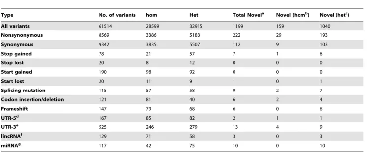
We used National Center for Biotechnology Information (NCBI) accession numbers, including NG_031875.1, NM_001252127.1 and NP_001239056.1 for the number of AP4E1 genomic DNA (gDNA), mRNA and protein sequences, respectively. Genomic DNA was isolated from the leukocytes of the patients and relatives, as previously described [31]. The 59 -cacagcaactgcaccatttt-39 and 59-tggggatctttcttgaccag–39 primers Table 2.Summary of whole-exome sequencing results.
Type No. of variants hom Het Total Novela Novel (homb) Novel (hetc)
All variants 61514 28599 32915 1199 159 1040
Nonsynonymous 8569 3386 5183 222 29 193
Synonymous 9342 3835 5507 112 9 103
Stop gained 78 21 57 7 1 6
Stop lost 20 8 12 0 0 0
Start gained 190 98 92 0 0 0
Start lost 20 11 9 1 0 1
Splicing mutation 115 57 58 9 2 7
Codon insertion/deletion 121 81 40 6 2 4
Frameshift 147 79 68 6 0 6
UTR-5d 167 85 82 2 1 1
UTR-3e 525 246 279 13 4 9
lincRNAf 129 71 58 3 0 3
miRNAg 117 42 75 10 0 10
aNumber of variants not found in dbSNP or 1000 Genomes or HapMap and,0.001% in our database; bHom: homozygous mutation;
cHet, heterozygous mutation;
dUTR-5: the five-prime untranslated region; eUTR-3: the three-prime untranslated region; f
lincRNA: long non-coding RNA; gmiRNA: microRNA.
doi:10.1371/journal.pone.0058286.t002
Figure 2. mRNA and protein levels for the subunits of the AP-4 complex.A). RT-qPCR to assess mRNA levels for the components of the AP-4 complex in EBV-B cells from P1. B). RT-PCR to assess the splicing ofAP4E1mRNA. C). Western blot: whole-cell homogenates from EBV-B cells from P1 and a healthy control were subjected to western blotting for clathrin heavy chain (CHC; loading control), AP-4e, AP-4bor AP-4m. The loss of AP-4eresults in a concomitant decrease in the levels of AP-4band AP-4m(specific bands are indicated by an arrow). These experiments were carried out at least twice.
were used to amplify and sequenceAP4E1exon 21 and its flanking intron regions. Total RNA from EBV-B cells was used to generate cDNA with the SuperScript III First-Strand Synthesis System (Invitrogen), according to the manufacturer’s protocol. We amplified exons 20 to 24 from the AP4E1 cDNA with the following primers: Forward primer: 59-agctg ctggcatcatcatta-39 and reverse primer: 59 tggggatctttcttgaccag-39. Polymerase chain reaction (PCR) was performed with Taq polymerase (Applied Biosystems) under the following conditions: 95uC for 10 minutes, 35 cycles of denaturation at 95uC for 30 seconds, annealing at 62uC for 30 seconds and extension at 72uC for 1 min. PCR products were analyzed by electrophoresis in agarose gels, purified on Sephadex, sequenced with the Big Dye Terminator cycle sequencing kit (Applied Biosystems, Foster City, CA) and analyzed on an ABI Prism 3730 (Applied Biosystems, Foster City, CA). We assessed the mRNA levels for the components of the AP-4 complex by reverse transcription- quantitative real-time PCR (RT-qPCR) on an ABI7500. All samples were analyzed in with triplicate, with TaqMan gene expression assays (AP4B1, Hs01017576_m1; AP4E1, Hs00989109_m1; AP4M1, Hs00428091_m1; AP4S1, Hs00393029_m1, Applied Biosystems), with GUS (b-glucuroni-dase) gene expression used for the normalization of results.
Immunofluorescence
Fibroblasts were plated on glass-bottomed dishes (Mattek), fixed with 3% formaldehyde and permeabilized with 0.1% Triton X-100. Cells were labeled with antibodies against AP-4e (mouse monoclonal BD 612019; raised against amino acids 685–794) and tepsin (rabbit polyclonal [32]), and incubated with Alexa Fluor 594-conjugated mouse and Alex Fluor 488 conjugated anti-rabbit secondary antibodies. All antibodies were used at a
concentration of 1mg/ml. The cells were imaged with a Zeiss Axiovert 200 inverted microscope with a Zeiss Plan Achromat
663 oil immersion objective, a Hamamatsu ORCA-ER2 camera
and IMPROVISION OPENLAB software.
Western Blotting and Immunoprecipitation
For western blotting, cell pellets were washed in phosphate-buffered saline (PBS), lysed in sample buffer, sonicated and equal amounts of protein were then loaded into the wells of a gel, for electrophoresis. For immunoprecipitation under native conditions, cell pellets containing equal amounts of protein were washed in PBS and then lysed in 1% IGEPAL in PBS supplemented with protease inhibitors. Insoluble material was removed by centrifu-gation at 20,0006gfor 20 min, and the sample was precleared with protein-A Sepharose. Immunoprecipitation was then per-formed with antibodies against subunits of AP-4 and the immune complexes were then isolated with protein-A sepharose. SDS– PAGE and western blots were performed by standard methods with antibodies against various AP-4 subunits [11]. All antibodies were used at a concentration of 1mg/ml. The polyclonal antibody against AP-4ewas raised in-house against amino acids 607–717. The bands were detected by enhanced chemiluminescence (ECL) and quantified with IMAGEJ software (http://rsb.info.nih.gov/ij).
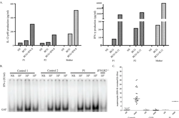
Whole-blood Assay of the IL-12/IFN-cCircuit, Electrophoretic Mobility Shift Assay (EMSA) and Oxidative Burst Test
Whole-blood assays were performed as previously described [33]. Briefly, whole blood diluted 1:2 in RPMI was either left unstimulated or was stimulated with BCG or BCG+IFN-c. Figure 3. Immunoprecipitation and immunofluorescence of the AP-4 complex in P1.A). EBV-B cells and SV40 fibroblasts from P1 and a healthy control were subjected to immunoprecipitation under native conditions with antibodies against AP-4eor AP-4b, and the immunoprecipitates were then western blotted with antibodies against AP-4eor AP-4b. Equal amounts of protein were used for immunoprecipitation from patient and control cells. A small amount of AP-4bis coassembled with AP-4e, and the AP-4eassembled with AP-4bin P1 appears to be slightly smaller (AP-4e*) than the AP-4ein control cells. B). Fibroblasts from P1 and a healthy control were double-labeled for AP-4eand the AP-4-associated protein tepsin. Note the specific loss of AP-4eand tepsin from the cells of P1. Bar 20mm. These experiments were carried out at least three times.
Supernatants were recovered 48 h later and the levels of IL-12p40 and IL-12p70 production were assessed by ELISA. EMSA was carried out as previously described [21,23]. Briefly, EBV-B cells were either left unstimulated or were stimulated with various doses of IFN-c or IFN-a for 20 min. Nuclear proteins were extracted and quantified by the Bradford method. We then incubated 10mg (stimulated with IFN-c) or 5mg (stimulated with IFN-a) of nuclear proteins with a radiolabeled GAS probe corresponding to the Fc-cR1 promoter and subjected the mixture to electrophoresis in a polyacrylamide gel. Oxidative burst assays were performed on EBV B cells as previously described [34].
Results
Identification of AP4E1 Deficiency in P1 and P2
We investigated twins (P1 and P2) born to first-cousin Moroccan parents with HSP associated with mycobacterial disease (Figure 1A). We performed WES on genomic DNA from P1 to identify the underlying genetic etiology. We found only one homozygous nonsense mutation inAP4E1 (Figure 1B, Table 2), resulting in the replacement of an arginine residue in position 1105 with a premature stop codon (p.R1105X). The homozygous mutation was confirmed by Sanger sequencing on gDNA isolated from EBV-B cells and leukocytes (Figure 1C). We collected and sequenced gDNA from peripheral blood samples from other
family members and found that both P1 and P2 carried the same homozygous mutation, whereas the parents were heterozygous and a healthy sibling was wild-type at this locus (Figure 1A). This variant was not found in the sequences of 1,050 healthy controls from 51 ethnic groups from the Centre d’E´tude du Polymorphisme Humain(CEPH) panel [35,36] or in an additional 69 Moroccan controls. Furthermore, no polymorphic premature stop codon in AP4E1was reported in the databases of the NCBI and the 1000-genome project. These results suggest that theAP4E1variant is not a polymorphism, but is instead a rare mutation segregating with autosomal recessive disease in this kindred.
Molecular Characterization of theAP4E1Mutant
We then investigated mRNA levels forAP4E1and for the other components of the AP-4 complex (AP4B1,AP4M1andAP4S1), by RT-qPCR, in EBV-B cells from P1. The levels of mRNA for AP4E1, AP4B1, AP4M1 and AP4S1 did not differ significantly between cells from P1 and control cells (expressed relative to GUS; Figure 2A). The portion of the cDNA corresponding to exons 20 to 24 ofAP4E1was also amplified and no aberrant splicing form of AP4E1mRNA was found in the cells of P1 (Figure 2B). Together, the RT-qPCR and RT-PCR results suggest that the homozygous p.R1105X mutation had no qualitative or quantitative effect on AP4E1 gene transcription, at least in EBV-B cells. We then investigated protein levels by western blotting. AP-4ewas barely Figure 4. Functional study of the IFN-c/IL-12 axis and oxidative burst in P1.A). Whole-blood IL-12/IFN-cpathway screening. P1 and P2 had normal responses to IFN-cand IL-12. B). EMSA detecting GAF DNA-binding activity after IFN-cstimulation in the EBV-B cells from two healthy controls, P1 and individuals with complete recessive IFN-cR2 deficiency, used as a negative control. Cells were stimulated for 15 minutes with the indicated dose of IFN-c. C). Oxidative burst. The production of superoxide in the EBV-B cells from healthy controls, P1 and patients with chronic granulomatous disease, used as negative controls, was determined following stimulation with PMA. These experiments were carried out at least twice.
detectable in EBV-B cells from P1, but was clearly present in control cells (Figure 2C). In addition, the loss of AP-4eresulted in a concomitant decrease in the levels of both the AP-4mand AP-4b proteins in the patient’s cells. These results suggest that the p.R1105X mutation greatly impairs production of the AP-4e protein, through effects on either protein stability or translation, and that the AP-4esubunit plays an important role in maintaining the stability of the other subunits of the AP-4 adaptor complex.
The AP-4 Adaptor Complex in AP4E1 Deficiency
We also investigated the integrity of the AP-4 complex in EBV-B cells and SV40-transformed fibroblasts from P1, by carrying out native immunoprecipitation with antibodies against AP-4eor AP-4b. Western-blot analysis showed that the levels of assembled AP-4 were much lower in both fibroblasts and EBV-B cells from P1 than in those from healthy controls (IMAGE J quantification indicated that AP-4 assembly levels were more than 95% lower than those of healthy controls). The residual AP-4eseemed to be slightly smaller than the corresponding control, possibly reflecting its lower molecular weight, consistent with C-terminal truncation. The loss of AP-4 was confirmed by immunofluorescence staining to detect the AP-4 complex in fibroblasts from P1. In addition, a recently identified AP-4 binding partner, tepsin, which binds to the C-terminal appendage domain of AP-4b [32], was detectable in control fibroblasts, but not in those of P1 (Figure 3B). Overall, we have demonstrated a severe impairment of AP-4 complex formation in both the EBV-B cells and fibroblasts of P1. These results suggest that both patients display autosomal recessive AP4E1deficiency, due to an almost complete loss of expression of the AP-4 complex.
Genetic and Functional Exploration of the IL-12/IFN-c Pathways
We first investigated whether the twins (P1 and P2) had any potential immunological abnormalities that might be caused by AP-4 deficiency and would also explain the presence of mycobacterial disease. P1 and P2 had normal counts of neutrophils, monocytes, CD19+
B cells, CD3+ , CD4+
, CD8+ T cells and NK cells. No immunoglobulin or complement defect was found (data not shown). We searched the WES data for mutations in the known MSMD-causing genes (IFNGR1, IFNGR2, STAT1, IRF8,CYBB,NEMO,IL12RB1,IL12B,ISG15). We identified only one heterozygous missense variant in IFNGR1, the Q190R mutation. We then carried out functional assays to investigate the IL-12/IFN-ccircuit, which has been shown to be crucial for antimycobacterial immunity [16,33]. However, the peripheral blood cells of both patients produced normal amounts of IFN-cin response to stimulation with IL-12 plus BCG, and of IL-12p40 in response to stimulation with IFN-cplus BCG (Figure 4A). We also searched for any subtle defects in the IFN-c pathway of the patients [23,24,25]. EMSA showed that the levels of IFN-c-induced gamma-activated factor (GAF) DNA-binding activity in the EBV-B cells from P1 were normal, ruling out the possibility of the IFNGR1variant being deleterious (Figure 4B). Furthermore, superoxide production was normal in EBV-B cells from P1, ruling out mutations in genes involved in the phagocyte NADPH oxidase pathway (Figure 4C) [34]. Autosomal dominant IL12RB2 deficiency has also recently been shown to be responsible for severe primary TB [37]. A heterozygous rare variant was identified in exon 16 of IL12RB2in P1. This missense mutation replaced a lysine residue with an asparagine residue at position 768 (K768N). However, this variant was found to have no functional impact in EBV-B cells transduced with the mutant allele and in fresh blood cells stimulated with IL-12 [37]. We were
therefore unable to identify any abnormal immunological pheno-type able to explain directly the mycobacterial disease in the patients.
Search for Other Potential Disease-causing Mutations by WES
As mycobacterial disease has not been reported in other patients with AP-4 deficiency, it remained possible that another genetic disorder in the twins would explain the immunological phenotype. We continued to explore this possibility by carrying out WES analysis for P1. We detected 29 homozygous, novel, nonsynon-ymous mutations in P1, one homozygous nonsense mutation (in AP4E1), two homozygous mutations affecting splicing and two homozygous insertions/deletions (indels) (Table 2). However, none of these 34 mutations were considered likely to be related to the clinical phenotypes observed. Only four of these variants affected proteins involved in the immune system: peptidoglycan recogni-tion protein 3 (PGLYRP3; R225Q/R225Q), SLAM family member 6 (SLAMF6; K71Q/K71Q), granzyme M (GZMM; S207L/S207L), leukocyte immunoglobulin-like receptor subfamily B (with TM and ITIM domains) member 1 (LILRB1; P496L/ P496L). Polyphen and HumVar analysis predicted thePGLYRP3, SLAM6 and LILRB1 mutations to be benign, and the GZMM S207L/S207L mutation to be probably damaging [38,39,40]. However, no link between these molecules and mycobacterial infections has ever been reported. Thus, only theAP4E1mutation appeared to be a good candidate for the causal mutation responsible for mycobacterial disease.
Discussion
The neurological phenotypes in our study, together with those in five other independent studies [3–7], are highly consistent, suggesting that these patients can be considered to have ‘‘AP-4 deficiency syndrome’’ [4], a subtype of HSP. In total, 27 patients from nine kindreds, including nine with AP4E1mutations, nine withAP4B1mutations, six withAP4S1mutations, and three with AP4M1mutations [4,5,6,7,8], have a uniform clinical phenotype of type I complex HSP, characterized by severe intellectual disability, microcephaly, progressive spastic paraplegia, growth retardation and a stereotypical laugh. WES-based diagnosis should therefore be considered to check for suspected mutations affecting the AP-4 complex in patients with similar clinical phenotypes. Furthermore, WES on a single identical twin is both a reasonable and practical approach to genetic diagnosis. HSP is characterized by a length-dependent distal axonopathy of the corticospinal tracts [1,2]. Axons crossed by corticospinal and lower motor neurons may extend for up to 1 m in length and their axoplasm comprises .99% of the total cell volume. Complex intracellular machineries are required for the sorting and distribution of proteins, lipids, mRNA, organelles and other molecules over such long distances [1,2,9]. The biological basis of AP-4 deficiency remains unclear, but the severity of the phenotype suggests that AP-4 plays a unique role in a very specific pathway acting on a specific cargo or its sorting. Indeed, it has been reported that AP-4 plays a key role in polarized protein trafficking in neurons [41], and has a neuro-protective function in Alzheimer’s disease [42]. AP-4 has been shown to interact with the transmembrane AMPA glutamate receptor regulatory proteins (TARPs) [41], the d2 orphan glutamate receptor [43], and amyloid precursor protein [42], although the basis of these interactions and their physiological relevance in HSP are not understood.
abnormal-ities accounting for mycobacterial disease in these patients. First, the co-occurrence of neurological disorders and primary immune deficiency is not uncommon [44]. Indeed, patients with a mutation affecting the related AP-3 complex (AP3B1 mutation, OMIM*603401), or Hermansky-Pudlak syndrome 2 (HPS2) [45,46], have recently been reported to present with both recurrent bacterial infections and developmental delay, poor balance and intention tremor [47]. Second, the clinical penetrance of mutations, in terms of infectious diseases can be low, as observed in MSMD for patients with complete IL-12Rb1 deficiency [20], or even extremely low, as in patients with autosomal dominant IFN-cR2 deficiency [25]. In addition, at least four AP4E1-deficient patients are known not to have been vaccinated with live BCG and the vaccination histories of the other three patients are unknown (personal communications from Dr. Colleaux and Dr. Martin). Nonetheless, one patient with an AP4M1mutation [8] died from pneumonia at a very young age, although the causal pathogen was not clearly identified. Third, we identified no genetic or functional defects in the known MSMD-causing genes or IFN-c/IL-12 axis related genes, and the other homozygous mutations do not appear to be realistic candidates. Fourth, it is also possible that clinical phenotype depends on the nature of the mutation concerned and the predicted functionality of the complex. Most of the mutations in AP4E1 are probably virtually null mutations, due to early truncations or a charge substitution in a critical folded domain [4,5,6,7,8]. In our case, the nonsense mutation p.R1105X, which is located close to the normal translation termination codon (p.X1137), does not
generate a transcript subject to nonsense-mediated mRNA decay (NMD) [48]. This escape from NMD may lead to the production of a C-terminally truncated protein, which may display a dominant-negative activity or a deleterious gain of function. No functional assays for AP-4 are currently available for further investigations of the potential differences between patients with AP-4 deficiencies. We thus propose that theAP4E1mutation is the most likely cause of the mycobacterial disease in our patients. The identification of more AP-4-deficient patients and detailed characterization of their clinical and immunological features are required for a full understanding of the role of AP-4 in neurons and, possibly, in immune cells.
Acknowledgments
We thank Laurence Colleaux, Christa L Martin and Grazia M.D. Mancini for helpful discussions. We thank Margaret Robinson for initial discussions and Georg Borner for the tepsin antibody. We thank Yelena Nemir-ovskaya, Eric Anderson, Tiffany Nivare, Tatiana Kochetkov and Jacque-line Rose for technical and secretarial assistance and all members of the Laboratory of Human Genetics of Infectious Diseases for helpful discussions.
Author Contributions
Evaluated and took care of the patients: AB AR FA. Conceived and designed the experiments: XFK JH JLC SBD. Performed the experiments: XFK YI AA VB SO JB JH. Analyzed the data: XFK YI AA SBD. Wrote the paper: XFK JLC JH SBD.
References
1. Blackstone C (2012) Cellular pathways of hereditary spastic paraplegia. Annu Rev Neurosci 35: 25–47.
2. Blackstone C, O’Kane CJ, Reid E (2011) Hereditary spastic paraplegias: membrane traffic and the motor pathway. Nat Rev Neurosci 12: 31–42. 3. Schule R, Schols L (2011) Genetics of hereditary spastic paraplegias. Semin
Neurol 31: 484–493.
4. Abou Jamra R, Philippe O, Raas-Rothschild A, Eck SH, Graf E, et al. (2011) Adaptor protein complex 4 deficiency causes severe autosomal-recessive intellectual disability, progressive spastic paraplegia, shy character, and short stature. Am J Hum Genet 88: 788–795.
5. Bauer P, Leshinsky-Silver E, Blumkin L, Schlipf N, Schroder C, et al. (2012) Mutation in the AP4B1 gene cause hereditary spastic paraplegia type 47 (SPG47). Neurogenetics 13: 73–76.
6. Moreno-De-Luca A, Helmers SL, Mao H, Burns TG, Melton AM, et al. (2011) Adaptor protein complex-4 (AP-4) deficiency causes a novel autosomal recessive cerebral palsy syndrome with microcephaly and intellectual disability. J Med Genet 48: 141–144.
7. Najmabadi H, Hu H, Garshasbi M, Zemojtel T, Abedini SS, et al. (2011) Deep sequencing reveals 50 novel genes for recessive cognitive disorders. Nature 478: 57–63.
8. Verkerk AJ, Schot R, Dumee B, Schellekens K, Swagemakers S, et al. (2009) Mutation in the AP4M1 gene provides a model for neuroaxonal injury in cerebral palsy. Am J Hum Genet 85: 40–52.
9. Hirst J, Irving C, Borner GH (2012) Adaptor protein complexes AP-4 and AP-5: New players in endosomal trafficking and progressive spastic paraplegia. Traffic. 10. Dell’Angelica EC, Mullins C, Bonifacino JS (1999) AP-4, a novel protein
complex related to clathrin adaptors. J Biol Chem 274: 7278–7285. 11. Hirst J, Bright NA, Rous B, Robinson MS (1999) Characterization of a fourth
adaptor-related protein complex. Mol Biol Cell 10: 2787–2802.
12. Lee PP, Chan KW, Jiang L, Chen T, Li C, et al. (2008) Susceptibility to mycobacterial infections in children with X-linked chronic granulomatous disease: a review of 17 patients living in a region endemic for tuberculosis. Pediatr Infect Dis J 27: 224–230.
13. Patel SY, Doffinger R, Barcenas-Morales G, Kumararatne DS (2008) Genetically determined susceptibility to mycobacterial infection. J Clin Pathol 61: 1006–1012.
14. Nunn PP, McAdam KP (1988) Mycobacterial infections and AIDS. Br Med Bull 44: 801–813.
15. Mussaffi H, Rivlin J, Shalit I, Ephros M, Blau H (2005) Nontuberculous mycobacteria in cystic fibrosis associated with allergic bronchopulmonary aspergillosis and steroid therapy. Eur Respir J 25: 324–328.
16. Alcais A, Quintana-Murci L, Thaler DS, Schurr E, Abel L, et al. (2010) Life-threatening infectious diseases of childhood: single-gene inborn errors of immunity? Ann N Y Acad Sci 1214: 18–33.
17. Filipe-Santos O, Bustamante J, Chapgier A, Vogt G, de Beaucoudrey L, et al. (2006) Inborn errors of IL-12/23- and IFN-gamma-mediated immunity: molecular, cellular, and clinical features. Semin Immunol 18: 347–361. 18. Bogunovic D, Byun M, Durfee LA, Abhyankar A, Sanal O, et al. (2012)
Mycobacterial Disease and Impaired IFN-gamma Immunity in Humans with Inherited ISG15 Deficiency. Science.
19. Bustamante J, Boisson-Dupuis S, Jouanguy E, Picard C, Puel A, et al. (2008) Novel primary immunodeficiencies revealed by the investigation of paediatric infectious diseases. Curr Opin Immunol 20: 39–48.
20. de Beaucoudrey L, Samarina A, Bustamante J, Cobat A, Boisson-Dupuis S, et al. (2010) Revisiting human IL-12Rbeta1 deficiency: a survey of 141 patients from 30 countries. Medicine (Baltimore) 89: 381–402.
21. Dupuis S, Dargemont C, Fieschi C, Thomassin N, Rosenzweig S, et al. (2001) Impairment of mycobacterial but not viral immunity by a germline human STAT1 mutation. Science 293: 300–303.
22. Hambleton S, Salem S, Bustamante J, Bigley V, Boisson-Dupuis S, et al. (2011) IRF8 mutations and human dendritic-cell immunodeficiency. N Engl J Med 365: 127–138.
23. Kong XF, Ciancanelli M, Al-Hajjar S, Alsina L, Zumwalt T, et al. (2010) A novel form of human STAT1 deficiency impairing early but not late responses to interferons. Blood 116: 5895–5906.
24. Kong XF, Vogt G, Chapgier A, Lamaze C, Bustamante J, et al. (2010) A novel form of cell type-specific partial IFN-gammaR1 deficiency caused by a germ line mutation of the IFNGR1 initiation codon. Hum Mol Genet 19: 434–444. 25. Kong XF, Vogt G, Itan Y, Macura-Biegun A, Szaflarska A, et al. (2012)
Haploinsufficiency at the human IFNGR2 locus contributes to mycobacterial disease. Hum Mol Genet.
26. Bolze A, Byun M, McDonald D, Morgan NV, Abhyankar A, et al. (2010) Whole-exome-sequencing-based discovery of human FADD deficiency. Am J Hum Genet 87: 873–881.
27. Byun M, Abhyankar A, Lelarge V, Plancoulaine S, Palanduz A, et al. (2010) Whole-exome sequencing-based discovery of STIM1 deficiency in a child with fatal classic Kaposi sarcoma. J Exp Med 207: 2307–2312.
28. Tumilowicz JJ, Gallick GE, East JL, Pathak S, Trentin JJ, et al. (1984) Presence of retrovirus in the B95–8 Epstein-Barr virus-producing cell line from different sources. In Vitro 20: 486–492.
29. Ozer HL, Banga SS, Dasgupta T, Houghton J, Hubbard K, et al. (1996) SV40-mediated immortalization of human fibroblasts. Exp Gerontol 31: 303–310. 30. Li H, Durbin R (2009) Fast and accurate short read alignment with
Burrows-Wheeler transform. Bioinformatics 25: 1754–1760.
32. Borner GH, Antrobus R, Hirst J, Bhumbra GS, Kozik P, et al. (2012) Multivariate proteomic profiling identifies novel accessory proteins of coated vesicles. J Cell Biol 197: 141–160.
33. Feinberg J, Fieschi C, Doffinger R, Feinberg M, Leclerc T, et al. (2004) Bacillus Calmette Guerin triggers the IL-12/IFN-gamma axis by an IRAK-4- and NEMO-dependent, non-cognate interaction between monocytes, NK, and T lymphocytes. Eur J Immunol 34: 3276–3284.
34. Bustamante J, Arias AA, Vogt G, Picard C, Galicia LB, et al. (2011) Germline CYBB mutations that selectively affect macrophages in kindreds with X-linked predisposition to tuberculous mycobacterial disease. Nat Immunol 12: 213–221. 35. Buetow KH, Weber JL, Ludwigsen S, Scherpbier-Heddema T, Duyk GM, et al. (1994) Integrated human genome-wide maps constructed using the CEPH reference panel. Nat Genet 6: 391–393.
36. Cann HM (1992) CEPH maps. Curr Opin Genet Dev 2: 393–399. 37. Bryant VL, Okada S, Vasseur E, Camcioglu Y, Pedraza S, et al. (2012)
Haploinsufficiency at the IL12RB2 locus in patients with severe primary tuberculosis. Paper in preparation.
38. Capriotti E, Calabrese R, Casadio R (2006) Predicting the insurgence of human genetic diseases associated to single point protein mutations with support vector machines and evolutionary information. Bioinformatics 22: 2729–2734. 39. Ramensky V, Bork P, Sunyaev S (2002) Human non-synonymous SNPs: server
and survey. Nucleic Acids Res 30: 3894–3900.
40. Pao LI, Sumaria N, Kelly JM, van Dommelen S, Cretney E, et al. (2005) Functional analysis of granzyme M and its role in immunity to infection. J Immunol 175: 3235–3243.
41. Matsuda S, Miura E, Matsuda K, Kakegawa W, Kohda K, et al. (2008) Accumulation of AMPA receptors in autophagosomes in neuronal axons lacking adaptor protein AP-4. Neuron 57: 730–745.
42. Burgos PV, Mardones GA, Rojas AL, daSilva LL, Prabhu Y, et al. (2010) Sorting of the Alzheimer’s disease amyloid precursor protein mediated by the AP-4 complex. Dev Cell 18: 425–436.
43. Yap CC, Murate M, Kishigami S, Muto Y, Kishida H, et al. (2003) Adaptor protein complex-4 (AP-4) is expressed in the central nervous system neurons and interacts with glutamate receptor delta2. Mol Cell Neurosci 24: 283–295. 44. Dehkordy SF, Aghamohammadi A, Ochs HD, Rezaei N (2012) Primary
immunodeficiency diseases associated with neurologic manifestations. J Clin Immunol 32: 1–24.
45. Jung J, Bohn G, Allroth A, Boztug K, Brandes G, et al. (2006) Identification of a homozygous deletion in the AP3B1 gene causing Hermansky-Pudlak syndrome, type 2. Blood 108: 362–369.
46. Shotelersuk V, Dell’Angelica EC, Hartnell L, Bonifacino JS, Gahl WA (2000) A new variant of Hermansky-Pudlak syndrome due to mutations in a gene responsible for vesicle formation. Am J Med 108: 423–427.
47. Badolato R, Parolini S (2007) Novel insights from adaptor protein 3 complex deficiency. J Allergy Clin Immunol 120: 735–741; quiz 742–733.