2010 Sept-Oct;18(5):919-27 www.eerp.usp.br/rlae
Corresponding Author:
Cristina Maria Garcia de Lima Parada
Universidade Estadual Paulista “Júlio de Mesquita Filho”. Faculdade de Medicina de Botucatu. Departamento de Enfermagem
Campus Universitário de Rubião Júnior, s/n Bairro Rubião Júnior
CEP: 18618-970 Botucatu, SP, Brasil E-mail: cparada@fmb.unesp.br
Abnormal Vaginal Flora in Low-Risk Pregnant Women Cared for by
a Public Health Service: Prevalence and Association with Symptoms
and Findings from Gynecological Exams
Danielle Cristina Alves Feitosa Gondo
1Marli Teresinha Cassamassimo Duarte
2Márcia Guimarães da Silva
3Cristina Maria Garcia de Lima Parada
4This study identifies the prevalence of vaginal flora alterations in low-risk pregnant women
and their association with reported symptoms and gynecological exams. This quantitative,
descriptive, cross-sectional study was conducted in public primary care service units
in Botucatu, SP, Brazil from 2006 to 2008 with 289 pregnant women from a stratified
sample obtained by sampling by care unit. Tests of vaginal content were performed
using Gram’s method and testing for Trichomonas vaginalis using Diamond’s medium.
The prevalence of altered vaginal flora was 49.5%, of which bacterial vaginosis (20.7%),
vaginal candidiasis (11.8%) and intermediate flora (11.1%) were the most frequent, not
considering associations. Results revealed a high prevalence of vaginal flora alterations with
little relation to symptoms, but in agreement with findings from the gynecological exams.
Considering undesirable maternal and perinatal outcomes and feasible laboratory practices,
the establishment of a routine for diagnosing vaginal flora alterations in low-risk pregnant
women is suggested.
Descriptors: Vaginosis, Bacterial; Candidiases, Vulvovaginal; Pregnancy; Prevalence.
1 RN, M.Sc. in Nusing, Secretaria Municipal de Saúde de Botucatu, SP, Brazil. E-mail: dcafeitosa@hotmail.com.
2 RN, M.Sc. in Nursing, Assistant Professor, Faculdade de Medicina de Botucatu, Universidade Estadual Paulista “Júlio de Mesquita
Filho”, SP, Brazil. E-mail: mtduarte@fmb.unesp.br.
3 Biologist, Ph.D. in Pathology, Assistant Professor, Faculdade de Medicina de Botucatu, Universidade Estadual Paulista “Júlio de
Mesquita Filho”, SP, Brazil. E-mail: mgsilva@fmb.unesp.br.
4 RN, Ph.D. in Nursing, Adjunct Professor, Faculdade de Medicina de Botucatu, Universidade Estadual Paulista “Júlio de Mesquita Filho”, SP,
www.eerp.usp.br/rlae
Alteração de flora vaginal em gestantes de baixo risco, atendidas em
serviço público de saúde: prevalência e associação à sintomatologia e achados do exame ginecológico
Objetivou-se identificar a prevalência das alterações de flora vaginal em gestantes de baixo risco, sua associação à sintomatologia referida e exame ginecológico. É estudo quantitativo, descritivo e transversal, desenvolvido no serviço público de atenção básica de Botucatu, SP, no período de 2006 a 2008, com 289 gestantes, amostradas de forma estratificada por unidade. Realizou-se exame do conteúdo vaginal, utilizando-se coloração pelo método de Gram e pesquisa de Trichomonas vaginalis em meio líquido de Diamond. Desconsiderando-se as associações, a prevalência de flora vaginal alterada foi de 49,5%, sendo as mais frequentes: vaginose bacteriana (20,7%), candidíase vaginal (11,8%) e flora intermediária (11,1%). Os dados apontam elevada prevalência das alterações de flora vaginal, com pouca associação à sintomatologia, mas associação com achados do exame ginecológico. Considerando-se as repercussões maternas e perinatais indesejáveis e a prática laboratorial exequível, sugere-se o estabelecimento de rotina para diagnóstico das alterações de flora vaginal em gestantes de baixo risco.
Descritores: Vaginose Bacteriana; Candidíase Vulvovaginal; Gravidez; Prevalência.
Alteración de la flora vaginal en gestantes de bajo riesgo atendidas en
servicio público de salud: prevalencia y asociación a la sintomatología y hallazgos del examen ginecológico
Se tuvo por objetivo identificar la prevalencia de las alteraciones de flora vaginal en gestantes de bajo riesgo, su asociación a la sintomatología referida y examen ginecológico. Estudio cuantitativo, descriptivo y transversal, desarrollado en el servicio público de atención básica de Botucatu/SP, en el período de 2006 a 2008, con 289 gestantes, el muestreo fue realizado de forma estratificada por unidad. Se realizó examen del contenido vaginal utilizándose coloración por el método de Gram e investigación de Trichomonas
vaginalis en medio líquido de Diamond. Desconsiderándose las asociaciones, la prevalencia de flora vaginal alterada fue de 49.5%, siendo las alteraciones más frecuentes: vaginitis bacteriana (20.7%), candidiasis vaginal (11.8%) y flora intermediaria (11.1%). Los datos apuntan elevada prevalencia de las alteraciones de flora vaginal, con poca asociación a la sintomatología, pero con asociación a hallazgos del examen ginecológico. Considerándose las repercusiones maternas y perinatales indeseables y la práctica de laboratorio ejecutable, se sugiere el establecimiento de rutina para diagnóstico de las alteraciones de flora vaginal en gestantes de bajo riesgo.
Descriptores: Vaginosis Bacteriana; Candidíasis Vulvovaginal; Embarazo; Prevalencia.
Introduction
Bacterial vaginosis (BV) is a relevant issue during
pregnancy because it is associated with a higher risk of late
miscarriage, infection of the amniotic cavity, premature
rupture of membranes, preterm labor, prematurity, and
infant low birth weight(1). The mechanism through which these obstetrical complications occur is not yet totally clarified, though it is known that BV consists of changes in vaginal flora, which produces endotoxins, making
some women more vulnerable to inflammatory responses with the production of cytokines and prostaglandins that
trigger labor. Microorganisms might ascend and invade
the chorioamniotic membranes, decidua and amniotic fluid. Even the possibility of protease production by the microorganism that compose BV is possible, which
would participate in the pathogenesis of the premature
It is also known that some bacterial species that
colonize the Lower Genital Tract (LGT), especially
those associated with BV, release sialidase and
prolidase. Sialidase are enzymes that cleave sialic
acid from glycoproteins, among them the IgA, mucins
and cell receptors and thus is associated with evasion
of innate and acquired immunity, due to degradation
of cervical IgA and changes in the cellular membrane
receptors(3). Prolidase are proteolytic enzymes that degrade the extracellular matrix, encourage cell infiltration and thus contribute to breaking down the protective mucosal barrier(4). Women in the second trimester of pregnancy, with higher activities of
sialidase and prolidase, accompanied by increased
vaginal pH, are at a higher risk of premature delivery
because of the synergistic interrelationship among the
virulence factors produced by bacteria in the altered
vaginal microbiota with a consequent risk of an adverse
pregnancy outcome(5).
The metabolism resulting from the proliferation of
these bacteria promote increased production of aromatic
amines, the putrescine and cadaverine, which evaporate
and induce a bad genital odor(6). Another frequent symptom is a grayish and fluid vaginal discharge with small bubbles(7).
The Lactobacillus SP is a gram-positive bacillus that
produces components such as lactic acid, bacteriocins
and hydrogen, and which has properties that protect the vaginal flora. These components determine an acidic vaginal pH, lower than 4.5, inhibiting the growth of pathogenic bacteria. The vaginal flora is also composed of other commensal microorganisms, which in certain
situations can become pathogenic(8).
We must take into account that under normal
conditions, progesterone increases the number of
intermediate epithelial cells, resulting in a higher
availability of glycogen and decreased vaginal pH. Even
though these factors favor the presence of lactobacilli,
they are also associated with the development of
Candida sp(9).
Vulvovaginal candidiasis is caused by several
species of Candida sp, while Candida albicans is considered a commensal of the vaginal flora. The high levels of vaginal glycogen, and local heat
and humidity, are conducive to fungus activation,
especially in the second trimester of pregnancy.
Its most common symptoms are itching, urinary
discomfort, perineal irritation, and whitish discharge
in plates, while between 25% and 40% of women are
asymptomatic(10). Although it is highly prevalent, it is
not related to relevant perinatal complications(11). Vaginal trichomoniasis is an important sexually
transmitted disease, caused by Trichomonas Vaginalis
(TV), an agent that mainly infects the LGT squamous
epithelium. It is an anaerobic organism that grows
well in the absence of oxygen, in pH between 5.0 and
5.7(12).
Trichomoniasis symptoms depend on individual
conditions, aggressiveness and the number of parasites
and vary in intensity depending on the type of infection, which can be classified as acute or chronic. In acute cases, there is the classical sign of abundant, foamy,
yellowish mucopurulent discharge(13). This infection is related to various complications such as infertility,
increased risk of HIV transmission, and undesirable
pregnancy intercurrences such as the premature rupture
of membranes, premature labor, and low birth weight
newborns(12).
Even though cytolytic vaginosis is not an infectious
pathology, it is a condition frequently diagnosed as vaginal
candidiasis because of its similar symptoms. Complaints
reported by patients include whitish discharge, vaginal
and vulvar itching, dyspareunia, dysuria, and perineal
burning, markedly in the luteal phase of the menstrual
cycle. These symptoms would be determined by the
elimination of irritating substances from the cytoplasm
of intermediate cells, arising from the cytolysis process
from the action of lactobacilli. Diagnosis should include
microscopic examination of samples directly from
vaginal content through Gram’s method, to exclude
the possibility of infection by Candida sp, observing a significant increase of the number of lactobacilli, generally adhered to epithelial cells, which are also in
a larger number, while leucocytes, when present, are
rare(14).
Aerobic vaginitis is associated with aerobic
microorganisms, especially Streptococcus agalactiae
and Escherichia coli, while its characteristics are different
from those observed in bacterial vaginosis. Aerobic vaginitis raises an important inflammatory response that can cause complications during pregnancy such
as ascendant chorioamnionitis, premature rupture of
membranes and premature labor(15).
Therefore, diagnosing and treating changes in the vaginal flora and their respective associations is especially relevant during pregnancy. Despite this relevance,
health services frequently do not have the means to provide etiological treatment. Aiming to seek scientific evidence to base clinical practice during prenatal care,
www.eerp.usp.br/rlae Rev. Latino-Am. Enfermagem 2010 Sep-Oct;18(5):919-27.
general objective is to identify the prevalence of vaginal flora alterations in low-risk pregnant women based on gold-standard tests and association with reported
symptoms and gynecological exams.
Method
This quantitative, descriptive and cross-sectional
study was carried out in Botucatu with approximately
120,000 inhabitants, located in the central region of the
state of São Paulo, Brazil. The city has public primary
health care delivered through three polyclinics, three
health centers and two school center units and another
eight Family Health Units with 10 teams. In addition
to providing primary care within their scope areas,
the polyclinics are also referral centers in pediatrics,
gynecology and obstetrics, and general practice for the
Family Health units and Health centers.
Based on a prevalence of altered vaginal flora of 20% with confidence interval of 95% and a margin of error of 5%, the sample minimum size was determined by:
n= 1.96 p ˆ (1−ˆ p )
d
2
= 1.96 0.20x0.80
0.05
2
≅245
With a total of 1,006 pregnant women cared for in the various health units in 2005, a stratified sample was obtained by unit, considering the coefficient:
c = 245 1006
= 0.2435
Half way through the study, the prevalence found
was used to recalculate the sample size. As the value
obtained was lower than that initially computed, we
opted for keeping the original computation.
A total of 289 pregnant women cared for during
the prenatal period in the primary care public service
in Botucatu, of any pregnancy stage, regardless of
gynecological complaint, with at least 72 hours of sexual
abstinence and/or vaginal procedures and absence of
antibiotherapy in the 30 days prior to the collection were
included in the study.
Data Collection
Data were collected from October 2006 to March
2008 by one of the authors. To characterize the studied
women, socio-demographic information and personal
and obstetrical antecedents were obtained.
Patients included in the study were submitted to
speculum examination using a bi-valve vaginal Collins
speculum, sterilized and without any lubricating.
After this procedure, the macroscopic characteristics
of the vaginal content were noted, followed by a pH
measurement with Merk tape graduated between four
and seven, pressed against the middle third of the
lateral vaginal wall for one minute, then proceeding to
the reading according to the manufacturer’s instructions.
Then, the material of the lateral vaginal wall was collected
with a sterile swab and that content smeared onto glass
slides. After this procedure, two drops of 10% KOH were
added to the swab with vaginal content to perform a
Whiff test(18-19).
Microscopy of vaginal content was performed using
Gram’s method. Collection of vaginal content for testing
for TV was performed with an Ayre spatula and cultured
in Diamond’s medium(18-19).
The characteristics of vaginal content, a foul odor,
vaginal itching, dyspareunia and bleeding after sexual
intercourse were considered symptoms and signs when
reported by the women(18-19).
Diagnosis of abnormal vaginal flora
The proportion between Lactobacillus SP and
other microorganisms determine the type of vaginal
microbiota: Flora I indicates there is a predominance of lactobacilli and diminished ancillary flora; Flora
II is intermediary, with a diminished number of
lactobacilli coexisting with other bacteria; and Flora II
or bacterial vaginitis, occurs with marked diminished or
absence of lactobacilli and predominance of bacterial
morphotypes(20).
The diagnosis of Candidiasis was based on the
presence of blastoconidia or pseudohyphae and
neutrophils in the microscopy exam of the vaginal
content stained by Gram’s method. The diagnosis of
aerobic(15) and cytolytic(14) vaginitis was also performed
according to criteria previously mentioned.
Abnormal vaginal flora was defined as the absence
or predominance of lactobacilli (BV and intermediate vaginal flora) or a positive culture for Candidasp or TV through microscopy. Mixed flora was defined as positive for BV and VC, intermediate flora and TV and BV and
trichomoniasis. All the slides with vaginal content
were evaluated by an experienced examiner from the
Universidade Estadual Paulista (UNESP) Medical Faculty
Studied variables
The studied socio-demographic variables were: age
(years), marital status (married, single, stable union
and others), schooling (years), occupation and children
(number). The following reported signs and symptoms
were evaluated: discharge (yes, no); intensity (low,
moderate, high), bad odor (yes, no, sometimes, does
not know), genital itching (yes, no, sometimes, does not
know), dyspareunia (yes, no, sometimes) and bleeding
after intercourse (yes, not, sometimes).
The following were observed during the gynecological
exam: vaginal discharge (yes, no), intensity (low,
moderate, high), pH was measured, Wiff test or amines
test was performed (positive, negative, dubious) and
ectopia was observed (yes, no).
Data analysis
A database were created in the Excel and analyzed
using the EpiInfo software. One of the researchers
input the entire data and data consistency was checked through verification and comparison of the distribution of frequencies in associated questions; errors were
corrected. Statistical analysis was performed through the χ2 test, whose level of significance was fixed at α=0.05, with computation of respective odds ratio and confidence interval (CI=95%). Whenever relevant, correction of Yates was performed.
Ethical Procedures
This study was evaluated and approved by the
local Research Ethics Committee (of 85-2006-CEP) and
complied with recommendations for research involving
human subjects. After the study’s objectives were clarified, women were invited to participate in the study and those who consented signed free and informed
consent forms.
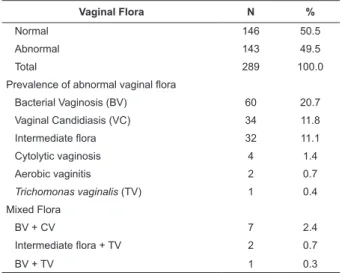
Vaginal Flora N %
Normal 146 50.5
Abnormal 143 49.5
Total 289 100.0
Prevalence of abnormal vaginal flora
Bacterial Vaginosis (BV) 60 20.7
Vaginal Candidiasis (VC) 34 11.8
Intermediate flora 32 11.1
Cytolytic vaginosis 4 1.4
Aerobic vaginitis 2 0.7
Trichomonas vaginalis (TV) 1 0.4
Mixed Flora
BV + CV 7 2.4
Intermediate flora + TV 2 0.7
BV + TV 1 0.3
Table 1 – Occurrence of abnormal vaginal flora in the studied pregnant women (n=289). Botucatu, SP, Brazil
2008
Among the study’s participants with abnormal vaginal flora, 69.9% reported discharge, 28.7%
complained of bad genital odor, 29.4% itching, 28.7%
dyspareunia and 4.9% complained of bleeding after
recent sexual intercourse. There was an association only between itching and abnormal vaginal flora (Table 2).
Results
The median age of the 289 pregnant women studied
was 25 years (14-43). Most of the women reported they
had a partner (79.2%), nine of more years of schooling
(55.4%), no paid job (56.1%) and 42.2% of them did
not have children.
The prevalence of abnormal vaginal flora was 49.5% and the most frequent ones were
BV, VC and intermediate flora: 20.7%, 11.8% and 11.1%, respectively, not considering associations. Mixed flora totaled 3.4% of the cases (Table 1).
Table 2 – Relationship between reported signs and symptoms and abnormal vaginal flora (n=289). Botucatu, SP, Brazil 2008
Abnormal vaginal flora
Signs and symptoms Yes % No % Total % p-value OR (CI 95%)
Discharge
Yes 100 69.9 91 62.3 191 66.1 0.1723 1.4 (0.9-2.3)
No 43 30.1 55 37.7 98 33.9
Bad odor
Yes 41 28.7 30 20.5 71 24.6 0.1087 1.5 (0.9-2.7)
No 102 71.3 116 79.5 218 75.4
Itching
Yes 42 29.4 24 16.4 66 22.8 0.0088 2.1 (1.2-3.7)
No 101 70.6 122 83.6 223 77.2
Rev. Latino-Am. Enfermagem 2010 Sep-Oct;18(5):919-27.
Table 2 - (continuation)
Abnormal vaginal flora
Signs and symptoms Yes % No % Total % p-value OR (CI 95%)
Dyspareunia
Yes 41 28.7 35 24.0 76 26.3 0.3643 1.3 (0.7-2.1)
No 102 71.3 111 76.0 213 73.7
Bleeding*
Yes 7 4.9 4 2.7 11 3.8 0.5157 1.8 (0.5-6.4)
No 136 95.1 142 97.3 278 96.2
* Yates Correction
The gynecological exam among women with abnormal vaginal flora indicated vaginal content in 91.6% of cases, pH was normal in 30.1% of them, the
Wiff test was positive for 36.4%, and 41.3% presented
ectopia. There was association between abnormal vaginal flora and vaginal content observed by the examiner, altered vaginal pH, positive amines test and
ectopia (Table 3).
Table 3 – Relationship between data of gynecological exams and abnormal vaginal flora (n=289). Botucatu, SP, Brazil 2008
Abnormal vaginal flora
Exam Yes % No % Total % p-value OR (CI 95%)
Vaginal content
Yes 131 91.6 108 74.0 239 82.7 0.0000 3.8 (1.9-7.7)
No 12 8.4 38 26.0 50 17.3
Altered pH
Yes 100 69.9 23 15.8 123 42.6 0.0000 12.4 (7.0-22.0)
No 43 30.1 123 84.2 166 57.4
Wiff test
Yes 52 36.4 4 2.7 56 19.4 0.0000 20.3 (7.1-58.0)
No 91 63.6 142 97.3 233 80.6
Ectopia
Yes 59 41.3 82 56.2 141 48.8 0.0112 0.5 (0.3-0.9)
No 84 58.7 64 43.8 148 51.2
Analysis concerning BV evidenced that 66.2% of the pregnant women with this type of flora alteration complained of discharge; 20.6% and 29.4% of them
reported itching and bad genital odor, respectively;
92.6% of the women presented vaginal content in the
gynecological exam; 94.1% presented altered pH; and
70.6% a positive Wiff test (Table 4).
Table 4 – Relationship of reported signs and symptoms, data from the gynecological exam and BV (n=289). Botucatu,
SP, Brazil 2008
VB
Variables Yes % No % Total % p-value OR (CI 95%)
Signs and Symptoms
Discharge
Yes 45 66.2 146 66.1 191 66.1 0.9862 1.0 (0.6-1.8)
No 23 33.8 75 33.9 98 33.9
Itching
Yes 14 20.6 52 23.5 66 22.8 0.6133 0.8 (0.4-1.6)
No 54 79.4 169 76.5 223 77.2
Bad odor
Yes 20 29.4 51 23.1 71 24.6 0.2886 1.4 (0.7-2.5)
No 48 70.6 170 76.9 218 75.4
Table 4 - (continuation)
VB
Variables Yes % No % Total % p-value OR (CI 95%)
Exam
Vaginal content*
Yes 63 92.6 176 79.6 239 82.7 0.0216 3.2 (1.2-8.5)
No 5 7.4 45 20.4 50 17.3
Altered pH*
Yes 64 94.1 59 26.7 123 42.6 0.0000 43.9 (15.3-125.9)
No 4 5.9 162 73.3 166 57.4
Wiff test
Positive 48 70.6 8 3,6 56 19.4 0.0000 63.9 (26.6-153.7)
Negative 20 29.4 213 96.4 223 80.6
* Yates correction
The women with candidiasis reported 80.5% vaginal
discharge, 56.1% itching and 43.9% bad genital odor.
Vaginal content was observed during the gynecological
exam in 90.2% of the women, altered pH in 61,0% and
positive Wiff test in 14.6% (Table 5).
Table 5 – Relationship between reported signs and symptoms, data from the gynecological exam and candidiasis
(n=289). Botucatu, SP, Brazil 2008
Candidiasis
Variables Yes % No % Total % p-value OR (CI 95%)
Signs and Symptoms
Discharge
Yes 33 80.5 158 63.7 191 66.1 0.0355 2.3 (1.0-5.3)
No 8 19.5 90 36.3 98 33.9
Itching
Yes 23 56.1 43 17.3 66 22.8 0.0000 6.1 (3.0-12.2)
No 18 43.9 205 82.7 223 77.2
Bad odor (n=282)*
Yes 15 44.1 53 21.4 68 24.1 0.0036 2.9 (1.4-6.1)
No 19 55.9 195 78.6 214 75.9
Exam
Vaginal content†
Yes 37 90.2 202 81.5 239 82.7 0.2477 2.1 (0.7-6.2)
No 4 9.8 46 18.5 50 17.3
Altered pH (n=282)*
Yes 18 52.9 98 39.5 116 41.1 0.1357 1.7 (0.8-3.5)
No 16 47.1 150 60.5 166 58.9
Wiff test
Positive 6 14.6 50 20.2 56 19.4 0.4068 0.7 (0.3-1.7)
Negative 35 85.4 198 79.8 233 80.6
* Cases of association with bacterial vaginosis were excluded † Yates correction.
Discussion
This study enabled the identification of the prevalence of abnormal vaginal flora in low-risk pregnant women, regardless of the reported complaint, using
gold-standard exams with a sample of pregnant women
from a medium sized city in the interior of São Paulo,
Brazil.
Taking into account the general prevalence (49.5%), the results reveal a high rate of abnormal vaginal flora, which is in agreement with other recent studies involving
www.eerp.usp.br/rlae Rev. Latino-Am. Enfermagem 2010 Sep-Oct;18(5):919-27.
findings are consonant with other studies that indicate it is the most prevalent vaginal flora change in the world. Intermediate vaginal flora or Flora II was found in 21.6% of the women, a frequency much higher than that found
in other studies: 13.1%(21) and 5.2%(18).
Candidiasis presented an isolated prevalence
of 11.8% and 14.2% with associations, an value
intermediate to those obtained in the same city by other
authors: 34.5%(19) and 10.2%(18).
The prevalence of trichomoniasis, considering all
cases, was low (1.4%). It is believed that hyperdiagnosis
and random treatment, especially of BV, might be leading
to a near eradication of the protozoan, which ends up
being indirectly treated(21).
The most frequent complaint of the women studied
was vaginal discharge, reported by 66.1% of them, which
is higher than the 51.6% reported by a study carried
out with pregnant women from a peripheral area of an
urban area in the south of Brazil(22). It is known that 20% or more women present mucoid and milky content
during the gestational period, which wets the underwear without, however, constituting a pathological flow(23).
Even though vaginal discharge was the most
frequent complaint among the pregnant women, it was not associated with the diagnosis of altered vaginal flora in this study. Similar to other studies’ findings(21,23), other genital complaints such as bad odor, dyspareunia and
bleeding after sexual intercourse were not associated with altered vaginal flora, either. Itching was associated with altered flora but one should consider that this symptom is frequently reported both in VC cases and
in cytolytic vaginosis, whose treatments are distinct. Hence, the non-specificity of these signs and symptoms is apparent, suggesting that their presence does not contribute to the diagnosis of abnormal vaginal flora.
Taking into account all the women examined in
this study, there was an association between observed
vaginal content and abnormal vaginal flora. There was also an association between altered measured pH and alteration of vaginal flora, while the odds ratio obtained was 12 times greater (OR=12.4). A study conducted
with pregnant women cared for in general prenatal care
in an obstetrical outpatient clinic of a university hospital
used vaginal pH for diagnosing BV and observed that pH was significantly higher in a group of pregnant women with such a diagnosis(24).
Ectopia was also associated with abnormal vaginal flora similar to the positive test of amines. However, some authors consider this test lacking the sensitivity for
diagnosing BV, subjectivity is a disadvantage; its result
depends on the examiner’s personal interpretation(9). Specifically in relation to BV, there was not any association between reported signs and symptoms and abnormal vaginal flora, though, data from the gynecological exam indicated an association with the
Amsel criteria(25).
There was association of all the reported
symptoms – vaginal discharge, bad genital odor
and itching – with VC, though when data from the
gynecological exam were considered, no association
was evidenced. We stress that the release of aromatic
amines, accountable for the bad genital odor, does not
occur in VC cases.
Taken as a whole, data from this study indicates a high prevalence of alterations in vaginal flora of low-risk pregnant women and that the reported symptoms were
not associated or unexpected associated with these
alterations, which might be explained by the subjectivity
inherent to the perception of signs and symptoms. Thus,
given the high prevalence, undesirable maternal and
perinatal repercussions and feasible laboratorial practice,
the establishment of a routine to diagnose alterations in the vaginal flora in low-risk prenatal care in outpatients is suggested.
References
1. Guerra B, Chi T, Quarta S, Morselli-Labate AM, Lazzarotto T, Pilu G, et al. Pregnancy outcome after early detection of bacterial vaginosis. Eur J Obstet Gynecol Reprod Biol. 2006; 128(1):40-5.
2. Fachini AM, Giraldo PC , Eleutério J Jr, Jacyntho C, Gonçalves
AK, Linhares I. Vaginose bacteriana e trabalho de parto
prematuro: uma Associação não muito bem compreendida. J
Bras Doenças Sex Transm. 2005;17(2):149-52.
3. Cauci S, Monte R, Driussi S, Lanzafame P, Quadrifoglio F. Impairment of the mucosal immune system: IgA and IgM cleavage detected in vaginal washings of a subgroup of patients with bacterial vaginosis. J Infect Dis. 1998;178(6):1698-706.
4. McGregor JA, French JI, Jones W, Milligan K, McKinney PJ, Patterson E, et al. Bacterial vaginosis is associated with prematurity and vaginal fluid mucinase and sialidase: results of a controlled trial of topical clindamicyn cream. Am J Obstet Gynecol. 1994;170(4):1048-59.
5. Cauci S, McGregor J, Thorsen P, Grove J, Guaschino S. Combination of vaginal sialidase and prolidase activities for prediction of low birth weight and preterm birth. Am J Obstet Gynecol. 2005;192(2):489-96.
6. Peixoto S, Ramos LO. Vulvovaginites. In: Peixoto, S. Infecção
7. Varma R, Gupta JK, James DK, Kilby MD. Do screening-preventative interventions in asymptomatic pregnancies reduce the risk of preterm delivery – A critical appraisal of the literature. Eur J Obstet Gynecol Reprod Biol. 2006; 127(2):145-59. 8. Giraldo PC, Fachini AMD, Pereira RTG, Pereira S, Nowakonski AV, Passos MRL. A pertinência do Lactobacillus sp na flora vaginal durante o trabalho de parto prematuro. J Bras Doenças
Sex Transm. 2006;18:200-3.
9. Simões JA, Discacciati MG, Silva MG. Flora vaginal normal e anormal. In: Peixoto S. Infecção genital na mulher. São Paulo:
Roca; 2007. p. 27-36.
10. Giraldo PC, Simões JA, Duarte G. Doenças sexualmente
transmissíveis. In: Neme B. Obstetrícia básica. São Paulo (SP): Sarvier; 2000. p. 481-503.
11. Simões JÁ. Complicações perinatais em gestantes assintomáticas com e sem infecções cervicovaginais [tese de
doutorado]. Campinas (SP): Universidade de Campinas; 1997. 12. Maciel GP, Tasca T, Carli GA. Aspectos clínicos, patogênese e diagnóstico de Trichomonas vaginalis. J Bras Patol Med Lab. 2004;40(3):152-60.
13. Simões JA, Discacciati MG, Brolazo EM, Portugal PM, Dini
DV, Dantas MCM. Clinical diagnosis of bacterial vaginosis. Int J Gynecol Obstet. 2006;94(1):28-32.
14. Cibley LJ, Cibley LJ. Cytolytic vaginosis. Am J Obstet Gynecol 1991;165(4):1245-9.
15. Donders GG. Definition of a type of abnormal vaginal flora that is distinct from bacterial vaginosis: aerobic vaginitis. BJOG 2002;109(1):34-43.
16. Dotto LMG, Moulin NM, Mamede MV. Prenatal care: difficulties experienced by nurses. Rev. Latino-Am. Enfermagem.
[internet]. 2006. [acesso 31 ago 2009];14(5):682-8.
Disponível em: http://www.scielo.br/scielo.php?script=sci_ arttext&pid=S0104-11692006000500007&lng=pt.doi:10.1590/ S0104-11692006000500007.
17. Feitosa DCA, Silva MG, Parada CMGL. Accuracy of simple urine tests for diagnosis of urinary tract infections in low-risk pregnant women. Rev. Latino-Am. Enfermagem. [internet]. 2009. [acesso 01 abril 2010];17(4):507-13. Disponível em:
http://www.scielo.br/scielo.php?script=sci_arttext&pid=S0104-1692009000400012&lng=pt. doi:10.1590/S0104-11692009000400012.
18. Gondo F. Prevalência das infecções do trato genital inferior em gestantes de baixo risco da Estratégia de Saúde da Família da Atenção Primária em Saúde [dissertação de mestrado]. Botucatu
(SP): Faculdade de Medicina de Botucatu da Universidade Estadual Paulista; 2007.
19. Tristão AR. Busca ativa e tratamento das infecções do
trato genital inferior de gestantes com rastreamento positivo
para diabete gestacional: repercussões maternas e perinatais [tese de doutorado]. Botucatu (SP): Faculdade de Medicina de
Botucatu daUniversidade Estadual Paulista; 2008.
20. Nugent RP, Krhon MA, Hillier SLL. Reliability of diagnosing bacterial vaginosis improved by standartization method of gram stain interpretation. J Clin Microbiol. 1991;29(2):297-301. 21. Gomes FAM. Valor do exame clínico especular e da anamnese
para o diagnóstico do corrimento vaginal [tese de doutorado].
Campinas (SP): Universidade de Campinas; 2003.
22. Fonseca TMV, César JA, Hackenhaar AA, Ulmi EE, Neumann
NA. Corrimento vaginal referido entre gestantes em localidade
urbana no sul do Brasil: prevalência e fatores associados. Cad
Saúde Pública. 2008;24(3):558-66.
23. Menezes ML, Faúndes AE. Validação do fluxograma de corrimento vaginal em gestantes. J Bras Doenças Sex Transm.
2004;16(1):38-44.
24. Carvalho MHB, Bittar RE, Maganha PPAS, Pereira SVP, Zugaib
M. Associação da vaginose bacteriana com o parto prematuro
espontâneo. Rev Bras Ginecol Obstet. 2001; 23(8):529-33. 25. Nugent RP, Krohn MA, Hillier SL. Reliability of diagnosing bacterial vaginosis is improved by a standardized method of gram stain interpretation. J Clin Microbiol. 1991; 29(2):297-301.
Received: Sep. 3rd 2009