Proton spectroscopy study of the masseter in patients
with systemic sclerosis*
Análise do masseter, por espectroscopia de próton, em pacientes com esclerose sistêmica
Marcelo Marcucci1, Nitamar Abdala2
OBJECTIVE: To evaluate metabolite concentration in the masseter of patients with systemic sclerosis, by analyzing creatine, choline, lipid and lactate levels, and correlating them with the presence of mandibular osteolysis. MATERIALS AND METHODS: The sample included 25 individuals, 15 of them with diagnosis of systemic sclerosis, divided into two groups according to the presence (group I) or absence (group II) of osteolysis, and 10 healthy individuals (group III, control). All of them were submitted to proton magnetic resonance spectroscopy with PRESS sequence and 3D acquisition. RESULTS: Metabolite analysis showed that the creatine and lipid levels were the same for the three groups. Patients in group I presented higher levels of choline when compared with group III. On the other hand, lower lactate levels were observed in groups I and II when compared with the healthy individuals. Creatine/lipid and choline/lactate ratios were the same in the three groups. CONCLUSION: Lower lactate levels were observed in the patients with systemic sclerosis (groups I and II). Choline levels were increased in the patients with mandibular osteolysis (group I). Creatine/choline, lipid/lactate and choline/lipid ratios were different among the three groups. Further studies are necessary to understand the role played by the masseter in the development of mandibular osteolysis. Keywords: Masseter; Systemic sclerosis; Proton spectroscopy.
OBJETIVO: Avaliar a concentração de metabólitos no masseter em portadores de esclerose sistêmica, ana-lisando os índices de creatina, colina, lipídio e lactato, e relacionar com a presença de osteólise mandibular. MATERIAIS E MÉTODOS: Foram selecionados 25 pacientes, sendo 15 com diagnóstico de esclerose sistê-mica e agrupados de acordo com a presença (grupo I) ou ausência (grupo II) de osteólise, e 10 indivíduos normais (grupo III, controle). Todos foram submetidos a exame de espectroscopia de próton por ressonância magnética, com técnica PRESS e aquisição tridimensional. RESULTADOS: O estudo dos metabólitos dos três grupos apresentou os mesmos valores absolutos de creatina e lipídio. Os pacientes do grupo I apresenta-ram maior quantidade de colina em relação aos do grupo III. Já os indivíduos dos grupos I e II apresentaapresenta-ram menor quantidade de lactato em relação aos indivíduos normais. Os índices creatina/lipídio e colina/lactato foram os mesmos em todos os grupos. CONCLUSÃO: Observamos menor quantidade de lactato nos pacien-tes com esclerose sistêmica (grupos I e II). A colina está aumentada nos pacienpacien-tes com osteólise mandibular (grupo I). Os índices creatina/colina, creatina/lactato, lipídio/lactato e colina/lipídio foram diferentes entre os grupos estudados. Mais estudos são necessários para a compreensão da participação do masseter no de-senvolvimento da osteólise mandibular.
Unitermos: Masseter; Esclerose sistêmica; Espectroscopia de próton.
Abstract
Resumo
* Study developed at the Department of Imaging Diagnosis – Universidade Federal de São Paulo/Escola Paulista de Medicina (Unifesp/EPM), São Paulo, SP, Brazil.
1. PhD, Supervisor of the Technical Team of the Division of Stomatology and Oromaxillofacial Surgery, Hospital Heliópolis – SUS, São Paulo, SP, Brazil.
2. PhD, Professor of Radiology, Department of Imaging Diag-nosis – Universidade Federal de São Paulo/Escola Paulista de Medicina (Unifesp/EPM), São Paulo, SP, Brazil.
Raynaud’s phenomenon is usually the first clinical manifestation among several clinical findings such as skin thickening(4),
esophageal dysmotility, restrictive pulmo-nary disease(5–7), pulmonary hypertension,
arthralgias, myopathies, myocardiopathy, and progressive renal failure(1–3,8).
Musculoskeletal system involvement is one of the most relevant causes of disabil-ity in systemic sclerosis(9). Frequently,
ero-sion of the intermediate and terminal por-tions of the phalanges (acroosteolysis) is observed, secondary to the involvement of Marcucci M, Abdala N. Proton spectroscopy study of the masseter in patients with systemic sclerosis. Radiol Bras. 2009; 42(3):145–150.
sition of collagen and glycosaminoglycans in the connective tissue of the skin and in-ternal organs(1–3). This disease has a low
in-cidence disease, affecting 19 individuals per million inhabitants, with prevalence in women (3:1). The most affected age group is between the third and fifth decade of life(2).
INTRODUCTION
Systemic sclerosis is a chronic autoim-mune inflammatory disease of unknown etiology, characterized by excessive
depo-Mailing address: Dr. Marcelo Marcucci. Rua Cônego Xavier, 276, sala 200. São Paulo, SP, Brazil, 04231-030. E-mail marcucci21@gmail.com
the fingertips skin. Less frequently, the sec-ond and fifth ribs, cervical vertebrae, the distal portion of the clavicle, radius and ulna(10,11) are affected.
Systemic sclerosis presents characteris-tic alterations in the maxillomandibular complex, such as thickening of periodon-tal ligament and areas of osteolysis in the mandible as described by Taveras(12) in
1959, coinciding with the zones of inser-tion of the lateral pterygoid, temporal and mainly the masseter muscles. Only a few studies have evaluated the involvement of the masseter muscle in systemic sclerosis as well as the possible association between this involvement and areas of mandibular osteolysis by means of electroneuromyo-graphy(13) and magnetic resonance imaging
(MRI)(14). Based on studies on mandible
microvascularization, Ramón et al.(15) have
proposed that osteolysis originates from muscle ischemia resulting from microvas-culopathy typical of systemic sclerosis.
Muscular involvement in systemic scle-rosis is probably caused by ischemia, which would lead to an insufficient oxygen and nutrient supply, inflammation, or to the sclerotic process itself. Thus, myopathy would be a primary process of the disease, and not necessarily resulting from the superjacent skin involvement(16,17). Studies
about capillary and arteriolar circulation have shown thickening of the lumen, flow reduction, stenosis and alterations of the capillary architecture(18).
The present study was aimed at evalu-ating the creatine, choline, lipid and lactate concentrations in the masseter muscle of patients with systemic sclerosis, by means of hydrogen spectroscopy, and correlating the results with the presence of mandibu-lar osteolysis.
MATERIALS AND METHODS
The present study included 25 male and female individuals, 15 of them (mean age = 43.72 ± 7.59 years) with diffuse systemic sclerosis, and 10 (mean age = 31.82 ± 12.64 years) without the disease forming a con-trol group. Patients with limited systemic sclerosis or in association with other rheu-matic diseases were excluded. The 15 pa-tients with systemic sclerosis were divided into two groups — group I, with seven
patients with systemic sclerosis and man-dibular osteolysis (Figure 1), and group II, with eight patients with systemic sclerosis without mandibular osteolysis — and the group III included the ten healthy individu-als constituting the control group. The present study was approved by the Com-mittee for Ethics in Research, and all the individuals signed a term of free and in-formed consent.
All the individuals were submitted to bilateral MRI study of the masseter (Figure 2) in a 1.5 T Sonata® (Siemens Medical
Figure 1. Panoramic radiography detail showing a concave area in angle and an ascending branch of the mandible (arrow), corresponding to osteolytic area.
Figure 2. Coronal plane, masseter visualization (arrows) in a patient with systemic sclerosis.
Systems; Erlangen, Germany) equipment with 40 mT gradient. Turbo spin echo (TSE) T2-weighted sequences were ac-quired with relaxation in the coronal plane, 2810 ms repetition time (TR), 84 ms echo time (TE), number of excitations (NEX) 2, field of view (FOV) of 230 mm and 5 mm interval. T1-weighted sequences were ac-quired with relaxation in the axial plane (479 ms TR; 13 ms TE; NEX 2; FOV 230 mm; 5 mm interval) with and without fat suppression (420 ms TR; 13 ms TE; NEX 2; FOV 230 mm; 5 mm interval). Total ac-quisition time was 15 minutes, and total ex-amination time was 25 minutes.
Magnetic resonance spectroscopy (MRS) with PRESS sequence was per-formed with a 3D-positioned single voxel. The region of interest (ROI) was repre-sented by a cubic volume visually posi-tioned at the central region of the muscle that anatomically is the site with largest tis-sue mass (Figure 3). Thus, the frequencies reading was focused exclusively on the masseter muscle, without interferences from surrounding tissues. Values for both masseters were obtained in groups I and II, while in group III, the acquisition was uni-lateral. Only the metabolites related to the muscle tissue were recorded with the re-spective frequency scales, in parts per mil-lion: creatine (3.14), choline (3.30), lactate (1.14) and lipid (1.42).
The variance analysis (ANOVA) was utilized for statistical analysis of the me-tabolites creatine, choline, lipid and lactate ratios, followed by the Bonferroni or Tamhane multiple comparisons, as neces-sary, besides Brown-Forsythe test for equality of groups variance corrections. The adopted rejection level for null hypoth-esis was 0.10 (10%). The software SPSS 11.5 was utilized for statistical analysis.
RESULTS
Figure 3. Coronal, axial and sagittal planes. Voxel position-ing on the masseter.
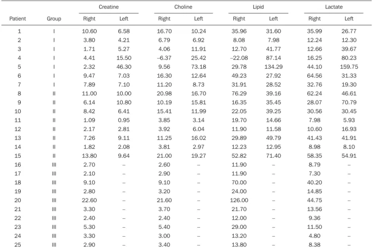
Table 1 Pure metabolites values found in the masseter as related to the groups.
systemic sclerosis, with or without osteoly-sis, as compared with the healthy individu-als.
The analysis of metabolite ratios dem-onstrated differences among the groups for creatine/choline, creatine/lactate, lipid/lac-tate and choline/lipid (Table 3).
DISCUSSION
MRS was developed early in the nine-ties, as a noninvasive method for evaluat-ing metabolites concentration in different tissues, such as the brain, heart and muscles(19). This method has been of
in-valuable help for studying the facial muscles metabolism, considering that
con-ventional methods required biopsies, with great limitations for this type of study(20).
Proton magnetic resonance spectros-copy (1H-MRS) was chosen considering
that hydrogen is the most abundant atom in the organism, with higher sensitivity, easy performance and interpretation, besides the capacity to investigate a higher number of metabolites as compared with phosphorus spectroscopy (31P-MRS)(21,22).
In the present study, creatine was the evaluated component of the energy chain. It acts as a creatine kinase substrate trans-ferring the phosphate group of the triphos-phate adenosine molecule to creatine for the production of phosphocreatine. Phos-phocreatine, on its turn, is the storing
com-pound utilized for energy production dur-ing the muscular effort(23). Creatine values
were statistically equivalent in the three groups, but a previous study with 1H-MRS
found a higher creatine concentration in the masseter muscle of patients with temporo-mandibular joint disorder, indicating higher intensity and/or frequency of bio-chemical reactions necessary for sustaining the muscular function(24).
Creatine levels remain stable in the brain tissue in several diseases(21,23),
how-ever this assertion is not valid for the mus-culoskeletal system, considering that some studies about the energy metabolism in rheumatic diseases have demonstrated vari-able results. By submitting lower limb muscles to 31P-MRS, some studies have
observed increased inorganic phosphate/ phosphocreatine levels in patients with systemic sclerosis and polymyositis(15,25),
which indicates the presence of ischemic muscle disease. On the other hand, studies with 1H-MRS have shown a decrease in
creatine concentration in the calf of patients with eosinophilia-myalgia syndrome(26)
and in patients with Duchenne muscular dystrophy(27).
As regards to the presence of lactate, the patients with systemic sclerosis presented lower concentration than the healthy indi-viduals. On the other hand, in the present study no difference was observed between patients with and without osteolysis. An in vitro study about muscular metabolism in
Duchenne muscular dystrophy has demon-strated a decrease in lactate levels in healthy individuals: the decrease in lactate concentration indicates a reduction of gly-colytic activity, or even a decrease in lac-tate concentration in the muscle(27). In the
present study, the interpretation of this find-ing can be based on the assumption that the vascular phenomena related to systemic sclerosis are not reducing the aerobic ca-pacity of the masseter because of the ab-sence of anaerobic glycolysis (lactic acid system), in the same way that there was no change in the energy demand (creatine). So, these phenomena would not significantly act on the masseter, or would act in a par-tial way, so that the extensive vascular net-work of the masseter would, in a way, be meeting its oxidative energy needs. Then, both biochemical factors do not appear to
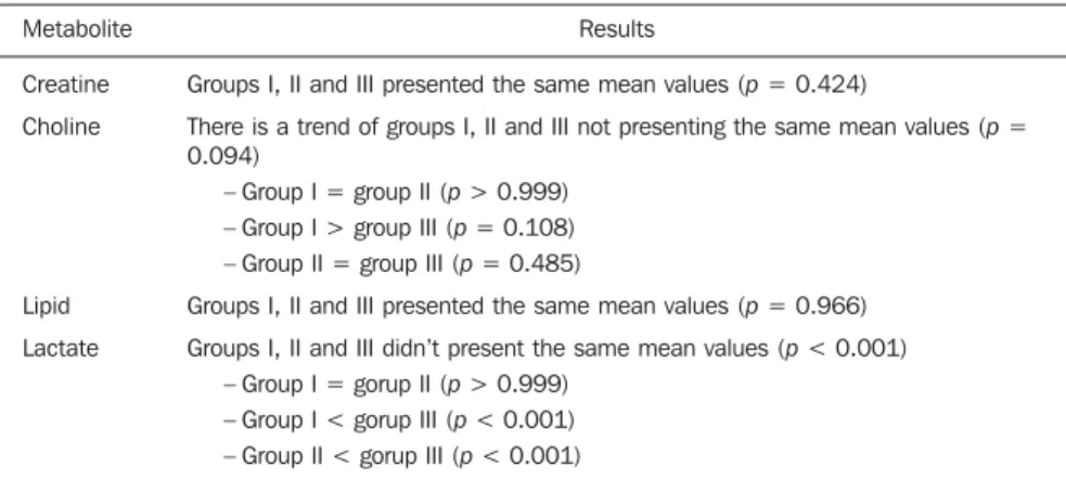
Table 2 Pure metabolite concentration as related to the groups.
Metabolite
Creatine
Choline
Lipid
Lactate
Results
Groups I, II and III presented the same mean values (p = 0.424)
There is a trend of groups I, II and III not presenting the same mean values (p = 0.094)
– Group I = group II (p > 0.999) – Group I > group III (p = 0.108) – Group II = group III (p = 0.485)
Groups I, II and III presented the same mean values (p = 0.966)
Groups I, II and III didn’t present the same mean values (p < 0.001) – Group I = gorup II (p > 0.999)
– Group I < gorup III (p < 0.001) – Group II < gorup III (p < 0.001)
Table 3 Results from metabolite ratios as related to groups.
Variable
Creatine/choline
Creatine/lipid
Creatine/lactate
Lipid/lactate
Choline/lactate
Choline/lipid
Results
Groups I, II and III didn’t present the same mean values (p < 0.001) – Group I = group II (p = 0.784)*
– Group I < group III (p < 0.001)* – Group II < group III (p < 0.001)*
Groups I, II and III presented the same mean values (p = 0.314)
Groups I, II and III didn’t present the same mean values (p = 0.019)† – Group I = group II (p = 0.423)‡
– Group I = group III (p = 0.355)‡ – Group II < group III (p = 0.040)‡
Groups I, II and III didn’t present the same mean values (p = 0.005) – Group I = group II (p > 0.999)*
– Group I < group III (p = 0.009)* – Group II < group III (p = 0.025)*
Groups I, II and III presented the same mean values (p = 0.790)
Groups I, II and III didn’t present the same mean values (p = 0.004) – Group I = group II (p > 0.999)*
– Group I > group III (p = 0.007)* – Group II > group III (p = 0.029)*
significantly contribute to the osteolysis genesis, as observed in the present study.
In the same way as creatine, no signifi-cant difference in lipid levels was observed among the three groups. Increased lipid concentration is related to cell degradation and necrosis(21), and this increase was
ob-served in patients with temporomandibu-lar joint disorder(24).
Choline was the only metabolite that showed increased level in healthy individu-als. The choline level increase reflects a process of cellular proliferation and evi-dence of cell membrane synthesis phenom-ena like those observed in neoplastic dis-eases, being increased in relation to healthy muscles(21,28). On the other hand, a decrease
in choline levels may demonstrate abnor-malities in the membrane, as observed in patients with Duchenne muscular dystro-phy(27). Increased choline levels may be
related to cell proliferation and/or increased density, and besides that, inflammatory processes may also demonstrate peaks of this metabolite resulting from the presence of a large population of inflammatory cells(28). Thus, the higher choline
concen-tration in patients with osteolysis may be related to pathophysiological mechanisms that affect the fibroblast in systemic scle-rosis(29) and to the growth of conjunctive
tissue in the epimysium and perimysium(1).
Additionally, it was observed that, in sys-temic sclerosis, one observed that that in systemic sclerosis the tissue hypoxia in-duces neoangiogenesis and that there is an intense proliferation of the vascular intima layer(30). Therefore, increased choline
lev-els may be a product of the above men-tioned phenomena, as well as expressing an inflammatory activity in the masseter muscle, with these factors as possible par-ticipants in the genesis of osteolysis.
The metabolites ratios were analyzed to evaluate their activity as a whole. The cre-atine/lipid and choline/lactate ratios were statistically equivalent in the three groups. On the other hand, the other ratios pre-sented variances suggesting significance. Previous studies have showed that choline/ lipid and creatine/lipid ratios are decreased in patients with polymyositis(26), while in
the present study, an increase was observed in the choline/lipid ratio and absence of variation in the creatine/lipid ratio in
pa-tients with systemic sclerosis. Papa-tients with systemic sclerosis also presented lower cre-atine/choline and lipid/lactate ratios when compared with healthy individuals. As re-gards creatine/lactate ratio, only the pa-tients without osteolysis showed a decrease in relation to healthy individuals.
Findings at 1H-MRS should be carefully
interpreted, as they may be a consequence of interaction of factors such as the disease activity degree, level of muscle involve-ment and may even be influenced by medi-cines such as corticosteroids utilized in the management of the disease(22). On the other
hand, this method demonstrates biochemi-cal alterations even in patients with no clinical or morphological evidence of muscle alterations(25).
CONCLUSIONS
Lower lactate levels were observed in the masseter muscle of patients with sys-temic sclerosis as compared with healthy individuals, while choline levels tend to be increased in patients with osteolysis. Cre-atine/choline, creatine/lactate, lipid/lactate and choline/lipid ratios showed a trend to-wards difference among the groups in the present study. Further studies are necessary to understand the role played by the mas-seter in the development of mandibular osteolysis.
REFERENCES
1. Kayser C, Andrade LEC. Esclerose sistêmica. In: Sato E. Guias de medicina ambulatorial e hospi-talar – reumatologia. São Paulo: Ed. Manole; 2004. p. 111–20.
2. Mayes MD, Lacey JV Jr, Beebe-Dimmer J, et al. Prevalence, incidence, survival, and disease char-acteristics of systemic sclerosis in a large US population. Arthritis Rheum. 2003;48:2246–55. 3. Badea I, Taylor M, Rosenberg A, et al. Pathogen-esis and therapeutic approaches for improved topical treatment in localized scleroderma and systemic sclerosis. Rheumatology (Oxford). 2009;48:213–21.
4. Bouer M, Chammas MC, Messina MCL, et al. Correlação clínica e ultra-sonográfica na esclero-dermia localizada cutânea. Radiol Bras. 2008;41: 87–91.
5. Azevedo ABC, Guimarães SMM, Tavares Jr WC, et al. Avaliação da tomografia de alta resolução versus radiografia de tórax na doença intersticial pulmonar na esclerose sistêmica. Radiol Bras. 2005;38:95–9.
6. Gasparetto EL, Pimenta R, Inoue C, et al. Escle-rose sistêmica progressiva: aspectos na tomogra-fia computadorizada de alta resolução. Radiol Bras. 2005;38:329–32.
7. Santos MK, Faria FB, Trad CS. Comprometi-mento pulmonar na esclerose sistêmica: revisão de casos. Radiol Bras. 2006;39:181–4. 8. Avouac J, Kowal-Bielecka O, Landewé R, et al.
European League Against Rheumatism (EULAR) Scleroderma Trial and Research group (EUSTAR) recommendations for the treatment of systemic sclerosis: methods of elaboration and results of systematic literature research. Ann Rheum Dis. 2009;68:629–34.
9. Pope JE. Musculoskeletal involvement in sclero-derma. Rheum Dis Clin North Am. 2003;29:391– 408.
10. Bassett LW, Blocka KLN, Furst DE, et al. Skel-etal findings in progressive systemic sclerosis (scleroderma). AJR Am J Roentgenol. 1981;136: 1121–6.
11. Rosa ACF, Costa EN, Machado MM, et al. Calci-nose peritendínea do tendão calcâneo associada a dermatomiosite: correlação entre radiografia con-vencional, ultra-sonografia, ressonância magné-tica e macroscopia cirúrgica. Radiol Bras. 2006; 39:75–8.
12. Taveras JM. The interpretation of radiographs. In: Schwartz L, editor. Disorders of the temporoman-dibular joint. Philadelphia: Saunders; 1959. p. 154–62.
13. Pogrel MA. Unilateral osteolysis of the mandibu-lar angle and coronoid process in scleroderma. Int J Oral Maxillofac Surg. 1988;17:155–6.
14. Ruprecht A, Dolan K, Lilly GE. Osteolysis of the mandible associated with progressive systemic sclerosis. Dentomaxillofac Radiol. 1990;19:31–3. 15. Ramón Y, Samra H, Oberman M. Mandibular condylosis and apertognathia as presenting symp-toms in progressive systemic sclerosis (sclero-derma). Pattern of mandibular bony lesions and atrophy of masticatory muscles in PSS, presum-ably caused by affected muscular arteries. Oral Surg Oral Med Oral Pathol. 1987;63:269–74. 16. King LE Jr, Olsen NJ, Puett D, et al. Quantitative
evaluation of muscle weakness in scleroderma pa-tients using magnetic resonance imaging and spectroscopy. Arch Dermatol. 1993;129:246–7. 17. Olsen NJ, King LE Jr, Park JH. Muscle abnormali-ties in scleroderma. Rheum Dis Clin North Am. 1996;22:783–96.
18. Schmidt WA, Wernicke D, Kiefer E, et al. Colour duplex sonography of finger arteries in vasculi-tis and in systemic sclerosis. Ann Rheum Dis. 2006;65:265–7.
19. Ross B, Kreis R, Ernst T. Clinical tools for the 90s: magnetic resonance spectroscopy and metabolite imaging. Eur J Radiol. 1992;14:128–40. 20. Al-Farra ET, Vandenborne K, Swift A, et al.
Mag-netic resonance spectroscopy of the masseter muscle in different facial morphological patterns. Am J Orthod Dentofacial Orthop. 2001;120:427– 34.
21. Ramin SL, Tognola WA, Spotti AR. Proton mag-netic resonance spectroscopy: clinical application in patients with brain lesions. São Paulo Med J. 2003;121:254–9.
22. Chung YL, Smith EC, Williams SCR, et al. In vivo proton magnetic resonance spectroscopy in poly-myositis and dermatopoly-myositis: a preliminary study. Eur J Med Res. 1997;2:483–7. 23. Miller BL. A review of chemical issues in H NMR
24. Nasri LFG. Análise comparativa entre os achados de ressonância magnética e de eletromiografia do músculo facial masseter, em indivíduos com e sem disfunção têmporo-mandibular [tese de dou-torado]. São Paulo: Universidade Federal de São Paulo; 2005.
25. Doornbos J, Luyten PR, Janssen M, et al. P-31 MR spectroscopy of skeletal and cardiac muscle metabolism in patients with systemic sclerosis: a multiple case study. J Magn Reson Imaging. 1994;4:165–8.
26. Schick F, Duda S, Dürk H, et al. Eosinophilia-myalgia syndrome: findings at MR imaging and proton spectroscopy of the lower leg. Magn Reson Imaging. 1994;12:513–22.
27. Sharma U, Atri S, Sharma MC, et al. Skeletal muscle metabolism in Duchenne muscular dys-trophy (DMD): an in-vitro proton NMR spectros-copy study. Magn Reson Imaging. 2003;21:145– 53.
28. Maheshwari SH, Mukherji SK, Neelon B, et al. The choline/creatine ratio in five benign
neo-plasms: comparison with squamous cell carci-noma by use of in vitro MR spectroscopy. AJNR Am J Neuroradiol. 2000;21:1930–5.