Inhibition
Gilad W. Vainer1*, Ann Saada2, Juliane Kania-Almog1, Adir Amartely3, Jacob Bar-Tana4, Rachel Hertz4
1Department of Pathology, Tel-Aviv Sourasky Medical Center, Tel-Aviv, Israel,2Monique and Jacques Roboh Department of Genetic Research, and Department of Genetics and Metabolic Diseases, Hadassah-Hebrew University Medical Center, Jerusalem, Israel,3Department of Pathology, Hadassah-Hebrew University Medical Center, Jerusalem, Israel,4Human Nutrition and Metabolism, Hebrew University Medical School, Jerusalem, Israel
Abstract
The P70 ribosomal protein S6 kinase 1 (P70S6K1) is activated by the mammalian target of rapamycin (mTORC1) and regulates proliferation, growth, and metabolism. PF-4708671 is a novel, cell-permeable, has been proposed to be a highly specific inhibitor of p70S6K1. It is used in micromolar concentration range to dissect signaling pathways downstream of mTORC1 and to study the function of p70S6K1. Here we show that PF-4708671 induces AMP-activated protein kinase (AMPK) phosphorylation and activation in immortalized mouse embryonic fibroblasts (MEF) independently of p70S6K1, due to specific inhibition of mitochondrial respiratory chain Complex I.
Citation:Vainer GW, Saada A, Kania-Almog J, Amartely A, Bar-Tana J, et al. (2014) PF-4708671 Activates AMPK Independently of p70S6K1 Inhibition. PLoS ONE 9(9): e107364. doi:10.1371/journal.pone.0107364
Editor:Petras Dzeja, Mayo Clinic, United States of America
ReceivedSeptember 23, 2013;AcceptedAugust 15, 2014;PublishedSeptember 9, 2014
Copyright:ß2014 Vainer et al. This is an open-access article distributed under the terms of the Creative Commons Attribution License, which permits unrestricted use, distribution, and reproduction in any medium, provided the original author and source are credited.
Funding:This work was financed in part by the Tel-Aviv Sourasky Medical Center and Mr. Daniel Levin donation (GWV); the Israeli Ministry of Health grant#5938 (GWV); The Israeli Cancer Association (GWV); and Israeli Science Foundation (ISF) grant#1462 (AS). The funders had no role in study design, data collection and analysis, decision to publish, or preparation of the manuscript.
Competing Interests:The authors have declared that no competing interests exist.
* Email: giladwv@tlvmc.gov.il
Introduction
P70 ribosomal protein S6 kinases (p70S6K) 1 & 2 (S6K1, S6k2) are two isoforms of the AGC kinase (protein kinase A/protein kinase G/protein kinase C) family. The AGC kinase family consists of about 60 members that share a conserved catalytic kinase domain [1], making it difficult to find a specific inhibitor for each individual member of this family.
p70S6K isoforms are activated by the phosphorylation of Thr389 catalyzed by the rapamycin-sensitive mammalian TOR complex 1 (mTORC1) [2–5]. Activated p70S6K1 is considered a key kinase in body metabolism. Activated p70S6Ks phosphory-lates rpS6 on five clustered residues, allowing the initiation of protein translation [6]. Also, mitochondria number and size is increased in p70S6K1-deficient mice, accompanied by enhanced beta-oxidation, increase in muscle AMP-activated protein kinase (AMPK), and increased life span [7,8].
PF-4708671 is a cell-permeate p70S6K1-specific inhibitor [9]. In vitro, PF-4708671 inhibits p70S6K1 at mid nanomolar concentrations (IC50of 160 nM). In a panel of,90 protein and
lipid kinases of most closely related AGC family members, PF-4708671 was found to be highly selective for p70S6K1, being 400-fold less potent for inhibiting S6K2. Furthermore, PF-4708671 does not inhibit pyruvate dehydrogenase kinase, isozyme 1 (PDK1), which acts upstream of several AGC kinases, including p70S6K1. Among the AGC kinase family, mitogen- and stress-activated protein kinase 1 (MSK1) was found to be the next most sensitive to PF-470867, with an IC50 of 950 nM in vitro.
However, PF-470867 did not appear to inhibit cellular MSK1 within the 1 to 10 micromolar concentration range, where it strongly inhibits p70S6K1 [9]. Due to its high specificity,
PF-470867 is extensively used by studies that focus on verifying the role of p70S6K1 and mTOC1 in regulating metabolism.
Here we show that PF-4708671 activates AMPK in p70S6K 1 & 2 double knockout (DKO) immortalized mouse embryonic fibroblasts (MEF), implying AMPK activation independently of P70S6K1 inhibition. It does so directly by inhibiting mitochon-drial respiratory chain Complex I. Since AMPK inhibits mTORC1 and p70S6K1 activities, its direct activation by PF-4708671 may amplify p70S6K inhibition by PF-PF-4708671.
Methods
Cell culture
Immortalized mouse embryonic fibroblasts (MEF) derived from S6K double knockout or S6K wild type cells were a kind gift of Mario Pende (Inserm, Paris, France) [10] (figure S1). MEFs were cultured in DMEM (GIBCO) supplemented with 10% fetal calf serum and Pen-Strep (Biological industries, Beit HaEmek, Israel), Rapamycin and PF-4708671(Sigma-Aldrich) as indicated. A validation batch of original PF-4708671 was kindly provided by Dario Alessi (University of Dundee, Dundee, Scotland). The compound was dissolved in dimethyl sulphoxide (DMSO) and 10 mM stock solution aliquots were kept in280uC.
Isolation of Mitochondria
buffer A containing 0.02% digitonin (Sigma-Aldrich) and re-centrifuged. The mitochondrial pellet was washed again twice with buffer A and kept at –80uC until use.
Activity of mitochondrial electron transport components
Enzymatic activities of respiratory chain complexes were measured at 37C by standard spectrophotometry, as previously
described [11]. Briefly, Complex I was measured as rotenone-sensitive NADH-CoQ reductase, by monitoring the oxidation of NADH at 340 nm in the presence of coenzyme Q1. Complex II was measured at 600 nm by succinate-mediated phenazine methosulfate reduction of dichloroindophenol by succinate dehy-drogenase (SDH). Complexes II+III were measured at 550 nm by succinate-mediated cytochrome c reduction. Complex IV (cyto-chrome c oxidase) was measured by the oxidation of reduced cytochrome c at 550 nm.
Citrate synthase (CS), a ubiquitous mitochondrial matrix enzyme serving as normalizer, was measured at 412 nm by the release of CoASH coupled to 50,50-dithiobis (2-nitrobenzoic) acid, in the presence of acetyl-CoA and oxaloacetate. Activities of mitochondrial respiratory chain complexes in the presence of added PF-4708671 are presented relative to respective activities in the presence of vehicle (DMSO). Protein concentration was determined by the Lowry method and calculated according to a bovine serum albumin (BSA) standard curve.
Seahorse oxygen consumption assay
L-15medium (Beit-Haemek biological industries; http://www. bioind.com/page_14372) containing 5% FCS (Beit-Haemek biological industries) was used as the standard medium in the XF and referred to as ‘‘assay medium’’. The Seahorse platform can calculate the extracellular acidification rate (ECAR) in a non-buffered medium. Thus, because L-15 is HEPES non-buffered extracellular acidification rate (ECAR) was not used.
MEF cells were seeded in XF 24-well cell culture microplates (Seahorse Bioscience) at 206103 cells/well (0.32 cm2) in 500ml
complete growth medium (DMEM +10% FCS) and then incubated at 37uC/5% CO2for 16 h, followed by discarding the
growth medium and washing the cells three times with 750ml of assay medium. The cells were further incubated at 37uC with no CO2 supplementation for 4 hours to allow temperature and pH
equilibration. Prior to each measurement, the XF24 Analyzer mixed the assay media for 3 min followed by 2 minutes wait to allow for oxygen pressure equilibration. Then oxygen consump-tion rate (OCR) was measured for 3 min. This was repeated to establish the OCR baseline. PF-4708671 or Rapamycin were injected into each well to reach the final working concentration of 2.5/5mM and 50 nM, respectively. DMSO prepared in assay medium was injected as control. After the compounds injection, OCR measurements were made. Responses were expressed as a percentage of the baseline rate prior to compound addition. Statistical analysis was done using Seahorse analysis software using OCR area under the curve ANOVA test, as recommended by the manufacturer.
Western blot
Cells were washed briefly in ice cold PBS, followed by adding lysis buffer (0.1M Tris pH6.8 containing 1% SDS). Lysate was incubated on ice for 30 minutes, and then centrifuged at 14,000 g at 4uC for 10 minutes. For Western blot analysis, 20mg of protein extract was subjected to 8% or 10% SDS/PAGE and transferred to nitrocellulose membranes. After blocking in 5% non-fat milk, blots were incubated with AMPK (#2532), Phospho-AMPK (#2531), ACC (#3662), Phospho-ACC (#3661), P70S6K1 (#9202), Phospho-S6 ribosomal protein 240/244 (#5364), Figure 1. AMPK activation by PF-4708671.Wild type MEF (A) and
p70S6K double knockout MEF (B) were treated with PF-4708671 as indicated. Phospho-ACC (Ser79), ACC, phospho-AMPK(Thr172), AMPK, phospho-S6 and (235/236 and 240/244 were determined by SDS-PAGE as described in Methods. Tubulin and beta-Catenin served as loading controls.
doi:10.1371/journal.pone.0107364.g001
Phospho-S6 ribosomal protein 235/236 (#4858), beta-actin (#4967) antibodies for overnight, at 4uC. All antibodies were from Cell Signaling Technology. Blots were washed in Tris buffered saline - 0.2% Tween20 (TBS-T) three times, and incubated for one hour at room temperature with anti-Rabbit or anti-Mouse secondary antibody diluted 1:350 in 5% no-fat milk (ImmPress peroxidase, Vector). Detection was performed using chemiluminescence reagent on microchemi digital platform (DNR bio-imaging systems). Protein concentration in extracts was determined using the BCA protein reagent (Pierce).
Results
In line with previous reports [9], phosphorylation of ribosomal protein S6 (rpS6) at positions 235/236 and 240/244 was dose-dependently inhibited in wild type MEF cultured in the presence of PF-4708671 (Figure 1A and S3 for densitometry data). Concomitantly with P70S6K1 inhibition, added PF-4708671 resulted in dose-dependent phosphorylation of AMPK(Thr172) and its ACC(Ser79) downstream substrate to an extent similar to that induced by oligomycin (Figure 1A). Phosphorylation of AMPK(Thr172) and ACC(Ser79) by added PF-4708671 was similarly observed in p70S6K 1/2 double knockout MEF (S6K DKO MEF ) (Figure 1B and S3 for densitometry data), indicating that AMPK activation by PF-4708671 was p70S6K1 independent. Of note, PF-4708671 decreased the phosphorylation level of rpS6(235/236, 240/244) in S6K DKO MEF, namely, indepen-dently of p70S6K1. Furthermore, the effect of PF-4708671 is not MEF specific as other cell lines, as BT-474, a human breast cancer cell line, show elevation in ACC(Ser79) phosphorylation (Figure S2).
Concomitantly with AMPK activation, PF-4708671 inhibited dose dependently the oxygen consumption of DKO MEF (Figure 2; and of BT-474 see Figure S2), resulting in 10 and 20% decrease by PF-4708671 concentrations of 2.5mM and 5mM, respectively. PF-4708671 effect in suppressing oxygen
consumption rate was immediate, steady, and maintained for over one hour.
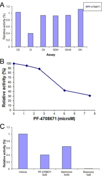
Inhibition of oxygen consumption by PF-4708671 was further pursued by studying PF-4708671 effects in isolated mitochondria. Mitochondrial citrate synthase, Complex II, succinate dehydro-genase, Complex II+III, and complex IV activities were unaffected by PF-4708671 (Figure 3A). However, mitochondrial respiratory chain Complex I activity was inhibited in a concentration dependent manner, with an IC50 of 5.2mM (Figure 3A–B).
Complex I inhibition was highly statistically significant between DMSO and 7.5mM PF-4708671 (t-Test 1.4*1026; Table S1). Inhibition of mitochondrial Complex I by 5mM PF-4708671 was comparable to that of 5 mM metformin. Mitochondrial Complex I activity was similarly inhibited by an original batch of PF-4708671 (data not shown), implying that inhibition was inherently due to PF-4708671, rather than an impurity in the commercial sample.
Discussion
The present study describes activation of AMPK by PF-4708671, a novel cell-permeable P70S6K1 inhibitor. AMPK activation by PF-4708671 is independent of p70S6K1, and may be ascribed to mitochondrial Complex I inhibition. Thus, AMPK activation and suppression of oxygen consumption by PF-4708671 were both observed in p70S6K null cells. The calculated IC50of
PF-4708671 in inhibiting mitochondrial Complex I was,5mM,
namely, within the working concentration range of 3–10mM used
to effectively block p70S6K1 [9].
p70S6K1 is activated by phosphorylation of its Thr389 by mTORC1 [3]. However, mTORC1 is one of the downstream targets of AMPK, being inhibited by AMPK via multiple mechanisms [12,13]. Hence, inhibition of p70S6K1 by PF-4708671 may be a result of both, direct inhibition of its kinase activity complemented by inhibition of its upstream mTORC1 activator by PF-4708671-activated AMPK. In line with that, Figure 2. Inhibition of oxygen consumption by PF-4708671.(A) S6K double knockout MEF were treated with vehicle (red line); rapamycin 50 nM (turquoise); PF-4708671 2.5mM (blue) and 5mM (pink). Oxygen consumption rate (OCR) of was determined by the Seahorse platform as
described in Methods. (B) ANOVA statistical analysis of the OCR area under the curve showed that after 2.5 uM or 5 uM PF-4708671 treatment the OCR was significantly decreased (p = 2.6*1025and 1.97*1029, respectively; see Table S1).
AMPK activation by 4708671 may partially account for PF-4708671 suppression of phospho-rpS6(235/235, 240/244), inde-pendently of P70S6K1 (Fig 1B). Indeed, in addition to p70S6K,
rpS6 may directly be phosphorylated by p90S6K (RSK) [6,14] that may cross talk with AMPK, independently of p70S6K1.
In conclusion, direct activation of AMPK by PF-4708671, combined with inhibition of P70S6K1 activity, may open new prospects for PF-4708671 in modifying mTORC1 and p70S6K1 control of metabolism, protein synthesis, autophagy, and prolifer-ation.
Supporting Information
Figure S1 DKO and WT MEFs comparison. p70S6K
double knockout MEF show lower levels of phospho-rpS6.Wild type MEF and p70S6K double knockout MEF were grown in complete medium. p70S6K1 and phospho-S6 (235/236 and 240/244) were determined by SDS-PAGE as described in Methods. Tubulin served as loading control.
(TIF)
Figure S2 BT-474 show pACC elevation due to PF-4708671. BT-474 were treated with PF-4708671 as indicated. Phospho-ACC (Ser79), phospho- AKT (Ser473), AKT, and phospho-S6 (235/236 and 240/244) were determined by SDS-PAGE as described in Methods. Tubulin served as loading control. (TIF)
Figure S3 Densitometry of Figure 1. AMPK activation by PF-4708671. Wild type MEF (A) and p70S6K double knockout MEF (B) were treated with PF-4708671 as indicated. Densitometry of Phospho-ACC (Ser79), ACC, phospho-AMPK(Thr172), AMPK, and phospho-S6 (235/236 and 240/ 244) were determined by chemiluminescence as described in Methods. ACC, AMPK or Tubulin served as loading controls, as indicated in the figures.
(TIF)
Figure S4 Inhibition of BT-474 oxygen consumption by PF-4708671.BT-474 were treated with vehicle (turquoise line) or 3mM of PF-4708671(pink). Oxygen consumption rate (OCR) of was determined by the Seahorse platform as described in Methods. As shown before, PF-4708671 effect in suppressing oxygen consumption rate was immediate, steady, and maintained for over one hour.
(TIF)
Table S1 Seahorse platform and mitochondrial assays statistics.ANOVA statistical analysis of the OCR area under the curve showed that after 2.5 uM or 5 uM PF-4708671 treatment the OCR was significantly decreased (manufacturer output), and mitochondrial assay statistics.
(XLSX)
Author Contributions
Conceived and designed the experiments: GWV JBT RH. Performed the experiments: GWV AS JKA AA RH. Analyzed the data: GWV AS JKA AA. Contributed reagents/materials/analysis tools: GWV AS RH. Wrote the paper: GWV JBT RH.
References
1. Pearce LR, Komander D, Alessi DR (2010) The nuts and bolts of AGC protein kinases. Nat Rev Mol Cell Biol 11: 9–22.
2. Avruch J, Hara K, Lin Y, Liu M, Long X, et al. (2006) Insulin and amino-acid regulation of mTOR signaling and kinase activity through the Rheb GTPase. Oncogene 25: 6361–6372.
3. Weng Q, Kozlowski M, Belham C, Zhang A, Comb M, et al. (1998) Regulation
4. Ben-Sahra I, Howell JJ, Asara JM, Manning BD (2013) Stimulation of de novo pyrimidine synthesis by growth signaling through mTOR and S6K1. Science 339: 1323–1328.
5. Dowling RJ, Topisirovic I, Fonseca BD, Sonenberg N (2010) Dissecting the role of mTOR: lessons from mTOR inhibitors. Biochim Biophys Acta 1804: 433– 439.
Figure 3. Inhibition of mitochondrial complex I by PF-4708671.
MEF mitochondria were prepared as described in Methods and incubated in the presence of PF-4708671, metformin or rotenone as indicated. (A) Activities of citrate synthase and mitochondrial respira-tory chain complexes were determined as described in Methods. Activities of mitochondrial respiratory chain complexes in the presence of added PF-4708671 are presented relative to respective activities in the presence of vehicle. (B) Relative activity of mitochondrial Complex I in the presence of increasing PF-4708671 concentrations as indicated. (C) Relative activity of mitochondrial Complex I in the presence of PF-4708671, metformin or rotenone as indicated. Activities of mitochon-drial respiratory chain complexes are presented relative to (DMSO). doi:10.1371/journal.pone.0107364.g003
7. Selman C, Tullet JM, Wieser D, Irvine E, Lingard SJ, et al. (2009) Ribosomal protein S6 kinase 1 signaling regulates mammalian life span. Science 326: 140– 144.
8. Um SH, Frigerio F, Watanabe M, Picard F, Joaquin M, et al. (2004) Absence of S6K1 protects against age- and diet-induced obesity while enhancing insulin sensitivity. Nature 431: 200–205.
9. Pearce LR, Alton GR, Richter DT, Kath JC, Lingardo L, et al. (2010) Characterization of PF-4708671, a novel and highly specific inhibitor of p70 ribosomal S6 kinase (S6K1). Biochem J 431: 245–255.
10. Shima H, Pende M, Chen Y, Fumagalli S, Thomas G, et al. (1998) Disruption of the p70(s6k)/p85(s6k) gene reveals a small mouse phenotype and a new functional S6 kinase. EMBO J 17: 6649–6659.
11. Saada A, Bar-Meir M, Belaiche C, Miller C, Elpeleg O (2004) Evaluation of enzymatic assays and compounds affecting ATP production in mitochondrial respiratory chain complex I deficiency. Anal Biochem 335: 66–72.
12. Xu J, Ji J, Yan XH (2012) Cross-talk between AMPK and mTOR in regulating energy balance. Crit Rev Food Sci Nutr 52: 373–381.
13. Alers S, Loffler AS, Wesselborg S, Stork B (2012) Role of AMPK-mTOR-Ulk1/ 2 in the regulation of autophagy: cross talk, shortcuts, and feedbacks. Mol Cell Biol 32: 2–11.