MECHANISMS OF DRUG ACTION
Deep brain stimulation for treatment-resistant depression: an
integrative review of preclinical and clinical
fi
ndings and
translational implications
MP Dandekar
1, AJ Fenoy
2, AF Carvalho
3, JC Soares
4and J Quevedo
1,4,5,6Although deep brain stimulation (DBS) is an established treatment choice for Parkinson
’
s disease (PD), essential tremor and
movement disorders, its effectiveness for the management of treatment-resistant depression (TRD) remains unclear. Herein, we
conducted an integrative review on major neuroanatomical targets of DBS pursued for the treatment of intractable TRD. The aim of
this review article is to provide a critical discussion of possible underlying mechanisms for DBS-generated antidepressant effects
identified in preclinical studies and clinical trials, and to determine which brain target(s) elicited the most promising outcomes
considering acute and maintenance treatment of TRD. Major electronic databases were searched to identify preclinical and clinical
studies that have investigated the effects of DBS on depression-related outcomes. Overall, 92 references met inclusion criteria, and
have evaluated six unique DBS targets namely the subcallosal cingulate gyrus (SCG), nucleus accumbens (NAc), ventral capsule/
ventral striatum or anterior limb of internal capsule (ALIC), medial forebrain bundle (MFB), lateral habenula (LHb) and inferior
thalamic peduncle for the treatment of unrelenting TRD. Electrical stimulation of these pertinent brain regions displayed differential
effects on mood transition in patients with TRD. In addition, 47 unique references provided preclinical evidence for putative
neurobiological mechanisms underlying antidepressant effects of DBS applied to the ventromedial prefrontal cortex, NAc, MFB, LHb
and subthalamic nucleus. Preclinical studies suggest that stimulation parameters and neuroanatomical locations could influence
DBS-related antidepressant effects, and also pointed that modulatory effects on monoamine neurotransmitters in target regions or
interconnected brain networks following DBS could have a role in the antidepressant effects of DBS. Among several
neuromodulatory targets that have been investigated, DBS in the neuroanatomical framework of the SCG, ALIC and MFB yielded
more consistent antidepressant response rates in samples with TRD. Nevertheless, more well-designed randomized double-blind,
controlled trials are warranted to further assess the efficacy, safety and tolerability of these more promising DBS targets for the
management of TRD as therapeutic effects have been inconsistent across some controlled studies.
Molecular Psychiatry
(2018)
23,
1094
–
1112; doi:10.1038/mp.2018.2; published online 27 February 2018
INTRODUCTION
Major depressive disorder (MDD) is a chronic and disabling
condition associated with significant morbidity, with an estimated
lifetime prevalence of 14.6 and 11.1% in high- and
low-to-middle-income countries, respectively.
1,2Standard antidepressant drugs
are thought to primarily act inhibiting or otherwise modulating
monoamine neurotransmission.
3,4Only approximately a third of
patients with MDD achieve remission after an adequate trial with a
first-line antidepressant agent.
5,6The failure to respond to one or
more adequate antidepressant trials (that is, with adequate doses
and duration) indicates the presence of treatment-resistant
depression (TRD), although the definition for TRD has varied
across trials.
7–9Moreover, the use of
first-line antidepressants is
associated with safety and tolerability concerns.
10TRD is
associated with elevated health-care costs, morbidity, reduced
quality of life and work productivity, and thus meaningfully
contributes to the overall burden of MDD.
11Therefore, the search
for mechanistically novel therapeutic options for TRD is currently a
research priority.
12In last decade, accumulating evidence
indicates that ketamine is efficacious and may provide rapid
antidepressant effects for patients with TRD.
13,14Nevertheless, its
long-term efficacy remains unclear, and benefits should be
weighed against untoward effects including but not limited to
dissociative effects, potential for abuse and deleterious cognitive
side effects at higher or repeated doses.
15Given the significant
public health impact of TRD, and the limited effectiveness of
available psychological and pharmacological treatments for
chronic TRD patients, the
field has witnessed an increasing interest
in exploring the therapeutic potential of non-pharmacological
interventions like repetitive transcranial magnetic stimulation,
1
Translational Psychiatry Program, Department of Psychiatry and Behavioral Sciences, McGovern Medical School, The University of Texas Health Science Center at Houston (UTHealth), Houston, TX, USA;2Department of Neurosurgery, McGovern Medical School, The University of Texas Health Science Center at Houston (UTHealth), Houston, TX, USA; 3Department of Clinical Medicine and Translational Psychiatry Research Group, Faculty of Medicine, Federal University of Ceará, Fortaleza, Brazil;4Center of Excellence on Mood
Disorders, Department of Psychiatry and Behavioral Sciences, McGovern Medical School, The University of Texas Health Science Center at Houston, Houston, TX, USA; 5Neuroscience Graduate Program, The University of Texas Graduate School of Biomedical Sciences at Houston, Houston, TX, USA and6Laboratory of Neurosciences, Graduate
Program in Health Sciences, Health Sciences Unit, University of Southern Santa Catarina, Criciúma, Brazil. Correspondence: Dr J Quevedo, Translational Psychiatry Program, Department of Psychiatry and Behavioral Sciences, McGovern Medical School, The University of Texas Health Science Center at Houston, 1941 East Road, Suite 3216, Houston, TX 77054, USA.
E-mail: [email protected]
transcranial direct current stimulation, vagus nerve stimulation,
epidural cortical stimulation, electroconvulsive therapy (ECT) and
deep brain stimulation (DBS) as therapeutic options for TRD.
16–20Herein, we provide an integrative review of preclinical and clinical
studies that have assessed DBS, a relatively recent neuromodulatory
treatment modality, within different neuroanatomical targets as a
putative treatment for TRD. Details of the search strategy and
criteria for selection of references are provided in the supporting
online material.
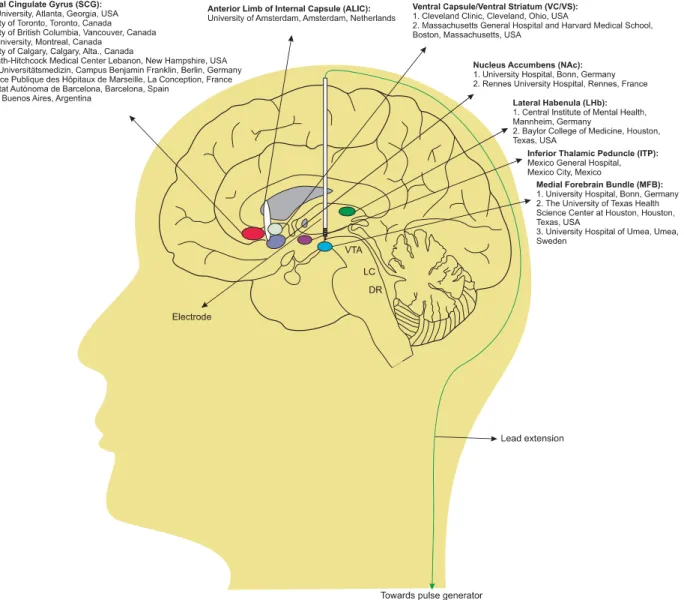
DEEP BRAIN STIMULATION
In DBS surgery, the electrode is stereotactically implanted into
specific neuroanatomical targets where stimulation is provided via
a pacemaker-like stimulator device that delivers continuous
electrical stimulation.
21A schematic representation of the
apparatus is provided in Figure 1. Benabid and Pollak pioneered
modern DBS over ablative surgery for the treatment of movement
disorders by targeting the thalamic nucleus ventralis intermedius,
globus pallidus internus and subthalamic nucleus (STN).
22,23Because of the tremendous clinical success of DBS as a treatment
for movement disorders and reported concurrent beneficial
effects on neuropsychiatric manifestations, this neuromodulatory
approach has also been explored as a possible treatment for many
mental disorders including obsessive
–
compulsive disorder and
intractable depression.
24–26Interestingly, ketamine
’
s rapid
anti-depressant and anti-anhedonic effects are associated with
alterations in glucose metabolism in brain structures, that are
also serving as potential targets for DBS, like the habenula, insula,
prefrontal cortex (PFC) and anterior cingulate cortex in patients
with TRD.
27,28Despite the incomplete understanding of the
underlying mechanisms of action (MOA) involved in the
therapeutic response to DBS among patients with TRD,
29several
brain targets have been tested, and thus DBS has evolved to
become a promising strategy for the management of TRD.
3,24,30–39CLINICAL AND PRECLINICAL OUTCOMES
Clinical studies have assessed putative therapeutic effects of DBS
in participants with TRD across several major brain targets namely
Brodmann area 25 or subcallosal cingulate gyrus (SCG), nucleus
accumbens (NAc), ventral capsule/ventral striatum (VC/VS) or
ventral part of anterior limb of the internal capsule (vALIC), medial
forebrain bundle (MFB), lateral habenular complex (LHb), and
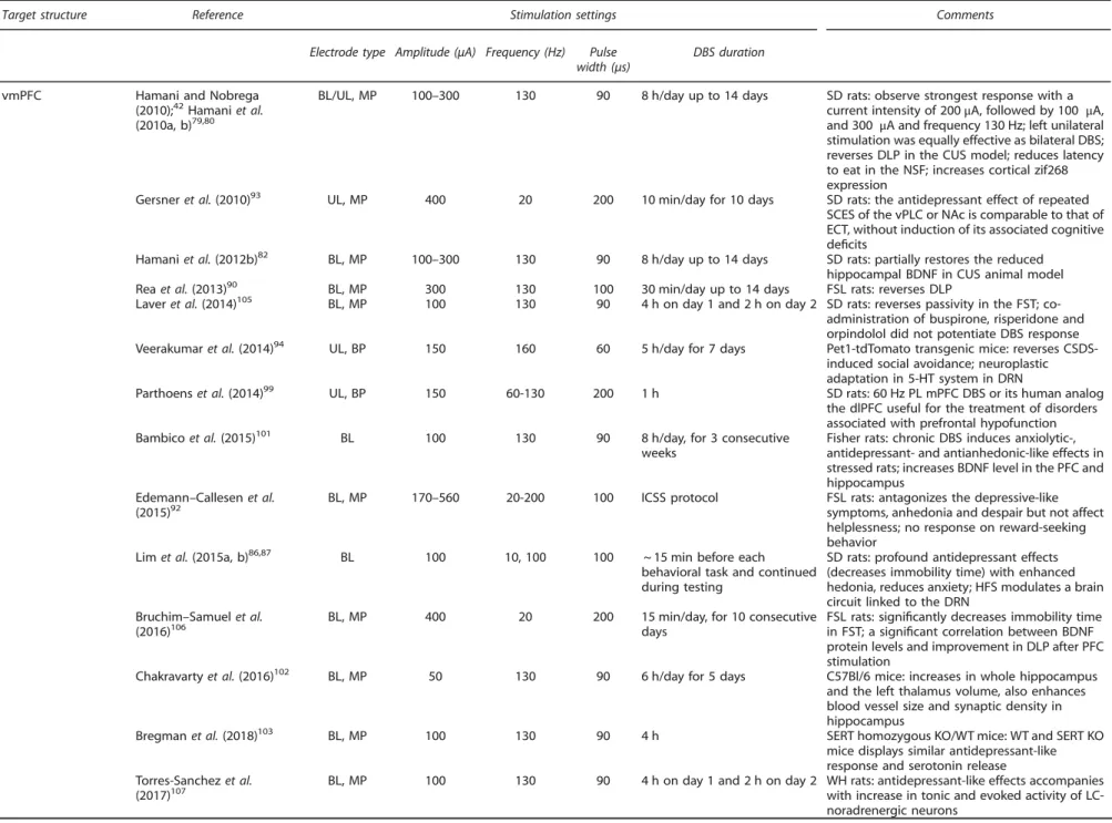
Figure 1.
Schematic representation of six DBS targets tested for the management of TRD. A quadripolar DBS electrode is implanted into the
selected brain targets, which is connected with long connecting lead extension wires to the pacemaker-like device (pulse generator) that is
mounted under the skin of the chest. The name of pioneering academic or research institutions that have tested each brain target are
provided. DSB, deep brain stimulation; DR, dorsal raphe; LC, locus coeruleus; TRD, treatment-resistant depression; VTA, ventral tegmental area.
MP Dandekaret al
1095
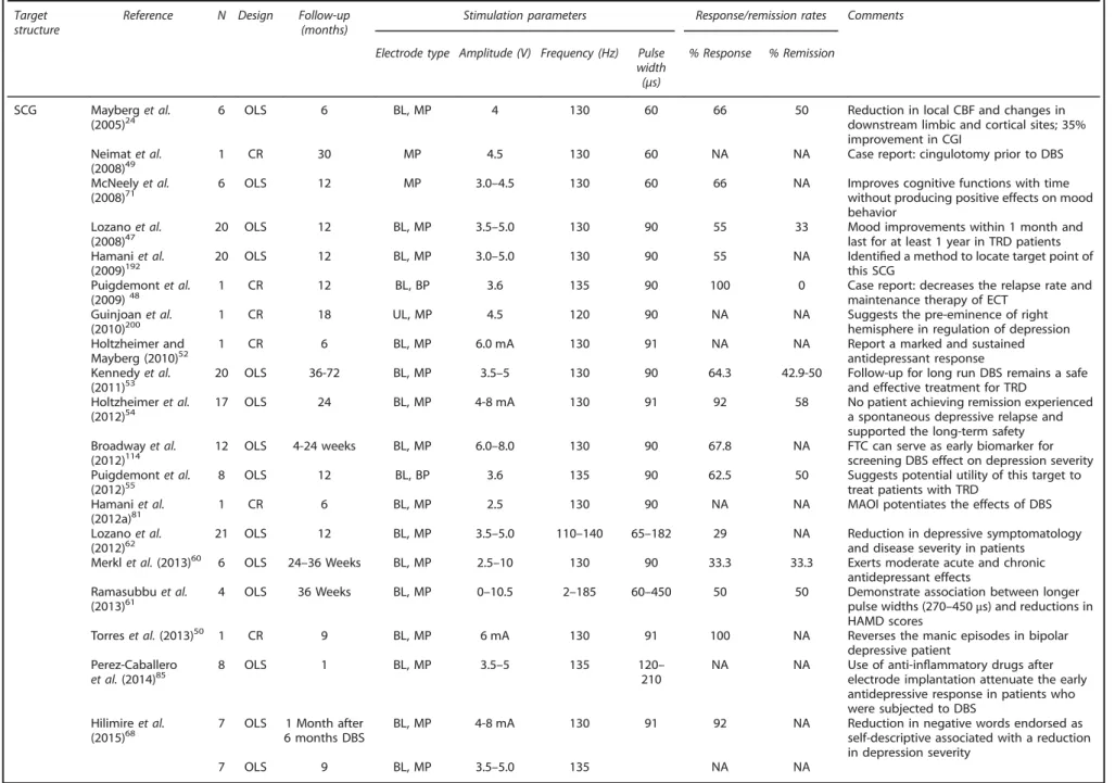
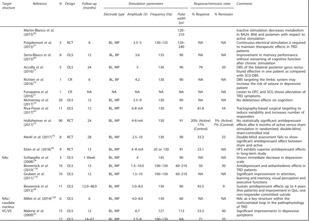
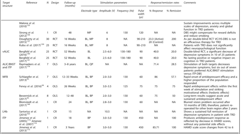
Table 1.
Summary of clinical trials and case reports of DBS applied to various brain targets for the management of TRD Targetstructure
Reference N Design Follow-up (months)
Stimulation parameters Response/remission rates Comments
Electrode type Amplitude (V) Frequency (Hz) Pulse width (μs)
% Response % Remission
SCG Mayberget al.
(2005)24 6 OLS 6 BL, MP 4 130 60 66 50 Reduction in local CBF and changes indownstream limbic and cortical sites; 35% improvement in CGI
Neimatet al. (2008)49
1 CR 30 MP 4.5 130 60 NA NA Case report: cingulotomy prior to DBS
McNeelyet al.
(2008)71 6 OLS 12 MP 3.0–4.5 130 60 66 NA Improves cognitive functions with timewithout producing positive effects on mood behavior
Lozanoet al. (2008)47
20 OLS 12 BL, MP 3.5–5.0 130 90 55 33 Mood improvements within 1 month and
last for at least 1 year in TRD patients Hamaniet al.
(2009)192
20 OLS 12 BL, MP 3.0–5.0 130 90 55 NA Identified a method to locate target point of
this SCG Puigdemontet al.
(2009)48
1 CR 12 BL, BP 3.6 135 90 100 0 Case report: decreases the relapse rate and
maintenance therapy of ECT Guinjoanet al.
(2010)200 1 CR 18 UL, MP 4.5 120 90 NA NA Suggests the pre-eminence of righthemisphere in regulation of depression Holtzheimer and
Mayberg (2010)52 1 CR 6 BL, MP 6.0 mA 130 91 NA NA Report a marked and sustainedantidepressant response Kennedyet al.
(2011)53 20 OLS 36-72 BL, MP 3.5–5 130 90 64.3 42.9-50 Follow-up for long run DBS remains a safeand effective treatment for TRD Holtzheimeret al.
(2012)54 17 OLS 24 BL, MP 4-8 mA 130 91 92 58 No patient achieving remission experienceda spontaneous depressive relapse and supported the long-term safety
Broadwayet al. (2012)114
12 OLS 4-24 weeks BL, MP 6.0–8.0 130 90 67.8 NA FTC can serve as early biomarker for screening DBS effect on depression severity Puigdemontet al.
(2012)55 8 OLS 12 BL, BP 3.6 135 90 62.5 50 Suggests potential utility of this target totreat patients with TRD Hamaniet al.
(2012a)81 1 CR 6 BL, MP 2.5 130 90 NA NA MAOI potentiates the effects of DBS
Lozanoet al.
(2012)62 21 OLS 12 BL, MP 3.5–5.0 110–140 65–182 29 NA Reduction in depressive symptomatologyand disease severity in patients Merklet al.(2013)60 6 OLS 24–36 Weeks BL, MP 2.5–10 130 90 33.3 33.3 Exerts moderate acute and chronic
antidepressant effects Ramasubbuet al.
(2013)61 4 OLS 36 Weeks BL, MP 0–10.5 2–185 60–450 50 50 Demonstrate association between longerpulse widths (270 –450μs) and reductions in HAMD scores
Torreset al.(2013)50 1 CR 9 BL, MP 6 mA 130 91 100 NA Reverses the manic episodes in bipolar
depressive patient Perez-Caballero
et al.(2014)85 8 OLS 1 BL, MP 3.5–5 135 120210– NA NA Use of anti-inelectrode implantation attenuate the earlyflammatory drugs after antidepressive response in patients who were subjected to DBS
Hilimireet al.
(2015)68 7 OLS 6 months DBS1 Month after BL, MP 4-8 mA 130 91 92 NA Reduction in negative words endorsed asself-descriptive associated with a reduction in depression severity
7 OLS 9 BL, MP 3.5–5.0 135 NA NA
Deep
brain
stimulation
for
treatment-res
istant
depression
MP
Dandekar
et
al
r
Psychiatry
(2018),
1094
–
1112
©
2018
Macmillan
Publishers
Limited,
part
of
Springer
Table 1.
(Continued ) Targetstructure
Reference N Design Follow-up (months)
Stimulation parameters Response/remission rates Comments
Electrode type Amplitude (V) Frequency (Hz) Pulse width (μs)
% Response % Remission
Martin-Blancoet al.
(2015)56 120210– Inactive stimulation decreases metabolismin BA24, BA6 and putamen with respect to active stimulation
Puigdemontet al.
(2015)57 5 RCT 6 BL, MP 3.5–5 130–135 120240– NA NA Continuous electrical stimulation is requiredto maintain therapeutic effects in TRD patients
Serra-Blascoet al. (2015)69
8 OLS 12 BL, BP 3.6 135 90 NA NA Improvement in memory performance
without worsening of cognitive function after chronic stimulation
Accollaet al.
(2016)77 5 OLS 24 BL, MP 5 130 90 79 20 DBS of the bilateral posterior gyrus rectusfound effective in one patient as compared with SCG-DBS
Richieriet al.
(2016)74 1 CR 6 BL, BP 4.2 130 90 NA NA DBS targeting the limbic system mayincrease the risk of seizure in depressive patient
Funayamaet al. (2016)51
1 CR NA NA NA NA NA NA NA Lesion to OFC and SCG shows alleviation of
TRD symptoms McInerneyet al.
(2017)70
20 OLS 12 BL, MP 2.5–9 130 90 NA NA No deleterious effects on cognition
Riva-Posseet al. (2017)45
11 OLS 12 BL, MP 6-8 mA 130 91 81.8 54 Tractography-based surgical targeting to
reduce variability and increases number of responders
Holtzheimeret al. (2017)75
90 RCT 24 BL, MP 4-8 mA 130 91 20% (Active)
17% (Control)
5% (Active) 7% (Control)
No statistically significant antidepressant effects after 6 months of active versus sham stimulation in randomized, double-blind, sham-controlled trial
Merklet al.(2017)76 8 RCT 28 BL, MP 2.5
–10 130 90 33.3 33 Double-blind assessment fails to show significant antidepressant effect between sham and active
Eitanet al.(2018)58 9 RCT 13 BL, MP 4–8 mA 20 or 130 91 23.1 NA HFS exhibits superior antidepressant effects in long-term study
NAc Schlaepferet al.
(2008)34 3 OLS 1 Week BL, MP 4 145 90 NA NA Shows immediate decrease in depressionscale
Bewernicket al. (2010)118
10 OLS 12 BL, MP 1.5–10.0 100–150 60–210 50 30 Antidepressant and antianhedonic effects in TRD patients
Grubertet al. (2011)119
10 OLS 12 BL, MP 1.5–10 100–150 60–210 NA NA Significant improvement in attention, learning and memory, visual perception and executive functions
Bewernicket al.
(2012)36 11 OLS 12.0–48.0 BL, MP 5.0–8.0 130 90 45.5 9 Sustain antidepressant effects up to 4 years(five patients) and improvement in QoL; one non-responder committed suicide
NAc/ caudate nucleus
Milletet al.(2014)120 6 OLS 6 BL, MP 4.0
–8.0 130 60 NA NA NAc as a key structure within the
corticostriatal loop in the pathophysiology of TRD
VC/VS Maloneet al. (2009)32
15 OLS 12 BL, MP 6.7 127 113 53.3 40 Significant improvements in depressive
symptoms
17 OLS 14–67 BL, MP 2.5–8 100–130 NA 71 35
Deep
brain
stimulation
for
treatment-res
istant
depression
MP
Dandekar
et
al
1097
2018
Macmillan
Publishers
Limited,
part
of
Springe
r
Nature.
Molecular
Psychiatry
(2018),
1094
–
Table 1.
(Continued ) Targetstructure
Reference N Design Follow-up (months)
Stimulation parameters Response/remission rates Comments
Electrode type Amplitude (V) Frequency (Hz) Pulse width (μs)
% Response % Remission
Maloneet al.
(2010)135 Sustain improvements across multiplescales of depression, anxiety and global
function in TRD patients Stronget al.
(2012)136
1 CR 48 MP 6 130 120 NA NA DBS might compensate for reward deficits
and reduce smoking Doughertyet al.
(2015)137 30 RCT 16 Weeks BL, MP 8 NA 90-210 23.3 (Active)20 (Control) 200 As per double-blind RCT VC/VS-DBS is notan ef
ficacious therapy for TRD
Kubuet al.(2017)139 25 RCT 16 Weeks BL, MP 8 NA 90–210 NA NA Patients with TRD does not significantly affect neuropsychological function vALIC Bergfeldet al.
(2016)140 25 RCT 52 Weeks BL 2.5–6.0 130–180 90 40.0 20.0 Double-blind RCT, a signidepressive symptoms in 10 of 25 patientsficant decrease of Bergfeldet al.
(2017)141 25 RCT 52 Weeks BL 2.5–6.0 130–180 90 40.0 20.0 No lasting positive or negative impact oncognition in TRD patients ALIC/BNST
and ITP
Raymaekerset al.
(2017)176 7 OLS 3–8 years BL, QP NA NA NA 71.4 28.5 Stimulation of both targets decreasesdepressive symptoms, but six out of seven patients preferred ALIC/BNST stimulation versus ITP-DBS
MFB Schlaepferet al. (2013)33
7 OLS 12–33 Weeks BL, BP 2.0–3.0 130 60 86 57.1 Rapid onset of antidepressant efficacy and a higher proportion of the population responded
Fenoyet al.(2016)39 4 OLS 26 Weeks BL, BP 3.0–3.5 125 75 75 75 Rapid antidepressant effects within thefirst week of stimulation and striking
motivational effects (hedonic effects) Bewernicket al.
(2017)35
8 OLS 12–48 BL, BP 2.0–3.0 130 60 75 50 Long-term results suggest acute and
sustained antidepressant effect Blomstedtet al.
(2017)146 1 CR 24 BL, BP 2.8–3.0 130 60 NA NA Blurred vision problem occurred after10 months of DBS; therefore, patient re-operated for other brain region after 2 years LHb Sartoriuset al.
(2010)163
1 CR 15 NA 10.5 NA NA NA NA Shows a sustained full remission of
depressive symptoms in patient with TRD ITP Jimenezet al.
(2005);173Jimenez et al.(2007)174
1 CR 18 BP 3.0–5.0 130 450 NA 100 Produces antidepressant response as
reflected by decrease in HAMD scores, without any potential side effects Jimenezet al.
(2013)175 1 CR 3 Years BP 3.0–5.0 130 450 85.71 100 HAMD scale score changes from 42 to 6
Abbreviations: BA24/6, Brodmann area 24/6; BL, bilateral; BNST, bed nucleus of the stria terminalis; BP, bipolar; CBF, cerebral bloodflow; CGI, clinical global impressions scale; CR, case report; DBS, deep brain stimulation; FTC, frontal theta cordance; HAMD, Hamilton depression rating scale; HFS, high-frequency stimulation; ITP, inferior thalamic peduncle; LHb, lateral habenula; MADRS, Montgomery–Asberg depression rating scale; MAOI, monoamine oxidase inhibitor; MFB, medial forebrain bundle; MP, monopolar; NA, not applicable; NAc, nucleus accumbens; OFC, orbital prefrontal cortex; OLS, open-label study; QoL, quality of life; QP, quadripolar; RCT, randomized controlled trial; SCG, subcallosal cingulate gyrus; TRD, treatment-resistant depression; vALIC, ventral part of the anterior limb of the internal capsule; VC/VS, ventral capsule/ventral striatum. Summary of key clinical DBS studies in depression.
Deep
brain
stimulation
for
treatment-res
istant
depression
MP
Dandekar
et
al
r
Psychiatry
(2018),
1094
–
1112
©
2018
Macmillan
Publishers
Limited,
part
of
Springer
Table 2.
Application of DBS in the various neuroanatomical targets usingin vivopreclinical modelsTarget structure Reference Stimulation settings Comments
Electrode type Amplitude (μA) Frequency (Hz) Pulse width (μs)
DBS duration
vmPFC Hamani and Nobrega
(2010);42Hamaniet al. (2010a, b)79,80
BL/UL, MP 100–300 130 90 8 h/day up to 14 days SD rats: observe strongest response with a current intensity of 200μA, followed by 100 μA, and 300μA and frequency 130 Hz; left unilateral stimulation was equally effective as bilateral DBS; reverses DLP in the CUS model; reduces latency to eat in the NSF; increases cortical zif268 expression
Gersneret al.(2010)93 UL, MP 400 20 200 10 min/day for 10 days SD rats: the antidepressant effect of repeated SCES of the vPLC or NAc is comparable to that of ECT, without induction of its associated cognitive deficits
Hamaniet al.(2012b)82 BL, MP 100–300 130 90 8 h/day up to 14 days SD rats: partially restores the reduced hippocampal BDNF in CUS animal model Reaet al.(2013)90 BL, MP 300 130 100 30 min/day up to 14 days FSL rats: reverses DLP
Laveret al.(2014)105 BL, MP 100 130 90 4 h on day 1 and 2 h on day 2 SD rats: reverses passivity in the FST; co-administration of buspirone, risperidone and orpindolol did not potentiate DBS response Veerakumaret al.(2014)94 UL, BP 150 160 60 5 h/day for 7 days Pet1-tdTomato transgenic mice: reverses
CSDS-induced social avoidance; neuroplastic adaptation in 5-HT system in DRN
Parthoenset al.(2014)99 UL, BP 150 60-130 200 1 h SD rats: 60 Hz PL mPFC DBS or its human analog the dlPFC useful for the treatment of disorders associated with prefrontal hypofunction Bambicoet al.(2015)101 BL 100 130 90 8 h/day, for 3 consecutive
weeks
Fisher rats: chronic DBS induces anxiolytic-, antidepressant- and antianhedonic-like effects in stressed rats; increases BDNF level in the PFC and hippocampus
Edemann–Callesenet al. (2015)92
BL, MP 170–560 20-200 100 ICSS protocol FSL rats: antagonizes the depressive-like symptoms, anhedonia and despair but not affect helplessness; no response on reward-seeking behavior
Limet al.(2015a, b)86,87 BL 100 10, 100 100 ~ 15 min before each behavioral task and continued during testing
SD rats: profound antidepressant effects (decreases immobility time) with enhanced hedonia, reduces anxiety; HFS modulates a brain circuit linked to the DRN
Bruchim–Samuelet al.
(2016)106 BL, MP 400 20 200 15 min/day, for 10 consecutivedays FSL rats: signiin FST; a signifificantly decreases immobility timecant correlation between BDNF protein levels and improvement in DLP after PFC stimulation
Chakravartyet al.(2016)102 BL, MP 50 130 90 6 h/day for 5 days C57Bl/6 mice: increases in whole hippocampus and the left thalamus volume, also enhances blood vessel size and synaptic density in hippocampus
Bregmanet al.(2018)103 BL, MP 100 130 90 4 h SERT homozygous KO/WT mice: WT and SERT KO
mice displays similar antidepressant-like response and serotonin release Torres-Sanchezet al.
(2017)107 BL, MP 100 130 90 4 h on day 1 and 2 h on day 2 WH rats: antidepressant-like effects accompanieswith increase in tonic and evoked activity of LC-noradrenergic neurons
Deep
brain
stimulation
for
treatment-res
istant
depression
MP
Dandekar
et
al
1099
2018
Macmillan
Publishers
Limited,
part
of
Springe
r
Nature.
Molecular
Psychiatry
(2018),
1094
–
Table 2.
(Continued )Target structure Reference Stimulation settings Comments
Electrode type Amplitude (μA) Frequency (Hz) Pulse width (μs)
DBS duration
IL-PFC Perez-Caballeroet al. (2014)85
BL, BP 100 130 90 4 h on day 1 and 2 h on day 2 WH rats: DBS of the prelimbic and infralimbic cortex exerts an antidepressant-like effect in the FST; inflammatory response following surgery might displayed early insertional antidepressive effect
Etievantet al.(2015)96 UL, BP 150 130 60 4 h on day 1 and 2 h on day 2 SD rats: decreases immobility duration; promote hippocampal mitosis and reverses the effects of stress on hippocampal synaptic metaplasticity; increases DRN 5-HTfiring activity and synaptogenesis
Inselet al.(2015)97 BL, UP 100 130 90 8 h/day for 10 days SD rats: therapeutic effects of DBS is independent of 5-HT levels, DBS disrupts communication between regions important for expectation-based control of emotion like infralimbic cortex and VH
Srejicet al.(2015)104 UL, BP 60 100 200 5 min SD rats: DRN cells significantly decreasesfiring rate (82%) during HFS
Jimenez–Sanchezet al.
(2016a)88 BL, BP 200 130 90 1 h WH rats: reverses hyperlocomotion,hyperemotionality and anhedonia, and increases social interaction in the OBX rats; increases synthesis of BDNF and GluA1 AMPA receptor, and stimulates mTOR, CREB
Jimenez–Sanchezet al. (2016b)89
BL, BP 200 130 90 1 h WH rats: shows antidepressant-like effects in FST
and NSF test; increases prefrontal efflux of glutamate and activate AMPA receptor Bezchlibnyket al.(2017)98 BL, BP 2.5 V 130 90 1 h WH rats: increases the complexity of apical
dendrites and the length of basal dendritic trees of pyramidal neurons located in the CA1 region of hippocampus
Prelimbic Mosheet al.(2016)95 UL 400 20 0.2 ms 10 min per session for 10
consecutive days
DRL and SD rats: reverses the depressive-like behaviors and increases the reduced BDNF levels vmPFC/NAc/
WMF Hamaniet al.(2014)126 UL, MP 100 130 90 4 h on day 1 and 2 h on day 2 SD rats: shows similar antidepressant-like effects at all three sites despite distinct impact in regional brain activity
Winteret al.(2015)130 BL, MP 100–300 130 90 1 h SD rats: neither vmPFC nor NAc DBS increases hippocampal neurogenesis
Cingulate cortex Dourneset al.(2013)100 BL 2.5 V 80, 120 90 1 h/day for 2 weeks BALB/c ByJ mice: normalizes the motivated-like responses, anxiety-related behaviors,
hyperactivity and aggressiveness
NAc Gersneret al.(2010)93 UL, MP 400 20 200 10 min/day for 10 days SD rats: reverses anhedonic-like behavior in CUS model, but no effect in FST assay
Sesiaet al.(2010)121 BL, BP 3, 30, 150 130 60 7 Days Lewis rats: increases levels of DA and 5-HT in the NAc, but not in mPFC; DBS of the NAc core has beneficial behavioral effects
Falowskiet al.(2011)129 UL, BP 2 V 130 200 ms 3 h/day for 14 day continuous WH rats: reduces anxiety, increases exploratory behavior and DA and NA in PFC
Deep
brain
stimulation
for
treatment-res
istant
depression
MP
Dandekar
et
al
r
Psychiatry
(2018),
1094
–
1112
©
2018
Macmillan
Publishers
Limited,
part
of
Springer
Table 2.
(Continued )Target structure Reference Stimulation settings Comments
Electrode type Amplitude (μA) Frequency (Hz) Pulse width (μs)
DBS duration
van Dijket al.(2011)122 UL, BP 300
–400 120 80 5 h WH rats: no significant effect on monoaminergic neurotransmitters or their metabolites in the stimulated region, that is, NAc core van Dijket al.(2012)123 BL, BP 300 120 80 1.45 h/day for 2 consecutive
days and 2 h in the NAc core
WH rats: increases the DA, NA and 5-HT in the mPFC and OFC after onset of stimulation in the NAc core
van der Plasseet al.(2012)125 BL 10, 50, 100 NA NA 1 h WH rats: NAc shell-DBS profoundly and selectively increases the chow intake; the intake of chow and motivation to work for palatable food can independently be modulated by DBS of subregions of the NAc shell
Schmuckermairet al.
(2013)91 UL, BP 100 130 60 1 h/day for 7 days HAB mice: antagonize the depressive- andanxiolytic-like phenotypes Hamaniet al.(2014)126 BL, MP 100 130 90 4 h on day 1; 2 h on day 2 SD rats: shows antidepressant-like effects in the
FST; increases zif268 expression in subcortical structures and piriform cortex
Limet al.(2015b)87 BL 100 10, 100 100 ~ 15 min before behavioral task and continued during testing
SD rats: reduces anxiety (decreased escape latency in the home-cage emergence test) and increases motivation for food intake
Winteret al.(2015)130 BL, MP 100, 300 130 90 1 h SD rats: no change in hippocampal neurogenesis Huguetet al.(2017)131 BL, BP 100 100 100 ~ 15 min before behavioral
task and continued during testing
SD rats: reduces CUS-induced increased c-Fos expression in the medial vestibular nucleus
Kimet al.(2016b)165 BL, BP 100 130 90 7 Days uninterrupted stimulation
WH rats: reduces immobility time (76%) in ACTH-treated animals; increases RCR in ACTH-ACTH-treated rats
Rummelet al.(2016)127 BL, MP 150 130 100 16 Days continuously FSL and congenitally LH rats: antidepressant response is associated with an increase in 5-HT turnover alongside site-specific reductions in 5-HT contents
MFB Bregmanet al.(2015)150 BL 100 20, 130 90 4 h on day 1; 2 h on day 2 SD rats: shows antidepressant-like effect; increases zif268 expressions in the piriform cortex, prelimbic cortex, NAc-Shell, caudate/ putamen and VTA
Edemann–Callesenet al. (2015)92
BL, MP 170–560 20–200 100 ICSS FSL rats: increases swimming time
(antidepressant effects); consume more sucrose solution (hedonic activity)
Furlanettiet al.(2015a)148 BL, BP 250 130 288 Continuously for 3–6 weeks SD rats: unilateral DA depletion do not preclude MFB-DBS in reversing depressive-like and anhedonic-like behavior
Furlanettiet al.(2015b)149 BL, BP 288 130 100 Continuously for 3
–6 weeks SD rats: increases and long-lasting c-fos expression in target regions of the mesolimbic/ mesocortical system
Furlanettiet al.(2016)147 BL, BP 250 130 100 Continuously for 3 weeks SD rats: rescues DLP; work through both DA dependent and independent mechanisms; activates distant structures involved in the neurocircuitry of depression
Dandekaret al.(2017)152 UL, BP 200 130 90 4 h on day 1; 2 h on day 2 Decreases passivity in the FST and increases expression DA D2 receptors in the PFC
Deep
brain
stimulation
for
treatment-res
istant
depression
MP
Dandekar
et
al
1101
2018
Macmillan
Publishers
Limited,
part
of
Springe
r
Nature.
Molecular
Psychiatry
(2018),
1094
–
Table 2.
(Continued )Target structure Reference Stimulation settings Comments
Electrode type Amplitude (μA) Frequency (Hz) Pulse width (μs)
DBS duration
LHb Friedmanet al.(2011)164 BP 200 10
–100 0.5 ms 15 min DBS and then ICSS SD rats: attenuates processes of positive reward-associated reinforcement
Menget al.(2011)84 UL, BP 80
–100 150 300 7–28 Days continuously WH rats: reduces DLP in CUS model; increases DA/NA/5-HT in blood serum and hippocampus Liet al.(2011)83 UL, BP 150, 300 130 40 ms FST: 1 h after and 1 h before
swimming sessions
LH: 1 h after baseline testing and 1 h before LH sessions
Congenital LH rats: antidepressant-like effects in the FST and LH
Limet al.(2015b)87 BL 100 10, 100 100 ~ 15 min before behavioral task and continued for the entire duration of testing
SD rats: HFS reduces anxiety in the home-cage emergence test and immobility time in the FST; also improves motivational behavior in food intake study
Kimet al.(2016a)128 BL, BP 200 130 90 3 Days prior to the FST WH rats: reduces immobility in ACTH-treated animals and phosphorylation of CaMKIIα/βand GSK3α/βin the LHb together with the downregulation of CaMKIIαβ/b, GSK3α/βand AMPK in the IL cortex
STN Temelet al.(2007)184 BL, BP 3–150 10–130 60 3 min SD rats: inhibits DRN 5-HT activity and
precipitates DLP
Creedet al.(2013)178 BP, MP 100 130 90 4 h/day for 21 days SD rats: induces and potentiates DLP; repeated DBS causes decreased levels of BDNF and trkB mRNA in hippocampus
Faggianiet al.(2015)177 BL, BP 170 130 60 10 min before starting the session and maintained during behavioral testing
SD rats: acute DBS improves DLP with bilateral depletion of DA, NA and 5-HT
Abbreviations: 5-HT, serotonin; ACTH, adrenocorticotropic hormone; AMPA,α-amino-3-hydroxy-5-methyl-4-isoxazolepropionic acid; AMPK, AMP-activated protein kinase; BDNF, brain-derived neurotrophic factor; BL, bilateral; BP, bipolar; CaMKIIα/β, Ca2+/calmodulin-dependent protein kinase; CREB, c-AMP response element binding; CSDS, chronic social defeat stress; CUS, chronic unpredictable mild stress; DA, dopamine; DLP, depression-like phenotype; dlPFC, dorsolateral prefrontal cortex; DRL, depressive rat line; DRN, dorsal raphe nucleus; ECT, electroconvulsive therapy; fMRI, functional magnetic resonance imaging; FSL, Flinders sensitive line; FST, forced swim test; GSK3α/β, glycogen synthase kinase 3; h, hour; HAB, high-anxiety behavior; HFS, high-frequency stimulation; IL-PFC, infralimbic prefrontal cortex; ICSS, intracranial self-stimulation; KO, knockout; LC, locus coeruleus; LFP, localfield potential; LH, learned helplessness; LHb, lateral habenula; MFB, medial forebrain bundle; Min, minute; MP, monopolar; mPFC, medial prefrontal cortex; mTOR, mammalian target of rapamycin; NA, noradrenaline; NAc, nucleus accumbens; NET, novelty exploration test; NSF, novelty suppressed feeding; OBX, olfactory bulbectomy; OFC, orbitofrontal cortex; PFC, prefrontal cortex; PL, prelimbic; RCR, respiratory control ratio; SCES, subconvulsive electrical stimulation; SCG, subcallosal cingulate gyrus; SD, Sprague–Dawley; SERT, serotonin transporter; SI, social interaction; STN, subthalamic nucleus; trkB, tropomyosin receptor kinase B; UL, unilateral; VH, ventral hippocampus; vmPFC, ventromedial prefrontal cortex; vPLC, ventral prelimbic cortex; VTA, ventral tegmental area; WMF, white matterfibers of the frontal region; WH, Wistar rats; WT, wild type. Summary of key preclinical DBS studies in depression.
Deep
brain
stimulation
for
treatment-res
istant
depression
MP
Dandekar
et
al
r
Psychiatry
(2018),
1094
–
1112
©
2018
Macmillan
Publishers
Limited,
part
of
Springer
inferior thalamic peduncle (ITP). The names of the respective
pioneering institutions that conducted DBS manipulations across
these brain targets for the treatment of TRD are presented in
Figure 1. The ideal settings for achieving optimum antidepressant
effects in humans remain unclear, although it is worthy to note
that therapeutic effects may vary as a function of respective DBS
targets and stimulation parameters, and also according to clinical
characteristics of individual patients.
40The exploratory
meta-analysis conducted by Smith
41suggests that active DBS applied to
some of the above brain targets could be 71% more efficacious
than sham treatment (summary effect size: 1.71; 95% confidence
interval: 1.47
–
1.96) for TRD. However, only eight studies were
available, and effect sizes could not be separated according to
specific brain targets. Detailed information pertaining to clinical
and preclinical central nervous system targets chosen for DBS as a
treatment for depression is provided in the following sections, and
is briefly summarized in Tables 1 and 2.
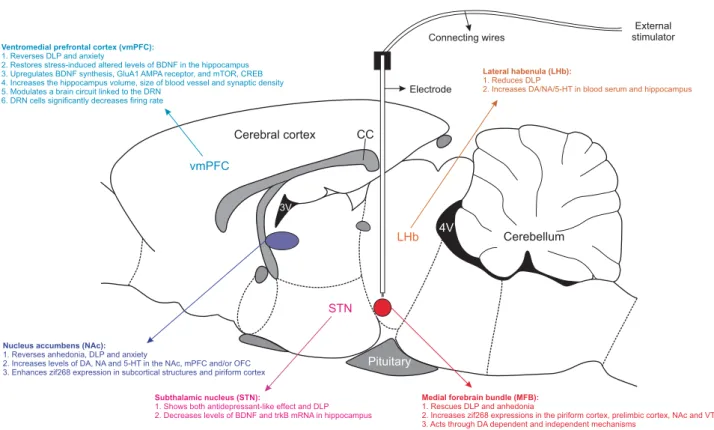
Details of neuroanatomical substrates tested across preclinical
DBS studies for depression-like phenotypes are diagrammatically
depicted in Figure 2. As presented in Table 2, stimulation
parameters markedly varied across preclinical investigations. Yet,
settings between 60 and 130 Hz for frequency, 60 and 200
μ
s for
pulse width and 50 and 300
μ
A for amplitude exhibited promising
antidepressant-like effects across various DBS targets. Preclinical
studies have also pointed that different stimulation parameters
and neuroanatomical locations may influence
antidepressant-related effects. However, at least in part because of the complex
and multifactorial pathophysiology of human depression,
cur-rently no animal model meets all validity criteria (including
predictive validities),
42,43and hence limitations of preclinical
models should be considered when inferences pertaining to
depression in humans are made.
DBS of the SCG
Clinical studies
.
The SCG may have a pivotal role in the regulation
of sadness and negative emotions occurring in both depressed
and healthy subjects.
24,44–46Clinical outcomes for this DBS target
are summarized in Table 1. Mayberg and coworkers
24initially
reported that four out of six patients with TRD achieved
antidepressant response after 6 months of open-label SCG-DBS.
Afterwards, Lozano
et al.
47reported that 40% of participants with
TRD (
n
= 20) achieved response after 1 week of stimulation,
whereas 60 and 55% of patients met response criteria at 6 and
12 months, respectively, in an open-label trial that tested
SCG-DBS. A few case reports also demonstrated efficacy for this target
in the TRD patients.
48–52Long-term outcomes of SCG-DBS for the
aforementioned open-label trial were subsequently reported, with
55
–
60 and 64.3% response rates after 1
–
3 and 3-6 years
’
follow-up
visits, respectively,
53and four participants either committed or
attempted suicide over the course of study, although because of
uncontrolled design of this study it could not be established
whether this serious adverse effect was related to DBS or to illness
evolution
per se
. Holtzheimer
et al.
54replicated those
findings in
an uncontrolled study involving 17 participants with TRD after
SCG-DBS. This trial reported 43.6
–
70.1% response rates (assessed
with the Hamilton depression rating scale) following 24-week,
1-year and 2-1-year follow-up visits. Although this study was
uncontrolled, it should be noted that the blinded discontinuation
Cerebral cortexvmPFC
LHb
STN
Nucleus accumbens (NAc):
1. Reverses anhedonia, DLP and anxiety
2. Increases levels of DA, NA and 5-HT in the NAc, mPFC and/or OFC 3. Enhances zif268 expression in subcortical structures and piriform cortex
Medial forebrain bundle (MFB):
1. Rescues DLP and anhedonia
2. Increases zif268 expressions in the piriform cortex, prelimbic cortex, NAc and VTA 3. Acts through DA dependent and independent mechanisms
Electrode
Connecting wires
External stimulator
Cerebellum 4V
3V
Pituitary CC Ventromedial prefrontal cortex (vmPFC):
1. Reverses DLP and anxiety
2. Restores stress-induced altered levels of BDNF in the hippocampus 3. Upregulates BDNF synthesis, GluA1 AMPA receptor, and mTOR, CREB 4. Increases the hippocampus volume, size of blood vessel and synaptic density 5. Modulates a brain circuit linked to the DRN
6. DRN cells significantly decreases firing rate
Lateral habenula (LHb):
1. Reduces DLP
2. Increases DA/NA/5-HT in blood serum and hippocampus
Subthalamic nucleus (STN):
1. Shows both antidepressant-like effect and DLP 2. Decreases levels of BDNF and trkB mRNA in hippocampus
Figure 2.
Diagrammatic representation of DBS brain targets that have been tested in preclinical investigations. The outline of the sagittal
diagram through the rat brain indicates the vmPFC, NAc, STN, MFB and LHb, which are not in scale. As denoted, monopolar or bipolar
electrodes are stereotactically implanted in any one of the brain targets. At the time of stimulation, implanted electrodes are connected with
long connecting wires to external stimulators, which delivers electric current as per protocol. Important functional and biochemical changes
following DBS applied to each brain target are highlighted. 3V, third ventricle; 4V, fourth ventricle; 5-HT, 5-Hydroxytryptamine; AMPA,
α
-Amino-3-hydroxy-5-methyl-4-isoxazolepropionic acid; BDNF, brain-derived neurotrophic factor; CC, corpus callosum; CREB, c-AMP-response
element-binding; DA, dopamine; DBS, deep brain stimulation; DLP, depression-like phenotype; DRN, dorsal raphe nucleus; LHb, lateral
habenula; MFB, medial forebrain bundle; mPFC, medial prefrontal cortex; mTOR, mammalian target of rapamycin; NA, noradrenaline; NAc,
nucleus accumbens; OFC, orbitofrontal cortex; STN, subthalamic nucleus; trkB, tropomyosin receptor kinase B.
MP Dandekaret al
1103
three patients in which this was attempted, whereas depressive
symptoms improved once stimulation was reinstated.
54Further-more, these robust therapeutic benefits and brain metabolic
changes have been further replicated in a preliminary
uncon-trolled observation;
55,56seven out of eight TRD patients achieved
antidepressant response after SCG-DBS at a 6-month follow-up
visit. In a more recent randomized, double-blind, sham-controlled
crossover trial Puigdemont
et al.
57reported reversal of depressive
scores after active SCG-DBS application in
five participants with
TRD,
57and long-term high frequency stimulation (HFS) exhibited
better antidepressant response.
58Berlim
et al.
59conducted an exploratory meta-analysis of four
observational studies, and verified that response and remission
rates following SCG-DBS were 36.6 and 16.7%, 53.9 and 24.1%,
and 39.9 and 26.3% at 3-, 6- and 12-month follow-up periods,
respectively. Moreover, open-label studies reported remission
rates ranging from 29 to 58% after chronic stimulation of the SCG
for up to 12
–
36 months,
60–62although evidence suggests that
longer pulse durations could influence pathways farther from the
SCG, and activation of the SCG
–
NAc network may contribute to
antidepressant response after SCG-DBS.
31Patient-specific
tracto-graphy modeling provided relevant insights for the identification
of electrode location and critical neuronal tracts that could
mediate antidepressant responses to SCG-DBS, and may also
mitigate inter-individual variability in the direct effects of
stimulation on brain circuitry.
45,63,64Using tractography-based
surgical targeting, Riva-Posse
et al.
45demonstrated 72.7% (8 of 11
participants) and 81.8% (9 of 11 participants) response rates at
6 months and 1 year after SCG-DBS, respectively, whereas six
patients were in remission at both time points.
SCG-DBS may also improve memory as well as executive and
motor functioning in participants with TRD, without meaningful
adverse effects upon cognitive measures.
65–71Moreover, patients
with TRD may exhibit transient emotional hypersensitivity and a
predictable worsening of depressive symptom scores at the initial
phases of SCG-DBS,
55thus long-term treatment could be critical
for central nervous system remodeling and neuroplasticity, and
hence to therapeutic benefits.
72In SCG-DBS-operated participants
with TRD, the most frequently observed surgery-associated
adverse
events
were
hardware-related
(11.4%),
suicidality
(9.3%)
73and risk of partial seizures.
74In addition, a multicenter,
randomized controlled trial (RCT) of SCG-DBS for TRD (BROADEN
study) was prematurely interrupted based on results of an interim
futility analysis (St Jude Medical Clinical Study).
38,75Recently,
Holtzheimer
et al.
75published results of multisite, randomized,
double-blind, sham-controlled SCG-DBS study in 90 TRD
partici-pants. They did not observe statistically significant effects in the
primary efficacy outcome between the stimulation (20%) and
control group (17%), similarly in a double-blind study of eight
participants Merkl
et al.
76reported no difference between active
versus sham group. However, 33
–
48% participants displayed an
antidepressant response and 25% achieved remission with up
to 2 years of open-label SCG-DBS.
75,76Nevertheless, the lack of
therapeutic response in TRD patients after SCG-DBS may be due
to the wrong placement of electrodes or misleading points of
stimulation.
45Preliminary evidence
for
site-specific
clinical
responses following SCG-DBS was provided in the small
open-label trial conducted by Accolla
et al.
77that enrolled
five
participants with TRD; noticeable antidepressant responses
following stimulation of the posterior gyrus rectus region in one
patient with TRD was observed, although none of the participants
in whom DBS was applied to the originally planned Brodmann
area 25 were considered responders.
Preclinical studies
.
The infralimbic cortex, which is part of the
rodent ventromedial PFC (vmPFC) is thought to represent the
rodent homologous of the human SCG (Brodmann area 25).
78lead to antidepressant-like effects across several preclinical
models including the forced swimming test (FST), sucrose
preference test and novelty suppressed feeding (Table 2).
79–90Electrical stimulation of this neuroanatomical target has also been
shown to increase hedonic and motivational states in animal
models of depression,
91,92and to reverse depressive-like
pheno-types induced by chronic unpredictable stress (CUS), chronic social
defeat stress, olfactory bulbectomy and in a putative
therapy-refractory depressive-like rat line.
82,87–89,93–95Hamani
et al.
79characterized the optimal brain stimulation
settings for DBS applied to the vmPFC in rats. DBS targeted to the
infralimbic cortex also resulted in antidepressant-like effects, may
be through a reversal of synaptic metaplasticity and increments in
mitosis.
85,96–98In another study, DBS applied to prelimbic mPFC
led to different functional brain alterations.
99Moreover, chronic
DBS applied to the vmPFC may reverse stress-induced behavioral
deficits in the sucrose preference test, FST, novelty suppressed
feeding and elevated plus maze models, and may also increase
brain-derived neurotrophic factor levels, blood vessel size,
synaptic density and astrocyte size in the hippocampus.
82,100–102Altogether, these data suggest that stimulation parameters need
to be precisely set for achieving meaningful responses after
prelimbic or infralimbic PFC-DBS.
Hamani and colleagues
80,82,103provided preclinical data to
support a putative role for the serotonergic system as a mediator
of the antidepressant-like effects of vmPFC-DBS. Moreover, vmPFC
stimulation was found to produce antidepressant, anxiolytic and
hedonic effects through the modulation of dorsal raphe nucleus
(DRN) circuitry in the CUS animal model of depression.
86,87,94,104Furthermore, adjuvant treatment with monoamine oxidase
inhibitors was reported to potentiate behavioral effects of
vmPFC-DBS in the FST.
81However, co-administration of buspirone,
pindolol or risperidone did not significantly alter
antidepressant-like effects of DBS.
105This further suggests that vmPFC-DBS might
involve the modulation of prefrontal projections to the DRN,
which is a brain region involved in serotonin (5-HT) synthesis and
release. Apart from the DRN, neurostimulation of the vmPFC may
also remotely affect activity of the ventral tegmental area (VTA)
and locus coeruleus.
106,107Possible MOA
.
Depression has been associated with increased
activity
of
the
SCG
and
dysregulated
corticolimbic
networks.
30,96,108–111Furthermore, SCG neurons are preferentially
more responsive to negative (unpleasant) emotions.
112SCG-DBS
may ameliorate depressive symptoms or more specifically
anhedonia in patients with TRD.
113Antidepressant response to
SCG-DBS in participants with TRD has been associated with frontal
asymmetry, higher frontal theta cordance (θ) and local
field
potential broad
α
-band activity.
114–116Suppression of gamma
oscillations and increased
θ-gamma coupling by active SCG-DBS
stimulation
may
also
enhance
gamma-aminobutyric
acid
neurotransmission.
117Therefore, it seems that SCG-DBS
stimula-tion may have a key role in normalizing spectral rhythms in brain
networks related to depression neurobiology.
DBS applied to the NAc
Clinical studies
.
Anhedonia was one of the
first manifestations to
improve during NAc stimulation in participants with TRD, which
also
reported
a
heightened
perception
of
pleasurable
activities.
34,36,113,118The ideal parameters of electrical stimulation
that could provide adequate antidepressant responses are
summarized in Table 1. Schlaepfer
et al.
34reported short-term
outcomes in three patients with TRD who underwent NAc-DBS.
Twelve months of chronic, open-label NAc-DBS led to a decrease
in the metabolism of the SCG, amygdala and prefrontal regions
with 45 and 9% response and remission rates, respectively, in 10
participants with TRD.
118The long-term open-label trial conducted
by Bewernick
et al.
36reported a sustained antidepressant effect
for NAc-DBS in 11 participants with TRD (45.5% response rate at
48-month follow-up). Yet, only
five participants completed this
4-year trial.
36Moreover, there was no evidence of cognitive
deterioration in agreement with data from the same research
group.
36,119An open-label trial with six participants reported 50%
responders, and did not show signs of cognitive deterioration.
120Altogether, these data suggest that NAc-DBS could be efficacious
for the management of TRD.
Preclinical studies
.
Application of DBS to the NAc-shell triggered
impulsive behaviors accompanied by significant increases in
dopamine and 5-HT levels in the NAc. However, DBS applied to
the NAc core led to antidepressant-like effects without
signifi-cantly altering levels of 5-HT and dopamine,
121,122although 2
consecutive days of bilateral stimulation elicited a rapid increase
in dopamine and 5-HT release in the orbital PFC.
123HFS or low
frequency stimulation of the NAc-DBS also produced distinct
region-specific and frequency band-specific changes in local
field
potential oscillations,
124therefore, suggesting that different
stimulation parameters may engage distinct brain areas, which
could then influence antidepressant responses to NAc-DBS.
Furthermore, a recent study reported that DBS applied to the
lateral NAc-shell reduced motivation for sucrose, whereas
stimulation of the medial NAc
–
shell selectively increased the
intake of chow.
125These
findings suggest that subdivisions of the
NAc
–
shell may influence motivational eating behavior, and may
point to dissociable effects of NAc-DBS in alleviating anhedonia in
depression. Yet, the
field awaits further investigations. Recently,
Lim
et al.
87observed reduced anxiety-like behaviors and increase
in motivation for chow intake in the CUS depression model after
HFS of the NAc
–
core as compared to that in the NAc
–
shell.
Accumulating evidence indicates that NAc-DBS may decrease
depressive-like behavior in CUS-induced animal model of
depression.
86,87,93,126,127Similar results were documented in the
high anxiety-related behavior mouse model and in a chronic
adrenocorticotropic
hormone
model
of
TRD
after
NAc
stimulation.
91,128Hamani
et
al.
126observed
comparable
antidepressant-like effects after stimulation either the vmPFC or
the NAc in the FST. Nevertheless, only NAc-DBS influenced
different subcortical relay centers in the brain reward circuitry. In
contrast, in another study vmPFC-DBS outperformed NAc-DBS.
127Antidepressant-like effects were significantly higher after
inter-rupted stimulation of the NAc compared with intermittent
stimulation,
128,129which was associated with decreased levels of
tyrosine hydroxylase, dopamine and norepinephrine in the PFC.
Although acute DBS-NAc did not significantly alter hippocampal
neurogenesis,
130DBS-NAc
–
core lowered CUS-induced increase in
c-Fos expression in the magnocellular part of the medial vestibular
nucleus compared with CUS sham.
131Possible MOA
.
Consistent with imaging studies in humans, a
significant increase in blood oxygenation level-dependent signal
in the insula, thalamus and parahippocampal cortex and a
decrease in the SCG and PFC during stimulation of the NAc
functional magnetic resonance imaging was reported in a pig
model.
132Moreover, modulation of the NAc may normalize
disease-related hypermetabolism in the SCG and in prefrontal
regions including the orbitofrontal cortex, with possible
procog-nitive effects,
118which are similar metabolic decreases observed
in patients undergoing SCG-DBS.
47Thus, it has been hypothesized
that effects on the SCG could also mediate antidepressant effects
of NAc-DBS.
118In addition, as reviewed in the section above,
site-specific effects on monoamine neurotransmission have been
implicated as a putative antidepressant mechanism of NAc-DBS.
DBS applied to the VC/VS or vALIC
Clinical studies
.
Application of DBS to the VC/VS (also referred as
vALIC in some studies) significantly decreased anxiety and
depressive symptoms in participants with obsessive
–
compulsive
disorder, thus providing a rationale for testing its efficacy in
samples
with
TRD.
65,133,134Obsessive
–
compulsive
disorder
patients who underwent DBS of the VC/VS showed a reduction
of cerebral blood
flow in the SCG, which appears to be
metabolically hyperactive in patients with MDD.
30An open-label
pilot trial conducted by Malone
et al.
32,135assessed the efficacy of
VC/VS-DBS in 17 patients with TRD. Response rates of 53 and 71%
at 12 month and last (14
–
67 months) follow-up visits, respectively,
and a 40% remission rate at last follow-up (6
–
51 months) were
observed. A case report described smoking cessation in a single
responder after VC/VS-DBS.
136However, double-blind,
rando-mized, sham-controlled trials of VC/VS-DBS for MDD have thus
far provided inconsistent
findings.
137,138In a 16-week
sham-controlled randomized trial followed by an open-label
continua-tion phase, Dougherty
et al.
137did not observe significant
differences in treatment response rates in the active DBS group.
The same research group subsequently reported that vALIC-DBS
did not influence cognitive function compared with sham.
139In
addition, adverse events were more severe for vALIC-DBS
compared with the sham group (Table 3). Nevertheless, 25
participants underwent 52-week open-label vALIC-DBS
(optimiza-tion phase), and 10 participants out of 25 with TRD were classified
as responders (40%).
140,141Sixteen participants were subsequently
randomized to active-sham or sham-active groups in a cross-over
design, and participants scored significantly lower during active
rather than during sham DBS.
140Therefore, the antidepressant
efficacy of DBS primarily applied to the VC/VS (or vALIC, a brain
structure slightly anterior and ventral to the VC/VS) remains to be
established.
Preclinical studies
.
As ALIC is not well developed in rodents,
Hamani
et al.
126had chosen white matter
fibers of the frontal
region for electrical stimulation as this neuroanatomical structure
resemble the ALIC in human. Application of DBS in white matter
fiber influenced the large brain regions of the cortical and
subcortical
structures,
without
producing
a
significant
antidepressant-like effect in FST.
126Possible MOA
.
Neuroimaging studies conducted in participants
with obsessive
–
compulsive disorder who underwent DBS in this
target demonstrated modulation of different nodes of the cortico
–
striatal
–
thalamic
–
cortical circuitry, including the orbitofrontal
cortex, basal ganglia, along with a reduction in metabolic
hyperactivity of the SCG, observed particularly in participants
with co-occurring MDD.
142,143In addition, the VS encompasses
structures like the bed nucleus of the stria terminalis (BNST) and
the NAc, which are regions putatively involved in the regulation of
stress and reward-motivational pathways in individuals with
depression.
144,145Nevertheless, the antidepressant efficacy as well
as possible MOA of VC/VS-DBS remains unclear.
DBS of the MFB
Clinical studies
.
Three different academic institutions have
assessed the efficacy of MFB-DBS in samples with TRD (Figure 1
and Table 1). However, evidence for putative antidepressant
effects of MFB-DBS remains relatively unexplored as only data
from 11 participants with TRD were provided from two
uncontrolled studies.
33,35,39Despite these limitations,
findings
suggest that MFB-DBS could confer rapid and long-lasting
antidepressant effects. Short-term bilateral stimulation of the
superolateral-MFB showed a rapid reduction in the severity of
depressive symptoms in six out of seven participants within 2 days
of stimulation, and four out of seven patients met criteria for
MP Dandekaret al1105
treatment response after 1-week stimulation,
33whereas at the last
observation (after 12
–
33 weeks) six participants (85.7%) were
treatment responders. Fenoy
et al.
39also reported in their interim
analysis a robust and rapid antidepressant response in an
open-label trial of bilateral MFB-DBS, in which three out of four
participants with TRD were responders after 1 week of DBS
initiation, and two of four participants displayed
4
80% decrease
in MADRS scores after 26 weeks of stimulation. Recently,
Bewernick
et al.
35provided long-term data for their open-label
trial.
33At the time of analysis, six out of eight participants (75%)
Table 3.
Summary of most common observed adverse effects after DBS of various brain targets Target structure Reference Adverse effectsSCG-DBS Mayberget al.(2005)24 Infections with hardware removal (33%) and skin erosion (17%)
Lozanoet al.(2008)47 Infections (25%), seizure, perioperative headaches (20%), pain at pulse generator site and worsening of mood/irritability (10%)
Guinjoanet al.(2010)200 Sign of orthostatic hypotension when stimulated adjacent to subcallosal cingulate gray matter Kennedyet al.(2011)53 Patients suffer from nonpsychotic unipolar major depression; two of the 20 patients
committed suicide and two others made suicide attempts
Lozanoet al.(2012)62 One patient committed suicide out of 21, and another patient attempted suicide, and presented tremor, spasms, muscle stiffness, nausea, vomiting, dizziness, headache, polyuria, superficial skin erosion, buzzing in ears, insomnia and agitation mainly after increase of amplitude
Holtzheimeret al.(2012)54 Infection, anxiety, worsening depression, suicidal ideation, suicide attempt
Puigdemontet al.(2012)55 Cephalalgia in two patients; three out of eight participants reported pain in the neck at the site of the subdermal cable and one patient attempted suicide
Merklet al.(2013)60 Headaches, pain and scalp tingling at the surgical site, dizziness, sore throat, feeling of tenseness in the neck region (hardware-related)
Ramasubbuet al.(2013)61 The long pulse width (450μs) stimulation induced insomnia, anxiety, confusion and drowsiness, decreases battery life of the pulse generator and risk of tissue damage due to higher electrical charge density
Puigdemontet al.(2015)57 Headache, dizziness, gastrointestinal disturbances, paresthesias
Holtzheimeret al.(2017)75 Twenty-eight participants experienced 40 serious adverse events. Study device or surgery-related: six infections (five patients), one skin erosion and one postoperative seizure, and other were primary mood disorder-related like anxiety, suicidal ideation, suicide attempt, seizure, headache
Merklet al.(2017)76 Headaches, pain and scalp tingling at the surgical site, dizziness and sore throat due to anesthesia, three out of eight participants removed macroelectrodes and internal pulse generator due to either inconvenience at movement or lack of effect
Eitanet al.(2018)58 Twenty-eight of forty adverse events related to the device/procedure; only one of these events was serious adverse event
NAc-DBS Bewernicket al.(2010)118 Surgical procedure (swollen eye, dysphagia and pain), to parameter change (erythema, transient increase in anxiety or tension and sweating)
Bewernicket al.(2012)36 One patient out of eleven committed suicide and one patient attempted suicide duringfirst year
Milletet al.(2014)120 Each of the following in 25% patients: suicide attempt, suicidal thoughts, worsening effects on mood/anxiety and sleep, memory problems, excessive food intake, increases appetite for sweets, slightly increases libido, headache or pain near the device, paresthesia
Richieriet al.(2016)74 DBS targeting the limbic system may increase the risk of seizure in depressive patient VC/VS-DBS or
vALIC-DBS
Maloneet al.(2009)32 VC/VS-DBS: pain at incision site (6.7%), lead fracture (6.7%), hypomania (6.7%)
Malone (2010)135 VC/VS-DBS: infection at lead or battery implantation site, paresthesias, anxiety, mood changes and autonomic effects
Doughertyet al.(2015)137 VC/VS-DBS: electrical stimulated subjects versus control subjects
—worsening depression (5 versus 3), insomnia (4 versus 3), irritability (3 versus 0), suicidal ideation (2 versus 0), hypomania (2 versus 0), disinhibition (2 versus 0) and mania (1 versus 0), early-morning awakening and purging (0 versus 1). Out of thirty participants, eight showed worsening depression, followed by suicidal ideation in 5 subjects, suicide attempt (four participants) implant site infection (five participants)
Richardsonet al.(2015)138 VC/VS-DBS: worsening depression, insomnia, irritability, suicidal ideation, hypomania, disinhibition and mania
Bergfeldet al.(2016)140 vALIC-DBS out of total 25 patients : severe nausea during surgery (one patient), suicide attempt (four patients), suicidal ideation (two patients) and hypomania
ALIC/BNST and ITP Raymaekerset al.(2017)176 Seventy-five adverse events and eleven serious adverse events (for example, conversely labeled leads, infections around neurostimulator site, damage of electrode), psychiatric (increase in depressive symptoms, sleep disturbances), suicide (two participants)
MFB-DBS Schlaepferet al.(2013)33 Blurred vision and strabismus at higher amplitudes (as MFB target site is in close proximity to the oculomotor nervefibers), dizziness and increased sweating
Fenoyet al.(2016)39 Vertical diplopia, transient headache postoperatively
Bewernicket al.(2017)35 Blurred vision, and double vision, a small strabism, small Intracranial bleeding (one patient) Blomstedtet al.(2017)146 Blurred vision following 10 months of DBS treatment
Overall DBS Saleh and Fontaine (2015)73
Hardware-related adverse effects 11.4% and suicidality 9.3%
Abbreviations: BNST, bed nucleus of the stria terminalis; DBS, deep brain stimulation; ITP, inferior thalamic peduncle; MFB, medial forebrain bundle; NAc, nucleus accumbens; SCG, subcallosal cingulate gyrus; vALIC, ventral part of anterior limb of the internal capsule; VC/VS, ventral capsule/ventral striatum.