Skin Commensal Staphylococci May Act as
Reservoir for Fusidic Acid Resistance Genes
Wei-Chun Hung1, Hsiao-Jan Chen2, Yu-Tzu Lin2, Jui-Chang Tsai3,4, Chiao-Wei Chen5, Hsiao-Hung Lu5, Sung-Pin Tseng6, Yao-Yu Jheng2, Kin Hong Leong2, Lee-Jene Teng2,7*
1Department of Microbiology and Immunology, Kaohsiung Medical University, Kaohsiung, Taiwan, 2Department of Clinical Laboratory Sciences and Medical Biotechnology, National Taiwan University College of Medicine, Taipei, Taiwan,3Center for Optoelectronic Medicine, National Taiwan University College of Medicine, Taipei, Taiwan,4Division of Neurosurgery, Department of Surgery, National Taiwan University Hospital, Taipei, Taiwan,5Taipei First Girls High School, Taipei, Taiwan,6Department of Medical Laboratory Science and Biotechnology, Kaohsiung Medical University, Kaohsiung, Taiwan, 7Department of Laboratory Medicine, National Taiwan University Hospital, Taipei, Taiwan
*ljteng@ntu.edu.tw
Abstract
We analyzed the occurrence and mechanisms of fusidic acid resistance present in staphy-lococci isolated from 59 healthy volunteers. The fingers of the volunteers were screened for the presence of staphylococci, and the collected isolates were tested for resistance to fusi-dic acid. A total of 34 fusifusi-dic acid resistant staphylococcal strains (all were coagulase-nega-tive) were isolated from 22 individuals (22/59, 37.3%). Examination of the resistance genes revealed that acquiredfusBorfusCwas present inStaphylococcus epidermidis, Staphylo-coccus capitissubsp.urealyticus,Staphylococcus hominissubsp.hominis, Staphylococ-cus warneriandStaphylococcus haemolyticus. Resistance islands (RIs) carryingfusB were found inS.epidermidisandS.capitissubsp.urealyticus, while staphylococcal chro-mosome cassette (SCC)-related structures harboringfusCwere found inS.hominissubsp. hominis. Genotypic analysis ofS.epidermidisandS.hominissubsp.hominisindicated that thefuselements were disseminated in diverse genetic strain backgrounds. ThefusC ele-ments inS.hominissubsp.hominisstrains were highly homologous to SCCfusCin the epi-demic sequence type (ST) 239/SCCmecIII methicillin-resistantS.aureus(MRSA) or the pseudo SCCmecin ST779 MRSA. The presence of acquired fusidic acid resistance genes and their genetic environment in commensal staphylococci suggested that the skin com-mensal staphylococci may act as reservoir for fusidic acid resistance genes.
Introduction
Fusidic acid is a steroid antibiotic that is used to treat skin infections caused by staphylococci in some countries [1]. The major target of fusidic acid is elongation factor G (EF-G), which is involved in protein synthesis [2–4]. Two major mechanisms of fusidic acid resistance have been reported. One mechanism is alteration of the drug target site, which is due to mutations infusA(encoding EF-G) orfusE(encoding ribosome protein L6) [2,5,6]. The other
OPEN ACCESS
Citation:Hung W-C, Chen H-J, Lin Y-T, Tsai J-C, Chen C-W, Lu H-H, et al. (2015) Skin Commensal Staphylococci May Act as Reservoir for Fusidic Acid Resistance Genes. PLoS ONE 10(11): e0143106. doi:10.1371/journal.pone.0143106
Editor:Herminia de Lencastre, Rockefeller University, UNITED STATES
Received:March 9, 2015
Accepted:October 30, 2015
Published:November 18, 2015
Copyright:© 2015 Hung et al. This is an open access article distributed under the terms of the
Creative Commons Attribution License, which permits unrestricted use, distribution, and reproduction in any medium, provided the original author and source are credited.
Data Availability Statement:The nucleotide sequences (accession numbers AB930126 to AB930128) have been deposited in the GenBank database and will be available after acceptance.
Funding:This work was supported by grants from the National Science Council of Taiwan (NSC 100-2320-B-002-014-MY3) and from Ministry of Science and Technology of Taiwan (MOST 103-2320-B-002-056-MY3).
mechanism is protection of the drug target site, which is mediated by the FusB-family proteins [7–10]. The FusB proteins bind to EF-G on the ribosome, thereby allowing the dissociation of stalled ribosomeEF-GGDP complexes that form in the presence of fusidic acid [3,4,6,9,10]. As a result, the ribosome clearance mediated by the FusB-family proteins rescues the stalled translation [6,9,10].
The FusB-family proteins are encoded by thefusB,fusC,fusDorfusFgene and usually cause low levels of fusidic acid resistance [7,8,11]. ThefusBgene has been found inStaphylococcus aureusand other staphylococcal species, either carried on a plasmid [12,13] or on phage-related resistance islands (RIs) integrated into the chromosome [14–16]. ThefusCgene has been found in the staphylococcal chromosome cassette (SCC), such as SCCfusC[17], SCC476
[18], SCCmecN1[19] and pseudo SCCmec[20]. ThefusDandfusFgenes are found exclusively
in the chromosome ofStaphylococcus saprophyticusandStaphylococcus cohniisubsp. urealyti-cus, respectively, which explains the intrinsic fusidic acid resistance of both organisms [7,11].
Coagulase-negative staphylococci (CoNS), which constitute a major element of the com-mensal microflora of human skin, comprise a multitude of species includingStaphylococcus capitis,S.cohnii,Staphylococcus epidermidis,Staphylococcus haemolyticus,Staphylococcus hominis,S.saprophyticusandStaphylococcus warneri[21–23]. CoNS have been identified as playing an important role as reservoirs of gene pools, which can facilitate pathogen infection. For example,S.epidermidisandS.haemolyticusmay act as a source of the SCCmec, thereby allowingS.aureusto become methicillin-resistantS.aureus(MRSA), which is responsible for several difficult-to-treat infections [24,25]. As another example, horizontal transfer of the argi-nine catabolic mobile element (ACME) fromS.epidermidisto MRSA USA300 may provide multiple fitness advantages [26].
The rate of resistance to fusidic acid in staphylococci varies in different countries. ForS. aureus, fusidic acid resistance rates ranged from 1.4% to 52.5% in European countries [27], 7% in Canada and Australia [28] and<0.35% in the United States [28,29]. In Asian countries, the fusidic acid-resistantS.aureusrates were relatively low (<10%), except in Kuwait, Pakistan and South Korea [30]. Higher fusidic acid resistance rates in CoNS than inS.aureushas been reported in some European countries (12.5% to 50.0%), the United States (7.2%), Canada (20.0%) and Australia (10.8%) [27,28]. In Taiwan, the proportion of fusidic acid-resistantS. aureusat the National Taiwan University Hospital ranged from 3 to 6% [13], which is much lower than the resistance rates inS.epidermidis(39 to 46%) [15] or in CoNS (48.9%, data from National Taiwan University). However, it has been reported that the novel SCCfusChas replaced point-mutatedfusAas the dominant in fusidic acid resistance mechanism in ST239/ SCCmecIII MRSA in Taiwan after 2008 [13,17]. Sequence homology analysis suggests that part of SCCfusCmay originate inS.epidermidis[17]. In contrast, the major resistance determi-nant of clinical fusidic acid-resistantS.epidermidisin Taiwan wasfusBcarried by RIs [15,16].
Materials and Methods
Sample collection
A total of 59 healthy, 16- to 18-year-old volunteers with no recent record of hospitalization or diseases from a senior high school in Taipei, Taiwan were enrolled in this study. The isolates were obtained during school hours in August 2010 by drawing the fingers of the right hand (index finger, middle finger and ring finger) with gentle pressure across the surface of mannitol salt agar (Difco Laboratories, Detroit, MI). After incubation at 37°C for 24 h, a total of 853 iso-lates were collected. This study was approved by the National Taiwan University Hospital Institutional Review Board (201307006RIN), waiving the requirement for written informed consent.
Identification and genotyping of fusidic acid-resistant staphylococci
The isolates were first screened by subculturing on Mueller-Hinton II agar (Difco Laboratories, Detroit, MI) containing 1μg/ml fusidic acid. The susceptibility of the growing isolateswere tested by the agar dilution method, and the minimal inhibition concentration (MIC) val-ues≧2μg/ml would be interpreted as fusidic acid-resistant. Species identification was
per-formed by Gram staining, the catalase test and the molecular methods described below. The isolates with no characteristics of staphylococci were excluded from this study. Bacterial DNA was purified with a DNA isolation kit (Puregene, Gentra Systems) according to the manufac-turer’s instructions. The following three PCR-based methods were carried out to identify the isolates at the species or subspecies level: (i)dnaJPCR-restriction fragment length polymor-phism (RFLP) analysis [32]; (ii)S.epidermidis-specific PCR [33]; (iii) 16S rRNA gene sequenc-ing. ThednaJPCR-RFLP analysis allows for the differentiation between subspecies pairs ofS. capitis,S.cohniiandS.hominis[32].
Pulsed-field gel electrophoresis (PFGE) was performed as previously described [13]. In brief, The DNA was digested with SmaI (New England BioLabs, Ipswich, MA) and then was separated using a CHEF-DRIII apparatus (Bio-Rad Laboratories). PFGE was carried out at 200 V and 12°C for 20 h with the pulse times ranging from 5 to 60 s.
Antimicrobial susceptibility testing
Antimicrobial susceptibility testing was performed by the agar dilution method according to CLSI 2014 guidelines [34]. Bacterial inocula were prepared by direct colony suspension to a turbidity of 0.5 McFarland standards. A bacterial density of 104CFU/spot was inoculated onto Mueller-Hinton II agar (BBL) with various concentrations of fusidic acid (0.03 to 256μg/ml)
using a Steers replicator, and the plates were incubated at 35°C for 16 to 20 h.S.aureusATCC 29213 was used as the reference strain. The breakpoint used to indicate fusidic acid resistance was≧2μg/ml [11,13,15,35], although the breakpoint is defined as>1μg/ml by EUCAST [36].
Detection of acquired fusidic acid resistance determinants and their
genetic environments
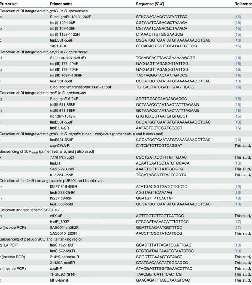
Table 1. Primers used in this studya.
Primer set Primer name Sequence (5'-3') Reference
Detection of RI integrated intogroELinS.epidermidis
a S. epi groEL 1213-1232F CTKGAAGAAGGTATYGTTGC [15]
int (I) 109-128F CGTAAATCAGACGCTAAACA [15]
b int (I) 109-128F CGTAAATCAGACGCTAAACA [15]
int (I) 1139-1122R CTAAACTTGTGGGAAGCG [15]
c fusB531-559F CGGATGGTCAATATGTAAAAAAAGGTGAC [13]
185 LA 3R CTCACAGAGGTTCTATAATGTTGG [15]
Detection of RI integrated intosmpBinS.epidermidis
d S.epi ssra407-429 (F) TCAAGCACTTAAAGAAAAAGCGG [16]
int (III) 175–194F GACGAGTTAGAGGGTATTGG [16]
e int (III) 175–194F GACGAGTTAGAGGGTATTGG [16]
int (III) 1087–1068R TACTAGGGTACAAATGACCG [16]
f fusB531-559F CGGATGGTCAATATGTAAAAAAAGGTGAC [13]
S.epi sodium transporter 1146–1168F TCTCACTATGGATTTAACTTCCG [16]
Detection of RI integrated intorpsRinS.epidermidis
g S epi rpsR 6-24F AGGTGGACCAAGAAGAGGC [15]
int(II) 541-565F GCTAAACGTAATAACTATTTAGAAG [15]
h int(II) 541-565F GCTAAACGTAATAACTATTTAGAAG [15]
int 1061-1042R GTGTGACGTAATGTGTGCGT [15]
i fusB531-559F CGGATGGTCAATATGTAAAAAAAGGTGAC [13]
fusB LA-2R AATACTCCTGGATGGCGT [15]
Detection of RI integrated intogroELinS.capatissubsp.urealyticus(primer sets a and b also used)
j fusB531-559F CGGATGGTCAATATGTAAAAAAAGGTGAC [13]
cap-CWA-R CYTCMTCTTCGTCAGGAT This study
Sequencing of ScRIfusB(primer sets a, b, and j also used)
k 7778 PstI up2F CGCTGATACCTTTGTTGAAC This study
fusBR ACAATGAATGCTATCTCGACA [13]
j Sepi 2793up2F AAAGTGCTGTATGGCGTG This study
ri17 284-265R TCCATAGCATTTAATCCGTG This study
Detection of thefusB-carrying plasmid pUB101 and its relatives
m IS257 518-499R ATATGACGGTGATCTTGCTC [13]
fusB 283-254R AGGTAGTTCAAAAG [13]
n IS257 33-52F GGATGTTATCACTGT [13]
fusB 530-558F CGGATGGTCAATATGTAAAAAAAGGTGAC [13]
Detection and sequencing SCCfusC
o orfX-uF ACTTCGTCTTCGTCATTGG This study
hsdR_593R CTCCAATAAAACATTTGTCCC [17]
p (inverse PCR) SAS0044dn382R GGATTCAGAATGGTTTCC [17]
SAS0046_226R AACCTTCGGTATCATCCG This study
Sequencing of pseudo SCC and itsflanking region
q (LA PCR) fusC 162-183F GGACTTTATTACATCGATTGAC [13]
fusC 572-550R CTGTCATAACAAATGTAATCTCC [13]
r (inverse PCR) 21429-helicase-R CGGCTTGAAACTGTAACC This study
21429A-copBR GTATGACAAGTATCGCAGCG This study
s (inverse PCR) copB-F ATACGAGTTGGTGAAACCTTAC This study
TFGfusC 7914F TAACGGTCATTTCACTCG This study
t MFS-transF GAACAGATTTAGCAAAGTCAC This study
covering almost the entire region as previously described [17]. The schematic diagram of SCCfusCmapping was shown inS2A Fig.
Sequencing of the
fus
elements ScRIfusB, SCC
fusC
and pseudo SCC
The sequence of ScRIfusBwas determined by five PCRs covering the entire region using the
primers listed inTable 1and illustrated inS1D Fig. The sequence of SCCfusCwas mined by the 21 primer sets used for PCR mapping, and the extreme right region was deter-mined by inverse PCR as shown inS2A Fig. To determine the sequence of pseudo SCC, general PCR, the LA PCR in vitro cloning kit (Takara Shuzo Co.) or inverse PCR were used as shown inTable 1andS2B Fig. In brief, PCR primers specific for the known sequence were used, and the PCR product was subsequently sequenced. To obtain the full sequence of the corresponding mobile elements, the fragments were used as probes for Southern blot hybrid-ization to determine a suitable restriction enzyme to use for further cloning. The sequence was collected by aligning and combining the amplification fragments obtained by LA PCR or inverse PCR.
Multilocus sequence typing (MLST)
The MLST was analyzed in 14fusB-positiveS.epidermidisstrains and 5fusC-positiveS. hominissubsp.hominisstrains according to the methods described previously [37,38]. The new sequence types were deposited in theS.epidermidisandS.hominisMLST databases. The eBURST method was used to infer the evolutionary relatedness of STs (http://www.mlst. net).
Detection of virulence genes associated with invasive infection in
S
.
epidermidis
isolates
We detected theicaABof theicalocus, IS256 andmecAto discriminate between virulent and non-virulent isolates as previously described [16].
Nucleotide sequences
The nucleotide sequences of SCCfusCofS.hominissubsp.hominisTFGsh1, ScRIfusBofS. capitissubsp.urealyticusTFGsc1 and pseudo SCC ofS.hominissubsp.hominisTFGsh5-1 have been deposited in the GenBank database under accession numbers AB930126 to AB930128.
Table 1. (Continued)
Primer set Primer name Sequence (5'-3') Reference
speG-7F CTAAGAGCATTAGAGTATAGTG [17]
u (inverse PCR) up speG-R GATTTGTATGAATGGCACTC This study
speG 408R TGTTTTAAATCCTTGTGACTCG [17]
v speGR408R TGTTTTAAATCCTTGTGACTCG [17]
hominis-afSCC-R TTCTTCTGAAACTATCTGCTGG This study
aThe positions of the primers are indicated inS1 Fig(fusB-carrying elements) orS2 Fig(fusC-carrying elements).
Results
Species distribution of fusidic acid-resistant staphylococci from hand
skin flora
Among the 853 isolates collected from 59 volunteers, a total of 70 isolates recovered from 22 individuals (22/59 = 37.3%) were found to be fusidic acid-resistant staphylococci (MIC val-ues≧2μg/ml). The isolates obtained from the same person exhibiting identical PFGE patterns
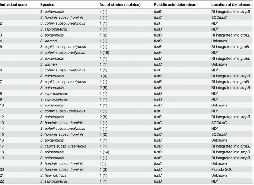
were considered to be the same strain. Therefore, 34 fusidic-acid resistant CoNS strains were obtained, and no fusidic acid-resistantS.aureuswas found. Among the 34 fusidic-acid resistant CoNS strains, the most common species wasS.epidermidis(14 strains in 38 isolates), followed byS.cohniisubsp.urealyticus(5 strains in 14 isolates),S.hominissubsp.hominis(5 strains in 8 isolates),S.saprophyticus(4 strains/isolates),S.capitissubsp.urealyticus(3 strains/isolates), S.warneri(2 strains/isolates) andS.haemolyticus(1 strains/isolate) (Table 2). ThreeS.capitis subsp.urealyticusstrains isolated from three different individuals were phylogenetically related because the DNA restriction patterns produced by PFGE had less than two-band differences. Among the 22 individuals who harbored fusidic acid-resistant staphylococci, five were colo-nized by two species, and one was colocolo-nized by four species. There were three individuals who were colonized with multiple strains ofS.epidermidis, and the strains isolated from the same person displayed very limited differences in the PFGE patterns.
Fusidic acid resistance determinants
PCR detection offusB-type genes (fusB,fusC,fusDandfusF) was performed on the 34 fusidic acid-resistant staphylococci. As shown inTable 2, allS.epidermidisandS.capitissubsp. urealy-ticusisolates possessedfusB, whereas theS.hominissubsp.hominisandS.haemolyticusisolates carried thefusCgene.S.warneriharboredfusBorfusC. ThefusDandfusFgenes were found exclusively inS.saprophyticusandS.cohniisubsp.urealyticusstrains, respectively.
Genetic environments of
fusB
The locations offusB, which appears to be associated with mobile genetic elements, were exam-ined by PCR based on the known sequences of RIs or plasmids (Table 1). Of the 14S. epidermi-disstrains, 12 strains carriedfusBby RIs either integrated intogroEL(n = 2) orsmpB(n = 10) (Table 2). The location offusBin the remaining twoS.epidermidisstrains and oneS.warneri strain remains unknown.
The threeS.capitissubsp.urealyticusstrains were found to acquirefusBby a RI integration intogroELusing PCR primer sets designed in this study (Table 1). Because there is no report on the structure of thefusBelement inS.capitis, strain TFGsc1 (isolated from individual No. 5) was subjected to sequencing to confirm the PCR results. Sequence analysis revealed a
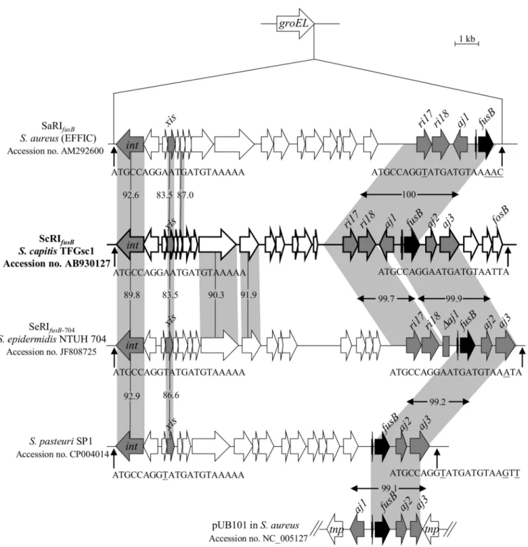
16,916-bp RI integrated intogroEL, which was referred to as ScRIfusB, where“Sc”signifies“S. capitis”. ScRIfusBhad 24 putative open reading frames (ORFs) (Fig 1andS1 Table). It carried
theri17-ri18-aj1-fusB-aj2-aj3locus, which is always fragmented in other reported mobile genetic elements, such as SaRIfusB, SeRIfusB-704, RI inS.pasteuriSP1 and pUB101 (Fig 1).
One person (individual No. 5) was colonized byS.capitissubsp.urealyticusandS. epidermi-dis. Both of the species carriedfusB-related RIs with the same integration sites ingroEL. To know if horizontal gene transfer has occurred between the two species, the structures of the fusBelement inS.epidermidiswas further analyzed using the resolved sequence of ScRIfusB
(16,916-bp RI) from theS.capitissubsp.urealyticus. However, PCR mapping revealed that the fusBsurrounding region inS.epidermidiswas different from ScRIfusB, indicating the two
Genotypic analysis of fusidic acid-resistant
S
.
epidermidis
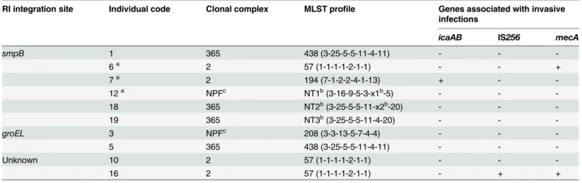
To further understand the genetic relatedness of the 14fusB-positiveS.epidermidisstrains, we determined the sequence types by MLST and the presence of genes associated with invasive infections (icaAB, IS256andmecA) [16]. As shown inTable 3, 7 sequence types were identified and were clustered by eBURST algorithm into three clonal complexes (CCs): CC2 (n = 7), CC365 (n = 4) and a CC with no predicted founder (n = 3). The prevalence oficaAB, IS256and mecAwas low for the 14S.epidermidisstrains. The strains isolated from individual No. 6, 10 and 16 were ST57, but exhibited differences in the virulence gene patterns and the mechanism by which thefusBelements were acquired. The strains isolated from individuals No. 1 and 5 were ST438 and shared identical virulence gene patterns, although they acquiredfusB-carrying RIs integrated into different sites. The strain isolated from individual No. 18 was ST-NT2, Table 2. Fusidic acid-resistant CoNS found in 22 individuals.
Individual code Species No. of strains (isolates) Fusidic acid determinant Location offuselement
1 S.epidermidis 1 (1) fusB RI integrated intosmpB
S.hominissubsp.hominis 1 (1) fusC SCCfusC
2 S.cohniisubsp.urealyticus 1 (1) fusF NDa
S.saprophyticus 1 (1) fusD NDa
3 S.epidermidis 1 (2) fusB RI integrated intogroEL
4 S.warneri 1 (1) fusB Unknown
5 S.capitissubsp.urealyticus 1 (1) fusB RI integrated intogroEL
S.cohniisubsp.urealyticus 1 (10) fusF NDa
S.epidermidis 1 (1) fusB RI integrated intogroEL
S.warneri 1 (1) fusC Unknown
6 S.cohniisubsp.urealyticus 1 (1) fusF NDa
S.epidermidis 3 (4) fusB RI integrated intosmpB
7 S.capitissubsp.urealyticus 1 (1) fusB RI integrated intogroEL
S.epidermidis 2 (5) fusB RI integrated intosmpB
8 S.saprophyticus 1 (1) fusD NDa
9 S.saprophyticus 1 (1) fusD NDa
10 S.epidermidis 1 (1) fusB Unknown
11 S.cohniisubsp.urealyticus 1 (1) fusF NDa
12 S.epidermidis 2 (8) fusB RI integrated intosmpB
13 S.hominissubsp.hominis 1 (1) fusC SCCfusC
14 S.cohniisubsp.urealyticus 1 (1) fusF NDa
15 S.hominissubsp.hominis 1 (2) fusC SCCfusC
16 S.epidermidis 1 (1) fusB Unknown
17 S.capitissubsp.urealyticus 1 (1) fusB RI integrated intogroEL
18 S.epidermidis 1 (14) fusB RI integrated intosmpB
19 S.epidermidis 1 (1) fusB RI integrated intosmpB
S.hominissubsp.hominis 1(1) fusC Unknown
20 S.hominissubsp.hominis 1 (3) fusC Pseudo SCC
21 S.haemolyticus 1 (1) fusC Unknown
22 S.saprophyticus 1 (1) fusD NDa
aNot determined becausefusDandfusFhave been reported to be intrinsic in theS.saprophyticusandS.cohniisubsp.urealyticuschromosome, respectively [7,11].
which is a single locus variant of ST-NT3 found in a strain isolated from individual No. 19. The S.epidermidisstrains isolated from the same individual (No. 6, 7 and 12) shared identical genetic patterns within the same person.
Fig 1. Structure of ScRIfusBinS.capitissubsp.urealyticusTFGsc1.ScRIfusBwas compared to SaRIfusB, SeRIfusB-704, the RI in theS.pasteurigenome
(the above RIs are inserted intogroEL) and the plasmid pUB101. The ORFs are shown as arrows, and the genes of interest are indicated as grey or black arrows. The homologous regions are shaded, and the numbers in the shadow show the percent homology between the corresponding sequences in comparison to ScRIfusB. The predictedattsites are indicated by vertical arrows. Th divergent nucleotides in the 21-bpattsequences are underlined.
Genetic environments of
fusC
A total of 7fusC-positive strains, including 5S.hominissubsp.hominis, 1S.haemolyticus, and 1S.warneri, were found (Table 2). AsfusCwas mostly found within the SCC structure in staphylococci [17–20], we first detected theccrgenes (encoding cassette chromosome recombi-nases) by PCR. ThreeS.hominissubsp.hominisstrains and 1S.warneristrain were positive forccrA1B1. Further PCR mapping revealed that all threeccrA1B1-positiveS.hominisstrains carried SCCfusC. Because the SCCfusChas only been reported in ST239/SCCmecIII MRSA [17], we subsequently determined the sequence of SCCfusCin theS.hominissubsp.hominis strain TFGsh1 to confirm the PCR mapping results. Nucleotide sequence analysis indicated 99.9% similarity (16 bp mismatch) compared to SCCfusCin ST239/SCCmecIII MRSA strain NTUH-4729 (Fig 2AandS2 Table).
To determine the location and structure of thefusCelement in the other twoS.hominis subsp.hominisstrains which do not carry knownccrABorccrC, the flanking region offusCin one strain (TFGsh5-1) was cloned and sequencing. The result revealed thatfusCin TFGsh5-1was present in a pseudo SCC structure and was inserted at the 30
-end ofrlmH(Fig 2BandS3 Table). The pseudo SCC is 10,252-bp in length and consists of 8 ORFs. The IS431-dinA-orf -orf-fusC-copBlocus (partialcopBbecause 1 to 575 bp was truncated in ST779 MRSA) showed 99.7% nucleotide sequence similarity to the pseudo SCCmecin ST779 MRSA. A 17.8-kb frag-ment was found immediately downstream of the pseudo SCC. The region from the secondatt site tospeGshowed 97.9% nucleotide sequence similarity to the SCC in ST779 MRSA.
For anotherfusC-positive,ccr-negativeS.hominissubsp.hominisstrain in individual No. 19, thedinA-orf-orf-fusC-copBlocus was detected, but the chromosome/plasmid location remains unknown because no amplification product could be generated betweenrlmHand the fusCflanking region.
ForfusC-positiveS.warneri(n = 1) andS.haemolyticus(n = 1), the genetic environments of fusCwere unknown.
Table 3. Genetic characteristics ofS.epidermidiscarryingfusBelements at different integration sites.
RI integration site Individual code Clonal complex MLST profile Genes associated with invasive infections
icaAB IS256 mecA
smpB 1 365 438 (3-25-5-5-11-4-11) - -
-6a 2 57 (1-1-1-1-2-1-1) - - +
7a 2 194 (7-1-2-2-4-1-13) + -
-12a NPFc NT1b(3-16-9-5-3-x1b-5) - -
-18 365 NT2b(3-25-5-5-11-x2b-20) - -
-19 365 NT3b(3-25-5-5-11-4-20) - -
-groEL 3 NPFc 208 (3-3-13-5-7-4-4) - -
-5 365 438 (3-25-5-5-11-4-11) - -
-Unknown 10 2 57 (1-1-1-1-2-1-1) - -
-16 2 57 (1-1-1-1-2-1-1) - + +
aThe multiple strains obtained from the same individual (No. 6, No. 7 or No. 12) display identical patterns in each person.
bNovel allele or ST found in this study. NT3 represents novel combination of known alleles, while NT1 and NT2 represent combinations containing novel allele sequences oftpi. The novel allele sequences and ST have been submitted to theS.epidermidisMLST database (http://sepidermidis.mlst.net). The two novel allele sequences oftpican be found inS1 Text.
cNPF: the ST-NT1 and ST208 were clustered in a clonal complex with no predicted founder.
Genotyping of fusidic acid-resistant
S
.
hominis
subsp.
hominis
MLST was carried out to understand the phylogenetic relationships between the five fusidic acid-resistantS.hominissubsp.hominisstrains. As shown inTable 4, two SCCfusC-carrying strains isolated from individuals No. 13 and 15 shared the same sequence type. The other strains were of diverse genetic backgrounds.
Fig 2. Genetic organization offusC-related elements inS.hominissubsp.hominis.(A) Schematic maps of SCCfusCin strain TFGsh1 and (B) the composite SCC structure in strain TFGsh5-1 are shown. The ORFs are shown as arrows, and the drug resistance genesfusCandmecAare shown as black arrows. The homologous regions are shaded, and the numbers in the shadow show the percent homology. Theattsequence is indicated by vertical arrows.
doi:10.1371/journal.pone.0143106.g002
Table 4. Genotypes offusC-positiveS.hominissubsp.hominis.
Structure offusCelement Individual code MLST profile
SCCfusC 1 41a(11a-2-13-1-6-3)
13 42a(2a-5-13-4-6-3)
15 42a(2a-5-13-4-6-3)
Pseudo SCC 20 44a(17a-5-13-4-7-3)
Unknown 19 43a(1a-3-3-12a-6-6a)
aNovel allele or ST found in this study.
Discussion
This is the first report to study fusidic acid resistance in staphylococci among the skin flora of healthy volunteers. In this study, we did not find fusidic acid-resistantS.aureusamong the skin flora, although we have isolated fusidic acid-susceptibleS.aureus(data not shown). The result is in accord with the low resistance rate to fusidic acid in clinical isolates ofS.aureusin Taiwan (0 to 6%) [15,30]. The overall resistance rate of fusidic acid in CoNS from skin flora in the community was 37.3%, which was lower than those isolated from hospitalized patients in Taiwan (48.9%, introduction) but is still higher than CoNS isolated from hospitals in the United States (7.2%), Canada (20.0%), Australia (10.8%) or some European countries (Greece, Israel, Italy, Poland, Spain and Turkey, 12.5% to 32.0%) [27,28].
In the present study, the breakpoint of 2μg/ml was used to interpret fusidic acid resistance
as we used before [11,13,15], instead of1μg/ml recommended by the EUCAST [36].
There-fore, the overall fusidic acid resistance rate would probably slightly increase if the EUCAST breakpoint is applied.
The fusidic acid resistance among 34 resistant strains was mostly mediated byfusB and fusC. ThefusBgenes in the present study were mainly chromosomally encoded within resis-tance islands (RIs) in specific chromosomal locations, whilefusCwas carried by different SCC elements, including the previously described SCCfusCand a new pseudo SCC element. The fusB-related RI elements have been reported inS.aureus,S.pasteuri, andS.epidermidisclinical isolates [15,16]. In the present study, we first found thatS.capitissubsp.urealyticuscarried fusB-RI. Both RIs and pathogenicity islands (PIs) are phage-related chromosomal islands that produce phage-like infectious particles by hijacking the capsids of phages [39]. To date, several PI-related accessory virulence genes have been described, such astst(encoding toxic shock syn-drome toxin) andseb(encoding staphylococcal enterotoxin B). Orthologues of these genes have been reported in different PIs, but most of them are still restricted in theS.aureusgenome [39]. Unlike the PI-related virulence genes, thefusBwas found not only in the RIs of four dif-ferentStaphylococcusspecies but also on aS.aureus-derived plasmid, pUB101 [12,14–16] (Fig 1). Comparison of the immediate flanking regions offusBamong the different species revealed high sequence similarities (>99.1%), even though some deletions were observed (Fig 1). The low G + C content (26.6%) of the ScRIfusBri17-ri18-aj1-fusB-aj2-aj3locus compared to theS. capitisgenome (32.76%) [40] implies that the element may originate in other bacterial species and then disseminate into staphylococci by RIs or plasmids.
We have previously reported a novel emerging SCCfusCin fusidic acid-resistant ST239/ SCCmecIII MRSA in Taiwan [17]. In the present study, we unexpectedly found three of fiveS. hominisstrains carried the SCCfusC, and the sequence of SCCfusCin strain TFGsh1 was nearly identical to that in ST239/SCCmecIII MRSA. To date onlyS.hominisand no other CoNS are known to carry SCCfusC. Thus, the commensalS.hominismay act as an important reservoir for horizontal gene transfer for the dissemination offusCto ST239/SCCmecIII MRSA in Tai-wan, although the origin of SCCfusCremains to be explored.
pseudo SCC by anattsequence (Fig 2B). The 17.8-kb fragment carried virulence-related genes, speGandarsCBRAD, which are usually components of mobile genetic elements but are not present in the core chromosome ofS.hominis[26,42]. Hence, this 17.8-kb fragment behaved just like an SCC, although it did not contain theccrgenes. The mosaic structures of pseudo SCC implied that multiple recombination events have occurred after acquisition of the foreign genetic element intoS.hominisorS.aureus.
MultipleS.epidermidisstrains isolated from the same person exhibited identical sequence types as well as virulence gene patterns associated with invasive infections (Table 3); only lim-ited differences were observed in the PFGE profiles. They also acquiredfusBelements that were integrated into the same site (Table 2). This suggested that these strains may have acquiredfusB-carrying RIs once and then undergone evolutionary changes to produce slightly different PFGE patterns in the same host.
Previous studies have shown that the CC2 was the most commonS.epidermidisin both hos-pital (87.3% to 100%) and community (58%) [43–46]. However, prevalence oficaAB, IS256 andmecAin CC2S.epidermidisisolated from community environment was lower than that in clinical isolates even they shared similar genetic background [44,46]. For the 14fusB-positiveS. epidermidisstrains which were CC2 or other minor clonal lineages, the overall prevalence of icaAB, IS256andmecAwas low (Table 3). It implies that thefusB-positiveS.epidermidisin skin flora is not originated in hospital.
In conclusion, fusidic acid resistance in commensal staphylococci was found to be mainly mediated by thefusB-family genes. At least four types of mobile genetic elements carryingfusB orfusCwere responsible for the fusidic acid resistance in CoNS, suggesting multiple events of horizontal gene transfer have occurred among various species or lineages in community. The structures of the acquired resistance elements were similar to the structures in clinical isolates, implying that commensals may act as reservoir for the pathogens. Furthermore, the high simi-larities of SCCfusCprovide evidence for possible horizontal transfer between commensalS. hominisand ST239/SCCmecIII MRSA.
Supporting Information
S1 Fig. Carton representation of PCR mapping and sequencing forfusB-carrying elements. Schematic maps of RI inS.epidermidisintegrated intogroEL(A),smpB(B) andrpsR(C), RI in S.capitissubsp.urealyticusintegrated into (D)groELand plasmid pUB101 (E) are shown. The arrows below the structures indicate PCR primers, which are listed inTable 1.
(PDF)
S2 Fig. Carton representation of PCR mapping and sequencing for fusC-carrying elements. Schematic maps for SCCfusC(A) and pseudo SCC with its flanking region (B) are shown. The arrows below the structures indicate PCR primers, which are listed inTable 1.
(PDF)
S1 Table. Genetic organization of ScRIfusB. (XLS)
S2 Table. Genetic organization of SCCfusC. (XLS)
S3 Table. Genetic organization of pseudo SCC and its flanking region. (XLS)
Author Contributions
Conceived and designed the experiments: WCH LJT. Performed the experiments: WCH HJC YTL CWC HHL SPT YYJ KHL. Analyzed the data: WCH HJC. Contributed reagents/materi-als/analysis tools: WCH HJC YTL CWC HHL JCT LJT. Wrote the paper: WCH LJT.
References
1. Dobie D, Gray J. Fusidic acid resistance inStaphylococcus aureus. Arch Dis Child. 2004; 89: 74–77.
PMID:14709515
2. Besier S, Ludwig A, Brade V, Wichelhaus TA. Molecular analysis of fusidic acid resistance in Staphylo-coccus aureus. Mol Microbiol. 2003; 47: 463–469. PMID:12519196
3. Borg A, Holm M, Shiroyama I, Hauryliuk V, Pavlov M, Sanyal S, et al. Fusidic acid targets elongation factor G in several stages of translocation on the bacterial ribosome. J Biol Chem. 2015; 290: 3440–
3454. doi:10.1074/jbc.M114.611608PMID:25451927
4. Wilson DN. Ribosome-targeting antibiotics and mechanisms of bacterial resistance. Nat Rev Microbiol. 2014; 12: 35–48. doi:10.1038/nrmicro3155PMID:24336183
5. Norstrom T, Lannergard J, Hughes D. Genetic and phenotypic identification of fusidic acid-resistant mutants with the small-colony-variant phenotype inStaphylococcus aureus. Antimicrob Agents Che-mother. 2007; 51: 4438–4446. PMID:17923494
6. Koripella RK, Chen Y, Peisker K, Koh CS, Selmer M, Sanyal S. Mechanism of elongation factor-G-mediated fusidic acid resistance and fitness compensation inStaphylococcus aureus. J Biol Chem. 2012; 287: 30257–30267. doi:10.1074/jbc.M112.378521PMID:22767604
7. O'Neill AJ, McLaws F, Kahlmeter G, Henriksen AS, Chopra I. Genetic basis of resistance to fusidic acid in staphylococci. Antimicrob Agents Chemother. 2007; 51: 1737–1740. PMID:17325218
8. O'Neill AJ, Chopra I. Molecular basis offusB-mediated resistance to fusidic acid inStaphylococcus aureus. Mol Microbiol. 2006; 59: 664–676. PMID:16390458
9. Guo X, Peisker K, Backbro K, Chen Y, Koripella RK, Mandava CS, et al. Structure and function of FusB: an elongation factor G-binding fusidic acid resistance protein active in ribosomal translocation and recycling. Open Biol. 2012; 2: 120016. doi:10.1098/rsob.120016PMID:22645663
10. Cox G, Thompson GS, Jenkins HT, Peske F, Savelsbergh A, Rodnina MV, et al. Ribosome clearance by FusB-type proteins mediates resistance to the antibiotic fusidic acid. Proc Natl Acad Sci U S A 2012; 109: 2102–2107. doi:10.1073/pnas.1117275109PMID:22308410
11. Chen HJ, Hung WC, Lin YT, Tsai JC, Chiu HC, Hsueh PR, et al. A novel fusidic acid resistance determi-nant,fusF, inStaphylococcus cohnii. J Antimicrob Chemother. 2015; 70: 416–419. doi:10.1093/jac/ dku408PMID:25313205
12. O'Brien FG, Price C, Grubb WB, Gustafson JE. Genetic characterization of the fusidic acid and cad-mium resistance determinants ofStaphylococcus aureusplasmid pUB101. J Antimicrob Chemother. 2002; 50: 313–321. PMID:12205055
13. Chen HJ, Hung WC, Tseng SP, Tsai JC, Hsueh PR, Teng LJ. Fusidic acid resistance determinants in Staphylococcus aureusclinical isolates. Antimicrob Agents Chemother. 2010; 54: 4985–4991. doi:10. 1128/AAC.00523-10PMID:20855746
14. O'Neill AJ, Larsen AR, Skov R, Henriksen AS, Chopra I. Characterization of the epidemic European fusidic acid-resistant impetigo clone ofStaphylococcus aureus. J Clin Microbiol. 2007; 45: 1505–1510.
PMID:17344365
15. Chen HJ, Tsai JC, Hung WC, Tseng SP, Hsueh PR, Teng LJ. Identification offusB-mediated fusidic acid resistance islands inStaphylococcus epidermidisisolates. Antimicrob Agents Chemother. 2011; 55: 5842–5849. doi:10.1128/AAC.00592-11PMID:21968364
16. Chen HJ, Chang YC, Tsai JC, Hung WC, Lin YT, You SJ, et al. New Structure of Phage-related Islands CarryingfusBand a Virulence Gene in Fusidic Acid-ResistantStaphylococcus epidermidis. Antimicrob Agents Chemother. 2013; 57: 5737–5739. doi:10.1128/AAC.01433-13PMID:23979742
17. Lin YT, Tsai JC, Chen HJ, Hung WC, Hsueh PR, Teng LJ. A Novel Staphylococcal Cassette Chromo-somal Element, SCCfusC, CarryingfusCandspeGin Fusidic Acid-Resistant Methicillin-Resistant Staphylococcus aureus. Antimicrob Agents Chemother. 2014; 58: 1224–1227. doi:10.1128/AAC. 01772-13PMID:24277045
19. Ender M, Berger-Bachi B, McCallum N. Variability in SCCmecN1spreading among injection drug users in Zurich, Switzerland. BMC Microbiol. 2007; 7: 62. PMID:17605795
20. Kinnevey PM, Shore AC, Brennan GI, Sullivan DJ, Ehricht R, Monecke S, et al. Emergence of sequence type 779 methicillin-resistantStaphylococcus aureusharboring a novel pseudo staphylococ-cal cassette chromosomemec(SCCmec)-SCC-SCCCRISPRcomposite element in Irish hospitals. Anti-microb Agents Chemother. 2013; 57: 524–531. doi:10.1128/AAC.01689-12PMID:23147725
21. Huebner J, Goldmann DA. Coagulase-negative staphylococci: role as pathogens. Annu Rev Med. 1999; 50: 223–236. PMID:10073274
22. Piette A, Verschraegen G. Role of coagulase-negative staphylococci in human disease. Vet Microbiol. 2009; 134: 45–54. doi:10.1016/j.vetmic.2008.09.009PMID:18986783
23. Becker K, Heilmann C, Peters G. Coagulase-negative staphylococci. Clin Microbiol Rev. 2014; 27: 870–926. doi:10.1128/CMR.00109-13PMID:25278577
24. Berglund C, Soderquist B. The origin of a methicillin-resistantStaphylococcus aureusisolate at a neo-natal ward in Sweden-possible horizontal transfer of a staphylococcal cassette chromosomemec between methicillin-resistantStaphylococcus haemolyticusandStaphylococcus aureus. Clin Microbiol Infect. 2008; 14: 1048–1056. doi:10.1111/j.1469-0691.2008.02090.xPMID:19040477
25. Barbier F, Ruppe E, Hernandez D, Lebeaux D, Francois P, Felix B, et al. Methicillin-resistant coagu-lase-negative staphylococci in the community: high homology of SCCmecIVa between Staphylococ-cus epidermidisand major clones of methicillin-resistantStaphylococcus aureus. J Infect Dis. 2010; 202: 270–281. doi:10.1086/653483PMID:20550456
26. Planet PJ, Larussa SJ, Dana A, Smith H, Xu A, Ryan C, et al. Emergence of the epidemic methicillin-resistantStaphylococcus aureusstrain USA300 coincides with horizontal transfer of the arginine cata-bolic mobile element andspeG-mediated adaptations for survival on skin. MBio. 2013; 4: e00889–13.
doi:10.1128/mBio.00889-13PMID:24345744
27. Castanheira M, Watters AA, Mendes RE, Farrell DJ, and Jones RN. Occurrence and molecular charac-terization of fusidic acid resistance mechanisms amongStaphylococcusspp. from European countries (2008). J Antimicrob Chemother. 2010; 65: 1353–1358. doi:10.1093/jac/dkq094PMID:20430787
28. Castanheira M, Watters AA, Bell JM, Turnidge JD, and Jones RN. Fusidic acid resistance rates and prevalence of resistance mechanisms amongStaphylococcusspp. isolated in North America and Aus-tralia, 2007–2008. Antimicrob Agents Chemother. 2010; 54: 3614–3617. doi:10.1128/AAC.01390-09
PMID:20566766
29. Jones RN, Mendes RE, Sader HS, and Castanheira M. In vitro antimicrobial findings for fusidic acid tested against contemporary (2008–2009) gram-positive organisms collected in the United States. Clin
Infect Dis. 2011; 52 Suppl 7: S477–486. doi:10.1093/cid/cir163PMID:21546624
30. Wang JL, Tang HJ, Hsieh PH, Chiu FY, Chen YH, Chang MC, et al. Fusidic acid for the treatment of bone and joint infections caused by meticillin-resistantStaphylococcus aureus. Int J Antimicrob Agents. 2012: 40:103–107. doi:10.1016/j.ijantimicag.2012.03.010PMID:22612900
31. den Heijer CD, van Bijnen EM, Paget WJ, Stobberingh EE. Fusidic acid resistance inStaphylococcus aureusnasal carriage strains in nine European countries. Future Microbiol. 2014; 9: 737–745. doi:10. 2217/fmb.14.36PMID:25046521
32. Hauschild T, Stepanovic S. Identification ofStaphylococcusspp. by PCR-restriction fragment length polymorphism analysis ofdnaJgene. J Clin Microbiol. 2008; 46: 3875–3879. doi: 10.1128/JCM.00810-08PMID:18832127
33. Liu D, Swiatlo E, Austin FW, Lawrence ML. Use of a putative transcriptional regulator gene as target for specific identification ofStaphylococcus epidermidis. Lett Appl Microbiol. 2006; 43: 325–330. PMID: 16910940
34. Clinical & Laboratory Standards Institute. 2014. Performance standards for antimicrobial susceptibility testing. Twenty-fourth informational supplement M100-S24. Clinical and Laboratory Standards Insti-tute, Wayne, PA, USA.
35. Coutant C, Olden D, Bell J, Turnidge JD. Disk diffusion interpretive criteria for fusidic acid susceptibility testing of staphylococci by the National Committee for Clinical Laboratory Standards method. Diagn Microbiol Infect Dis. 1996; 25: 9–13. PMID:8831039
36. European Committee on Antimicrobial Susceptibility Testing. Breakpoint tables for interpretation of MICs and zone diameters, Version 5.0, 2015.http://www.eucast.org/clinical_breakpoints/
38. Zhang L, Thomas JC, Miragaia M, Bouchami O, Chaves F, d'Azevedo PA, et al. Multilocus sequence typing and further genetic characterization of the enigmatic pathogen,Staphylococcus hominis. PLoS One. 2013; 8:e66496. doi:10.1371/journal.pone.0066496PMID:23776678
39. Novick RP, Christie GE, Penades JR. The phage-related chromosomal islands of Gram-positive bacte-ria. Nat Rev Microbiol. 2010; 8: 541–551. doi:10.1038/nrmicro2393PMID:20634809
40. Qin N, Ding W, Yao J, Su K, Wu L, Li L. Genome sequence ofStaphylococcus capitisQN1, which causes infective endocarditis. J Bacteriol. 2012; 194: 4469–4470. doi:10.1128/JB.00827-12PMID: 22843597
41. Mahillon J, Chandler M. Insertion sequences. Microbiol Mol Biol Rev. 1998; 62: 725–774. PMID: 9729608
42. Yu D, Pi B, Chen Y, Wang Y, Ruan Z, Otto M, et al. Characterization of the staphylococcal cassette chromosome composite island ofStaphylococcus haemolyticusSH32, a methicillin-resistant clinical isolate from China. PLoS One 2014; 9: e87346. doi:10.1371/journal.pone.0087346PMID:24466348
43. Mendes RE, Deshpande LM, Costello AJ, Farrell DJ. Molecular epidemiology ofStaphylococcus epi-dermidisclinical isolates from U.S. hospitals. Antimicrob Agents Chemother 2012; 56: 4656–4661. doi: 10.1128/AAC.00279-12PMID:22687512
44. Rolo J, de Lencastre H, Miragaia M. Strategies of adaptation ofStaphylococcus epidermidisto hospital and community: amplification and diversification of SCCmec. J Antimicrob Chemother 2012; 67: 1333–
1341. doi:10.1093/jac/dks068PMID:22422509
45. Iorio NL, Caboclo RF, Azevedo MB, Barcellos AG, Neves FP, Domingues RM, et al. Characteristics related to antimicrobial resistance and biofilm formation of widespread methicillin-resistant Staphylo-coccus epidermidisST2 and ST23 lineages in Rio de Janeiro hospitals, Brazil. Diagn Microbiol Infect Dis 2012; 72: 32–40. doi:10.1016/j.diagmicrobio.2011.09.017PMID:22100013
46. Du X, Zhu Y, Song Y, Li T, Luo T, Sun G, et al. Molecular analysis ofStaphylococcus epidermidis strains isolated from community and hospital environments in China. PLoS One 2013; 8: e62742. doi: