the INS-GAS Mouse
Melanie J. Thomson1, D. Mark Pritchard2, Sally A. Boxall1, Abdul A. Abuderman2, Jonathan M. Williams2, Andrea Varro2, Jean E. Crabtree1*
1Molecular Gastroenterology, Leeds Institute of Molecular Medicine, St. James’s University Hospital, Leeds, United Kingdom,2Institute of Translational Medicine, University of Liverpool, Liverpool, United Kingdom
Abstract
There is increasing evidence from clinical and population studies for a role ofH. pyloriinfection in the aetiology of iron deficiency. Rodent models ofHelicobacterinfection are helpful for investigating any causal links and mechanisms of iron deficiency in the host. The aim of this study was to investigate the effects of gastricHelicobacterinfection on iron deficiency and host iron metabolism/transport gene expression in hypergastrinemic INS-GAS mice. INS-GAS mice were infected with Helicobacter felisfor 3, 6 and 9 months. At post mortem, blood was taken for assessment of iron status and gastric mucosa for pathology, immunohistology and analysis of gene expression. ChronicHelicobacterinfection of INS- GAS mice resulted in decreased serum iron, transferrin saturation and hypoferritinemia and increased Total iron binding capacity (TIBC). Decreased serum iron concentrations were associated with a concomitant reduction in the number of parietal cells, strengthening the association between hypochlorhydria and gastricHelicobacter-induced iron deficiency. Infection with H. felisfor nine months was associated with decreased gastric expression of iron metabolism regulators hepcidin,Bmp4and Bmp6but increased expression of Ferroportin 1, the iron efflux protein, iron absorption genes such as Divalent metal transporter 1, Transferrin receptor 1 and alsoLcn2a siderophore-binding protein. The INS-GAS mouse is therefore a useful model for studyingHelicobacter-induced iron deficiency. Furthermore, the marked changes in expression of gastric iron transporters followingHelicobacter infection may be relevant to the more rapid development of carcinogenesis in the Helicobacterinfected INS-GAS model.
Citation:Thomson MJ, Pritchard DM, Boxall SA, Abuderman AA, Williams JM, et al. (2012) GastricHelicobacterInfection Induces Iron Deficiency in the INS-GAS Mouse. PLoS ONE 7(11): e50194. doi:10.1371/journal.pone.0050194
Editor:Yoshio Yamaoka, Veterans Affairs Medical Center (111D), United States of America
ReceivedJune 21, 2012;AcceptedOctober 22, 2012;PublishedNovember 19, 2012
Copyright:ß2012 Thomson et al. This is an open-access article distributed under the terms of the Creative Commons Attribution License, which permits unrestricted use, distribution, and reproduction in any medium, provided the original author and source are credited.
Funding:This study was funded under the Sixth Framework Programme of the European Union, Project CONTENT (INCO-DEV-3-032136) and a PhD studentship to AA from King Fahd Medical City, Saudi Arabia. The funders had no role in study design, data collection and analysis, decision to publish, or preparation of the manuscript.
Competing Interests:The authors have declared that no competing interests exist.
* E-mail: j.crabtree@leeds.ac.uk
Introduction
Iron deficiency is the most common nutritional disorder in the world with iron deficiency anemia (IDA) affecting 500–600 million people globally (WHO). Manifestations of clinically advanced IDA include increased childhood mortality, reduced growth, cognitive function and susceptibility to infectious diseases [1]. Accumulating evidence from clinical [2,3,4,5,6] and population [7,8,9,10,11] studies implicates gastricHelicobacter pyloriinfection in the aetiology of iron deficiency and IDA. Iron deficiency inH. pyloriinfection is resistant to iron supplementation, but is reversible by bacterial eradication [5,7,12,13,14,15].H. pylori-associated IDA is particu-larly prevalent in children, who have smaller iron stores compared with adults [16].
There are several mechanisms by which H. pylori might contribute to iron deficiency and IDA. These include competition between the pathogen and host for limited iron stores [13,17,18], particularly in individuals with iron poor diets, thus exacerbating the effects of reduced iron intake. Iron is essential for H. pylori growth and survival, but this pathogen does not produce siderophores conventionally used by other bacteria to acquire iron [19]. The ability ofH. pylorito sequester iron facilitates its colonisation of the acidic gastric environment via enhanced
protection from oxidative stress [20]. Inactivation of iron transport and storage proteins in H. pylori renders it incapable of host colonisation. H. pylori iron binding proteins have also been implicated in the aetiology of IDA [18,21]. Hypochlorhydria associated with acuteH. pyloriinfections may predispose to IDA due to the inability to generate the bioavailable, reduced form of iron (ferrous, Fe2+
infection and dietary iron deficiency on host iron homeostasis has been studied in wild type C57BL/6 mice [29,30]. H. pylori diminished further iron stores, measured by serum ferritin and liver iron, in mice on an iron deficient diet [30]. GastricHelicobacter felis, but notH. pyloriinfection, caused iron deficiency assessed by serum iron and ferritin levels in non pregnant and pregnant mice [29]. Taken together, these and other studies provide evidence that iron acquisition byH. pylorimay have a significant role in the development of IDA.
The transgenic INS-GAS mouse model over-expresses the human gastrin (hGAS) gene controlled by the rat insulin promoter, resulting in hypergastrinemia. Male INS-GAS mice on the FVB/ N background spontaneously develop gastric adenocarcinoma after 20 months of age and this process is accelerated to 8 months by gastricHelicobacterinfection [31]. A positive correlation between circulating gastrin and transferrin saturation has been seen in hypergastrinemic patients [32]. Gastrin may play a direct role in modulating iron homeostasis as hepatic transcripts of the iron regulator hepcidin (Hamp) were increased in Gastrin-resistant CCK2 receptor knockout (CCK2RKO) mice, which have in-creased levels of circulating gastrin and dein-creased in gastrin knockout (GasKO) mice maintained on an iron deficient diet [33]. The aim of this study was therefore to investigate the haematological and molecular changes relating to iron deficiency resulting from gastric Helicobacter infection in hypergastrinemic INS-GAS mice. This was achieved by using a series of clinically relevant iron assays combined with molecular investigations of the expression of host iron metabolism and absorption genes in the gastric mucosa. As with the earlier studies from Wanget al[31], we have usedH. felisinfection rather thanH. pylorias our earlier studies have demonstrated this cause greater inflammation than H. pyloriin the murine model [35].
Methods
Ethics Statement
All experimental procedures and breeding of the INS-GAS transgenic mice were undertaken at the University of Liverpool according to UK Home Office guidelines. The study was undertaken on project licence number PPL 40/2833 which was approved by the local ethics committee of the University of Liverpool and the UK Home Office. The welfare of the mice was monitored daily.
Bacteria
Helicobacter felis ATCC 49179 was cultured as previously described [46] at 37uC for 2–3 days in a microaerophilic atmosphere. Bacteria were harvested and suspended in Tryptone Soya Broth (TSB, Oxoid) to a density of approx 56108CFU/ml and were used immediately.
H. felis Infection of Mice
Male transgenic INS-GAS mice on a FVB/N genetic background, kindly supplied by Professor Timothy C. Wang (Columbia University, New York, USA), were bred at the University of Liverpool conventional animal facility. Male FVB/ N mice were purchased from Harlan UK Ltd, (Bichester, UK). INS-GAS mice (Helicobacterspecies free) at 6–8 weeks of age were inoculated three times by oral gavage with H. felis (.108 CFU) suspended in tryptone soya broth (Oxoid) over a period of 1 week. Infected mice (n = 16219 per group) and uninfected controls (n = 19220 per group) were sacrificed at 3, 6 and 9 months post-inoculation.
Following euthanasia with CO2, whole blood was collected via cardiac puncture; serum and plasma were stored at220uC. The stomach was removed, incised along the greater curvature and rinsed with phosphate buffered saline. Gastric corpus tissue from the 9 month cohort was stored in ‘RNA Later’ (Invitrogen) at 220uC prior to total RNA extraction. Antral and corpus tissue were fixed in 4% formal saline and embedded in paraffin wax for histological analysis.
For histological assessment of parietal cell numbers the whole stomach was dissected from the mouse. Ligatures were tied around the distal oesophagus and proximal duodenum, and the stomach lumen was infiltrated with 4% formal saline. This procedure distended the stomach and fixed gastric glands in a reliable manner for histological assessment. Tissue was paraffin embedded and sections were cut at a thickness of 4mm. Histology andH. felis infection were assessed on Haematoxylin and Eosin and Giemsa stained sections. Gastric histopathology was assessed in a blind, randomised fashion by a Veterinary Pathologist (JMW) and graded according to the scoring system of Rogerset al. [36].
Immunohistology
Immunohistochemistry was used to detect and quantify parietal cells. Following permeabilization by incubating dewaxed and rehydrated sections with 1% Triton6in phosphate buffered saline for 45 minutes at room temperature, sections were incubated overnight at 4uC in rabbit anti-rat H+/K+ ATPase polyclonal antibody (1:1000, Calbiochem) diluted in 10% goat serum in Tris buffered saline (TBS) with 1% bovine serum albumin (BSA). Sections were washed in TBS-Tween20 and incubated with HRP-labelled anti-rabbit antibody (Envision kit (Dako UK Ltd) for 30 minutes before washing and substrate development according to the manufacturer’s instructions. Sections were counterstained with Gill’s haematoxylin. To quantify cell numbers in the gastric corpus mucosa, ten areas were chosen from each section and H+
-K+ -ATPase-positive cells were counted using a Leica light microscope.
Iron Parameters
Serum iron concentration and unsaturated iron binding capacity (UIBC) were measured directly using a microtitre plate assay based on change in absorbance on addition of Ferrozine (Sigma) according to the standard protocols in Practical Haema-tology [37]. Total Iron binding capacity (TIBC) was calculated from Serum Iron+UIBC. Transferrin saturation was calculated by Serum Iron/TIBC6100. Taken together, these three parameters form an important part of the routine diagnostic assessment for iron deficiency anaemia and anaemia of chronic disease in clinical settings.
Serum Ferritin
Serum ferritin protein concentrations were measured by a mouse specific enzyme-linked immunosorbent assay (ELISA) supplied by Kamiya Biochemical Company (Seattle, WA, USA) according to the manufacturer’s instructions. Clinically, the assessment of circulating ferritin levels is used as a surrogate marker for stored iron in the host.
Real Time PCR
expression of target genes was assessed as relative abundance compared toGapdhcontrol gene by theDDCt method [38]. Primer sequences are listed in Table 1 and were designed to cross an intron/exon boundary or taken from published literature [39].
Statistics
Statistical analysis was performed using GraphPad PRISM software, utilising Student’s t test (unpaired with Welch’s correction) or Mann-Whitney U test as appropriate after assessing distribution of data with the Kruskal-Wallis test. Data were considered significant if p#0.05.
Results
Effects of H. felis Infection on INS-GAS Iron Parameters
Serum iron concentration decreases in chronically
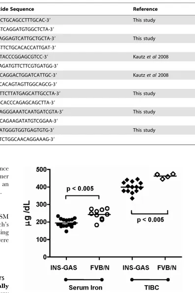
Helicobacter infected INS-GAS mice. The serum iron assay measures the amount of circulating iron that is bound to transferrin, one of the host iron carriers. Initial studies showed that uninfected INS-GAS mice have no difference in transferrin saturation (data not shown) but a significantly reduced concentra-tion of serum iron (p,0.005) and TIBC (p,0.005) compared to uninfected FVB/N mice at 10-11 months age (Figure 1). Although some bias may have been introduced due to the purchased FVB/ N control mice being obtained from a different breeding colony to background strain used to produce the INS-GAS mice, this is still evidence that there is a reduction in iron parameters in this transgenic line, making it a suitable model for further investigation of Helicobacter-induced iron deficiency. In uninfected INS-GAS mice there was no significant change in serum iron concentrations at 3, 6 or 9 months (Figure 2a, white boxes). In contrast, at 6 months, serum iron concentrations inH. felis infected mice were significantly lower (p,0.005) than in 3 month H. felis infected mice, and also significantly reduced (p = 0.05) relative to 6 month uninfected controls (Figure 2a). No further reduction in serum iron concentration was evident at 9 months in H. felis infected mice (Figure 2a).
Total iron binding capacity increases over time in
Helicobacterinfected INS-GAS mice. The total iron-binding capacity (TIBC) assay saturates the sample to measure the total
amount of transferrin in circulation. At 3 months TIBC inH. felis infected mice was significantly increased (p,0.05) compared to controls (Figure 2b). TIBC in infected mice was further increased (p,0.05) at 9 months compared to 6 month infected mice (Figure 2b). In uninfected INS-GAS mice there was no significant change in TIBC at 3, 6 or 9 months (Figure 2b, white boxes).
Transferrin saturation is reduced in chronically
Helicobacterinfected INS-GAS mice. The transferrin satura-tion assay measures the extent to which sites on transferrin molecules are occupied by iron ions. The percentage of transferrin saturation was also decreased in infected mice, becoming significantly reduced (p,0.05) by 9 months post-infection com-pared to control mice (Figure 2c). No age related changes were evident in uninfected INS-GAS mice.
Table 1.Real time PCR primers.
Locus Namea Nucleotide Sequence Reference
Total Hepcidin HampF 59-AGAGCTGCAGCCTTTGCAC-39 This study
HampR 59-GAGGTCAGGATGTGGCTCTA-39
Ferroportin 1 Fpn1F 59-TTGCAGGAGTCATTGCTGCTA-39 This study
Fpn1R 59-GGAGTTCTGCACACCATTGAT-39
Bone Morphogenic Protein 4 Bmp4F 59-TGAGTACCCGGAGCGTCC-39 Kautzet al2008
Bmp4R 59-CTCCAGATGTTCTTCGTGATGG-39
Bone Morphogenic Protein 6 Bmp6F 59-ATGGCAGGACTGGATCATTGC-39 Kautzet al2008
Bmp6R 59-CCATCACAGTAGTTGGCAGCG-39
Divalent metal transporter 1 Dmt1F 59-GGCTTTCTTATGAGCATTGCCTA-39 This study
Dmt1R 59-GGAGCACCCAGAGCAGCTTA-39
Transferrin receptor 1 Tfr1F 59-CATGAGGGAAATCAATGATCGTA-39 This study
Tfr1R 59-GCCCCAGAAGATATGTCGGAA-39
Lipocalin 2 Lcn2F 59-CTGAATGGGTGGTGAGTGTG-39 This study
Lcn2R 59-GCTCTCTGGCAACAGGAAAG-39
doi:10.1371/journal.pone.0050194.t001
Figure 1. Iron parameters in INS-GAS and FVB/N mice at 11 months of age.Scatter plots of individual serum iron concentrations (circles) and total iron binding capacity (TIBC) (diamonds) in transgenic INS-GAS mice (solid markers) (n = 17) and genetic age and gender-matched FVB/N control mice (open markers) (iron, n = 10; TIBC, n = 4). Insufficient serum was available for TIBC analysis in 6 FVB/N control mice. Statistical analysis by Mann-Whitney U test.
Serum Ferritin increases then decreases over time in
Helicobacterinfected INS-GAS mice. Ferritin is a ubiquitous intracellular protein that sequesters iron and releases it in a controlled manner. The amount of circulating serum is used clinically as a surrogate biomarker for decreased iron stores in the host. In the early phase (3 months) of theH. felisinfection, serum ferritin concentration was significantly increased compared to uninfected mice (p,0.005, Figure 2d). In contrast, at the later stages of the infection, serum ferritin concentrations were significantly reduced (Figure 2d, p,0.005 at 6 months; p,0.001 at 9 months), perhaps reflecting the overall lack of iron absorbed by the host due to the hypochlorhydria associated with parietal cell loss.
Reduced Parietal Cell Numbers in INS-GAS Mice with Gastric Helicobacter Infection
As gastrin may have a direct role in modulating iron homeostasis [32,34] and the hypochlorhydria associated with H. pylori-induced corpus atrophy is associated with iron deficiency [22], gastric parietal numbers were assessed in the INS-GAS mice
and the relationship between iron status and the plasma gastrin levels previously determined in the mice was assessed. There was no significant correlation between iron concentrations and plasma gastrin concentrations in this cohort (data not shown). The decrease in serum iron concentrations at 6 months in H. felis infected mice however was associated with a concomitant decrease in gastric parietal cell numbers relative to controls (p = 0.05), which was also evident at 9 months (p,0.05) (Figure 3).
Effects of H. felis Infection on INS-GAS Gastric Mucosa AllH. felisinoculated mice with available pathology at 3 and 6 months were histologically positive for the bacterium. At 9 months, H. felis could be detected in 55% of the inoculated animals, however, all mice showed marked inflammation and mucosal hyperplasia, indicative of successful infection. Loss of gastric Helicobacter infection with corpus atrophy has been frequently document both clinically and in rodent models. Helicobacter felis infected INS-GAS mice exhibited increasing severity of gastric mucosal lesions proportional with the duration of infection (Figure 4). Lesions were characterised by mucosal Figure 2. Iron parameters in INS-GAS mice at 3, 6 and 9 months post-infection withH. felis.Box and whisker plots of serum iron (A), total iron binding capacity (TIBC) (B), transferrin saturation (C) and serum ferritin (D) in uninfected (white boxes) andH. felisinfected (hatched boxes) INS-GAS mice. Time points as marked, group size n = 16220, whiskers represent maximum/minimum values. Statistical analysis by unpairedttest with Welch’s correction.
hyperplasia, loss of parietal cells and chief cells (oxyntic gland atrophy), mucous metaplasia, dilated gastric glands, and in-flammatory infiltrates consisting predominantly of lymphocytes, plasma cells, and to a lesser extent neutrophils. Gastric glands of longer term infected animals often exhibited features of dysplasia with disorganised branching, cell crowding with loss of polarity, hyperchromatic enlarged nuclei and increased mitotic figures.
Significantly increased inflammation (p,0.01) was evident at both 6 months and 9 months inH. felisinfected INS-GAS mice compared to INS-GAS control mice (Figure 4a). Oxyntic gland atrophy inH. felisinfected INS-GAS mice increased progressively with time being significantly greater than controls at both 6 months (p,0.01) and 9 months post-infection (p,0.001) (Figure 4b). In contrast, in the control INS-GAS mice no significant increase in inflammation was observed over the 9 months and only a small increase in oxyntic gland atrophy at 6 and 9 months was observed. None of these changes were statistically significant from the 3 month controls. A significant increase (p,0.01) in gastric dysplasia was evident in H. felis infected INS-GAS mice compared to INS-GAS control mice (Figure 4c) consistent with earlier studies [31]. No significant differences were observed temporarily between the control groups.
Effects of H. felis Infection on INS-GAS Iron Metabolism Genes
Chronic H. felis infection reduces the expression of the iron metabolism regulator, Hepcidin, in the gastric mucosa. Hepcidin is a hormone that controls the levels of iron in the labile iron pool for use in iron metabolism by negatively regulating the expression of Ferroportin 1 on iron trafficking cells and modulating the absorption of iron from ingested food via the small intestine. Hepcidin expression is regulated by IL6 in response to inflammation. A significant increase (p,0.01) in the abundance of gastric corpus Il6transcripts was observed in the INS-GAS mice compared to the FVB/N controls but no significant difference (p = 0.57) was observed between theH. felis infected and uninfected control mice (data not shown). Again, it is possible that the significant difference between the INS-GAS transgenic mice and the FVB/N control mice could be due to a possible bias introduced to the experimental system, as they
originate from different breeding colonies. INS-GAS mice infected with H. felis for 9 months (Figure 5a, hatched bar) showed a significant reduction in hepcidin transcripts in the gastric mucosa compared to both the uninfected INS-GAS and FVB/N mice. The abundance of transcripts for the upstream regulators of Figure 3. Parietal cell number in INS-GAS mice at 3, 6 and 9
months post-infection withH. felis.Box and whisker plots of gastric parietal cell number in uninfected (white boxes) andH. felisinfected (hatched boxes) INS-GAS mice. Group size n = 527 mice, whiskers represent maximum/minimum values. Statistical test is an unpairedt test with Welch’s correction.
doi:10.1371/journal.pone.0050194.g003
Figure 4. Gastric histopathology in INS-GAS mice at 3,6 and 9 months post-infection withH. felis.Grade of gastric inflammation (A), corpus atrophy (B) and dysplasia (C) in uninfected control (white bars) andH. felisinfected INS-GAS mice (hatched bars). Group size n = 5–7. Error bars represent mean 6 SEM, when error present. Statistical analysis by the Mann Whitney U test.
Hepcidin,Bmp4(p,0.05, Figure 5c) andBmp6(p,0.01, Figure 5d) were also significantly reduced following 9 months of H. felis infection in INS-GAS gastric corpus mucosa. Gastric corpusBmp6 expression in FVB/N control mice was lower but not significantly different (p = 0.06) than uninfected INS-GAS mice.
Ferroportin 1 is an efflux protein that is expressed on the basolateral membranes of gastrointestinal cells. This protein is negatively regulated by hepcidin, which binds to Ferroportin1 on the surface of cells inducing degradation. A significant increase (p = 0.05) in the abundance of the corpus transcripts ofFpn1was observed in the INS-GAS mice 9 months following H. felis infection (Figure 5b, hatched bar). Gastric corpusFpn1expression in FVB/N control mice was slightly lower (p = 0.28) than uninfected INS-GAS mice.
ChronicH. felisinfection increases the expression of iron absorption genes in the gastric mucosa. Divalent metal transporter 1 (DMT1), is expressed on the apical membrane of gastrointestinal cells and absorbs reduced ferrous iron (Fe2+
) from
the gut lumen. There was a significant increase (p,0.05) in relativeDmt1transcripts in the gastric corpus mucosa of INS-GAS mice infected withH. felis for 9 months (Figure 6a, hatched bar) compared to uninfected controls. Gastric corpusDMT1expression in FVB/N control mice was higher but not significantly different (p = 1.0) from uninfected INS-GAS mice.
Transferrin receptor 1 (TFR1) is a protein that is widely expressed on mammalian tissues. It binds and internalises (via endocytosis) circulating iron-loaded transferrin. Hepatocytes, which modulate hepcidin concentrations, sense the saturation of transferrin (via an unknown mechanism) and release the regulator hepcidin in response. Tfr1 transcripts were significantly up-regulated in the gastric mucosa ofH. felisinfected INS-GAS mice at 9 months (Figure 6b, hatched bar, p,0.005). Gastric corpus Tfr1 expression in FVB/N control mice was lower but not significantly different (p = 0.20) from uninfected INS-GAS mice.
Lipocalin 2 (Lcn2, also known as Neutrophil gelatinase-associated lipocalin [NGAL] or Holo-24p3) is a host outer Figure 5. Expression of iron metabolism regulation genes in murine gastric mucosa.Relative expression levels of genes involved in the regulation of iron metabolism were measured by qRT-PCR in biopsies of the gastric corpus mucosa of INS-GAS mice at 9 months post-infection with H. felisand in age-matched uninfected INS-GAS mice and FVB/N controls. The graphs represent the mRNA abundance of the test genes relative to the house keeping gene,Gapdh. Uninfected (white bars) (n = 10) andH. felisinfected (hatched bars) transgenic INS-GAS mice (n = 10) were compared to the uninfected FVB/N genetic background control (solid bars) (n = 5). The relative levels of total hepcidin genes (Hamp 1and2) (A), Ferroportin 1 (Fpn1) gene (B), bone morphogenic protein gene 4 (Bmp4) (C) and 6 (Bmp6) gene (D) were assessed. Error bars represent mean6SEM. Statistical analysis by unpairedttest with Welch’s correction.
membrane protein that binds to bacterial siderophores loaded with iron and is thought to bind an endogenous host equivalent (yet to be identified). In H. felis infected INS-GAS mice, there was a significant increase (Figure 6c, hatched bar, p,0.001) in Lcn2 transcripts compared to the uninfected INS-GAS control cohort at 9 months. This shows the inverse relationship between hepcidin transcripts (low in infected group) and the increased expression of a gene whose product is associated with host iron metabolism. Gastric corpusLcn2expression in FVB/N control mice was higher (not significantly, p = 0.32) than uninfected INS-GAS mice.
Discussion
The aim of this study was to use the INS-GAS mouse model to explore the hematological and molecular changes relating to iron deficiency resulting from gastric Helicobacter infection. INS-GAS mice with chronic gastric Helicobacterinfection have significantly raised circulating amidated gastrin concentrations compared to uninfected INS-GAS controls at 9 months post infection [40]. This increase is over and above the existing elevated concentrations observed in uninfected INS-GAS mice compared to their FVB/N genetic background controls. These data concur with previous observations resulting from gastricHelicobacterinfection in the INS-GAS model [41]. Conversely, parietal cell numbers in H. felis infected INS-GAS mice were reduced significantly at 6 and 9 months post-inoculation concomitant with the decrease in serum iron concentrations. The reduction in the number of parietal cells and associated hypochlorhydria induced by gastric Helicobacter infection may lead to poor ferrous iron (Fe2+
) absorption. Gastric acid is important for reducing dietary ferric (Fe3+) iron to ferrous (Fe2+) iron. In the achlorhydric Atp4a2/2 mouse, which has a mutation in the 4a subunit of H+/K+ATPase, the failure of iron absorption is considered to be the combined effects of poor solubility of ferric iron and impaired Dmt1 function due to the reduced levels of protons in the stomach [42].
The haematological parameters at the chronic stages of Helicobacterinfection in the present study are indicative of poor iron absorption. At 6 months post-infection, serum iron concen-trations were significantly lower due toH. felisinfection and by 9 months post-infection, the transferrin saturation was also markedly lower in the infected cohort. This reflects the systemic iron deficiency of these animals when compared to their uninfected counterparts, as they have reduced circulating iron loaded onto the transferrin protein. Iron storage capability, as measured by serum ferritin, increased in the acute phase of the infection at 3 months, as the host attempted to sequester iron so that it that it could not be used by the pathogen. However, by the chronic stage of the infection (6 and 9 months post-inoculation) serum ferritin concentrations were significantly reduced, perhaps in response to the overall lack of systemic iron, rendering the requirement for iron storage obsolete. Keenanet alobserved a similar pattern of raised serum ferritin concentrations at 10 weeks post-inoculation followed by a reduction at 30 weeks afterH. pyloriinfection of non-transgenic mice fed on an iron-restricted diet [30].
In this study,H. felisinfection modified the abundance of gastric mucosal mRNA of various iron metabolism genes. Some of these genes, such asFpn1, Dmt1and Tfr1are regulated post-transcrip-tionally and future studies are required confirm relative protein levels. Immunohistological studies confirmed Dmt1 protein was Figure 6. Expression of iron binding and transport genes in
murine gastric mucosa.Relative expression levels of genes involved in the binding and transport of iron were measured by qRT-PCR in tissue taken from the gastric corpus mucosa of INS-GAS mice at 9 months post-infection withH. felisand in age-matched uninfected INS-GAS mice and FVB/N controls. The graphs represent the mRNA abundance of the test genes relative to the house keeping gene, Gapdh. Uninfected (white bars) (n = 10) andH. felisinfected (hatched bars) transgenic INS-GAS mice (n = 10) were compared to the uninfected FVB/N genetic background control (solid bars) (n = 5). The relative levels of Divalent Metal transporter 1 gene (Dmt1) (A), Transferrin receptor 1 (Tfr1) gene (B) and Lipocalin 2 (Lcn2) gene (C)
were assessed. Error bars represent mean6SEM. Statistical analysis by unpairedttest with Welch’s correction.
increased in gastric epithelial cells in H. felis infection (data not shown).
Hepcidin is one of the major regulators of iron homeostasis in the host and due to its reported antimicrobial activity, is often expressed in epithelial tissues that form the first line of defence against bacterial colonisation, such as the lungs and the glandular stomach [43]. Hence it is not surprising that ampleHampmRNA was found in the gastric corpus mucosa of uninfected INS-GAS and FVB/N mice in this study. A recent study using hepcidin knockout mice [44] has shown that hepcidin can regulate acid secretion and vice versa. The absence of hepcidin resulted in a rise in gastric pH that was accompanied by bacterial overgrowth. In the present study, Hamp mRNA transcripts were significantly reduced in the gastric mucosa ofH. felis infected INS-GAS mice when compared to both uninfected INS-GAS mice and FVB/N controls. This may be due to the significant reduction in the number of parietal cells, which have recently been shown to be the site of hepcidin expression in the gastric mucosa [44]. The same study examined the effect of H. pylori infection on hepcidin expression using the human AGS epithelial cellin vitromodel and found that hepcidin expression increased in response to in-flammation in this system. This was reflected in parallel studies using human gastric tissue where, in contrast to the present study, increased hepcidin expression was observed in the antral and corpus mucosa in the presence of H. pylori infection [44]. In humans, H. pyloriinfection is not associated with an increase in serum hepcidin [44] or urinary hepcidin [45] suggesting that gastric Helicobacter–induced changes in hepcidin do not have a systemic correlate.
The effects of the decrease in gastric hepcidin expression in infected INS-GAS mice on the expression of various downstream iron absorption and efflux genes in the gastric mucosa were examined. Ferroportin 1, an iron efflux protein, is normally degraded after interacting with hepcidin. Consistent with this, we observed a significant concomitant increase in gastric Fpn1 transcripts which is likely to result directly from the reduced localised hepcidin. The transcripts of other iron binding and transport genes such asDmt1andTfr1were also significantly up-regulated in an indirect response to the decreased hepcidin levels in the 9 monthH. felisinfected INS-GAS mice. This is likely to be due to the decrease in cytosolic iron which is detected by the iron responsive transcriptional regulators IRP1 and 2; these in turn activate the expression of Dmt1 and Tfr1 genes. These data, combined with the fluctuation of the serum ferritin levels, reflect a shift in the host response from iron storage in the acute phase of the infection, to iron acquisition in the latter stages of infection. Despite this apparent increase in the host’s ability to absorb, release and transport iron, the chronically infected INS-GAS mice had poor iron status as measured by transferrin saturation and serum ferritin. The majority of iron absorption will be in the small intestine and the functional importance of hepcidin and iron transporters in the gastric mucosa remains to be investigated. Gastric and small intestinal expression of proteins involved in iron metabolism and absorption may be differentially regulated. It is well established that iron trafficking proteins in the duodenum control the rate of iron absorption, impacting on systemic iron parameters [46]. The functional importance of iron metabolism
proteins in the gastric mucosa and related changes to systemic iron parameters associated with gastricHelicobacterinfection, remains to be investigated.
Lipocalin 2 protein expression has been previously shown to increase in humans with gastritis in response toH. pyloriinfection [47,48]. This is thought to be related to the function of Lcn2 as a siderophore-binding protein and hence an innate host response to restrict bio-available iron from invading pathogens [49]. H. pyloridoes not produce siderophores in order to harvest iron from its environment [19] and hence is intrinsically resistant to this form of host defence. Lipocalin 2 may have evolved in the host to bind iron-rich cathecholamines (such as dopamine and epineph-rine) described as ‘mammalian siderophores’ [50]. It is intriguing to speculate thatHelicobacterspecies can in some way exploit this localised iron supply as they have siderophore-uptake outer membrane proteins (such as TonB, FeoB) even though they do not produce these molecules [51]. Strains of H. pylori with polymorphisms in thefeoBgene have been related to subsequent impaired iron status in a human population in Korea, although this study was statistically under-powered [18].
Previous studies have shown that expression of proteins from the bone morphogenic pathway (such as BMP4) in the human gastric mucosa can be altered byH. pyloriinfection [52,53]. Members of the BMP pathway (such as BMP6 and SMAD) also positively control hepcidin gene expression. The gastric expression ofBmp4 andBmp6in INS-GAS mice was significantly decreased following H. felis infection. The three fold decrease in Bmp6 expression correlated with the decrease in hepcidin expression observed. This is interesting as the source of hepcidin in the gastric mucosa is said to be the parietal cells [44], but the expression ofBmpgenes is part of the stem cell signalling network and is hence likely to occur in the stem cell compartment. Our results are supported by several other studies that have implicatedH. pyloriin the dysregulation of these cells [54,55,56].
In conclusion, chronic gastricHelicobacterinfection in INS-GAS mice results in decreased serum iron concentrations, hypoferriti-nemia and transferrin saturation and increased TIBC. Decreased serum iron concentrations were associated with a concomitant reduction in the number of parietal cells, strengthening the association between hypochlorhydria and gastric Helicobacter-in-duced iron deficiency. Infection withH. felisfor nine months was associated with decreased gastric expression of iron metabolism regulators such as hepcidin, Bmp4 and Bmp6 but increased expression of ferroportin 1, the iron efflux protein, and iron absorption genes such asDmt1, Transferrin receptor 1 and also Lcn2a siderophore-binding protein. The INS-GAS murine model is therefore a useful model for studyingHelicobacter-induced iron deficiency. Furthermore, the marked changes in gastric iron transporters withHelicobacterinfection identified in this study may be relevant to the more rapid development of carcinogenesis in INS-GAS mice followingHelicobacterinfection.
Author Contributions
Conceived and designed the experiments: JEC DMP. Performed the experiments: MJT SAB AAA JMW. Analyzed the data: MJT SAB AAA DMP JMW AV JEC. Wrote the paper: MJT DMP JEC.
References
1. Stoltzfus RJ (2001) Iron-deficiency anemia: reexamining the nature and magnitude of the public health problem. Summary: implications for research and programs. Journal of Nutrition 131: 697S–700S; discussion 700S–701S. 2. Barabino A, Dufour C, Marino CE, Claudiani F, De Alessandri A (1999)
Unexplained refractory iron-deficiency anemia associated withHelicobacter pylori
gastric infection in children: further clinical evidence. Journal of Pediatric Gastroenterology and Nutrition 28: 116–119.
4. Hershko C, Hoffbrand AV, Keret D, Souroujon M, Maschler I, et al. (2005) Role of autoimmune gastritis,Helicobacter pyloriand celiac disease in refractory or unexplained iron deficiency anemia. Haematologica 90: 585–595.
5. Kurekci AE, Atay AA, Sarici SU, Yesilkaya E, Senses Z, et al. (2005) Is there a relationship between childhoodHelicobacter pyloriinfection and iron deficiency anemia? Journal of Tropical Pediatrics 51: 166–169.
6. Yoshimura M, Hirai M, Tanaka N, Kasahara Y, Hosokawa O (2003) Remission of severe anemia persisting for over 20 years after eradication ofHelicobacter pylori
in cases of Menetrier’s disease and atrophic gastritis: Helicobacter pylori as a pathogenic factor in iron-deficiency anemia. Internal Medicine 42: 971–977. 7. Choe YH, Lee JE, Kim SK (2000) Effect ofHelicobacter pylorieradication on sideropenic refractory anaemia in adolescent girls with Helicobacter pylori
infection. Acta Paediatrica 89: 154–157.
8. Konno M, Muraoka S, Takahashi M, Imai T (2000) Iron-deficiency anemia associated withHelicobacter pylorigastritis. Journal of Pediatric Gastroenterology and Nutrition 31: 52–56.
9. Milman N, Rosenstock S, Andersen L, Jorgensen T, Bonnevie O (1998) Serum ferritin, hemoglobin, andHelicobacter pyloriinfection: a seroepidemiologic survey comprising 2794 Danish adults. Gastroenterology 115: 268–274.
10. Seo JK, Ko JS, Choi KD (2002) Serum ferritin andHelicobacter pyloriinfection in children: a sero-epidemiologic study in Korea. Journal of Gastroenterology and Hepatology 17: 754–757.
11. Yang YJ, Sheu BS, Lee SC, Yang HB, Wu JJ (2005) Children ofHelicobacter pylori-infected dyspeptic mothers are predisposed toH. pyloriacquisition with subsequent iron deficiency and growth retardation. Helicobacter 10: 249–255. 12. Choe YH, Kim SK, Son BK, Lee DH, Hong YC, et al. (1999) Randomized
placebo-controlled trial of Helicobacter pylori eradication for iron-deficiency anemia in preadolescent children and adolescents. Helicobacter 4: 135–139. 13. DuBois S, Kearney DJ (2005) Iron-deficiency anemia and Helicobacter pylori
infection: a review of the evidence. American Journal of Gastroenterology 100: 453–459.
14. Marignani M, Angeletti S, Bordi C, Malagnino F, Mancino C, et al. (1997) Reversal of long-standing iron deficiency anaemia after eradication ofHelicobacter pyloriinfection. Scandinavian Journal of Gastroenterology 32: 617–622. 15. Ciacci C, Sabbatini F, Cavallaro R, Castiglione F, Di Bella S, et al. (2004)
Helicobacter pyloriimpairs iron absorption in infected individuals. Dig Liver Dis 36: 455–460.
16. Hershko C, Ronson A (2009) Iron deficiency,Helicobacterinfection and gastritis. Acta Haematologica 122: 97–102.
17. Barabino A (2002)Helicobacter pylori-related iron deficiency anemia: a review. Helicobacter 7: 71–75.
18. Jeon BH, Oh YJ, Lee NG, Choe YH (2004) Polymorphism of theHelicobacter pylorifeoB gene in Korea: a possible relation with iron-deficiency anemia? Helicobacter 9: 330–334.
19. Velayudhan J, Hughes NJ, McColm AA, Bagshaw J, Clayton CL, et al. (2000) Iron acquisition and virulence inHelicobacter pylori: a major role for FeoB, a high-affinity ferrous iron transporter. Molecular Microbiology 37: 274–286. 20. Waidner B, Greiner S, Odenbreit S, Kavermann H, Velayudhan J, et al. (2002)
Essential role of ferritin Pfr inHelicobacter pyloriiron metabolism and gastric colonization. Infection and Immunity 70: 3923–3929.
21. Lee JH, Choe YH, Choi YO (2009) The expression of iron-repressible outer membrane proteins inHelicobacter pyloriand its association with iron deficiency anemia. Helicobacter 14: 36–39.
22. Annibale B, Capurso G, Lahner E, Passi S, Ricci R, et al. (2003) Concomitant alterations in intragastric pH and ascorbic acid concentration in patients with
Helicobacter pylorigastritis and associated iron deficiency anaemia. Gut 52: 496– 501.
23. Charlton RW, Bothwell TH (1983) Iron absorption. Annual Review of Medicine 34: 55–68.
24. Windle HJ, Kelleher D, Crabtree JE (2007) ChildhoodHelicobacter pyloriinfection and growth impairment in developing countries: a vicious cycle? Pediatrics 119: e754–759.
25. Merrell DS, Thompson LJ, Kim CC, Mitchell H, Tompkins LS, et al. (2003) Growth phase-dependent response of Helicobacter pylori to iron starvation. Infection and Immunity 71: 6510–6525.
26. Papini E, Satin B, Norais N, de Bernard M, Telford JL, et al. (1998) Selective increase of the permeability of polarized epithelial cell monolayers byHelicobacter pylorivacuolating toxin. Journal of Clinical Investigation 102: 813–820. 27. Senkovich O, Ceaser S, McGee DJ, Testerman TL (2010) Unique host iron
utilization mechanisms of Helicobacter pylori revealed with iron-deficient chemically defined media. Infection and Immunity 78: 1841–1849.
28. Tan S, Noto JM, Romero-Gallo J, Peek RM Jr, Amieva MR (2011)Helicobacter pyloriperturbs iron trafficking in the epithelium to grow on the cell surface. PLoS Pathog 7: e1002050.
29. Gobel R, Symonds EL, Kritas S, Butler RN, Tran CD (2006)Helicobacter felis
infection causes an acute iron deficiency in nonpregnant and pregnant mice. Helicobacter 11: 529–532.
30. Keenan JI, Peterson RA, Fraser R, Frampton CM, Walmsley TA, et al. (2004) The effect ofHelicobacter pyloriinfection and dietary iron deficiency on host iron homeostasis: a study in mice. Helicobacter 9: 643–650.
31. Wang TC, Dangler CA, Chen D, Goldenring JR, Koh T, et al. (2000) Synergistic interaction between hypergastrinemia andHelicobacterinfection in a mouse model of gastric cancer. Gastroenterology 118: 36–47.
32. Kovac S, Smith K, Anderson GJ, Burgess JR, Shulkes A, et al. (2008) Interrelationships between circulating gastrin and iron status in mice and humans. Am J Physiol Gastrointest Liver Physiol 295: G855–861.
33. Langhans N, Rindi G, Chiu M, Rehfeld JF, Ardman B, et al. (1997) Abnormal gastric histology and decreased acid production in cholecystokinin-B/gastrin receptor-deficient mice. Gastroenterology 112: 280–286.
34. Kovac S, Anderson GJ, Alexander WS, Shulkes A, Baldwin GS (2011) Gastrin-deficient mice have disturbed hematopoiesis in response to iron deficiency. Endocrinology 152: 3062–3073.
35. Court M, Robinson PA, Dixon MF, Crabtree JE (2002) GastricHelicobacter
species infection in murine and gerbil models: comparative analysis of effects of
H. pyloriandH. felison gastric epithelial cell proliferation. Journal of Infectious Diseases 186: 1348–1352.
36. Rogers AB, Taylor NS, Whary MT, Stefanich ED, Wang TC, et al. (2005)
Helicobacter pyloribut not high salt induces gastric intraepithelial neoplasia in B6129 mice. Cancer Research 65: (23) 10709–10715.
37. Warwood M (2006) Dacie and Lewis Practical Haematology. Philadelphia: Churchill Livingstone Elsevier.
38. Livak KJ, Schmittgen TD (2001) Analysis of relative gene expression data using real-time quantitative PCR and the 2(-Delta Delta C(T)) Method. Methods 25: 402–408.
39. Kautz L, Meynard D, Monnier A, Darnaud V, Bouvet R, et al. (2008) Iron regulates phosphorylation of Smad1/5/8 and gene expression of Bmp6, Smad7, Id1, and Atoh8 in the mouse liver. Blood 112: 1503–1509.
40. Steele IA, Dimaline R, Pritchard DM, Peek RM Jr, Wang TC, et al. (2007)
Helicobacter and gastrin stimulate Reg1 expression in gastric epithelial cells through distinct promoter elements. Am J Physiol Gastrointest Liver Physiol 293: G347–354.
41. Cui G, Koh TJ, Chen D, Zhao CM, Takaishi S, et al. (2004) Overexpression of glycine-extended gastrin inhibits parietal cell loss and atrophy in the mouse stomach. Cancer Research 64: 8160–8166.
42. Krieg L, Milstein O, Krebs P, Xia Y, Beutler B, et al. (2011) Mutation of the gastric hydrogen-potassium ATPase alpha subunit causes iron-deficiency anemia in mice. Blood 118: 6418–6425.
43. Pigeon C, Ilyin G, Courselaud B, Leroyer P, Turlin B, et al. (2001) A new mouse liver-specific gene, encoding a protein homologous to human antimicrobial peptide hepcidin, is overexpressed during iron overload. Journal of Biological Chemistry 276: 7811–7819.
44. Schwarz P, Kubler JA, Strnad P, Muller K, Barth TF, et al. (2012) Hepcidin is localised in gastric parietal cells, regulates acid secretion and is induced by
Helicobacter pyloriinfection. Gut 61: 193–201.
45. Cherian S, Forbes DA, Cook AG, Sanfilippo FM, Kemna EH, et al. (2008) An insight into the relationships between hepcidin, anemia, infections and inflammatory cytokines in pediatric refugees: a cross-sectional study. PLoS One 3: e4030.
46. Dupic F, Fruchon S, Bensaid M, Loreal O, Brissot P, et al. (2002) Duodenal mRNA expression of iron related genes in response to iron loading and iron deficiency in four strains of mice. Gut 51: 648–653.
47. Alpizar-Alpizar W, Laerum OD, Illemann M, Ramirez JA, Arias A, et al. (2009) Neutrophil gelatinase-associated lipocalin (NGAL/Lcn2) is upregulated in gastric mucosa infected withHelicobacter pylori. Virchows Archiv 455: 225–233. 48. Hornsby MJ, Huff JL, Kays RJ, Canfield DR, Bevins CL, et al. (2008)Helicobacter
pylori induces an antimicrobial response in rhesus macaques in a cag pathogenicity island-dependent manner. Gastroenterology 134: 1049–1057. 49. Flo TH, Smith KD, Sato S, Rodriguez DJ, Holmes MA, et al. (2004) Lipocalin 2
mediates an innate immune response to bacterial infection by sequestrating iron. Nature 432: 917–921.
50. Bao G, Clifton M, Hoette TM, Mori K, Deng SX, et al. (2010) Iron traffics in circulation bound to a siderocalin (Ngal)-catechol complex. Nat Chem Biol 6: 602–609.
51. Worst DJ, Maaskant J, Vandenbroucke-Grauls CM, Kusters JG (1999) Multiple haem-utilization loci inHelicobacter pylori. Microbiology 145 (Pt 3): 681–688. 52. Barros R, Pereira B, Duluc I, Azevedo M, Mendes N, et al. (2008) Key elements
of the BMP/SMAD pathway co-localize with CDX2 in intestinal metaplasia and regulate CDX2 expression in human gastric cell lines. Journal of Pathology 215: 411–420.
53. Bleuming SA, Kodach LL, Garcia Leon MJ, Richel DJ, Peppelenbosch MP, et al. (2006) Altered bone morphogenetic protein signalling in theHelicobacter pylori -infected stomach. Journal of Pathology 209: 190–197.
54. Ferrand J, Lehours P, Schmid-Alliana A, Megraud F, Varon C (2011)Helicobacter pyloriInfection of Gastrointestinal Epithelial Cells in vitro Induces Mesenchymal Stem Cell Migration through an NF-kappaB-Dependent Pathway. PLoS One 6: e29007.
55. Fukui T, Kishimoto M, Nakajima A, Yamashina M, Nakayama S, et al. (2011) The specific linker phosphorylation of Smad2/3 indicates epithelial stem cells in stomach; particularly increasing in mucosae ofHelicobacter-associated gastritis. Journal of Gastroenterology 46: 456–468.