Changes in Epithelial-Mesenchymal Transition
Teppei Shimamura1*, Seiya Imoto1, Yukako Shimada2, Yasuyuki Hosono2, Atsushi Niida1, Masao Nagasaki1, Rui Yamaguchi1, Takashi Takahashi2, Satoru Miyano1
1Human Genome Center, Institute of Medical Science, University of Tokyo, Minato-ku, Tokyo, Japan,2Nagoya University Graduate School of Medicine, Showa-ku, Nagoya, Japan
Abstract
Patient-specific analysis of molecular networks is a promising strategy for making individual risk predictions and treatment decisions in cancer therapy. Although systems biology allows the gene network of a cell to be reconstructed from clinical gene expression data, traditional methods, such as Bayesian networks, only provide an averaged network for all samples. Therefore, these methods cannot reveal patient-specific differences in molecular networks during cancer progression. In this study, we developed a novel statistical method called NetworkProfiler, which infers patient-specific gene regulatory networks for a specific clinical characteristic, such as cancer progression, from gene expression data of cancer patients. We applied NetworkProfiler to microarray gene expression data from 762 cancer cell lines and extracted the system changes that were related to the epithelial-mesenchymal transition (EMT). Out of 1732 possible regulators of E-cadherin, a cell adhesion molecule that modulates the EMT, NetworkProfiler, identified 25 candidate regulators, of which about half have been experimentally verified in the literature. In addition, we used NetworkProfiler to predict EMT-dependent master regulators that enhanced cell adhesion, migration, invasion, and metastasis. In order to further evaluate the performance of NetworkProfiler, we selected Krueppel-like factor 5 (KLF5) from a list of the remaining candidate regulators of E-cadherin and conducted in vitro validation experiments. As a result, we found that knockdown of KLF5 by siRNA significantly decreased E-cadherin expression and induced morphological changes characteristic of EMT. In addition,in vitroexperiments of a novel candidate EMT-related microRNA, miR-100, confirmed the involvement of miR-100 in several EMT-related aspects, which was consistent with the predictions obtained by NetworkProfiler.
Citation:Shimamura T, Imoto S, Shimada Y, Hosono Y, Niida A, et al. (2011) A Novel Network Profiling Analysis Reveals System Changes in Epithelial-Mesenchymal Transition. PLoS ONE 6(6): e20804. doi:10.1371/journal.pone.0020804
Editor:Eric J. Bernhard, National Cancer Institute, United States of America
ReceivedNovember 2, 2010;AcceptedMay 13, 2011;PublishedJune 7, 2011
Copyright:ß2011 Shimamura et al. This is an open-access article distributed under the terms of the Creative Commons Attribution License, which permits unrestricted use, distribution, and reproduction in any medium, provided the original author and source are credited.
Funding:This research was supported by "Research and Development of the Next-Generation Integrated Simulation of Living Matter" (a part of the Development and Use of the Next-Generation Supercomputer Project of MEXT) and "Integrative Systems Understanding of Cancer for Advanced Diagnosis, Therapy and Prevention" (Grant-in-Aid for Scientific Research on Innovative Areas from MEXT, Japan). The funders had no role in study design, data collection and analysis, decision to publish, or preparation of the manuscript.
Competing Interests:The authors have declared that no competing interests exist.
* E-mail: shima@ims.u-tokyo.ac.jp
Introduction
Currently, several large-scale omics projects, such as the National Cancer Institute’s Cancer Genome Atlas (http://cancergenome.nih. gov/) and the Sanger Institute’s Cancer Genome Project (http:// www.sanger.ac.uk/genetics/CGP/), produce large amounts of data, including genomic, epigenomic, and transcriptomic information, about cancer patients or cell lines. Two challenges in omics are to construct and analyze patient-specific molecular networks to develop a comprehensive understanding of the molecular mechanisms of tumorigenesis and to identify molecules that are critical for tumor proliferation and progression [1]. If these challenges can be overcome, it may be possible to personalize cancer therapy, improve its efficacy, and reduce its toxicity and cost [2,3].
Systems biology integrates various types of omics data and computational tools to represent and analyze complex biological systems. For example, gene network estimation that is based on Bayesian networks or mutual information networks can reconstruct biological systems from gene expression data [4]. However, most traditional gene network estimation methods construct a static network by using gene expression data from different cellular
conditions. As a result, these methods only produce an averaged network for all patients and cannot reveal patient-specific molecular mechanisms of cancer. In addition, it is very difficult to infer a patient-specific gene network from only a few gene expression profiles of the patient without making any assumptions about the network.
Network-Profiler is the first algorithm for constructing patient-specific gene regulatory networks from clinical cancer gene expression data to elucidate cancer heterogeneity.
We applied NetworkProfiler to gene expression microarray data from 762 cancer cell lines to determine system changes related to the epithelial-mesenchymal transition (EMT). The epithelial-mesenchy-mal transition (EMT) is a process that changes proliferating cells from an aplanetic state to a motile state [5], which allows cancer cells to leave the primary tumor and metastasize. The loss of E-cadherin, a cell adhesion molecule, is a biomarker of EMT [5]. NetworkProfiler identified 25 key regulators of E-cadherin, of which half have been previously described and the other half were novel candidates. NetworkProfiler also revealed regulatory changes inmiR-141,ZEB1, and E-cadherin. Specifically, our results suggested that decreased expression of miR-141 in mesenchymal cells disrupts the negative feedback loop betweenmiR-141andZEB1, which would allowZEB1 to decrease the expression of E-cadherin during the EMT. In addition, we predicted 45 EMT-dependent putative master regula-tors that control sets of genes involved in cell adhesion, migration, invasion and metastasis, namely, 17 of which are downstream targets of TGFB1, a master switch of the EMT. To further validate the performance of NetworkProfiler, we experimentally evaluatedin silico predictions obtained by NetworkProfiler. We consequently found that knockdown of KLF5, a new candidate regulator of E-cadherin, decreased E-cadherin expression and induced morphological changes characteristic of EMT. In addition, the functional involvement of miR-100 was validated in some EMT-related aspects, which was consistent with the predictions obtained by Network Profiler.
Results
Overview of NetworkProfiler
Here, we provide an overview of NetworkProfiler; please refer to the Methods section for a complete description. NetworkProfiler is a modulator-dependent graphical model because it includes a
modulator (M) variable in addition to regulator (R) and target (T) variables (genes). R controls the transcription of T and M is a cofactor that modulates the interaction betweenRandT. In this study, we definedMas a biological or a clinical feature that is related to cancer, such as drug response, survival risk, or a molecule or pathway that is related to cancer initiation, progression, or metastasis. The relationships betweenR,T, andMare illustrated in Figure 1a. As shown in Figure 1b, the strength of the relationship betweenRand Tvaries depending on the value ofM. Thus,Mdoes not affectR andTdirectly; instead, it influences the strength of the relationship betweenR andT. In contrast, existing graphical models, such as Bayesian networks and mutual information networks [4], do not consider the effect ofM(Figure 1c), so the strength of the relationship betweenRandTremains constant for all values ofM(Figure 1d). In addition, NetworkProfiler can infer the relationships between R and T, given a value of M. As a result, we could use NetworkProfiler to construct patient-specific networks with varying R-T relationships that reflect changes in the feature of interest in cancer patients. A simple example with synthetic data forR,T, and M is shown in Figure 2a. In this example, we assume that R regulatesT only with a high value ofM (Figure 2b). In this case, most existing methods that only considerRand T in all of the samples (Figure 2c) and ignoreMwould conclude thatRdoes not regulateT. In contrast, NetworkProfiler attempts to quantify the strength of the relationship betweenRandTfor a specific valuem ofMby reweighting the data according to the value ofMto identify the neighborhood of samples with values ofMthat are close tom. Then, NetworkProfiler measures the dependency betweenRandT on the basis of these neighboring samples. The optimization of the size of the neighborhood is explained in the Method section.
A schematic representation of the entire analytical process of NetworkProfiler is shown in Figure 3. NetworkProfiler used 2 inputs: (1) gene expression data and (2) the modulator for each sample (Figure 3a). The gene expression data was represented as a p|nmatrix, wherepis the number of genes andnis the number
Figure 1. The relationships between a regulator (R), a target(T), and a modulator (M) in NetworkProfiler and existing graphical models.(a). The relationships betweenR,T andMin NetworkProfiler. The directed solid-line edge fromRtoT represents ‘‘Rregulates the transcript ofT’’. The directed dot-line edge fromMto the edge betweenRandTdescribes ‘‘Mcontrols the strength of the relationship betweenR
andT’’. (b). The strength of the relationship betweenRandTin NetworkProfiler that varies depending on the value ofM. (c). The relationships betweenRandTin existing graphical models that do not consider the effect ofM. (d). The strength of the relationship betweenRandTin existing graphical models that remains constant for all values ofM.
of samples (patients). If the modulator was an observable variable, then we directly applied NetworkProfiler to these inputs. However, if the modulator was a variable that is difficult to observe, then we used a signature-based hidden modulator extraction algorithm to estimate the value of the modulator. The output of NetworkPro-filer is a set of gene networks for every value ofM (i.e., sample-specific gene networks) shown in Figure 3b.
Afterwards, we used 2 post-analysis techniques to extract biological information from the networks. The first technique identified upstream regulators of a target gene of interest in the constructed modulator-dependent gene networks. To evaluate the modulator-dependent strength of a regulator for the target gene, we created a measure called the regulatory effect. The regulatory effect profiles of the upstream regulators for specific target genes are shown in Figure 3c. The second technique discovered putative
master regulators that control downstream target gene sets with previously curated functions. To evaluate the enrichment of the target genes on a functional gene set, we created measure called the enrichment score. The resulting regulator-function matrix (Figure 3d) illustrates the candidate regulators (rows) of functions (columns) that are enhanced in the target genes.
Identification of system changes in the epithelial-mesenchymal transition
To identify system changes during the EMT, we applied NetworkProfiler to gene expression profiles of 762 cancer cell lines from the Sanger Cell Line Project (http://www.broadinstitute. org/cgi-bin/cancer/datasets.cgi). This dataset included the ex-pression profiles of 22,777 probes, which correspond to 13,006 mRNAs in these cancer cell lines from the Affymetrix GeneChip Figure 2. A regulatory change between a regulator (R) and a target (T) depending on the value of a modulatorM.(a). A simple example with synthetic data from 1000 samples forR,T, andMwherex-,y-, andz-axises correspond to the expressions ofRandT, and the values ofM, respectively. (b). The 3 scatter plots ofRandTthat are conditioned on the value ofM. The left, middle, and right figures represent the scatter plots from 1-st sample to 333-th sample, from 334-th sample to 666-th sample, and from 667-th sample to 1000-th sample in order of ascendingM, respectively. (c). The scatter plot ofRandTthat are not conditioned on the value ofM.
Human Genome U133 Array Set (HG-U133A) and the expression profiles of 502 human microRNAs from bead-based oligonucle-otide arrays. The MAS5-normalized mRNA dataset was further transformed to the log scale and quantile-normalized. During the mapping of the probes to genes, we selected 1 probe for each gene that had the largest variance, which produced a final 13,508 (genes)|762 (cancer cell lines) gene expression matrix.
In this study, we considered transcription factors, nuclear receptors, and microRNAs to be potential regulators. To identify transcription factors and nuclear receptors, we selected human genes that were annotated as a ‘‘transcription regulator’’ or ‘‘ligand-dependent nuclear receptor’’ from the Ingenuity Knowl-edge Base (IKB; http://www.ingenuity.com). We also included
some transcription factors that were not annotated in the IKB but were annotated in the Biobase Knowledge Library (BKL; http:// biobase-international.com/). We mapped a total of 1230 genes in the HG-U133A microarray gene set to 1183 transcription factors and 47 nuclear receptors (Table S1). In addition, we included 502 human miRNA probes (Table S2).
To calculate the modulator values for the EMT in the 762 cancer cell lines, we applied a signature-based hidden modulator extraction algorithm (see Methods for details) to the expression data. First, we selected 122 genes labeled ‘‘EMT_UP’’, ‘‘EMT_DN’’, ‘‘JECHLIN-GER_EMT_UP’’, and ‘‘JECHLINGER_EMT_DN’’ from Molec-ular Signatures Database v2.5 ([6]; http://www.broadinstitute.org/ gsea/msigdb/index.jsp). Then, this algorithm narrowed the set to Figure 3. A schematic representation of the entire analytical process of NetworkProfiler.(a). Inputs of NetworkProfiler: gene expression data matrix and the modulator for each sample. (b). Outputs of NetworkProfiler: a set of gene networks for every value ofM(i.e., sample-specific gene networks). (c). The regulatory effect profiles of the upstream regulators for a specific target gene. (d). The resulting regulator function matrix whose columns are the candidate regulators and rows are functions that are enhanced in the target genes.
50 functionally coherent genes with pv10{5 by using the extraction of expression module (EEM) [7] (Table S3) and computed the first principal component of these 50 genes as hidden
values of the EMT-related modulator (Table S4). Since the direction of the first principal component did not always correspond to that of the EMT, we changed the sign of the modulator values by Figure 4. Expression profiles of the 50 functionally coherent genes in ascending order of the EMT-related modulator values.The heatmap represents normalized expression profiles so that the mean and variance for each gene are 0 and 1, respectively. The red color represents positive expressions and the green color represents negative expressions. The upper strings indicate cell line names which are known to be epithelial or mesenchymal. The upper horizontal color bar represents the values of the EMT-related modulator with the signature-based hidden modulator extraction algorithm. The bottom horizontal color bar shows primary histories of 762 cancer cell lines whose color corresponds to one of the eight primary histories or the other histories (black). The bottom histograms represent frequencies of the primary histories between samples with the 200 lowest and 200 highest values of the EMT-related modulator, respectively.
multiplying either plus or minus one so that epithelial-like cells have lower modulator values than mesenchymal-like cells.
Figure 4 shows the expression profiles of the 50 functionally coherent genes in ascending order of the EMT-related modulator values. These modulator values clearly discriminated cell lines that were epithelial-like or mesenchymal-like. Specifically, cells with smaller or larger modulator values had more epithelial or mesenchymal phenotypes, respectively. Furthermore, many carci-nomas and squamous tumors had low modulator values, while many gliomas and melanomas had high values. By using these EMT-related modulator values, NetworkProfiler constructed 762 regulatory gene networks that are related to the EMT. The list of the estimated edges in each of these networks can be downloaded from the supporting web site (Files S1, S2, and S3; http://bonsai. hgc.jp/*shima/NetworkProfiler).
Identification of regulators of E-cadherin that induce the epithelial-mesenchymal transition
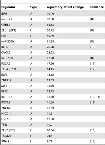
To identify possible regulators that might control the expression of E-cadherin during the EMT, we calculated the regulatory effects of the upstream regulators of E-cadherin. Out of 1732 potential regulators, NetworkProfiler inferred that 370 of them may control the expression of E-cadherin in any of the 762 cancer cell lines (Table S5). These putative regulators were ranked according to the change in their regulatory effect during the EMT. Although we did not include any information on known E-cadherin regulators, about half of the 25 highest ranked regulators were previously reported in the literature (Table 1). For example, 2 zinc finger transcription factors, ZEB1 and ZEB2, are direct repressors of E-cadherin and are involved in the EMT [9,15]. In addition, the miR-200 family indirectly suppresses the EMT by inhibiting the translation of ZEB1 and ZEB2 mRNAs [8]. Similarly, miR-192 inhibits the translation of ZEB2 [13,14]. In addition, SNAI2, a member of the Snail superfamily of zinc finger transcription factors, also is involved in the EMT [16]. Likewise, TCF4 (also known as E2-2), a class I bHLH transcription factor, is an EMT regulator; its isoforms induce the EMT in MDCK kidney epithelial cells [12]. In contrast, FOXA1 and FOXA2 are positive regulators of E-cadherin, which suppress the EMT in pancreatic ductal adenocarcinoma [11]. KLF4 also inhibits the EMT by regulating E-cadherin expression [10]. NetworkProfiler also identified several other known direct repressors of E-cadherin, such as TWIST1 [17] and TCF3 (also known as E47) [18]; however, these regulators were ranked 38th and 84th, respectively.
The other half of the 25 highest ranked regulators has not yet been reported and may be novel EMT-dependent regulators of E-cadherin. For example, although the relationship between GRHL2 and EMT is not known, GRHL2 is required for morphogenesis of epidermal and tracheal cells and plays an important role in regulating the expression levels of E-cadherin in Drosophilapost-embryonic neuroblasts [19]. ZNF217 binds the E-cadherin promoter [20], which suggests that ZNF217 might be a transcription factor for E-cadherin.
Next, we compared the performance of NetworkProfiler with that of a structural equation model (SEM) of E-cadherin that was inferred by the elastic net [22]. This model was equivalent to a regression model where the response variable is the expression of E-cadherin and the explanatory variables are the 1732 regulator expressions. The significance of each regulator was evaluated based on the number of non-zero regression coefficients in 1000 bootstrapped datasets. The SEM inferred 627 putative regulators (Table S6). Among these putative regulators, there were only 6 regulators, namely,ZEB1, miR-141, ZEB2, TCF3,miR-200b, and
miR-200c, in the 25 highest ranked regulators that were previously reported in the literature. This result suggested that NetworkPro-filer was superior to the traditional gene network estimation methods to identify regulators of E-cadherin that are involved in the EMT. Moreover, NetworkProfiler can reveal regulatory changes among genes during the EMT. Figures 5a and 5b show the regulatory profiles of putative regulators of E-cadherin when the lengths of the paths from the regulators to E-cadherin is 1 and 2, respectively.
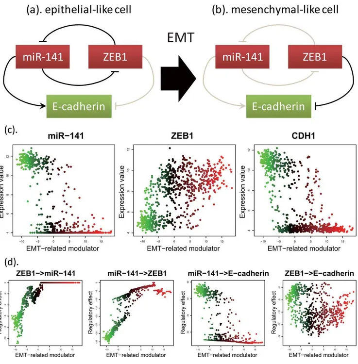
NetworkProfiler can also predict mechanistic interpretations of published experiments. For example, it is known that ZEB1 and ZEB2 induce EMT by repressing E-cadherin transcription and that ectopic expression of the miR-200 family (miR-200a, miR-200b, miR-200c, and miR-141) or miR-205 leads to downregulation of ZEB1 and ZEB2, upregulation of E-cadherin, and mesenchymal-epithelial transition (MET) in cells [8]. As the relationships between these genes, the prediction of NetworkProfiler provides the following results. As shown in Figures 6c and 6d, although the expression of miR-141 had a strong positive effect on that of E-cadherin in epithelial-like cells, this effect decreases during the EMT. In contrast, although the expression of ZEB1 had a weak negative effect on that of E-cadherin in epithelial-like cells, this effect increased during the EMT. Interestingly, miR-141 and ZEB1 had a strong, direct
Table 1.25 top-ranked regulators of E-cadherin for the change in the regulatory effect change among the EMT with published evidence.
regulator type regulatory effect change Evidence
IRF6 A 101.04
miR-141 A 87.58 [8]
GRHL2 A 64.13
ZEB1 (SIP1) I 50.72 [9]
LSR I 46.89
miR-200b A 31.55 [8]
KLF4 A 26.28 [10]
OVOL2 A 22.08
miR-200a A 17.70 [8]
FOXA2 A 17.26 [11]
TCF4 (E2.2) I 14.15 [12]
ELF3 A 13.58
ZNF217 A 13.53
MYB A 12.50
KLF5 A 12.42
miR-192 A 12.30 [13, 14]
FOXA1 A 11.69 [11]
ZNF165 A 11.39
NKX2-1 A 11.21
HNF1B A 11.08
TFE3 A 11.01
ZEB2 (dEF) I 10.66 [15]
TRIM29 I 9.87
SNAI2 I 9.74 [16]
The labels ‘‘A’’ and ‘‘I’’ indicate 2 types of the regulator: activator (A) and inhibitor (I). See Table S5 for the complete table of the 370 putative regulators for E-cadherin.
negative effect on each other only when the EMT-related modulator values were low. This implied that there is a negative feedback loop between miR-141 and ZEB1 in epithelial-like
cells, which is consistent with a previous study [23]. Further-more, during the EMT, the expression levels of miR-141 and E-cadherin decreased, while the expression level of ZEB1 Figure 5. Regulatory effect profiles of the putative regulators of E-cadherin among the EMT.(a). The regulatory effect profiles of the 13 putative regulators among the EMT when the length of the paths from the regulators to E-cadherin is 1 where rows indicate the putative regulators of E-cadherin and columns indicate samples (cancer cell lines). The positive (red) and negative (green) regulatory effect indicate that the parent regulator controls the transcript of E-cadherin positively and negatively, respectively. (b). The regulatory effect profiles of the 13 putative regulators among the EMT when the length of the paths from the regulators to E-cadherin is 2.
doi:10.1371/journal.pone.0020804.g005
Figure 6. Regulatory changes among miR-141, ZEB1, and E-cadherin among the EMT.(a). The relationship among miR-141, ZEB1, and E-cadherin in epithelial-like cells. (b). The relationship among miR-141, ZEB1, and E-cadherin in mesenchymal-like cells. (c). The expression profiles of miR-141 (left), ZEB1 (middle), and E-cadherin (right) in order of ascending the EMT-related modulator values. The green and red colors indicate epithelial- and mesenchymal-like cells, respectively. (d). The regulatory effects from ZEB1 to miR-141, from miR-141 to ZEB1, from miR-141 to E-cadherin, and from ZEB1 to E-cadherin when the length of the paths is 1.
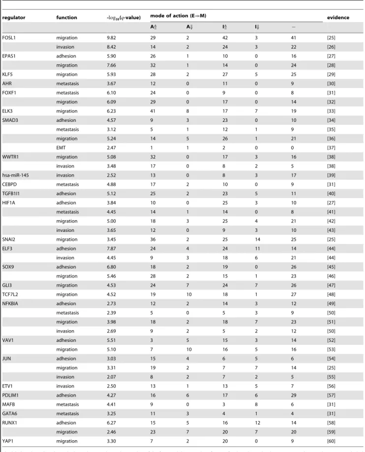
Table 2.Selected relationships between the 47 putative master regulators and the 5 functional categories with published evidence.
regulator function -log10(q-value) mode of action (E[M) evidence
AX AY IX IY {
FOSL1 migration 9.82 29 2 42 3 41 [25]
invasion 8.42 14 2 24 3 22 [26]
EPAS1 adhesion 5.90 26 1 10 0 16 [27]
migration 7.66 32 1 14 0 24 [28]
KLF5 migration 5.93 28 2 27 5 25 [29]
AHR metastasis 3.67 12 0 11 0 9 [30]
FOXF1 metastasis 6.10 24 0 9 0 8 [31]
migration 6.09 29 0 17 0 14 [32]
ELK3 migration 6.23 41 8 17 7 19 [33]
SMAD3 adhesion 4.57 9 3 23 0 10 [34]
metastasis 3.12 5 1 12 1 9 [35]
migration 5.24 14 5 26 1 21 [36]
EMT 2.47 1 1 2 0 0 [37]
WWTR1 migration 5.08 32 0 17 3 16 [38]
invasion 3.48 17 0 8 2 5 [38]
hsa-miR-145 invasion 2.52 13 0 8 3 17 [39]
CEBPD metastasis 4.88 17 2 10 0 9 [31]
TGFB1I1 adhesion 5.12 25 2 23 5 11 [40]
HIF1A adhesion 3.84 10 0 25 3 10 [27]
metastasis 4.45 14 1 14 0 8 [41]
migration 5.00 18 3 25 4 21 [42]
invasion 3.65 12 0 9 3 10 [43]
SNAI2 migration 3.45 36 2 25 14 25 [25]
ELF3 adhesion 7.87 24 4 24 11 14 [44]
invasion 4.45 9 3 18 6 21 [44]
SOX9 adhesion 6.80 18 2 19 0 26 [45]
migration 5.46 28 2 15 1 23 [46]
GLI3 migration 4.53 24 7 24 7 26 [47]
TCF7L2 migration 4.52 19 10 18 1 27 [48]
NFKBIA adhesion 2.73 12 2 14 3 12 [49]
metastasis 2.39 5 0 5 3 9 [50]
migration 3.98 18 2 18 7 23 [51]
invasion 2.69 9 2 5 2 12 [50]
VAV1 adhesion 5.51 3 5 15 3 14 [52]
migration 5.10 7 10 16 5 16 [53]
JUN adhesion 3.03 15 4 6 5 6 [54]
migration 3.31 19 2 7 7 14 [25]
invasion 2.07 8 2 7 2 5 [55]
ETV1 invasion 2.50 13 1 13 5 7 [56]
PDLIM1 adhesion 4.27 16 6 17 6 29 [57]
MAFB metastasis 4.41 9 0 3 8 6 [31]
GATA6 metastasis 3.25 11 3 4 1 4 [31]
RUNX1 adhesion 6.27 15 5 16 12 14 [58]
migration 2.46 23 7 20 7 20 [59]
YAP1 migration 3.30 7 2 20 0 9 [60]
The labels ‘‘AX’’, ‘‘AY’’, ‘‘IX’’, and ‘‘IY’’, and ‘‘{’’ indicate the number of the five modulator modes of action for the relationship between a regulator and its target included in the functional gene set: ‘‘the activation of a regulator on the expressions of its target genes with the functional category was increased by the modulator’’, ‘‘inhibition increased’’, ‘‘activation decreased’’, ‘‘inhibition decreased’’, and ‘‘the modulator mode of action is not determined’’, respectively.
increased. These results suggested that reduced expression of miR-141 disrupts the negative feedback loop between miR-141 and ZEB1 (Figures 6a and 6b), which would allow ZEB1 to decrease the expression of E-cadherin, as illustrated in Figure 6c. It should be noted that these results cannot be predicted by traditional graphical models which infer a static gene network structure.
Identification of relationships between regulators and epithelial-mesenchymal transition-related functional gene sets
The EMT-dependent relationships between downstream target genes for each regulator and previously curated functional gene sets in each sample were analyzed by applying gene set analysis (see Methods for details) to the constructed gene networks for 762
cancer cell lines. We tested five curated gene sets included in Ingenuity Knowledge Base (IKB; http://www.ingenuity.com). These gene sets were related withadhesion,migration,invasion, and metastasiswhich were hallmarks of EMT [5], and EMT itself. By using gene set analysis, the statistical significances (q-values) for the enrichments of downstream genes for the 1732 regulators on the five functional gene sets were calculated in each of the 762 cell lines. These results can be downloaded from the supporting web site (File S4; http://bonsai.hgc.jp/*shima/NetworkProfiler).
To search for regulators that strongly affected the five EMT-related functional gene sets, the change in the enrichment score during the EMT and their integralq-value were calculated. The result was summarized by a regulator function matrix (Table S7). We focused on 45 regulators with the integralq-values less than 10{10 as putative master regulators that strongly enhanced the
Figure 7. Induction of EMT by KLF5 knockdown in A549 NSCLC cell line.(a) Phase contrast images of A549 cells 72 hours after siRNA transfection, showing a fibroblast-like morphology in siKLF5 treated cells. TGF-btreatment serves as a positive control for EMT induction in A549 cells. (b) Representative immunofluorescence staining images, showing reduced E-cadherin expression in siKLF5-treated A549 cells. (c) Western blot analysis of E-cadherin and vimentin, showing EMT-related changes in their expression in A549 cells treated with two differenct siRNAs.
doi:10.1371/journal.pone.0020804.g007
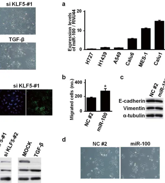
Figure 8. miR-100-induced changes in biologic characteristics in A549 NSCLC cell line.(a) Quantitative real-time RT-PCR analysis of miR-100 in six NSCLC cell lines, showing low miR-100 expression in A549, NCI-H727 and NCI-H1437. (b) Motility assay showing increased migration in miR-100-transfected A549 cells. Error bars indicate SE in three independent experiments (*,pv0:05). NC#2, negative control. (c)
Western blot analysis of E-cadherin, vimentin anda-tubulin, showing lack of noticeable changes in miR-100-transfected A549 cells (d) Representative phase contrast microscopic images showing negligible changes in miR-100-trasfected A549 cells.
functional gene sets related with the EMT. Interestingly, among the 45 regulators, 17 regulators were downstream targets of transforming growth factorb-1 (TGFB1), a master switch of EMT [24], with published evidence (Table S8). This result suggests that these regulators have crucial roles in TGFB1-induced EMT.
As a control, we tested how well the NetworkProfiler analysis identified known relationships between regulators and functional gene sets in the Ingenuity Knowledge Base. The known functional relationships of the 45 putative master regulators are shown in Table 2. For example, FOSL1 increases the migration of MDA-MB-436 cells [25] and the invasion of A549 cells [26]. SMAD3 increases the adhesion [34], the metastasis [35], and the migration [36] of cells, respectively. Similarly, HIF1A increases the adhesion of undifferentiated trophoblast stem cells [27], the metastasis of LM2 cells [41], the migration of HUVEC cells [42], and the invasion of Achn cells [43], respectively.
Although some of the 47 putative master regulators have not been reported to enhance the EMT-related functions in IKB, some predictions were supported by other resent works which were not included in IKB. For example, the prediction of NetworkProfiler suggested that PTRF regulates gene sets related with migration (q-value =2:45|10{8) and with metastasis (q -value =2:03|10{6) during the EMT. Consistent with the in
silico result, PTRF expression inhibits migration and correlates with metastasis in PC3 prostate cancer cells [61]. Similarly, NetworkProfiler predicted that miR-146 contributes to migra-tion (q-value =3:27|10{9) and invasion (q-value =1:01|10{4) during the EMT. Thisin silico result is comparable with thein vitro result that miR-146 inhibits invasion and migration, and acts as a metastasis suppressor [62]. In addition, some predictions between miRNAs and functions seem reasonable based on the known functions of the miRNA host genes. For example, the prediction of NetworkProfiler provided the hypothesis that miR-143 and miR-145 promotes metastasis (q -value =7:17|10{4 and 3:15|10{5) and migration (q -value =1:37|10{6 and 6:10|10{8), respectively. miR-143 and miR-145 cooperatively target a network of transcription factors, such as KLF4, to control smooth muscle phenotype switching [63]. Since KLF4 increases the migration of cells [29] and induces EMT [10], these miRNAs might be related with EMT-related functions or control EMT by targeting KLF4. Again, it should be noted that these relationships between regulators and functions cannot be predicted from one gene network constructed by traditional graphical models, and only the results of multiple network comparison between epithelial-like and mesenchymal-epithelial-like cells based on NetworkProfiler enables us to support the recent biological knowledge and new hypotheses about unknown relationships.
Comparison betweenin silico predictions andin vitro results
To validate the performance of NetworkProfiler, in silico predictions obtained by NetworkProfiler were evaluated experi-mentally. We first conducted in vitro experiments of a new candidate regulator of E-cadherin listed in Table 1, KLF5, to investigate whether KLF5 affects E-cadherin expression and induces morphologic changes characteristic of EMT using A549 lung adenocarcinoma cell line, which is well known to exhibit EMT in response to TGF-b [64]. KLF5 knockdown markedly altered a cobblestone epithelial morphology of A549 cells and induced a more fibroblast-like morphology with reduced cell-cell contact, which was similar to that seen in TGF-b-treated A549 cells (Figure 7a and Figure S1). Immunofluorescence analysis showed significant reduction of E-cadherin expression in A549
cells knocked down for KLF5 (Figure 7b), which was also confirmed by western blot analysis (Figure 7c). Conversely, vimentin expression was shown to be modestly increased by siKLF5 treatment (Figure 7c). Consistent with thein vitroresults, the prediction of NetworkProfiler suggested that KLF5 affects E-cadherin expression as well as Vimentin expression during the EMT, since the changes in the regulatory effects from KLF5 to E-cadherin and Vimentin were much larger compared with the other regulators (12.42 and 16.57, respectively) which was ranked 15-th and 10-th among the 1732 regulators (Table S9). The result of gene set analysis (Table S7) also suggested that KLF5 affects EMT (q-value =1:60|10{24). Thus, we consequently found that
in silico predictions obtained by NetworkProfiler was confirmed with the results ofin vitroexperiments; KLF5, a newly identified candidate regulator of EMT, was shown to affect expressions of E-cadherin and Vimentin as well as morphologic characteristics related to EMT as a repressor of EMT.
We also conducted in vitro experiments to validate functional involvement of a novel candidate EMT-related microRNA, miR-100 whose expression was increased in 762 cancer cell lines during the EMT (Figure S2). miR-100 was found to be expressed at a low level in A549, NCI-H727 and NCI-H1439 NSCLC cell lines, which had low EMT-related modulator values among the 762 cell lines panel (Figure 8a). miR-100 was transiently introduced into A549 cells, resulting in a significant increase of cell migration activity (Figure 8b). However, overexpression of miR-100 did not affect expressions of an epithelial marker, E-cadherin, and a mesenchymal marker, vimentin (Figure 8c), and also did not influence cell morphology (Figure 8d). However, overexpression of miR-100 significantly increased cell migration without noticeably affecting morphology in NCI-H727 and NCI-H1437 cells (Figure S3). Consistent with the in vitro results, the prediction of NetworkProfiler suggested that miR-100 enhances migration (q -value =1:42|104) but does not affect EMT itself (q-value = 0.24) from gene set analysis (Table S7). It also suggested that miR-100 does not affect the expressions of E-cadherin and Vimentin during the EMT, since E-cadherin and Vimentin were not target genes of miR-100 in all the 762 cell line-specific gene networks related with the EMT(Files S1, S2, and S3) and the changes in the regulatory effects from miR-100 to E-cadherin and Vimentin were much smaller compared with the other regulators (0 and 1.72, respectively), which were ranked 371-th and 151-th among the 1732 regulators (Table S9). Thus, we conclude that several hypotheses of miR-100 functions provided by NetworkProfiler are consistent with the results ofin vitroexperiments; NetworkProfiler has the potential to uncover novel biological mechanisms.
Discussion
traditional SEM approach, the performance of NetworkProfiler was superior for identifying regulators of E-cadherin during the EMT. We also showed that NetworkProfiler can reveal regulatory changes of E-cadherin during the EMT. In particular, our results suggested that decreased expression of miR-141 disrupts the negative feedback loop between miR-141 and ZEB1, which would allow ZEB1 to decrease the expression of E-cadherin.
Furthermore, we also identified putative relationships between regulators and EMT-dependent functional gene sets, some of which had published evidence. Based on the significance of the enrichment of downstream target genes for the regulator on the 5 functional gene sets, we identified 45 putative master regulators for the EMT. We found that 17 regulators were downstream targets of TGFB1 that is a master switch of the EMT. We then showed that NetworkProfiler can not only predict the relationships between these regulators and functions that were supported by many published evidence, but also produce new hypotheses that some of them might enhance EMT-related functions or induce EMT.
Finally, it is of note that we were able to validate thein silico predictions obtained by NetworkProfiler in ourin vitroexperiments. KLF5, a newly identified candidate regulator of EMT, was experimentally shown to affect E-cadherin expression as well as morphologic characteristics related to EMT, validating the NetworkProfiler-based prediction that KLF5 is a negative regulator of EMT. We also conducted in vitro experiments of another regulator, miR-100, for which NetworkProfiler predicted its association with some EMT-associated functions. As a result, we found that the predicted miR-100 functions conformed to the results ofin vitroexperiments. Thus, we conclude that the effectiveness of the proposed method was validated not only from published literature but also from newin vitrovalidation experiments.
We anticipate several possible applications and extensions of NetworkProfiler. In this study, we only focused on the system changes that are associated with the EMT. NetworkProfiler also could be used to infer system changes and reconstruct modulator-dependent gene networks for other well-defined modulators, such as drug sensitivity and prognosis risk. Currently, a significant limitation of NetworkProfiler is that the modulator must be one-dimensional. However, cancer development is a multivariate process. It may be possible to use multivariate kernel functions in NetworkProfiler to overcome this limitation.
During the past decade, cancer therapy has become increasingly personalized [2,3]. Unlike the traditional ‘‘one-size-fits-all’’ approach to cancer therapy, patient-specific cancer therapy reduces the side effects of chemotherapy and predicts the odds of cancer recurrence more accurately by tailoring cancer treatment to specific genetic defects in the tumors of individual patients. However, this goal is not an easy task since cancer is an extremely complex and heterogeneous disease. We believe that NetworkProfiler will help elucidate the systems biology of cancer and facilitate personalized chemotherapy.
Materials and Methods
Cell lines and reagents
Human non-small cell lung cancer (NSCLC) cell lines, A549, NCI-H1437 and NCI-H727, were purchased from American Tissue Culture Collection, while other NSCLC cell lines, Calu1, Calu6 and SK-MES1, were generously provided by Dr. L. J. Old (Memorial Sloan-Kettering Cancer Center). Cells were maintained in RPMI 1640 supplemented with 10% fetal bovine serum. The anti-E-cadherin antibody was purchased from BD Transduction Labora-tories, anti-vimentin from Santa Cruz Biotechnology, anti-a-tublin from Sigma Aldrich, and anti-mouse IgG from Cell Signaling
Technology. The Alexa-conjugated anti-mouse IgG was purchased from Molecular Probes. siRNAs against KLF5 (siKLF5#1 and#2) and a negative control (siNC) were purchased from Sigma Genosys. Pre-miR has-miR-100 and negative control#2 were purchased from Ambion. Human TGF-bwas purchased from R&D Systems, Inc.
Immunostaining, western blot analysis and in vitro motility assay
2|104cells in 6-well plates were transiently transfected with either 20 nM siRNA or 10 nM Pre-miR molecules using Lipofectamine RNAiMAX (Invitrogen), as previously described [65]. Immunoflu-orescence staining was carried out after fixation with 3.7% formaldehyde and postfixing with 0.1% Triton X-100 each for 10 min at RT. Photographs were taken 72 hr after transfection. Cells were harvested 48 hr after transfection for western blot analysis. In vitro motility assay based on Transwell-chamber culture systems was performed, as previously described [66].
Quantitative real-time reverse transcription (RT)-PCR analysis
Quantitative real-time RT-PCR analysis of KLF5 was per-formed using Power SYBR Green (Applied Biosystems) and the following PCR primers:
59-CCCTTGCACATACACAATGC-39 and 59 -GGATGGA-GGTGGGGTTAAAT-39. Quantitative real-time RT-PCR anal-ysis of miR-100 and RNU44 was performed using TaqMan probes and 7500 Fast Real-Time PCR system (Applied Biosys-tems), essentially as previously described [67].
NetworkProfiler
NetworkProfiler employed a varying-coefficient structural equation model (SEM) to represent the modulator-dependent conditional independence between gene transcripts. Let there be q possible regulators,R1,. . .,Rq, that may control the transcription of thek-th target geneTkwhen the modulatorM~m. Then the varying-coefficient structural equation model forTk is
Tk~
Xq
j~0
bjk(m):Rjzek,
wherebjk(m)is the coefficient function that represents the effect of RjonTk,R0~1, andekis a noise term. IfTk~Rl, then the term
blk(m):Rlcan be omitted from the model, i.e.,blk(m)~0for allm. By estimating the parametersbjk(m), we obtain the transcriptional regulatory gene network atM~m.
We used a kernel-based method to estimate these parameters. Let there bensets of gene expression profiles. Then, the SEM for thea-th sample can be rewritten as
tak~
X
q
j~0
bjka:rajzeak,a~1,. . .,n,
wheretak,raj, andmaare the values of thek-th target gene, thej -th regulator, and -the modulator for -thea-th sample, respectively; r0k~1, andbjka~bjk(ma). Fornsamples, we obtainn modulator-dependent gene regulatory networks, i.e., the regulatory effects of Rj (j~1,. . .,q) on Tk (k~1,. . .,p) are determined by
^ b
b111,. . .,^bbqpn, whereb^bjkais the estimate ofbjka.
satisfiesjmi{majvdfor some constantcand smalld. Then, we estimated the parameters bjka for fixed a by minimizing a regularized kernel-based weighted residual sum of squares
Lk(b1ka,. . .,bqkajhk)~
1 2
Xn
i~1 ftik{
X
q
j~1
bjka:rijg2K(mi{majhk)
zlka X
q
j~1
wjka:bjka
z
cka 2
X
q
j~1
b2jka, ð1Þ
whereK(mi{majhk)is a Gaussian kernel function defined by
K(mi{majhk)~exp {
1 hk
(mi{ma)2
,
and lka and cka are hyperparameters that control the L1 (lasso [68]) andL2(ridge [69]) penalties, respectively. In addition,wjkais an importance weight for bjka, andhk is the bandwidth of the Gaussian kernel. The kernel functionK(mi{majhk) defines the neighborhood around thea-th sample in terms ofM; a large value of K(mi{majhk) means that the i-th sample is in the neighborhood of the a-th sample. By fixing lka, cka, wjka, and hk, we obtain the estimates
fbb^1ka,. . .,bb^qkag~arg min
bjkaLk(b1ka,. . .,bqka):
This parameter estimation method is a weighted version of the elastic net [22]. The L1 penalty zeroes some coefficients [68], which produces a sparse network structure. In contrast, the L2 penalty stabilizes the solution by a grouping effect that promotes the collective inclusion or exclusion of highly correlated variables in the model [22]. The importance weights wjka allow tuning parameters to take on different values for different coefficientsbjka. For example, ifwjkahas a large value, then an estimatorbb^jkatends to be zero. In contrast, ifwjkahas a small value that is nearly equal to zero,^bbjkatends to be non-zero. These weights create a sparser network structure than the lasso and elastic net methods. The parameters bjka were estimated by using a recursive procedure, and the weights wjka were updated by wjka~1=(~bbjkazj) [70], wherebb~jkais the estimate from the previous step andj~10{5to avoid dividing by zero. Then, the modulator-dependent networks for n samples can be derived from the estimates of bb^jka (j~1,. . .,q,k~1,. . .,p, anda~1,. . .,n).
For convenience of subsequent explanations, we introduce the following notations:
tka(hk)~
k1a(hk):t1k
.. .
kna(hk):tnk
0 B B B @ 1 C C C A ,and
Ra(hk)~
k1a(hk):r11 k1a(hk):r1q
.. .
P ...
kna(hk):rn1 kna(hk):rnq
0 B B B @ 1 C C C A ,
wherekia(hk)~
ffiffiffiffiffiffiffiffiffiffiffiffiffiffiffiffiffiffiffiffiffiffiffiffiffiffiffiffiffi K(mi{majhk)
p
.
In these expressions,tka(hk)andRa(hk)were normalized so that the means and variances fortka(hk) and each column ofRa(hk) were0and1, respectively. As a result, the interceptb0kawas not included in the loss function (1). For fixedhk, the loss function (1) can be minimized by using a kernel-based weighted version of the recursive elastic net [70]. The tuning parameterslkaandckawere selected by minimizing a modified version of the bias-corrected weighted Akaike information criterion (AIC) [71]:
mWAICcka~(na(hk)z1):log (2p^ss2ka)z
2na(hk)(ddf^ kaz1) na(hk){^ddfka{2 ,
wherena(hk)~Pni~1kia(hk), and^ss2kais estimated by
^ s s2ka~ 1
na(hk)
Etka(hk){Ra(hk)^bb kaE22,
with ^bbka~(^bb1ka,. . .,^bbqka)’. In addition, ddf^ka is the unbiased estimate of the degrees of freedom given by
^ d
dfka~tr (RR~(hk)’RR~(hk)zckaI)
{1RR~(hk)’RR~(hk)
,
whereIis the identify matrix andRR~(hk)is the submatrix ofR(hk), which has columns that correspond to the nonzero coefficients, respectively.
The NetworkProfiler algorithm is shown below: Algorithm: NetworkProfiler.
1:ww~jka/1 (j~1,. . .,q) 2: iter/1
3:forcka~c½r(r~1,. . .,G)do 4:repeat
5: Calculate bb^ka½l,r and mWAICcka½l,r corresponding to lka~lk½l(l~1,. . .,L).
6:zr½iter/minfmWAICcka(l,r);l~1,. . .,Lg 7:l/arg minlfmWAICcka(l,r);l~1,. . .,Lg 8:ifzr½iter{zr½iter{1w0then
9: Exit loop 10:else
11:z½r/zr½iter
12:bb~ka½r/bb^ka½l,r
13:ww~jka/1=(jbb~jka(r)jzj)(j~1,. . .,q) 14: iter/iterz1
15:end if
16: untilliter reaches toM. 17: end for
18:r/arg min
rfz½r;r~1,. . .,Gg 19: Return the coefficient vectorbb^ka~bb~ka½r.
The results from NetworkProfiler, which are the estimates ofq coefficients ^bbjka(j~1,. . .,q) for thek-th target gene of thea-th patient, depend on the values ofhk. We used cross-validation to select an optimal value of hk and estimate q|n coefficients,
b1k1,. . .,bqkn by minimizing the cross-validation error:
CVk~
X
a[S (tak{
X
q
j~0
^ b
b(jk{aa):raj)2, ð2Þ
whereSis a randomly selected set of samples and^bb({a) 1ka ,. . .,^bb
L{a
k (b1ka,. . .,bqkajhk)~
1 2
X
i[S=
ftik{
X
q
j~0
bjka:rijg2K(mi{majhk)
zlka Xq
j~1
wjka:jbjkajz cka
2 Xq
j~1
b2jka: ð3Þ
The algorithm in NetworkProfiler for minimizing this loss function (3) is shown below:
Algorithm: Conditional optimization with cross-validation.
1:forhk~hl(l~1,. . .,H)do 2:for allasuch thata[Sdo 3: Calculate^bb({a)
1ka ½hl,. . .,bb^( {a)
qka ½hlwith NetworkProfiler. 4:end for
5: CalculateCVk½hl. 6:end for
7:hk/argminhlfCVk½hl;l~1,. . .,Hg 8:fora~1,. . .,ndo
9: Calculate^bb1ka½h
k,. . .,^bbqka½hkwith NetworkProfiler. 10:end for
11: Return a sequence of the coefficient vectors ^
b bk1(h
k),. . .,^bbkn(hk).
Subsequently, the modulator-dependent gene networks for n samples are determined from the coefficient vectors ^bbk1(^hhk), . . .,
^ b
bkn(^hhk) (k~1,. . .,p) by applying the above algorithm for all
k~1,. . .,p. The computational cost of this algorithm rapidly increases as the number of samples and genes increase. Thus, for computers that only have a single central processing unit (CPU), this algorithm is only practical for medium-sized networks with up to several genes. However, since this algorithm can be executed in parallel for everyk, it can be run on a stand-alone workstation with multi-core CPUs and computer clusters. Figure S4 represents the histogram of computational times based on 12 core CPUs (Intel Xeon Processor E5450 (#of cores = 4, clock speed = 3.0GHz)|3) for calculating 762 cancer cell line-specific gene networks from 13,508 | 762 gene expression data through 100,000 iterations when 100 target genes were randomly selected among 13,508 genes and the number of regulators was not restricted, i.e., 1732 regulators were used. The average computational time was about 9 days. In this situation, we can find putative master regulators of the focused target genes related with a modulator of interest. Of course, for calculating gene networks of 762 samples for a large number of target genes, a supercomputer is required. In this study, we used the Super Computer System at the Human Genome Center, Institute of Medical Science, University of Tokyo, Japan, to analyze 762 gene networks with 13,508 target genes.
Signature-based hidden modulator extraction
When the modulator was a variable that is difficult to observe, we used a signature-based hidden modulator extraction algorithm to estimate the value of the modulator for each sample. This algorithm takes seed genes that are related to the modulator and computes the underlying latent variable of the modulator by using principal components and extraction of expression modules (EEM) [7]. LetMbe a gene set that is related to the modulator and let XM be an n|jMj matrix ofn expression levels ofM. Then, a linear model, which is a special case of the single factor model [72], relatesM, a subset ofM, to an underlying latent variableU as follows:
Xj~a0jza1jUze’j,j[M(M, ð4Þ
whereXjis the expression level of thej-th gene inM,a0jis the y-intercept,a1jis a coefficient, ande’jis a noise term. We assumed that other genes that do not include M (fXj;j6[Mg) are independent ofU.
The values ofU fornsamples,ui(i~1,. . .,n), can be estimated by the following procedure:
Algorithm: signature-based hidden modulator extraction.
1: For a given setM, find a subsetMbased on the expression coherence with the EEM algorithm [7].
2: GivenM, singular value decomposition of the data matrix XM estimatesuiby the largest principal component.
3: Return the valuesui(i~1,. . .,n).
In the first step, we estimateM. In the second step, we assume that the noise terms e’j have Gaussian distributions with equal variances. As a result, the singular value decomposition generates maximum likelihood estimates of ui for the single factor model [72].
Regulatory effect
To identify upstream regulators that had strong effects on the expression of a target gene of interest in the constructed modulator-dependent gene networks, we defined a measure, called the regulatory effect, of the effect of thej-th regulator on thek-th target gene in thea-th sample as
REjka~ X
l[p jka
^ b b(j?k)
l (ma):raj, ð5Þ
where pjka is the set of all possible paths from Rj to Tk, and
^ b b(j?k)
l (ma)is the product of the estimated coefficients on thel-th path that includespjka. For example, given all the possible paths fromR1toT2in thea-th sample (Figure S5), the setp12ais
p12a~fR1?T2,R1?R3?T2,R1?R3?R4?T2g, ð6Þ
and the regulatory effectRE12ais
RE12a~(^bb12azbb^13a:bb^32azbb^13a:^bb34a:bb^42a):raj: ð7Þ
In our analysis, the length of the paths fromRjtoTkis restricted to either1or2.
To determine how the modulator affects the regulatory effect REjka, we also defined the change in the regulatory effect of thej -th regulator on -thek-th target as
RECjk~maxfREjka;a~1,. . .,ng{minfREjka;a~1,. . .,ng: ð8Þ
Gene set analysis of downstream genes for a regulator To identify regulators that enhanced the functions of their targets, we calculated the statistical significance of the enrichment of targets for a given regulator in each sample. To test the enrichment, we use the degree of independence between the two properties:
Aja :gene is in the list of targets for thej-th regulator in the a{th sample
Bu :gene is a member of theu-th priori set
Testing the association between the properties Aja and Bu corresponds to Fisher’s exact test. Thep-value calculated by this test,Pjua, indicates the probability of observing at least the same amount of enrichment when downstream genes are randomly selected out of all genes. Thus, a very small p-value gives strong evidence for an association between Aja and Bu for the j-th regulator in thea-th sample. To correct for multiple hypotheses testing, Benjamini-Hochberg (BH)-corrected p-values (q-values) [73],Qjua, were calculated.
To determine how the modulator affects the functions of downstream genes for a regulator, we defined the enrichment score, ESju, as a change in the statistical significance of the enrichment of targets for thej-th regulator on theu-th function:
ESju~log ( maxfQjua;a~1,. . .,ng=minfQjua;a~1,. . .,ng):ð9Þ
Thus, a very large ESju indicates that the modulator causes a significant change of the enrichment of the targets for the j-th regulator on theu-th function.
To identify putative master regulators that control more functional gene sets than other regulators, we also calculated the total enrichment score, TESj, by combining independent enrichment scores, ESj1,. . .,ESjU, where U is the number of functional gene sets:
TESj~2
XU
u~1
ESju: ð10Þ
The total enrichment score is equivalent to the difference of the Fisher’s statistic{2Pk
i~1log Pk[74] which was used to combine independent tests obtained fromkstudies based on thep-values, P1,. . .,Pk. The Fisher’s method is based on the fact that the statistic{2Pk
i~1log Pifollows a chi-square distribution with2k degrees of freedom under the global null hypothesis that all null hypotheses are true. A small integral p-value for the hypothesis indicates that thej-th regulator controlled at least one or more functional gene sets during the change of the modulator.
Supporting Information
Figure S1 Quantitative real-time RT-PCR analysis of KLF5 in siKLF5-treated A549 cells.
(PDF)
Figure S2 Expression profiles of miR-100 in order of ascending the EMT-related modulator values.
(PDF)
Figure S3 miR-100-induced changes in biologic charac-teristics in NCI-H1437 and NCI-H727 NSCLC cell lines. (a) Representative phase contrast microscopic images showing negligible changes in morphology by miR-100 introduction in both NSCLC cells lines. NC#2, negative control. (b) Motility
assay showing increased migration by introduction of miR-100 in both NSCLC cell lines. *,Pv0:05:
(PDF)
Figure S4 Histogram of computational times for infer-ring cancer cell line-specific gene networks running on 12 core CPUs. The 762 cancer cell line-specific gene networks related with the EMT were calculated from 13,508|762 gene expression data when 100 target genes were randomly selected among 13,508 genes and the number of regulators was not restricted, i.e., 1,732 regulators were used. The comptational times were based on 12 core CPUs (Intel Xeon Processor E5450 (#of cores = 4, clock speed = 3.0 GHz)|3). The histogram was calculated by 100,000 iterations.
(PDF)
Figure S5 Example of paths among four genes,R1,T2,
R3, andR4: (PDF)
Table S1 List of candidate regulators mapped to 1183 transcription factors and 47 nuclear receptors.
(XLS)
Table S2 List of candidate regulators mapped to 502 human microRNAs.
(XLS)
Table S3 List of coherent genes(p-valuev10{5) related to EMT calculated by extraction of expression module (EEM).
(XLS)
Table S4 EMT-related modulator values of 762 cancer cell lines calculated by signature-based hidden modula-tor extraction.
(XLS)
Table S5 List of 370 putative master regulators of E-cadherin during the EMT which were estimated by NetworkProfiler.
(XLS)
Table S6 List of 627 putative master regulators of E-cadherin which were estimated by a structual equation model (SEM) with the elastic net.
(XLS)
Table S7 Regulator function matrix between 1732 regulators and 5 functions. The row and column indicate regulator and functional gene set, respectively. The (i,j)-th element represents the change during the EMT in the statistical significance (-log10(q-value)) for the enrichment of target genes of thei-th regulator on thej-th function. The last column indicate the integral q-value of each row regulator which were used to determine which regulator strongly affected the functional gene sets.
(XLS)
Table S8 List of 17 putative master regulators (integral q-valuev10{10
) which correlated at least one or more EMT-related functions and were known to be down-stream targets of TGFB1 with published evidence from Ingenuity Knowledge Base (http://www.ingenuity.com). (XLS)
Table S9 List of the changes in the regulatory effects from 1732 regulators to E-cadherin and vimentin during the EMT.
Acknowledgments
The supercomputing resource was provided by Human Genome Center (University of Tokyo).
Author Contributions
Conceived and designed the experiments: TT. Performed the experiments: YS YH. Analyzed the data: TS AN. Wrote the paper: TS. Organized the project: SM. Provided statistical expertise: SI RY. Provided computational expertize: AN MN. Provided experimental expertise: TT. Provided manuscript review: SI AN RY TT.
References
1. Wang E (2010) Cancer systems biology CRC Press.
2. Schisky RL (2010) Personalized medicine in oncology: the future is now. Nat Rev Drug Discov 9(5): 363–6.
3. Gonzalez-Angulo AM, Hennessy BT, Mills GB (2010) Future of personalized medicine in oncology: a systems biology approach. J Clin Oncol 28(16): 2777–83.
4. Bansal M, Belcastro V, Ambesi-Impiombato A, di Bernardo D (2007) How to infer gene networks from expression profiles. Mol Syst Biol 3: 78.
5. Thiery JP, Acloque H, Huang RY, Nieto MA (2009) Epithelial-mesenchymal transitions in development and disease. Cell 139(5): 871–90.
6. Subramanian A, Tamayo P, Mootha VK, Mukherjee S, Ebert BL, et al. (2005) Gene set enrichment analysis: a knowledge-based approach for interpreting genome-wide expression profiles. Proc Natl Acad Sci U S A 102(43): 15545–50. 7. Niida A, Smith AD, Imoto S, Aburatani H, Zhang MQ, et al. (2009) Gene set-based module discovery in the breast cancer transcriptome. BMC Bioinformatics 10: 71.
8. Gregory PA, Bert AG, Paterson EL, Barry SC, Tsykin A, et al. (2008) The miR-200 family and miR-205 regulate epithelial to mesenchymal transition by targeting ZEB1 and SIP1. Nat Cell Biol 10(5): 593–601.
9. Comijn J, Berx G, Vermassen P, Verschueren K, van Grunsven L, et al. (2001) The two-handed E box binding zinc finger protein SIP1 downregulates E-cadherin and induces invasion. Mol Cell 7(6): 1267–78.
10. Yori JL, Johnson E, Zhou G, Jain MK, Keri RA (2010) Kruppel-like factor 4 inhibits epithelial-tomesenchymal transition through regulation of E-cadherin gene expression. J Biol Chem 285(22): 16854–63.
11. Song Y, Washington MK, Crawford HC (2010) Loss of FOXA1/2 is essential for the epithelialto-mesenchymal transition in pancreatic cancer. Cancer Res 70(5): 2115–25.
12. Sobrado VR, Moreno-Bueno G, Cubillo E, Holt LJ, Nieto MA, et al. (2009) The class I bHLH factors E2-2A and E2-2B regulate EMT. J Cell Sci 122(Pt 7): 1014–24.
13. Kato M, Zhang J, Wang M, Lanting L, Yuan H, et al. (2007) MicroRNA-192 in diabetic kidney glomeruli and its function in TGF-beta-induced collagen expression via inhibition of E-box repressors. Proc Natl Acad Sci U S A 104(9): 3432–7.
14. Wang B, Herman-Edelstein M, Koh P, Burns W, Jandeleit-Dahm K, et al. (2010) E-cadherin expression is regulated by miR-192/215 by a mechanism that is independent of the profibrotic effects of transforming growth factor-beta. Diabetes 59(7): 1794–802.
15. Eger A, Aigner K, Sonderegger S, Dampier B, Oehler S, et al. (2005) DeltaEF1 is a transcriptional repressor of E-cadherin and regulates epithelial plasticity in breast cancer cells. Oncogene 24(14): 2375–85.
16. Hajra KM, Chen DY, Fearon ER (2002) The SLUG zinc-finger protein represses E-cadherin in breast cancer. Cancer Res 62(6): 1613–8.
17. Yang J, Mani SA, Donaher JL, Ramaswamy S, Itzykson RA, et al. (2004) Twist, a master regulator of morphogenesis, plays an essential role in tumor metastasis. Cell 117(7): 927–39.
18. Perez-Moreno MA, Locascio A, Rodrigo I, Dhondt G, Portillo F, et al. (2001) A new role for E12/E47 in the repression of E-cadherin expression and epithelial-mesenchymal transitions. J Biol Chem 276(29): 27424–31.
19. Almeida MS, Bray SJ (2005) Regulation of post-embryonic neuroblasts by Drosophila Grainyhead. Mech Dev 122(12): 1282–93.
20. Cowger JJ, Zhao Q, Isovic M, Torchia J (2007) Biochemical characterization of the zinc-finger protein 217 transcriptional repressor complex: identification of a ZNF217 consensus recognition sequence. Oncogene 26(23): 3378–86. 21. Yang Y, Goldstein BG, Chao HH, Katz JP (2005) KLF4 and KLF5 regulate
proliferation, apoptosis and invasion in esophageal cancer cells. Cancer Biol Ther 4(11): 1216–21.
22. Zou H, Hastie T (2005) Regularization and variable selection via the elastic net. J Roy Statist Soc Ser B 67: 301–20.
23. Bracken CP, Gregory PA, Kolesnikoff N, Bert AG, Wang J, et al. (2008) A double-negative feedback loop between ZEB1-SIP1 and the microRNA-200 family regulates epithelial-mesenchymal transition. Cancer Res 68(19): 7846–54. 24. Willis BC, Borok Z (2007) TGF-beta-induced EMT: mechanisms and implications for fibrotic lung disease. Am J Physiol Lung Cell Mol Physiol 293(3): L525–34.
25. Chen H, Zhu G, Li Y, Padia RN, Dong Z, et al. (2009) Extracellular signal-regulated kinase signaling pathway regulates breast cancer cell migration by maintaining slug expression. Cancer Res 69(24): 9228–35.
26. Adiseshaiah P, Lindner DJ, Kalvakolanu DV, Reddy SP (2007) FRA-1 proto-oncogene induces lung epithelial cell invasion and anchorage-independent growth in vitro, but is insufficient to promote tumor growth in vivo. Cancer Res 67(13): 6204–11.
27. Cowden Dahl KD, Robertson SE, Weaver VM, Simon MC (2005) Hypoxia-inducible Factor Regulates alphavbeta3 Integrin Cell Surface Expression. Mol Biol Cell 16(4): 1901–12.
28. Imtiyaz HZ, Williams EP, Hickey MM, Patel SA, Durham AC, et al. (2010) Hypoxia-inducible factor 2alpha regulates macrophage function in mouse models of acute and tumor inflammation. J Clin Invest 120(8): 2699–714. 29. Yang Y, Tetreault MP, Yermolina YA, Goldstein BG, Katz JP (2008)
Kruppel-like Factor 5Controls Keratinocyte Migration via the Integrin-linked Kinase. J Biol Chem 283(27): 18812–20.
30. Marlowe JL, Puga A (2005) Aryl hydrocarbon receptor, cell cycle regulation, toxicity, and tumorigenesis. J Cell Biochem 96(6): 1174–84.
31. Nakagawa H, Liyanarachchi S, Davuluri RV, Auer H, Martin EW, et al. (2004) Role of cancerassociated stromal fibroblasts in metastatic colon cancer to the liver and their expression profiles. Oncogene 23(44): 7366–77.
32. Malin D, Kim IM, Boetticher E, Kalin TV, Ramakrishna S, et al. (2007) Forkhead box f1 is essential for migration of mesenchymal cells and directly induces integrin-beta3 expression. Mol Cell Biol 27(7): 2486–98.
33. Buchwalter G, Gross C, Wasylyk B (2005) The Ternary Complex Factor Net Regulates Cell Migration through Inhibition of PAI-1 Expression. Mol Cell Biol 25(24): 10853–62.
34. Hayes SA, Huang X, Kambhampati S, Platanias LC, Bergan RC (2003) p38 MAP kinase modulatesSmad-dependent changes in human prostate cell adhesion. Oncogene 22(31): 4841–50.
35. Matsuzaki K, Kitano C, Murata M, Sekimoto G, Yoshida K, et al. (2009) Smad2 and Smad3 phosphorylated at both linker and COOH-terminal regions transmit malignant TGF-beta signal in later stages of human colorectal cancer. Cancer Res 69(13): 5321–30.
36. Sekimoto G, Matsuzaki K, Yoshida K, Mori S, Murata M, et al. (2007) Reversible Smad-Dependent Signaling between Tumor Suppression and Oncogenesis. Cancer Res 67(11): 5090–6.
37. Sato M, Muragaki Y, Saika S, Roberts AB, Ooshima A (2003) Targeted disruption of TGFbeta1/Smad3 signaling protects against renal tubulointerstitial fibrosis induced by unilateral ureteral obstruction. J Clin Invest 112(10): 1486–94.
38. Chan SW, Lim CJ, Guo K, Ng CP, Lee I, et al. (2008) A role for TAZ in migration, invasion, and tumorigenesis of breast cancer cells. Cancer Res 68(8): 2592–8.
39. Sachdeva M, Mo YY (2010) MicroRNA-145 suppresses cell invasion and metastasis by directly targeting mucin 1. Cancer Res 70(1): 378–87. 40. Matsuya M, Sasaki H, Aoto H, Mitaka T, Nagura K, et al. (1998) Cell adhesion
kinase beta forms a complex with a new member, Hic-5, of proteins localized at focal adhesions. J Biol Chem 273(2): 1003–14.
41. Lu X, Yan CH, Yuan M, Wei Y, Hu G, et al. (2010) In vivo dynamics and distinct functions of hypoxia in primary tumor growth and organotropic metastasis of breast cancer. Cancer Res 70(10): 3905–14.
42. Okuyama H, Krishnamachary B, Zhou YF, Nagasawa H, Bosch-Marce M, et al. (2006) Expressionof vascular endothelial growth factor receptor 1 in bone marrow-derived mesenchymal cells is dependent on hypoxia-inducible factor 1. J Biol Chem 281(22): 15554–63.
43. Kim KS, Sengupta S, Berk M, Kwak YG, Escobar PF, et al. (2006) Hypoxia Enhances Lysophosphatidic Acid Responsiveness in Ovarian Cancer Cells and Lysophosphatidic Acid Induces Ovarian Tumor Metastasis In vivo. Cancer Res 66(16): 7983–90.
44. Schedin PJ, Eckel-Mahan KL, McDaniel SM, Prescott JD, Brodsky KS, et al. (2004) ESX induces transformation and functional epithelial to mesenchymal transition in MCF-12A mammary epithelial cells. Oncogene 23(9): 1766–79. 45. Panda DK, Miao D, Lefebvre V, Hendy GN, Goltzman D (2001) The
transcription factor SOX9 regulates cell cycle and differentiation genes in chondrocytic CFK2 cells. J Biol Chem 276(44): 41229–36.
46. Mori-Akiyama Y, Akiyama H, Rowitch DH, de Crombrugghe B (2003) Sox9 is required for determination of the chondrogenic cell lineage in the cranial neural crest. Proc Natl Acad Sci U S A 100(16): 9360–5.
47. Tomioka N, Osumi N, Sato Y, Inoue T, Nakamura S, et al. (2000) Neocortical origin and tangential migration of guidepost neurons in the lateral olfactory tract. J Neurosci 20(15): 5802–12.
48. Jean C, Blanc A, Prade-Houdellier N, Ysebaert L, Hernandez-Pigeon H, et al. (2009) Epidermal growth factor receptor/beta-catenin/T-cell factor 4/matrix metalloproteinase 1: a new pathway for regulating keratinocyte invasiveness after UVA irradiation. Cancer Res 69(8): 3291–9.
50. Huang S, Pettaway CA, Uehara H, Bucana CD, Fidler IJ (2001) Blockade of NF-kappaB activity in human prostate cancer cells is associated with suppression of angiogenesis, invasion, and metastasis. Oncogene 20(31): 4188–97. 51. Shair KH, Schnegg CI, Raab-Traub N (2008) EBV Latent Membrane Protein 1
Effects on Plakoglobin, Cell Growth, and Migration. Cancer Res 68(17): 6997–7005.
52. Gakidis MA, Cullere X, Olson T, Wilsbacher JL, Zhang B, et al. (2004) Vav GEFs are required for beta2 integrin-dependent functions of neutrophils. J Cell Biol 166(2): 273–82.
53. Schymeinsky J, Sindrilaru A, Frommhold D, Sperandio M, Gerstl R, et al. (2006) The Vav binding site of the non-receptor tyrosine kinase Syk at Tyr 348 is critical for beta2 integrin (CD11/CD18)- mediated neutrophil migration. Blood 108(12): 3919–27.
54. Hong IK, Jin YJ, Byun HJ, Jeoung DI, Kim YM, et al. (2006) Homophilic Interactions of Tetraspanin CD151 Up-regulate Motility and Matrix Metallo-proteinase-9 Expression of Human Melanoma Cells through Adhesion-dependent c-Jun Activation Signaling Pathways. J Biol Chem 281(34): 24279–92.
55. Janulis M, Silberman S, Ambegaokar A, Gutkind JS, Schultz RM (1999) Role of mitogen-activated protein kinases and c-Jun/AP-1 trans-activating activity in the regulation of protease mRNAs and the malignant phenotype in NIH 3T3 fibroblasts. J Biol Chem 274(2): 801–13.
56. Cai C, Hsieh CL, Omwancha J, Zheng Z, Chen SY, et al. (2007) ETV1 Is a Novel Androgen Receptor-Regulated Gene that Mediates Prostate Cancer Cell Invasion. Mol Endocrinol 21(8): 1835–46.
57. Bauer K, Kratzer M, Otte M, de Quintana KL, Hagmann J, et al. (2000) Human CLP36, a PDZdomain and LIM-domain protein, binds to alpha-actinin-1 and associates with actin filaments and stress fibers in activated platelets and endothelial cells. Blood 96(13): 4236–45.
58. Zent CS, Mathieu C, Claxton DF, Zhang DE, Tenen DG, et al. (1996) The chimeric genes AML1/MDS1 and AML1/EAP inhibit AML1B activation at the CSF1R promoter, but only AML1/MDS1 has tumor-promoter properties. Proc Natl Acad Sci U S A 93(3): 1044–8.
59. Perry C, Sklan EH, Birikh K, Shapira M, Trejo L, et al. (2002) Complex regulation of acetylcholinesterase gene expression in human brain tumors. Oncogene 21(55): 8428–41.
60. Zhang X, Milton CC, Humbert PO, Harvey KF (2009) Transcriptional output of the Salvador/ warts/hippo pathway is controlled in distinct fashions in Drosophila melanogaster and mammalian cell lines. Cancer Res 69(15): 6033–41.
61. Aung CS, Hill MM, Bastiani M, Parton RG, Parat MO (2010) PTRF-cavin-1 expression decreases the migration of PC3 prostate cancer cells: role of matrix metalloprotease 9. Eur J Cell Biol, 2010 Aug 21. [Epub ahead of print]. 62. Hurst DR, Edmonds MD, Scott GK, Benz CC, Vaidya KS, et al. (2009) Breast
cancer metastasis suppressor 1 up-regulates miR-146, which suppresses breast cancer metastasis. Cancer Res 69(4): 1279–83.
63. Cordes KR, Sheehy NT, White MP, Berry EC, Morton SU, et al. (2009) miR-145 and miR-143 regulate smooth muscle cell fate and plasticity. Nature 460(7256): 705–10.
64. Kasai H, Allen JT, Mason RM, Kamimura T, Zhang Z (2005) TGF-b1 induces human alveolar epithelial to mesenchymal cell transition (EMT). Respir Res 6: 56.
65. Taguchi A, Yanagisawa K, Tanaka M, Cao K, Matsuyama Y, et al. (2008) Identification of hypoxiainducible factor-1 alpha as a novel target for miR-17-92 microRNA cluster. Cancer Res 68(14): 5540–5.
66. Kozaki K, Miyaishi O, Tsukamoto T, Tatematsu Y, Hida T, et al. (2000) Establishment and characterization of a human lung cancer cell line NCI-H460-LNM35 with consistent lymphogenous metastasis via both subcutaneous and orthotopic propagation. Cancer Res 60(9): 2535–40.
67. Tokumaru S, Suzuki M, Yamada H, Nagino M, Takahashi T (2008) let-7 regulates Dicer expression and constitutes a negative feedback loop. Carcino-genesis 29(11): 2073–7.
68. Tibshirani R (1996) Regression shrinkage and selection via the lasso. J Royal Statist Soc B 58(1): 267–88.
69. Hoerl AE, Kennard R (1970) Ridge regression: biased estimation for nonorthogonal problems. Technometrics 12: 55–67.
70. Shimamura T, Imoto S, Yamaguchi R, Fujita A, Nagasaki M, et al. (2009) Recursive regularization for inferring gene networks from time-course gene expression profiles. BMC Syst Biol 3: 41.
71. Shimamura T, Imoto S, Yamaguchi R, Nagasaki M, Miyano S (2010) Inferring dynamic gene networks under varying conditions for transcriptomic network comparison. Bioinformatics 26(8): 1064–72.
72. Mardia K, Kent J, Bibby J (1979) Multivariate Analysis Academic Press. 73. Benjamini Y, Hochberg Y (1995) Controlling the false discovery rate: a practical
and powerful approach to multiple testing. J Roy Statist Soc Ser B 57(1): 289–300.


