0939 – 5075/2009/1100 – 0824 $ 06.00 © 2009 Verlag der Zeitschrift für Naturforschung, Tübingen · http://www.znaturforsch.com · D Introduction
Terrestrial-derived microfungi have been the sources of several novel and pharmacologically active compounds over decades (Hormazabal et al., 2005; Schmeda-Hirschmann et al., 2008; Taka-hashi and Lucas, 2008). In this context, endo-phytes belonging to the Penicillium genus have been recognized as a rich source of bioactive sec-ondary metabolites (Fill et al., 2007). Recent ex-amples include the anticancer berkelic acid from Penicillium sp., polyketides with HIV-integrase inhibitory activity from P. chrysogenum and the insecticidal paraherquamides H and I from P. clu-niae (Singh, 2003; Stierle et al., 2006).
As part of our interest for bioactive metabo-lites from endophytes associated with Brazilian plants (Cafêu et al., 2005; Inácio et al., 2006; Silva et al., 2006; Teles et al., 2006), Alibertia macrophyl-la (Rubiaceae) was selected for our study, due to
the fact that this species accumulates a series of bioactive compounds (Bolzani et al., 1991; Silva et al., 2007).
Thus, the major aim of the current study was to identify potential bioactive compounds from Penicillium sp.1 and Penicillium sp.2, two endo-phytes associated with A. macrophylla leaves, by using antifungal and acetylcholinesterase inhibi-tory assays.
Material and Methods General
1H NMR (500 MHz), 13C NMR (126 MHz),
gHMBC, gHMQC and gCOSY experiments were recorded on a VARIAN DRX-500 spectrometer, using the solvents as internal standard. Mass spectra were measured on a Q-TOF Micromass spectrometer in the ESI mode using MeOH/H2O
(1:1) as solvent (cone voltage 25 V). TLC was
Two Endophytes Associated with
Alibertia macrophylla
(Rubiaceae)
Camila M. Oliveiraa, Geraldo H. Silvab, Luis O. Regasinia, Lisinéia M. Zanardia,
Alana H. Evangelistaa, Maria C. M. Youngc, Vanderlan S. Bolzania, and
Angela R. Araujoa,*
a NuBBE – Núcleo de Bioensaios, Biossíntese e Ecofi siologia de Produtos Naturais,
Departamento de Química Orgânica, Instituto de Química, Universidade Estadual Paulista, Rua Professor Francisco Degni, SN, Bairro Quitandinha, 14800-900 Araraquara, São Paulo, Brazil. Fax +55-16-33 22 79 32. E-mail: araujoar@iq.unesp.br
b Universidade Federal de Sergipe, Av. Vereador Olimpio Grande, SN,
49500-000 Itabaiana, Sergipe, Brazil
c Seção de Fisiologia e Bioquímica de Plantas, Instituto de Botânica, Av. Miguel Stéfano
3687, 04301-902 São Paulo, São Paulo, Brazil * Author for correspondence and reprint requests
Z. Naturforsch. 64 c, 824 – 830 (2009); received June 29, 2009
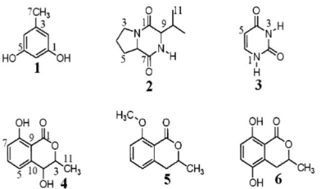
In the course of our continuous search for bioactive metabolites from endophytic fungi living in plants from the Brazilian fl ora, leaves of Alibertia macrophylla (Rubiaceae) were submitted to isolation of endophytes, and two species of Penicillium were isolated. The ac-etonitrile fraction obtained in corn from a culture of Penicillium sp.1 afforded orcinol (1). On the other hand, Penicillium sp.1 cultivated in potato-dextrose-broth furnished two differ-ent compounds, cyclo-(L-Pro–L-Val) (2) and uracil (3). The chromatographic fractionation of the acetonitrile fraction obtained from Penicillium sp.2 led to three dihydroisocoumarins, 4-hydroxymellein(4), 8-methoxymellein (5) and 5-hydroxymellein (6). Compounds 5 and 6
were obtained from the Penicillium genus for the fi rst time. Additionally, metabolites 1– 6
were evaluated for their antifungal and acetylcholinesterase (AChE) inhibitory activities. The most active compounds 1 and 4 exhibited detection limits of 5.00 and 10.0 µg against
Cladosporium cladosporioides and C. sphaerospermum, respectively. Compound 2 showed a detection limit of 10.0 µg, displaying potent AChE inhibitory activity.
performed using Merck silica gel 60 (230 mesh) and precoated silica gel 60 PF254 plates. Spots on
TLC plates were visualized under UV light and by spraying with anisaldehyde/H2SO4 reagent
fol-lowed by heating at 120 ºC. Preparative HPLC was performed on a Varian Prep-Star 400 system using a Phenomenex C-18 preparative column (250 mm × 21.2 mm). Analytical HPLC was per-formed on a Varian Pro Star 230 instrument using a Phenomenex C-18 column (250 mm × 4.6 mm). Column chromatography (CC) was performed over reversed-phase silica gel 230 – 400 mesh (Merck).
Plant material
Authenticated Alibertia macrophylla K. Schum. (Rubiaceae) was collected in Estação Ecológica Experimental de Mogi-Guaçu, Fazenda Campin-inha, Mogi-Guaçu, São Paulo, Brazil, in Novem-ber 2003. The botanical identifi cation was made by Ph.D. Maria Cláudia Marx Young (Institute of Botany, São Paulo, Brazil), and a voucher speci-men was deposited at the Herbarium of the Bo-tanic Garden of São Paulo, Brazil (voucher no. SP 370915).
Isolation and identifi cation of the endophytes For isolation of the endophytic fungi, adult and healthy leaves of A. macrophylla were selected and submitted to surface sterilization. They were fi rst washed with water and soap, and then im-mersed in an 1% aqueous sodium hypochlorite solution for 5 min and 70% aqueous ethanol for 1 min. A second washing with water and soap was performed and fi nally the leaves were immersed in sterile water for 10 min. Sterilized leaves were cut into 2 × 2 cm pieces and deposited on a Petri dish containing PDA (potato-dextrose-agar) and anthramicine sulfate (50 mg/mL) with approx. 3 to 4 pieces on each dish (Silva et al., 2006).
The endophytic fungi were identifi ed according to the characteristics of morphology of the cul-ture and spores, mechanism of spore production, and molecular taxonomy according to a modi-fi cation of the rapid preparation of DNA from fi lamentous fungi (Raeder and Broda, 1985). The fungi were identifi ed by Ph.D. Derlene Attili de Angelis (CPQBA-UNICAMP – Centro Plu-ridisciplinar de Pesquisas Químicas, Biológicas e Agrícolas, Campinas, Brazil) and deposited in our collection with culture numbers 05070032-C and
05-070032-A for Penicillium sp.1 and Penicillium sp.2, respectively.
Preparation of the crude extract in corn and potato-dextrose-broth of Penicillium sp.1
The cultivation in corn was accomplished in 9 Erlenmeyer fl asks (500 mL) each containing as medium 90 g of corn and 75 mL of water. The media were autoclaved four times (four consecu-tive days) at 121 ºC for 40 min. After cool, the media were inoculated with the endophyte and incubated at 25 ºC in static mode for 21 d. After the incubation period, the solid cultures were ground and extracted with MeOH (6 × 250 mL); after that the solvent was evaporated, providing the crude MeOH extract (10.6 g). The methanol extract was dissolved in water and then subjected to liquid-liquid partition with EtOAc. The EtOAc fraction was suspended in acetonitrile and parti-tioned with hexane. Evaporation of the organic phase resulted in the crude acetonitrile extract (804 mg).
The fungus was cultivated using potato-dex-trose-broth (PDB) as culture medium (4.0 g of potato extract, 20.0 g dextrose/L H2O). For the
PDB, 20 Erlenmeyer fl asks (500 mL) were used, each containing 100 mg of potato extract, 5.0 g dextrose and 250 mL distilled water, which were autoclaved at 125 °C for 15 min. Approx. 10 small pieces (1 cm2) of PDA medium from the Petri dish
containing biomass of Penicillium sp.1 were inoc-ulated into each fl ask, and the fl asks were sealed with cotton to permit aerobic growth. After in-cubation for 28 d at 25 °C on rotatory shakers at 150 rpm, the mycelia biomass accumulated in the fl asks was separated from the aqueous medium byfi ltration, and the fi ltrate was partitioned with EtOAc (3 × 2.5 L). Collection and evaporation of the organic phase under reduced pressure yielded a brown, solid residue (507 mg).
Preparation of the crude extract in corn of Penicillium sp.2
ground and extracted with EtOAc (6 × 250 mL); after that the solvent was evaporated, providing the crude EtOAc extract (15.0 g). The EtOAc ex-tract was dissolved in acetonitrile and then sub-jected to liquid-liquid partition with hexane. The acetonitrile fraction was evaporated resulting in the crude acetonitrile extract (6.0 g).
Isolation and identifi cation of compounds 1 – 6 The crude acetonitrile fraction obtained in corn of Penicillium sp.1 (804 mg) was fractionated by CC using silica gel C18 eluted with a H2
O/ac-etonitrile gradient (10 – 100% acetonitrile) af-fording 10 fractions (A – J). Fraction D (13.1 mg) afforded compound 1. The crude EtOAc extract obtained in PDB of Penicillium sp.1 (507 mg) was fractionated by CC using silica gel C18 eluted with a H2O/acetonitrile gradient (10 – 100%
ac-etonitrile) affording 5 fractions (K – O). Fraction C (30.5 mg), after recrystallization with MeOH, led to compound 2 (4.5 mg). Fraction L (40.2 mg) was further purifi ed using RP-HPLC [ = 254 nm, 12.0 mL/min, H2O/acetonitrile (9:1, 40 min)] and
supplied compound 3 (12.4 mg, Rt = 3 min).
The crude acetonitrile fraction obtained in corn of Penicillium sp.2 (6.0 g) was fractionated by CC using silica gel C18 eluted with a H2O/MeOH
gradient (5 – 100% MeOH) affording 16 fractions (Fr1–Fr16). Fraction Fr2 (218.0 mg) was further purifi ed using RP-HPLC [ = 254 nm, 10.0 mL/ min, H2O/acetonitrile (8:2, 80 min)] leading to
compounds 4 (3.4 mg, Rt = 24.5 min), 5 (5.3 mg,
Rt = 31.1 min) and 6 (6.3 mg, Rt = 62.3 min).
Orcinol (1): 1H NMR (500 MHz, DMSO-d 6):
= 8.97 (2H, brs, 1-OH and 5-OH), 6.01 (2H, d, J = 2.0 Hz, H-2 and H-4), 5.99 (1H, d, J = 2.0 Hz, H-6), 2.43 (3H, s, H-7). – 13C NMR (126 MHz,
DMSO-d6): = 158.1 (C-1, C-5), 138.9 (C-3), 106.9
(C-2, C-4), 99.7 (C-6), 21.1 (C-7).
Cyclo(L-Pro–L-Val) (2): 1H NMR (500 MHz,
DMSO-d6): = 7.92 (1H, brs, H-8), 4.12 (1H, t,
J = 7.5 Hz, H-6), 3.92 (1H, brs, H-9), 3.40 (2H, m, H-3), 2.35 (1H, dsept, J = 2.5, 6.5 Hz, H-10), 2.13 (2H, m, H-5), 1.78 (2H, m, H-4), 1.02 (3H, d, J = 6.5 Hz, H-12), 0.86 (3H, d, J = 6.5 Hz, H-11). –
13C NMR (126 MHz, DMSO-d
6): = 170.2 (C-7),
165.2 (C-1), 59.5 (C-9), 58.2 (C-6), 44.6 (C-3), 27.7 (C-5, C-10), 22.0 (C-4), 16.4 (C-11), 18.3 (C-12).
Uracil (3): 1H NMR (500 MHz, DMSO-d 6):
= 7.38 (1H, d, J = 8.0 Hz, H-6), 5.41 (1H, d, J =
8.0 Hz, H-5). – 13C NMR (126 MHz, DMSO-d 6):
= 164.4 4), 151.9 2), 142.9 6), 99.9 (C-5).
4-Hydroxymellein (4): 1H NMR (500 MHz,
DMSO-d6): = 11.0 (1H, brs, 8-OH), 7.45 (1H,
dd, J = 7.5, 8.5 Hz, H-6), 6.96 (1H, d, J = 7.5 Hz, H-7), 6.86 (1H, d, J = 7.5 Hz, H-5), 4.63 (1H, dq, J = 6.5, 2.0 Hz, H-3), 4.50 (1H, d, J = 2.0 Hz, H-4), 1.90 (1H, br 4-OH), 1.51 (3H, d, J = 6.5 Hz, H-9). – 13C NMR (126 MHz, DMSO-d
6): = 169.1
(C-1), 162.1 (C-8), 140.5 (C-4a), 136.7 (C-6), 118.5 (C-5), 118.2 (C-7), 106.8 (C-8a), 78.1 (C-3), 67.2 (C-4), 16.0 (C-9).
8-Methoxymellein (5): 1H NMR (500 MHz,
DM-SO-d6): = 7.35 (1H, t, J = 8.0 Hz, H-6), 6.83 (1H,
d, J = 8.0 Hz, H-7), 6.71 (1H, d, J = 8.0 Hz, H-5), 4.48 (1H, m, H-3), 3.88 (3H, s, H-10), 2.80 (2H, m, H-4), 1.40 (3H, d, J = 6.0 Hz, H-9). – 13C NMR
(126 MHz, DMSO-d6): = 162.5 1), 161.2
(C-8), 141.8 (C-4a), 134.3 (C-6), 119.1 (C-5), 113.0 (C-8a), 110.9 (C-7), 74.0 (C-3), 56.1 (C-10), 36.1 (C-4), 20.6 (C-9).
5-Hydroxymellein (6): 1H NMR (500 MHz,
DMSO-d6): = 10.5 (1H, brs, 8-OH), 6.92 (1H, d,
J = 9.0 Hz, H-6), 6.73 (1H, d, J = 9.0 Hz, H-7), 4.63 (1H, m, H-3), 3.08 (2H, dd, J = 3.5, 16.5 Hz, H-4), 2.62 (2H, dd, J = 11.5, 16.5 Hz, H-4), 1.49 (1H, d, J = 6.5 Hz, H-9). – 13C NMR (126 MHz,
DMSO-d6): = 161.5 (C-1), 156.5 (C-8), 143.0 (C-5), 125.0
(C-4a), 124.0 (C-6), 116.0 (C-7), 108.5 (C-8a), 76.0 (C-3), 28.0 (C-4), 21.0 (C-9).
Antifungal activity
Cladosporium cladosporioides (Fresen) de Vries SPC 140 and C. sphaerospermum (Perzig) SPC 491 were used in the antifungal assay. They have been maintained at the Institute of Botany, São Paulo, Brazil. Compounds 1 – 6 were applied on precoated silica gel TLC plates using a solu-tion (10 µL) containing 100, 50.0, 25.0, 10.0, 5.00 and 1.00 µg of each. After eluting with adequate solvent they were sprayed with the fungi (Rahali-son et al., 1991). Nystatin was adopted as positive control.
Acetylcholinesterase (AChE) inhibitory activity
Acetylcholinesterase (1000 U) was dissolved in 150 mL of 0.05 M Tris-hydrochloric acid buffer at
add-ed to the solution in order to stabilize the enzyme during the biochemical assay. The stock solution was kept at 4 °C. TLC plates were eluted with an appropriate solvent (acetone or isopropanol), in order to wash them, and were thoroughly dried just before use. After migration of the sample in a suitable solvent (or direct deposition of sam-ple), the TLC plate was dried for complete re-moval of the solvent. The plate was then sprayed with enzyme stock solution and thoroughly dried again. For incubation of the enzyme, the plate was laid fl at on plastic plugs in a plastic tank con-taining a little bit of water; by this means, water did not come directly into contact with the plate but the atmosphere was kept humid. The cover was placed on the tank and incubation was per-formed at 37 °C for 20 min. For detection of the AChE inhibitory activity, solutions of 1-naphthyl acetate (250 mg) in EtOH (100 mL) and of Fast Blue B salt (400 mg) in distilled water (160 mL) were prepared immediately before use. After in-cubation of the TLC plate, 10 mL of the 1-naph-thyl acetate solution and 40 mL of the Fast Blue B salt solution were mixed and sprayed onto the plate to give a purple colouration after 1 – 2 min. Galanthamine was employed as positive control (Marston et al., 2002).
Results and Discussion
The molecular structures of all isolates (Fig. 1) were confi rmed by the MS and NMR data as shown below. Assignments were based on 2D-NMR experiments including gCOSY, gHMQC, and gHMBC.
Compound 1 was isolated as a brown amor-phous powder and its ESI mass data suggested the molecular formula C7H8O2 ([M–H]– at m/z 123).
The 1H NMR spectrum of 1 showed two aromatic
signals, H 5.99 (d, J = 2.0 Hz, 1H, H-6) and H
6.01 (d, J = 2.0 Hz, 2H, H-2 and H-4) suggesting a trisubstituted aromatic ring. The 13C NMR and
DEPT 135o spectra showed fi ve signals, assigned
to one oxygenated aromatic groups at C 158.1
(C-1 and C-5), two methines attributed to signals at C 99.7 (C-6) and C 106.9 (C-2 and C-4) and
one methyl group at C 21.1 (C-7). The position
of the methyl substituent at position C-3 was at-tributed on the basis on the gHMBC experiment, which indicates a correlation of H 2.43 (H-7) with C 138.9 (C-3) and C 106.9 (C-2 and C-4). This
data suggests that 1 is orcinol (Monde, 1998).
Compound 2 was isolated as a white amor-phous solid and its ESI mass data indicated the molecular formula C10H16N2O2 ([M+H]+ at m/z
197). The 1H NMR spectrum of 2 showed signals
at H 0.86 (d, J = 6.5 Hz, 3H, H-11), H 1.02 (d,
J = 6.5 Hz, 3H, H-12), H 4.12 (t, J = 7.5 Hz, 1H,
H-6), and H 3.92 (brs, 1H, H-9). Also, three
mul-tiplets at H 3.40 (2H, H-3), 1.78 (2H, H-4), and
2.13 (2H, H-5) were observed. Analysis of the
13C NMR spectrum indicated two methyl groups,
three methylene carbon atoms and three methine carbon atoms. The signals at C 170.2 and C 165.2
were attributed to two amide carbonyl groups at C-7 and C-1, respectively, and 1H NMR signals
of an amino acid suggested that 2 belongs to the diketopiperazine class. The gHMBC cross-peaks between the signal for C-1 and the signals for H-3, H-6, H-9, and H-10, as well as the signal for C-7 and the signals for H-5, H-6, and H-9 confi rmed that 2 was biosynthesized by condensation of the amino acids proline and valine. The interpretation of these results combined with comparison with literature data (Furtado et al., 2005) suggest that diketopiperazine 2 is cyclo-(L-Pro–L-Val).
Compound 3 was isolated as yellow amorphous solid and its ESI mass data suggested the molecu-lar formula C4H4N2O2 ([M–H]– at m/z 111). The 1H NMR spectrum of 3 appeared to be relatively
simple, exhibiting two doublets at H 5.41 (d, J =
8.0 Hz, 1H, H-5) and H 7.38 (d, J = 8.0 Hz, 1H,
H-6). The 13C NMR spectrum showed only four
signals, for two carbonyl groups [ C 151.9 (C-2)
and C 164.4 (C-4)] and two methines [ C 99.9
(C-5) and C 142.9 (C-6)]. These data indicate that 3
Compound 4 was isolated as a white amorphous solid and its ESI mass data indicated the molecu-lar formula C10H10O4 ([M+Na]+ at m/z 217). The 1H NMR spectrum showed doublets for three
ar-omatic protons at H 6.86 (d, J = 7.5 Hz, 1H, H-5), H 6.96 (d, J = 7.5 Hz, 1H, H-7) and H 7.45 (dd,
J = 7.5, 8.5 Hz, 1H, H-6). Altogether these signals suggest that 4 presented a trisubstituted aromatic ring. The gCOSY cross-peaks of the doublet at H
1.51 (d, J = 6.5 Hz, 3H, H-9) coupled to a methine hydrogen atom with a signal at H 4.63 (dq, J =
6.5, 2.0 Hz, 1H, H-3) in turn coupled to a methine hydrogen atom at H 4.50 (d, J = 2.0 Hz, 1H, H-4),
suggesting an α-methylhydroxy unit in the molec-ular structure of 4. The broad singlet at H 11.0
(1H) indicated the presence of an intramolecular hydrogen bond between a hydroxy group (8-OH) and a carbonyl group (C-1). The 13C NMR and
DEPT 135o spectra showed the presence of ten
signals, assigned to one methyl [ C 16.0 (C-9)], fi ve
methine groups [ C 78.1 (C-3), C 67.2 (C-4), C
118.5 (C-5), C 136.7 (C-6), C 118.2 (C-7)], and
three aromatic quaternary carbon atoms [ C 162.1
(C-8), C 106.8 (C-8a), C 140.5 (C-4a)], and one
signal at C 169.1 (C-1), which was attributed to
a carbonyl group, suggesting that 4 is an dihydro-isocoumarin. This information coupled to data of the literature (Holler et al., 1999) assigne 4 to the structure of 4-hydroxymellein.
Compound 5 was isolated as a white amor-phous solid and its ESI mass data suggested the molecular formula C11H12O3 ([M+H]+ at m/z 193).
In particular, a high similarity between the NMR spectral data of compounds 5 and 4 was observed. The major difference was identifi ed in the
chemi-cal shifts attributed to H-4 of 5, which showed a multiplet at H 2.80 (2H, H-4). Furthermore, the
presence of a singlet at H 3.88 (3H, H-10)
sug-gested a methoxy substituent. A gHMBC correla-tion was observed between the signal at C 161.2
(C-8) and H-10, which confi rmed the position of the methoxy group at C-8. This information cou-pled to literature data (Kamisuki, 2007) assigne 5 to the structure of 8-methoxymellein.
The dihydroisocoumarin 6 was isolated as a white amorphous solid and its ESI mass data in-dicated the molecular formula C10H10O4 ([M–H]–
at m/z 193). Considering differences in the NMR spectral data between compounds 5 and 6, the 1H
NMR spectrum of 6 exhibited two ortho-coupled hydrogen atoms at H 6.73 (d, J = 9.0 Hz, 1H, H-7)
and H 6.92 (d, J = 9.0 Hz, 1H, H-6), suggesting a
tetrasubstituted aromatic ring. The 13C NMR and
DEPT 135o data indicated signals at
C 156.5 and C 143.0, which where attributed to the
hydroxy-lated aromatic carbon atoms C-8 and C-5, respec-tively. These data indicate a hydroquinone ring in the molecular structure of 6, which was identifi ed as 5-hydroxymellein (Harwood, 1984).
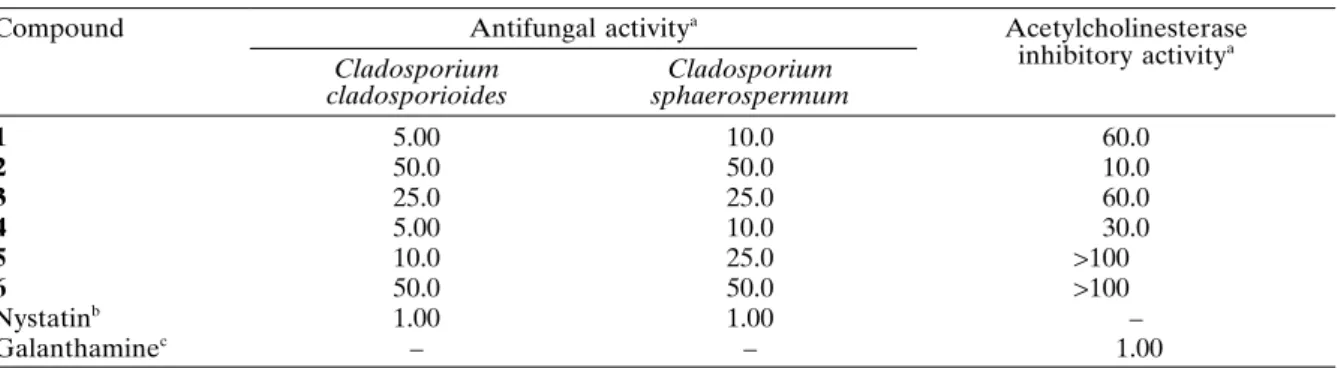
Compounds 1 – 6 were evaluated for their an-tifungal activity against Cladosporium cladospo-rioides and C. sphaerospermum using a direct bioautography assay (Table I). The most active compounds, orcinol (1) and 4-hydroxymellein (4), showed a potent effect towards the yeasts, exhib-iting a detection limit of 5.00 and 10.0 g against C. cladosporioides and C. sphaerospermum, re-spectively. 5 exhibited moderate fungitoxicity to-wards C. cladosporioides and C. sphaerospermum, showing a detection limit of 10.0 and 25.0 g,
re-Table I.Antifungal and acetylcholinesterase inhibitory activities ofcompounds 1 – 6.
Compound Antifungal activitya Acetylcholinesterase
inhibitory activitya Cladosporium
cladosporioides
Cladosporium sphaerospermum
1 5.00 10.0 60.0
2 50.0 50.0 10.0
3 25.0 25.0 60.0
4 5.00 10.0 30.0
5 10.0 25.0 >100
6 50.0 50.0 >100
Nystatinb 1.00 1.00 –
Galanthaminec – – 1.00
a Expressed as minimum amount required for inhibition of fungal growth on TLC plates (in µg). b Positive control employed for antifungal activity assay.
spectively. 5-Hydroxymellein (6) proved to be not potent, since the content at which this compound showed activity was 50 g. Altogether, these data indicate a clear positive correlation between the antifungal effect demonstrated by the dihydro-isocoumarins and free hydroxy groups at C-4 and C-8. The identifi cation of the compounds 1, 4 and 5, potentially bioactive against the phytopatho-genic fungi, indicates that they act as defence of the host species against possible attacks of patho-gens.
Although a broad spectrum of antimicrobial activities has already been demonstrated for compounds isolated from Penicillium, few stud-ies have been devoted to the acetylcholineste-rase (AChE) inhibitory activity. AChE catalyzes the hydrolysis of acetylcholine to terminate the impulse-transmitted action through cholinergic synapses (Stryer, 1995; Sigurdsson and Gudbjar-nason, 2007). Although the fundamental reason of Alzheimer’s disease (AD) is not clear so far, AD is fi rmly associated with impairment in the cholinergic transmission. A number of AChE in-hibitors have been considered as candidates for the symptomatic treatment of AD as the most useful relieving strategy (Barbosa-Filho et al., 2006; Alarcón et al., 2008). The diketopiperazine 2 exhibited the highest AChE inhibitory activity displaying a detection limit of 10.0 µg (Table I). Compounds 1 and 3 showed a detection limit of
60 µg, which was considered as weakly potent. The dihydroisocoumarin 4 displayed moderate AChE inhibitory activity, exhibiting a detection limit of 30.0 µg. On the other hand, compounds 5 and 6 were not active (detection limits over 100 µg). Similar to the observed antifungal activ-ity, free hydroxy groups at C-4 and C-8 corrobo-rate to increase the anti-AChE potency presented by dihydroisocoumarins.
In conclusion, six metabolites from Penicillium sp.1 and Penicillium sp.2, two endophytes associ-ated with A.macrophylla leaves, exhibited poten-tial bioactivity, including antifungal and AChE inhibitory activities. The results suggest that com-pounds 2 and 4 should be considered further in the development of new agrochemicals and/or hit compounds for the use in the drug design for Alzheimer’s disease treatment.
Acknowledgements
This work was funded by grants of the Fundação de Amparo à Pesquisa do Estado de São Paulo (FAPESP) as part of Biota-FAPESP, The Biodiver-sity Virtual Institute Program (www.biota.org.br), grant no. 03/02176-7, awarded to V. S. B.; C. M. O. and L. O. R. acknowledge FAPESP and L. M. Z. acknowledges Coordenação de Aperfeiçoamento de Pessoal de Nível Superior (CAPES) for MS and Ph.D. scholarships.
Alarcón J., Astudillo L., and Gutierrez M. (2008), In-hibition of acetylcholinesterase activity by dihydro--agarofuran sesquiterpenes isolated from Chilean Celastraceae. Z. Naturforsch. 63c, 853 – 856.
Barbosa-Filho J. M., Medeiros K. C. P., Diniz M. F. F. M., Batista L. M., Athayde-Filho P. F., Silva M. S., Cunha E. M. V., Almeida J. R. G. S., and Quintans-Junior L. J. (2006), Natural products inhibitors of the en-zyme acetylcholinesterase. Braz. J. Pharmacogn. 16, 258 – 285.
Bolzani V. S., Trevisan M. V. L., and Young M. C. M. (1991), Caffeic acid esters and triterpenes of Aliber-tia macrophylla. Phytochemistry 30, 2089 – 2091. Cafêu M. C., Silva G. H., Teles H. L., Bolzani V.
S., Araújo A. R., Young M. C. M., and Pfen-ning L. H. (2005), Substâncias antifúngicas de Xylaria sp., um fungo endofítico isolado de Palicourea marcgravii (Rubiaceae). Quim. Nova
28, 991 – 995.
Fill T. P., Pereira G. K., Santos R. M. G., and Rodrigues-Filho E. (2007), Four additional meroterpenes pro-duced by Penicillium sp. found in association with
Melia azedarach. Possible biosynthetic intermediates to austin. Z. Naturforsch. 62b, 1035 – 1044.
Furtado N. A. J. C., Pupo M. T., Carvalho I., Campo V. L., Duarte M. C. T., and Bastos J. K. (2005), Dike-topiperazines produced by an Aspergillus fumigatus
strain. J. Braz. Chem. Soc. 16, 1448 – 1453.
Harwood L. M. (1984), Access to phenolic fungal me-tabolites via the acid-catalysed Claisen rearrange-ment. The total synthesis of (+)-mellein, aurocitrin, and 5’,6’-dihydroaurocitrin. J. Chem. Soc. Perkin Trans. 1, 2577 – 2582.
Holler U., Konig G. M., and Wright A. D. (1999), Three new metabolites from marine-derived fungi of the genera Coniothyrium and Microsphaeropsis. J. Nat. Prod. 62, 114 – 118.
Hormazabal E., Schmeda-Hirschmann G., Astudillo L., Rodriguez J., and Theoduluz C. (2005), Metabolites from Microsphaeropsis olivacea, an endophytic fun-gus of Pilgerodendron uviferum. Z. Naturforsch. 60c, 11 – 21.
Kamisuki S. (2007), Nodulisporol and nodulisporone, novel specifi c inhibitors of human DNA polymer-ase k from a fungus, Nodulisporium sp. Bioorg. Med. Chem. 15, 3109 – 3114.
Marston A., Kissling J., and Hostettmann K. (2002), A rapid TLC bioautographic method for the detection of acetylcholinesterase and butyrylcholinesterase in-hibitors in plants. Phytochem. Anal. 13, 51 – 54. Martin F. P. J., Wang Y., Sprenger N., Holmes E., Lindon
J. C., Kochhar S., and Nicholson K. (2007), Effects of probiotic Lactobacillus paracasei treatment on the host gut tissue metabolic profi les probed via magic-angle-spining NMR spectroscopy. J. Proteome Res.
6, 1471 – 1481.
Monde K. (1998), Organochlorine compounds from a terrestrial higher plant: structures and origin of chlo-rinated orcinol derivatives from diseased bulbs of
Lilium maximowiczii. J. Nat. Prod. 61, 913 – 921. Raeder J. and Broda P. (1985), Rapid preparation of
DNA from fi lamentous fungi. Lett. Appl. Microbiol.
1, 17 – 20.
Rahalison L., Hamburger M., Hostettmann K., Mon-od M., and Frenk E. (1991), A bioautographic agar overlay method for the detection of anti-fungal compounds from higher plants. Phytochem. Anal. 2, 199 – 201.
Schindler M. (1998), Magnetic properties in terms of lo-calized quantities. The DNA bases and the protona-tion of adenine. J. Am. Chem. Soc. 110, 6623 – 6630. Schmeda-Hirschmann G., Hormazabal E., Rodriguez J.
A., and Theoduloz C. (2008), Cycloaspeptide A and pseurotin A from the endophytic fungus Penicillium janczewskii. Z. Naturforsch. 63c, 383 – 388.
Sigurdsson S. and Gudbjarnason G. (2007), Inhibition of acetylcholinesterase by extracts and constituents from Angelica archangelica and Geranium sylvati-cum. Z. Naturforsch. 62c, 689 – 693.
Silva G. H., Teles H. L., Zanardi L. M., Young M. C. M., Eberlin M. N., Haddad R., Pfenning L. H., Costa-Neto C., Castro-Gamboa I., Bolzani V. S., and Araújo A. R. (2006), Cadinane sesquiterpenoids of Pho-mopsis cassiae, an endophytic fungus associated with
Cassia spectabilis (Leguminosae). Phytochemistry 67, 1964 – 1969.
Silva V. C., Faria A. O., Bolzani V. S., and Lopes M. N. (2007), A new ent-kaurane diterpene from stems of
Alibertia macrophylla K. Schum. (Rubiaceae). Helv. Chim. Acta 90, 1781 – 1785.
Singh S. B. (2003), Isolation, structure, and HIV-1 inte-grase inhibitory activity of xanthoviridicatin E and F, two novel fungal metabolites produced by Penicil-lium chrysogenum. Helv. Chim. Acta 86, 3380 – 3385. Stierle A. A., Stierle D. B., and Kelly K. (2006), Berkelic
acid, a novel spiroketal with selective anticancer ac-tivity from an acid mine waste fungal extremophile. J. Org. Chem. 71, 5357 – 5360.
Stryer L. (1995), Biochemistry, 4th ed. Freeman, San Francisco, p. 1017.
Takahashi J. A. and Lucas E. M. A. (2008), Ocorrência e diversidade estrutural de metabólitos fúngicos com atividade antibiótica. Quim. Nova 31, 1807 – 1813. Teles H. L., Sordi R., Silva G. H., Castro-Gamboa I.,