oncogenic stress. Although p19 is not normally detected in tissues of young adult mice, a notable exception occurs in the male germ line, whereArfis expressed in spermatogonia, but not in meiotic spermatocytes arising from them. Unlike other contexts in which the induction ofArfpotently inhibits cell proliferation, expression of p19Arfin spermatogonia does not interfere with mitotic cell division. Instead, inactivation ofArftriggers germ cell–autonomous, p53-dependent apoptosis of primary spermatocytes in late meiotic prophase, resulting in reduced sperm production. Arf deficiency also causes premature, elevated, and persistent accumulation of the phosphorylated histone variant H2AX, reduces numbers of chromosome-associated complexes of Rad51 and Dmc1 recombinases during meiotic prophase, and yields incompletely synapsed autosomes during pachynema. Inactivation of Ink4a increases the fraction of spermatogonia in S-phase and restores sperm numbers inInk4a-Arfdoubly deficient mice but does not abrogatec-H2AX accumulation in spermatocytes or p53-dependent apoptosis resulting fromArfinactivation. Thus, as opposed to its canonical role as a tumor suppressor in inducing p53-dependent senescence or apoptosis,Arfexpression in spermatogonia instead initiates a salutary feed-forward program that prevents p53-dependent apoptosis, contributing to the survival of meiotic male germ cells.
Citation:Churchman ML, Roig I, Jasin M, Keeney S, Sherr CJ (2011) Expression ofArfTumor Suppressor in Spermatogonia Facilitates Meiotic Progression in Male Germ Cells. PLoS Genet 7(7): e1002157. doi:10.1371/journal.pgen.1002157
Editor:John C. Schimenti, Cornell University, United States of America
ReceivedMarch 28, 2011;AcceptedMay 11, 2011;PublishedJuly 21, 2011
Copyright:ß2011 Churchman et al. This is an open-access article distributed under the terms of the Creative Commons Attribution License, which permits unrestricted use, distribution, and reproduction in any medium, provided the original author and source are credited.
Funding:Virtually all experimental work was funded by Howard Hughes Medical Institute. Use of core facilities at St. Jude Children’s Research Hospital were supported in part by NCI Cancer Center Core Grant CA-21765 and by ALSAC, the fund-raising corporation supporting the hospital. MJ and SK are supported by NIH grant R01 HD-40916. The funders had no role in study design, data collection and analysis, decision to publish, or preparation of the manuscript.
Competing Interests:The authors have declared that no competing interests exist.
* E-mail: [email protected]
Introduction
The Cdkn2a-Cdkn2b gene cluster (also designated Ink4-Arf) encodes two polypeptide inhibitors (p16Ink4a and p15Ink4b) of cyclin D-dependent kinases (Cdk4 and Cdk6), as well as a third protein (p19Arf) that antagonizes the Mdm2 ubiquitin E3 ligase to activate p53 [1]. Although theInk4aand Ink4bgenes likely arose through gene duplication, the structure of theInk4-Arfgene cluster is highly unusual, as major portions of the p16Ink4a and p19Arf proteins are encoded by alternative reading frames of a shared exon [2]. Induction of p16Ink4a and p15Ink4b prevents the phosphorylation of the retinoblastoma protein (Rb), thereby maintaining Rb in its growth suppressive state and preventing entry into the DNA synthetic (S) phase of the cell division cycle. In contrast, p19Arf expression elicits a p53-dependent transcription program that either enforces cell cycle arrest or triggers apoptosis, depending on cell type, physiologic setting, and collateral modulating signals [1]. The Ink4-Arfgenes prevent cell prolifer-ation by implementing Rb- and p53-dependent programs that enforce cellular senescence and inhibit tissue regeneration as animals age, but their intimate genetic linkage facilitates their coordinate repression in embryonic and adult tissue stem cells, thereby allowing self-renewal [3,4]. Deleterious growth-promoting
stimuli conveyed by activated oncogenes induce Ink4-Arf gene expression and engage both p53 and Rb to counter untoward cellular proliferation. Not surprisingly, bi-allelic deletion of the
Ink4-Arfgene cluster abrogates this form of tumor suppression and is one of the more frequent events in human cancer.
Despite its canonical role as an inducer of p53 in response to oncogene signaling,Arfalso has p53-independent tumor suppres-sive activity. Deletion ofArftogether withMdm2andp53expands the spectrum and decreases the latency of cancers that spontaneously arise in mice lacking p53, p53 and Mdm2, orArf
alone [5]. Although highly basic p19Arf(,20% arginine) has been reported to physically interact with more than 25 different proteins other than Mdm2, the role of p19Arf, if any, in regulating the functions of these putative ‘‘target’’ proteins remains controversial [6]. Indeed, numerous reports that p19Arf regulates such diverse processes as ribosomal biosynthesis, transcription, DNA repair, apoptosis and autophagy in a p53-independent manner have generally relied on experiments performed with cultured cells but have not been buttressed by more extensivein vivoanalyses.
vitreous, so that Arf-null mice form a retrolenticular mass predominantly composed of pericytes; the abnormal accumulation of these cells disrupts the retina and lens and leads to blindness [8].
Arf inactivation also results in a significant reduction of sperm production through as yet poorly defined mechanisms, although young male mice remain fertile [9]. In contrast, Arf-null females have no discernable reproductive defects.
Spermatogenesis involves a stereotyped sequence of mitotic and meiotic divisions followed by sperm differentiation [10]. In mice, male germ cell progenitors (gonocytes) renew in the testis between days 1–7 postpartum (P1–P7) and generate spermatogonia that line the basement membranes of developing seminiferous tubules [11,12]. At P7–P10, spermatogonia divide to form preleptotene spermatocytes that detach from the basement membrane, are displaced toward the tubular lumen, and enter meiosis-I as primary leptotene spermatocytes. During the extended prophase of meiosis-I, homologous pairs of maternal and paternal chromosomes align to form synaptonemal complexes and exchange genetic information through homologous recombination [13]. Meiosis-I is completed by P18, and is followed rapidly by meiosis-II, and by spermiogenesis (P19–P35), after which the first mature spermatozoa enter the epididymis. As spermatogenesis continues throughout life, spermatogonia within mature seminiferous tubules remain localized on the peripheral tubular basement membrane, whereas spermatocytes, spermatids, and mature sperm are arranged in a sequential order from the periphery towards the lumen [10].
Intriguingly, p19Arf is transiently expressed in mitotically dividing spermatogonia, but not in the meiotic cells that arise from them [9]. Here, we provide genetic evidence demonstrating thatArfexpression initiates a germ cell autonomous program that protects meiotic spermatocytes from undergoing p53-dependent elimination. This physiologic function of p19Arfdirectly contrasts with its role as a tumor suppressor in inducing p53.
Results
ArfIs Expressed in Mitotically Dividing Spermatogonia
Lineage tracing experiments in the mouse previously revealed that all viable male germ cells are derived from spermatogonial
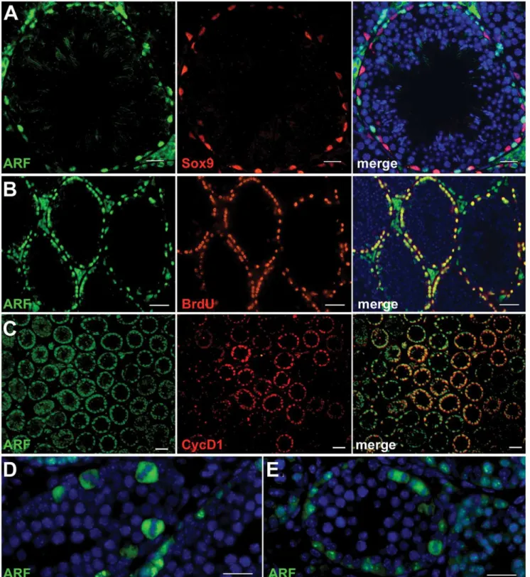
progenitors in which transient Arf expression neither inhibits proliferation nor subsequent meiotic commitment [9]. Underscoring these findings, expression of p19Arfin young adult mice is observed in all types of spermatogonia, but not in Sox9-expressing Sertoli cells on the tubular basement membrane or in DAPI-stained intratubular spermatocytes, spermatids, or sperm (Figure 1A). The fact that p19Arf is not detected in cells that have detached from the basement membrane implies thatArfexpression is extinguished at or near the primary spermatocyte stage of germ cell differentiation. Consistent with this interpretation, the Arf protein does not co-localize with Dmc1 [9], a meiotic recombinase expressed in leptotene spermato-cytes. In the mature testis, spermatogenesis occurs in waves along the length of the seminiferous tubules, so that cross sections capture tubules in which dividing spermatogonia are in synchronous phases of the cell cycle. When five month-old mice injected intraperitoneally with BrdU were sacrificed two hours later, dual immunofluorescence analysis revealed that many cells on the tubular basement membrane that had synthesized DNA also expressed p19Arf (Figure 1B). Similarly, at P12 when the number of mitotically cycling progenitors exceeds those of more differentiated germ cells, p19Arf was co-expressed with cyclin D1, a G1 phase marker of proliferating spermatogonia [14] (Figure 1C), and strikingly, was detected during all stages of mitosis (Figure 1D, 1E). Therefore, in spermatogonia, p19Arf is expressed throughout the cell division cycle without interfering with proliferation.
ArfDeficiency Compromises Sperm Production, But Is Compensated byInk4aInactivation
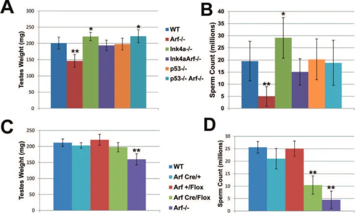
Total body weights of age-matched wild-type,Arf-null,Ink4a-null, andInk4a-Arfdouble-null mice are equivalent, but testis weights of
Arf-null animals were reduced relative to those of wild-type controls (Figure 2A), and this was associated with a significant reduction in numbers of mature sperm by the time Arf-null mice were two months old (Figure 2B). Nonetheless, youngArf-null males remain fertile, and despite the widespread use of independently derivedArf -null strains by us and others, there is no suggestion that young fertile males produce reduced litter sizes. Hence, defects in spermatogen-esis were not previously appreciated.
Knock-in of a cDNA encoding Cre recombinase in place of the firstArfexon creates a functionally nullArfallele that expresses Cre in lieu of p19Arfunder the control of theArfpromoter. Crossing
ArfCre/+ females to homozygous males containing Arf alleles
flanked by LoxP sites (‘‘floxed’’ alleles) specifically results in the inactivation ofArffunction in the testis of compound heterozygous
ArfCre/Floxmale offspring. Although penetrance of Cre expression is not complete, more than 90% of spermatogonia in the seminiferous tubules of P21 mice had no detectable anti-p19Arf fluorescence signals [9]. Overall, while p19Arfwas detected in the testes of haplo-insufficientArfCre/+
mice, any residual levels of the protein in ArfCre/Flox testes were too low to be detected by immunoblotting analysis (representative data illustrated in Figure 3), confirming significant Cre-mediatedArfdeletion in this setting. We therefore used this ‘‘targeted’’ deletion approach to compare theArfloss-of-function phenotypes ofArfCre/Flox males with those ofArf2/2males. Analysis of testis weights revealed no differences between those of ArfCre/Flox mice and wild-type controls (Figure 2C). However, the sperm counts ofArfCre/Flox
animals were reduced to levels approaching those ofArf2/2males (Figure 2D). Notably, theArf-CreorArf-Flox alleles alone had no significant effects in limiting sperm production unless coexpressed in compound heterozygotes. Therefore, tissue-restricted effects of
Arf inactivation independently recapitulated those seen in mice that completely lackArffunction.
Author Summary
Hormone signaling networks are involved in the proper control of spermatogenesis. Key regulatory gonadotrophins include luteinizing hormone (LH) and follicle-stimulating hormone (FSH) secreted by the anterior pituitary gland, and testosterone
produced by testicular interstitial Leydig cells. The considerable day-to-day and even hour-to-hour variation over a 30-fold range in plasma testosterone levels in age-matched mice of a single strain precluded accurate measurements of strain-specific differences, even in a relatively large sample size (Figure 4) [15]. Importantly, however, no discernable defects in pituitary or Leydig cell development have been observed in Arf-null, Ink4a-null, or
doubly-deficient mice, and no significant differences were observed in the ranges of serum FSH and LH among all genotypes examined (Figure 4). These findings suggest that spermatogenesis defects inArf-deficient mice are not a secondary consequence of hormonal imbalances.
Unlike Arf-null males, those lacking functional Ink4a instead exhibit increased testis weights and produce higher numbers of sperm than wild type mice (Figure 2A and 2B). Cdk4, the major target of p16Ink4aprotein inhibition in the adult testis, is expressed at maximal levels at the earlier stages of spermatogenesis, where spermatogonia undergoing mitotic cell divisions predominate [16,17], and Cdk4 inactivation compromises male fertility [18,19]. We therefore quantified thein vivoincorporation of BrdU in spermatogonia of young adult wild-type, Arf-null, Ink4a-null, andInk4a-Arf-null mice by counting stained cells that had entered S phase during a two-hour pulse. The S phase fractions of wild-type and Arf-null spermatogonia did not differ from each other (Figure 5), implying that the failure ofArf-null mice to produce normal numbers of sperm reflects a loss of meiotic cells or their Figure 2. Decreased sperm production inArf-null mice is compensated by loss ofInk4aorp53.(A, C) Testes were dissected from adult (2--6 month old) mice of the indicated genotypes and weighed as pairs. (B, D) Caudal epididymides were collected from corresponding mice, and recovered sperm were enumerated using a hemocytometer. Relative testes weights (A) and sperm counts (B) are reduced inArf-null males but increased inInk4a-null mice.Ink4a-Arfdouble-null andp532/2;Arf2/2double null mice exhibit increased testes weights (A) and sperm counts (B). While testes weights (C) are not significantly reduced inArfCre/Floxmales, reduced sperm counts (D) mimic theArfloss-of-function phenotype. [N = 20– 32 mice (A, B) and 5 mice (C, D)]. Bars represent standard deviations from the mean. P values were determined using a Student’s t-test (*p,0.001, **p,0.0001) and designate significant differences from the wild type genotype.
doi:10.1371/journal.pgen.1002157.g002
Figure 3. Loss of expression of p19Arf in ArfCre/Flox testes.
Immunoblotting analysis was performed using an antibody against p19Arf. Whole testis lysates were prepared from two month old mice of the indicated genotypes. Immunoblotting with an antibody to actin was used to control for equal protein loading per lane.
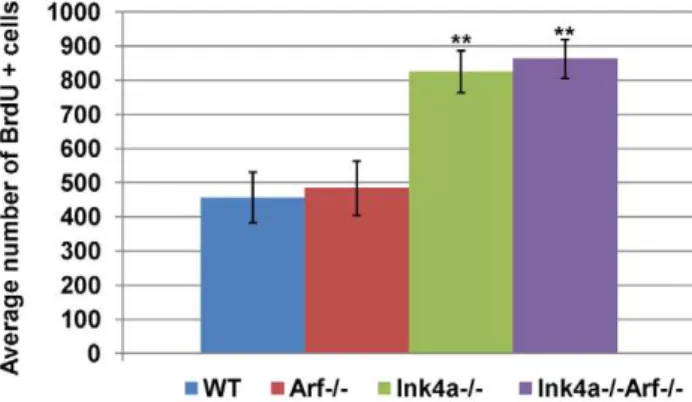
immediate progeny rather than spermatogonia. In contrast, we observed a significant two-fold increase (p,0.0001, Student’s t-test) in the relative number of S phase spermatogonia from both strains that lackInk4a(Figure 5). Looking only at testis weights and sperm counts inInk4a-Arfdouble-null animals,Ink4ainactivation
appears to compensate for loss ofArffunction (Figure 2A and 2B), presumably by fueling the production of a greater number of mitotic progenitors. Together, the consequences of these two independent loss-of-function effects rebalance testis size and sperm output in the doubly null strain. In this sense, these two ‘‘tumor suppressor’’ genes play opposing physiologic roles in male germ cell development.
ArfInactivation Leads to p53-Dependent Apoptosis in Primary Spermatocytes
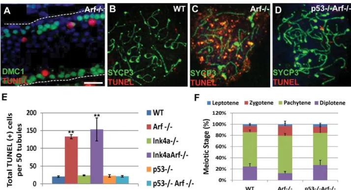
Testes from two month-oldArf-null mice exhibited a significant increase in the numbers of apoptotic (TUNEL-positive) cells when compared to age-matched wild-type controls (Figure 6A and 6E). The vast majority of apoptotic cells are spermatocytes as judged by the topological relation of TUNEL-positive cells to the expression of the meiosis-specific strand-exchange protein Dmc1, which is expressed during early prophase-I (Figure 6A). Notably, however, intratubular TUNEL-positive cells were not stained with antibod-ies to Dmc1, implying thatArf-null cells die during a later stage of germ cell development after Dmc1 expression is greatly dimin-ished. To examine this issue further, we conducted TUNEL staining of meiotic chromosome spreads. Characteristic stages of prophase during meiosis-I can be marked by staining chromo-somes with antibodies to synaptonemal complex proteins, such as the axial element component SYCP3 [20], and by several ancillary criteria (see Materials and Methods). Unlike pachytene cells from wild-type mice, those from theArf-null strain exhibited consider-able TUNEL staining (Figure 6C) with a concomitant reduction in the fraction ofArf-null diplotene spermatocytes (Figure 6F) that correlated with decreased sperm production (Figure 2B). Inacti-vation ofInk4aalone did not trigger spermatocyte apoptosis nor limit apoptosis in theArf-null background (Figure 6E) reinforcing the conclusion that the two closely linked genes play fundamen-tally different roles within the male germline.
Arf2/2; p532/2 doubly-deficient males are even more suscep-tible to spontaneous tumor development than mice lacking either
Arf or p53 alone [5]. However, the young tumor-free males produce sufficient viable sperm to remain fertile. Inactivation of
p53restored testis weights and sperm production inArf-null males (Figure 2A and 2B), prevented the apoptotic elimination ofArf-null germ cells (Figure 6D and 6E), and restored the number of Figure 4. Hormone levels in mice of different genotypes.Blood
was collected in the afternoon from 15 two to four month-old male mice of each indicated genotype, and sera were analyzed for hormone content. P values calculated using a Student’s t-test indicated no significant differences between samples taken from mice of different genotypes. Wide day-to-day and even hour-to-hour fluctuations in plasma testosterone levels yield largely unpredictable changes of as much as 30-fold between individual samples, as determined by radioimmunoassay [19]. Error bars indicate standard deviations from the mean.
doi:10.1371/journal.pgen.1002157.g004
Figure 5. Increased frequency of BrdU-incorporating sper-matogonia in Ink4a-null mice. Quantification of BrdU-positive spermatogonia in five month-old wild-type, Arf-null, Ink4a-null, and
doubly Ink4a and Arf-null mice was determined two hours after
intraperitoneal BrdU administration. BrdU-labeled cells were scored in 100 tubules in testis sections from seven different mice. Error bars indicate standard deviations from the mean. ** p,0.0001 vs wild-type by Student’s t-test.
diplotene spermatocytes (Figure 6F). Accordingly, higher levels of p53 were detected in whole testis lysates from Arf-null mice as compared to those in wild-type mice (Figure 7). Thus, in direct contrast to the role of p19Arfin triggering a p53 response following abnormal hyperproliferative stress in somatic cells, it is instead the absence of Arf expression in spermatogonial progenitors that impairs the fidelity of meiotic progression and ultimately leads to p53-dependent elimination ofArf-null primary spermatocytes.
Inappropriatec-H2AX Accumulation inArf-Null Germ Cells
Histone H2AX is phosphorylated at serine-139 in response to DNA strand breaks caused by ionizing radiation [21], UV irradiation [22], replication stress [23,24], failure of nucleotide excision repair [25], and at the leptotene stage of meiosis prior to synaptonemal complex formation [26]. H2AX phosphorylation also occurs in spermatocytes in a DNA damage-independent manner during formation of the sex body, a heterochromatic sub-nuclear domain encompassing the nonhomologous parts of the X and Y chromosomes [27]. Remarkably, staining of testis sections and immunoblotting of whole testis lysates revealed a profound increase in global c-H2AX levels when Arf was inactivated (Figure 8A and 8B). Again, inactivation of Ink4a neither recapitulated nor ameliorated thisArf-null defect (Figure 8A and 8B). Germ cells at the periphery ofArf-null seminiferous tubules exhibited the greatest increase inc-H2AX staining, suggesting that more immature cells were the ones most affected (Figure 8A). Microscopic quantification revealed that the number ofc -H2AX-positive spermatogonia, as well as the number ofc-H2AX foci per cell, were increased,2-fold whenArfwas inactivated (Figure 9), but the greatest increase in c-H2AX staining was observed in primaryArf-null spermatocytes (Figure 8A and 8B; see below). The increasedc-H2AX in meiotic cells is especially striking because these cells do not normally express p19Arf when the gene is present (Figure 1).
Figure 6. Arf deficiency leads to increased apoptosis of primary spermatocytes. (A) During spermatogenesis, germ cells move intraluminally as they differentiate, initially yielding primary meiotic spermatocytes (immunostained for Dmc1, green) that have detached from the basement membrane (represented by a dashed line). Dmc1-positive cells are visualized in a longitudinal section of a tubule from a two month-old Arf-null mouse. Spermatocytes within the tubular lumen are TUNEL-positive (red). DAPI (blue) marks the nuclei of all developing germ cells within the tubule. Neither DAPI-positive spermatogonia residing on the basement membrane nor more mature intraluminal cells express Dmc1 or are TUNEL-positive. (B–D) Surface spread spermatocytes from three month old wild-type (B),Arf2/2(C), andp532/2;Arf2/2(D) mice were immunostained for
SYCP3 (green) and assayed for TUNEL (red). Although pachytene and diplotene spermatocytes fromArf2/2mice were TUNEL-positive, few such cells
were identified in spreads from age-matched wildtype orp532/2;Arf2/2mice. (E) TUNEL assays were performed on testis sections from 2–3 month
old mice of the indicated genotypes. The total numbers of TUNEL-positive cells were enumerated in 50 tubules within three different sections taken from 3 different mice of each genotype. (F) One hundred surface spread spermatocytes (n = 3 mice) were categorized into the four phases of prophase I based on SYCP3 andc-H2AX immunostaining patterns, as described in Materials and Methods. Errors bars represent standard deviations from the mean. ** p,0.0001 and *p,0.003 vs wild-type levels by Student’s t-test.
doi:10.1371/journal.pgen.1002157.g006
Figure 7. Levels of p53 detected in wild-type andArf-null testis.
Immunoblotting analysis was performed using whole testis lysates from four representative 3–4 month-old mice of each genotype. Actin was used as a loading control.
Meiotic double-strand DNA breaks are induced by the Spo11 transesterase and its accessory factors, which are loaded onto chromatin during the final pre-meiotic S-phase [13]. Autosomalc -H2AX staining is normally observed during the leptotene and zygotene phases of meiosis-I, which are the early stages at which chromatids undergo DNA scission as a prelude to homologous recombination. In contrast, c-H2AX foci are not normally detected by early pachytene (except in the sex body) once homologous synapsis is complete (Figure 8C, top panels) [28]. Chromosome spreads from meiotic primary spermatocytes from
Arf-null males revealed that 150 of 382 individually enumerated pachytene cells (39%) exhibited persistent autosomalc-H2AX foci in addition to normal sex body staining, whereas very few such
cells (6.7%) were detected at the diplotene stage (Figure 8C, bottom panels). It could be that disappearance of c-H2AX is delayed until diplonema, or that cells with aberrantly elevatedc -H2AX are preferentially eliminated. The latter interpretation is supported by the prophase I apoptosis and depletion of diplotene cells observed inArf-null mice (Figure 6). Therefore, in the absence ofArf,c-H2AX accumulates to higher levels than normal starting in the least mature spermatogonia, continuing into meiotic prophase I, and persisting past the time when it would normally disappear from autosomes. Although inactivation ofp53 suppress-es the increased apoptosis of Arf-null spermatocytes, c-H2AX persists in cells lacking both of these genes (Figure 8A, lower right panel). In meiotic chromosome spreads fromp53;Arfdouble-null Figure 8.Arfdeficiency provokes elevated and persistentc-H2AX foci.(A) Testis sections from 3 month-old mice of the indicated genotypes were immunostained forc-H2AX, as visualized here at low magnification to demonstrate overall relative intensities of staining. The brightest c-H2AX-positive cells are primary spermatocytes. (B) Immunoblotting analysis of whole testis lysates from 3 month-old mice showing an accumulation of c-H2AX inArf-deficient testis. (C) Surface spread spermatocytes from three month old mice were immunostained for SYCP3 (red),c-H2AX (green), and with DAPI to visualize nuclei (blue). Representative spreads from each stage of prophase I are shown for wild type (top) andArf-null (bottom) strains, demonstrating persistence of autosome-associatedc-H2AX in pachytene spermatocytes fromArf-deficient mice. Staining of sex bodies persists throughout prophase in both genotypes. Images were captured with the same exposure times using a Zeiss Axioscope fluorescence microscope (A) or Intelligent Imaging Innovations Marianas spinning disc confocal microscope (C). Scale bars: (A) 100mm.
mice, 20.5% (34 of 166) of diplotene spermatocytes display persistant autosomal c-H2AX immunostaining as compared to 6.7% (5 of 74) of singlyArf-null and less than 1% (1 of 149) of wild-type diplotene spermatocytes. These data underscore the fact that the accumulation of c-H2AX in Arf-null spermatocytes is p53-independent, whereas the elimination of defective spermatocytes that retainc-H2AX inappropriately is p53-dependent.
Additional Meiotic Prophase Defects inArf-Null Primary Spermatocytes
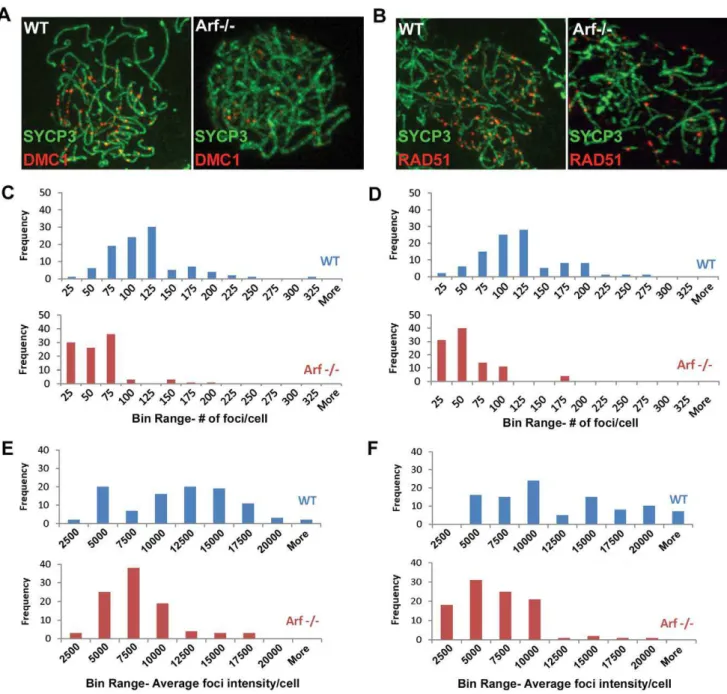
DNA double-strand breaks induced early in prophase I by Spo11 serve as substrates for the strand exchange proteins Rad51 and meiosis-specific Dmc1, which are required for double strand break repair during homologous recombination. Foci of staining using antibodies to Dmc1 (Figure 10A) and Rad51 (Figure 10B) were readily observed in zygotene spermatocytes from wild-type mice (left panels) but were fewer and less prominent in theirArf -null counterparts (right panels). The number and average fluorescence intensities of foci in 100 zygotene cells of each genotype were determined using commercial imaging software. In wild-type zygotene spermatocytes, the frequency of Dmc1 and Rad51 foci peaked at 100–125 per cell (Figure 10C and 10D, respectively, blue bars) and exhibited a broad distribution of relative intensities over a ,10-fold range (Figure 10E and 10F, blue bars). In contrast, both the number and intensities of Dmc1/ Rad51 foci were significantly reduced in Arf-null cells (average number of foci6S.D.: 103646 Dmc1 foci in wild-type vs. 44634 inArf2/2; 108645 Rad51 foci in wild-type vs. 45633 inArf2/2; average relative intensities 6 S.D.: 1023964959 Dmc1 foci in wild-type vs. 661163005 in Arf2/2; 1098065783 Rad51 foci in wild-type vs. 563763340 inArf2/2N = 100, p,0.0001, Student’s t-test; Figure 10C–10F). An accumulation of Arf-null spermato-cytes in zygonema (Figure 6F) suggests that there may be a delay at this stage before progression to pachytene.
Pachytene cells are normally characterized by well developed synaptonemal complexes that stretch the length of autosome axes and by knob-like accumulation of SYCP3 at telomeres (Figure 11A). However, 34% ofArf-null cells exhibited defects in synapsis (quantified in Figure 11D), including forked terminal
structures and interstitial bubbles on autosomes (Figure 11B) and complete asynapsis of sex chromosomes (arrow, Figure 11C). In addition, interrupted regions of SYCP3 staining (denoted by arrowheads in Figure 11C) were more frequently observed in meiotic chromosome spreads from Arf-null cells versus those in wild-type cells (191 versus 53 such segments, respectively, in 300 pachytene cells of each genotype). Because synapsis was complete in the majority ofArf-null pachytene cells, we could not distinguish whether the observed defects arose from regions in which synaptonemal complexes did not form at all, or where complexes had formed but subsequently disassembled. Taken together,Arf -deficiency results in a series of abnormalities during prophase I that include reduced loading of the Rad51 and Dmc1 recombi-nases, defects in synapsis, elevated and persistent c-H2AX expression, and p53-dependent apoptosis, ultimately associated with diminished production of mature sperm.
Discussion
With few exceptions, theArftumor suppressor is not expressed in normal tissues of healthy mice but is induced by abnormally sustained and elevated thresholds of proliferative signals, activating a p53 response that opposes the deleterious effects of oncogene activation. Notably, p53 responds to a much wider range ofArf -independent signal transduction cascades triggered by many other forms of cellular stress, including acute DNA damage, to which the
Arfpromoter does not respond [6]. By converging on p53, these different signaling pathways inhibit cell cycle progression or trigger apoptosis, acting to suppress tumor formation.
We now document a physiological role of Arf in mouse male germ cell development that is distinct from its tumor suppressive functions in key respects. First,Arfis expressed in spermatogonia, but not in the primary spermatocytes that arise from them. Expression of p19Arf neither arrests spermatogonial mitotic progression nor triggers their p53-dependent apoptosis. However, the absence of Arf expression in spermatogonia leads to p53-dependent apoptosis of spermatocytes before they exit meiosis-I. The defect in spermatogenesis is germ cell autonomous and results in a significant reduction in sperm counts by the timeArf-null mice are two months old, although residual sperm production maintains fertility in young males. Thus, expression of Arf in mitotic progenitor cells enhances the survival of their meiotic progeny in which Arf expression is normally extinguished. These features indicate that Arf expression initiates a salutary, feed-forward program that facilitates meiotic progression. Indeed, althoughArf
andInk4aare widely viewed to convey tumor suppressive functions that coordinate the activities of the p53 and Rb signaling ‘‘pathways,’’ inactivation of Arf and Ink4ain the testes leads to opposing outcomes. Disruption of Ink4a increases the mitotic activity of spermatogonial progenitors to enhance sperm output and, in this respect, compensates forArfloss of function without eliminating the cellular defects that arise in theArf-null setting. In short, loss of Ink4a increases the spermatogonial pool size, but withoutArfexpression, spermatocytes undergo increased apopto-sis, returning the number of mature sperm to normal levels.
Homologous recombination during meiosis exchanges genetic information between maternally and paternally derived chromo-somes and also guides proper segregation of chromosome pairs to maintain correct chromosome numbers in gametes [13]. During meiosis, in contrast to mitotically diving cells, homologous chromosomes are favored over sister chromatids as templates for recombinational DNA repair. Double-strand DNA breaks are formed by the topoisomerase-II-related transesterase Spo11. This process activates the Atm kinase and leads to phosphorylation of Figure 9. Arf deficiency leads to increased c-H2AX foci in
spermatogonia.The number of A and intermediate-type spermato-gonia with c-H2AX foci in 150 seminiferous tubules and the total number of foci contained within these cells were quantified in wild-type andArf-null mice at P17. Enumeration ofc-H2AX foci within 150 tubules in each of three different sections from each genotype was performed at high magnification for foci limited to spermatogonia distinguished by morphology and position along the basement membrane. P values were determined using a Student’s t- test; **p value,0.0001 vs wild-type. Error bars indicate standard deviations from the mean.
the H2AX histone variant near sites of strand breakage during early prophase I. Binding of the RecA family strand exchange proteins, Rad51 and meiosis-specific Dmc1, to Spo11-induced DNA ends generates filaments that search for and invade homologous duplex DNA molecules, leading to pairing of homologous chromosomes. Loading of Rad51 and Dmc1 is normally reversed by early pachytene when chromosomes are fully synapsed, after whichc-H2AX foci are no longer detected.
In theArf-null setting, a modest but significant increase in c -H2AX staining was first detected in the least mature spermato-gonia, and primary spermatocytes displayed accentuated signals
that persisted inappropriately into the pachytene stage. Arf-null cells also formed fewer Dmc1/Rad51 foci at zygotene and exhibited focal regions of asynapsis at pachytene. AberrantArf -null spermatocytes underwent apoptosis at pachytene, resulting in the emergence of fewer diplotene cells and a significant reduction in sperm output. Importantly, Arf2/2; p532/2 double-null pachytene cells also exhibited persistent c-H2AX staining, but these cells escaped elimination. Thus, apoptosis was p53-dependent, but aberrantc-H2AX accumulation was not.
Although the underlying mechanisms remain unknown, we consider here two plausible interpretations of this apoptotic arrest. Figure 10. Diminished Dmc1/Rad51 focus formation inArf-deficient spermatocytes.(A, B) Surface spread spermatocytes from three month old WT (left panels) andArf-null (right panels) mice were immunostained for SCP3 (A, B; green) and Dmc1 (A; red) or Rad51 (B; red). Images were captured with the same exposure time using a Marianas spinning disc confocal microscope. (C, D) Histograms showing the distribution of the number of Dmc1 (C) and Rad51 (D) foci found in wild-type (blue bars) andArf-null (red bars) spermatocytes. Foci were enumerated from one hundred zygotene spermatocytes immunostained for SYCP3 and either Dmc1 (C) or Rad51 (D) using Slidebook 5.0 SDC software. Actual values of foci per cell are plotted within bin ranges to display the distribution of frequencies. (E, F) Histograms showing the distribution of average intensities of Dmc1 (C) and Rad51 (D) foci found in wild-type (blue bars) andArf-null (red bars) spermatocytes analyzed in panels C and D.
First, it may be that reduced Rad51/Dmc1 focus formation and persistentc-H2AX staining inArf-null male germ cells connote a defect in DNA repair that then activates p53 through Arf -independent but Atm/Atr-dependent signaling pathways. In this scenario, Spo11-induced DSBs would form at normal levels but Rad51/Dmc1 loading would be impaired such that some DNA damage would persist into pachytene. This might conceivably involve the p53-independent ability of p19Arf to promote the sumoylation of numerous target proteins by inhibiting the SUMO2/3 protease Senp3 [29–31]. SUMO2/3 accumulates at sites of DNA damage in mammalian cells [32,33], and various aspects of DNA repair are regulated by the SUMO conjugation pathway [34]. There is fragmentary evidence that absence of p19Arf compromises nucleotide excision repair in cultured cells [35,36] raising the possibility thatArfmay play an as yet undefined role in promoting homologous recombination. All meiotic mutants that cannot properly synapse homologous chromosomes arrest during pachytene [37], and accompanying defects in sex body formation and failure to properly silence transcription of the sex chromosomes during prophase is itself sufficient to eliminate pachytene cells [27,38]. However, spermatocytes can also undergo
apoptosis in direct response to unrepaired Spo11-induced breaks even if sex body formation is normal [27,39]. Where tested, spermatocyte apoptosis in meiotic mutants with chromosome synapsis errors has been found to be p53-independent [40–42]. Moreover, Spo11-dependent activated phospho-p53 can be transiently detected from leptonema and zygonema in wild-type male mice, and in Drosophila, p53 activity is prolonged in cells defective for meiotic repair [43]. Thus, it remains a formal possibility that meiotic recombination defects can trigger p53-dependent apoptosis.
A second, alternative interpretation rests on the idea that the earlier and less profound accumulation of c-H2AX in Arf-null spermatogonia might be a symptom of an underlying defect affecting chromatin structure or Atm/Atr signaling. The appearance of c -H2AX reflects chromatin modifications that flank sites of DNA damage rather than strand breaks themselves, so the kinetics ofc -H2AX formation and dissolution do not necessarily coincide with the appearance and repair of DNA damage [44,45]. Moreover, aberrant Atm/Atr signaling is itself sufficient to activate p53, whether triggered by DNA breaks or not [46]. Thus, it may be thatArfdeficiency causes inappropriate Atm/Atr signaling that provokes p53-dependent apoptosis in a DNA damage-independent manner. In this view, the observed meiotic prophase defects in Arf-null spermatocytes may possibly be a separate downstream consequence of this earlier anomaly, and may not be the cause of apoptosis. Regardless of which interpretation is correct, it is important to note that our findings provide strong evidence that p53-dependent monitoring promotes proper meiotic maturation, in addition to the previously documented p53-independent pathway(s). Whatever the underlying mechanisms, the role of Arfin male germ cell development contrasts with the general paradigm of p19Arfacting as an activator of p53. Instead, it is the absence ofArfin spermatogonia that consequently leads to p53-dependent apoptosis of spermatocytes.
Materials and Methods
Ethics Statement
No human or non-human primates were studied. All animal work with mice was performed under established guidelines and supervision by the St. Jude Children’s Research Hospital’s Institutional Animal Care and Use Committee (IACUC), as required by the United States Animal Welfare Act and NIH policy to ensure proper care and use of laboratory animals for research. Experiments were undertaken in an accredited facility of the Association for Assessment of Laboratory Animal Care under the supervision of trained veterinary personnel and in strict compli-ance with Howard Hughes Medical Institute, St. Jude Children’s Research Hospital, and NIH institutional guidelines. The latter include detailed protocol submission and review of all animal care, monitoring, and experimental procedures prior to initiation of any experiments. Ongoing protocols for animal research not necessi-tating interim amendments are minimally subjected to annual review by the IACUC. All persons involved in the use of animals have read and understand all implications of pertinent protocols, have received training in the execution of relevant animal-related procedures prior to participation in the protocol, and have participated in educational or training programs deemed neces-sary by the IACUC or the Animal Resources Center personnel. Studies reported herein did not unnecessarily duplicate previous research, and were undertaken only because suitable non-animal models were unavailable. The number of animals used was consistent with good statistical design. Anesthesia, analgesia and tranquilization were used to relieve pain and distress in accordance with the IACUC recommendations.
Figure 11. Arf-null pachytene spermatocytes exhibit synaptic defects. (A–C) Staining of surface spread spermatocytes for SYCP3 marks the lateral elements of bivalents. (A) In wildtype pachytene spermatocytes, fully synapsed autosomal bivalents are observed, as judged by continuous SYCP3 staining along the axes. (B–C) Represen-tative synaptic defects inArf-null pachytene spermatocytes include (B) unsynapsed ends (arrow) and interstitial asynaptic ‘‘bubbles’’ (arrow heads), and (C) asynapsis of the X and Y chromosomes (arrow). Synapsed X and Y chromosomes are shown in (A, B). Synaptonemal complexes with segmental disruption of SYCP3 staining were more frequently observed (p = 0.051) inArf-null cells (C, arrowheads) versus their wild-type counterparts (191 versus 53 such segments in 300 pachytene chromosomal spreads from each genotype). (D) Quantifica-tion of synaptic defects observed in wild-type andArf-null pachytene spermatocytes. ‘‘Wide open’’ ends are illustrated in (B); less extensive asynapsis at telomeres was categorized as ‘‘slight’’ open ends. 100 pachytene spermatocytes from each of three mice of different genotypes yielded highly significant differences in overall defects (p = 0.0017 by Student’s t-test). Error bars indicate standard deviation from the mean.
Phenotypic Characterization of Mouse Testes and Sperm Count Analysis
Caudal epididymides were harvested before dissection of the testes. For each male mouse, two cauda were minced into 1 ml of Dulbecco’s modified Eagle’s medium (DMEM) containing 25 mM HEPES buffer (pH 7.5) and 4 mg/ml bovine serum albumin and incubated at 37uC for 20 minutes. Suspensions of sperm were fixed at a 1:25 dilution in 10% formalin and counted on a hemocytometer. All sperm counts were performed between 1:00– 3:00 PM. Dissected testes were weighed in pairs.
Immunofluorescence of Testes Sections
Mice were euthanized by CO2 asphyxiation, and testes were removed and fixed overnight at 4uC in 4% paraformaldehyde followed by saturation in 30% sucrose at 4uC overnight. Tissues were embedded in TBS Tissue Freezing Medium (Fisher Scientific, Pittsburg PA), and sliced with a HM500M Cryostat (Microm International, Walldorf, Germany) into 10mm sections. Fixed and frozen samples were sectioned and subjected to antigen retrieval in 0.1 M Na citrate buffer, pH 6.0, followed by one hour incubation at room temperature in a blocking solution of 10% normal goat serum (NGS), 0.1% Triton-X 100 in phosphate-buffered saline (PBS), and then by overnight incubation at 4uC in primary antibodies diluted in 3% NGS, 0.1% Triton-X 100 in PBS. Antibodies were directed to p19Arf [rat monoclonal immunoglobulin 5C3-1 [50], Sox9 (Millipore AB5535, 1:1000), BrdU (Santa Cruz sc32323, 1:100), cyclin D1 (Santa Cruz 72-13G, 1:750), Dmc1 (Santa Cruz H-100, 1:750), c-H2AX (Cell Signaling 2577, 1:200), and SUMO2/3 (Cell Signaling 18H8, 1:300). Slides were washed three times in PBS, and then incubated for 1 hour at room temperature in 3% NGS, 0.1% Triton-X 100 in PBS containing the relevant secondary antibodies conjugated to Ig-Alexa Fluor 555 or Ig-Alexa Fluor 488 (1:500 dilutions; Invitrogen). Slides were washed three times in PBS and mounted with Vectashield (Vector Labs) containing 49 -6-diamidino-2-phenylindol (DAPI). TUNEL assays were performed using an in situ cell death detection kit (TMR red, Roche) following the manufacturer’s protocol. Images of tissue sections were photo-graphed using a Zeiss Axioscope fluorescence microscope and assembled using Zeiss Axiovision software.
Analysis and Staging of Meiotic Spreads
Testes were decapsulated and minced in 5 ml of DMEM per testis and transferred to a 15 ml Falcon tube. After further dissociation of the tubules by pipeting up and down, large pieces were allowed to settle to the bottom of the tube by gravity for 10 minutes on ice. One ml of the supernatant, containing a suspension of spermatocytes, was transferred to a 1.5 ml Eppendorf tube and centrifuged for five minutes at 58006g. The pellet was
(Cell Signaling 2577, 1:500) to identify sex body formation and sites of DNA damage, and to Rad51 (Calbiochem Ab-1, 1:500) and Dmc1 (Santa Cruz H-100, 1:750) to demonstrate formation of complexes required for DNA strand exchange during homologous recombination. Slides were washed three times in PTBG at room temperature for 3 minutes with shaking. Secondary antibodies, also diluted in PTBG, were applied to slides which were covered with parafilm and incubated at 37uC for one hour in a humidity chamber. Slides were washed three times in PTBG for 3 minute intervals in the dark with shaking and mounted with Vectashield (Vector Labs) containing DAPI. Surface spread spermatocytes were visualized by a Marianas spinning-disc confocal microscope, and images were assembled and analyzed using Slidebook 5.0 SDC software (Intelligent Imaging Innovations, Denver CO). Meiotic spreads from three adult mice (age three months) were analyzed. One hundred spermatocytes were scored each from mouse.
Distinct staining patterns allow for classification of each stage of meiotic prophase [51,52]. Leptotene cells were categorized by short stretches of axial elements accompanied by intensec-H2AX staining throughout the nucleus and the absence of a distinct sex body. Zygotene cells also display intense c-H2AX staining throughout the nucleus and lack a sex body, but can be distinguished by longer stretches of SYCP3 staining, some of which are synapsed. Pachytene cells have fully formed and synapsed axes that appear as thick, continuous SYCP3-stained threads, while displaying intensec-H2AX staining only in the sex body. Dmc1 and Rad51 foci are normally present at leptotene and zygotene, and largely disappear by pachytene. Diplotene cells have c-H2AX localized only to the sex body, but fully formed axes are desynapsing and chiasmata are visible.
Immunoblotting
As previously described [53], detergent lysates were prepared, and protein concentration was quantified by bicinchoninic acid assay (Pierce). Samples (25–75mg protein per lane) were electrophoretically separated on 4% to 12% Bis-Tris NuPAGE gels (Invitrogen), transferred to polyvinylidene fluoride membranes (Millipore), and detected using antibodies to c-H2AX (Cell Signaling S139, 1:500), p19Arf (5C3-1; Bertwistle et al. 2004b), p53 (Cell Signaling 1C12, 1:500), and actin (Santa Cruz C-11, 1:500) to control for protein loading.
Acknowledgments
Author Contributions
Conceived and designed the experiments: MLC IR MJ SK CJS. Performed the experiments: MLC. Analyzed the data: MLC CJS.
Contributed reagents/materials/analysis tools: CJS SK MJ. Wrote the paper: MLC CJS SK MJ.
References
1. Lowe SW, Sherr CJ (2003) Tumor suppression by Ink4a-Arf: progress and puzzles. Curr Opin Genet Dev 13: 77–83.
2. Quelle DE, Zindy F, Ashmun RA, Sherr CJ (1995) Alternative reading frames of the INK4a tumor suppressor gene encode two unrelated proteins capable of inducing cell cycle arrest. Cell 83: 993–1000.
3. Pardal R, Molofsky AV, He S, Morrison SJ (2005) Stem cell self-renewal and cancer cell proliferation are regulated by common networks that balance the activation of proto-oncogenes and tumor suppressors. Cold Spr Harb Symp Quant Biol 70: 177–185.
4. Gil J, Peters G (2006) Regulation of the INK4b-ARF-INK4a tumour suppressor locus: all for one or one for all. Nat Rev Mol Cell Biol 7: 667–677. 5. Weber JD, Jeffers JR, Rehg JE, Randle DH, Lozano G, et al. (2000)
p53-independent functions of the p19ARF tumor suppressor. Genes Dev 14: 2358–2365.
6. Sherr CJ (2006) Divorcing ARF and p53: an unsettled case. Nat Rev Cancer 6: 663–673.
7. Zindy F, Williams RT, Baudino TA, Rehg JE, Skapek SX, et al. (2003)Arf
tumor suppressor promoter monitors latent oncogenic signalsin vivo. Proc Natl Acad Sci USA 100: 15930–15935.
8. McKeller RN, Fowler JL, Cunningham JJ, Warner N, Smeyne RJ, et al. (2002) The Arf tumor suppressor gene promotes hyaloid vascular regression during mouse eye development. Proc Natl Acad Sci USA 99: 3848–3853.
9. Gromley A, Churchman ML, Zindy F, Sherr CJ (2009) Transient expression of the Arf tumor suppressor during male germ cell and eye development in Arf-Cre reporter mice. Proc Natl Acad Sci USA 106: 6285–6290.
10. Russell LD, Ettlin RA, Sinha Hikim AP, Clegg ED (1990) Histological and histopathological evaluation of the testis Cache River Press. Clearwater, FL. 11. de Rooij DG (2001) Proliferation and differentiation of spermatogonial stem
cells. Reproduction 121: 347–354.
12. Culty M (2009) Gonocytes, the forgotten cells of the germ cell lineage. Birth Defects Res (Part C) 87: 1–26.
13. Cole F, Keeney S, Jasin M (2010) Evolutionary conservation of meiotic DSB proteins: more than just Spo11. Genes Dev 24: 1201–1207.
14. Beumer TL, Roepers-Gajadien HL, Gademan IS, Kal HB, de Rooij DG (2000) Involvement of the D-type cyclins in germ cell proliferation and differentiation in the mouse. Biol Reprod 63: 1893–1898.
15. Bartke A, Steele RE, Musto N, Caldwell BV (1973) Fluctuations in plasma testosterone levels in adult male rats and mice. Endocrinology 92: 1223–1228. 16. Zindy F, den Besten W, Chen B, Rehg JE, Latres E, et al. (2001) Control of spermatogenesis in mice by the cyclin D-dependent kinase inhibitors p18(Ink4c) and p19(Ink4d). Mol Cell Biol 21: 3244–3255.
17. Bartkova J, Lukas C, Sorenson CS, Meyts ER, Skakkebaek NE, et al. (2003) Deregulation of the RB pathway in human testicular germ cell tumors. J Pathol 200: 149–156.
18. Rane SG, Dubus P, Mettus RV, Galbreath EJ, Boden G, et al. (1999) Loss of Cdk4 expression causes insulin-deficient diabetes and Cdk4 activation results in b-islet cell hyperplasia. Nature Genet 22: 44–52.
19. Tsutsui T, Hesabi B, Moons DS, Pandolfi PP, Hansel KS, et al. (1999) Targeted disruption of CDK4 delays cell cycle entry with enhanced p27Kip1
activity. Mol Cell Biol 19: 7011–7019.
20. Dobson MJ, Pearlman RE, Karaiskakis A, Spyropoulos B, Moens PB (1994) Synaptonemal complex proteins: occurrence, epitope mapping, and chromo-some disjunction. J Cell Sci 107: 2749–2760.
21. Rogakou EP, Pilch DR, Orr AH, Ivanova VS, Bonner WM (1998) DNA double-stranded breaks induce histone H2AX phosphorylation on serine 139. J Biol Chem 273: 5858–5868.
22. Limoli CL, Giedzinski E, Bonner WM, Cleaver JE (2002) UV-induced replication arrest in the xeroderma pigmentosum variant leads to DNA double-strand breaks,c-H2AX formation, and Mre11 relocalization. Proc Natl Acad Sci USA 99: 233–238.
23. Ward IM, Chen J (2001) Histone H2AX is phosphorylated in an ATR-dependent manner in response to replicational stress. J Biol Chem 276: 47759–47762.
24. Furuta T, Takemura H, Liao ZY, Aune GJ, Redon C, et al. (2003) Phosphorylation of histone H2AX and activation of Mre11, Rad50, and Nbs1 in response to replication-dependent double-strand breaks induced by mammalian DNA topoisomerase I. J Biol Chem 278: 20303–20312. 25. Marti TM, Hefner E, Feeney L, Natale V, Cleaver JE (2006) H2AX
phosphorylation within the G1phase after UV irradiation depends on nucleotide
excision repair and not DNA double-strand breaks. Proc Natl Acad Sci USA 103: 9891–9896.
26. Mahadevaiah SK, Turner JM, Baudet FREP, de Boer P, Blanco-Rodriguez J, et al. (2001) Recombinational DNA double-strand breaks in mice precedes synapsis. Nat Genet 27: 271–2716.
27. Burgoyne PS, Mahadevaiah SK, Turner JM (2009) The consequences of asynapsis for mammalian meiosis. Nat Rev Genet 10: 207–216.
28. Inagaki A, Schoenmakers S, Baarends WM (2010) DNA double strand break repair, chromosome synapsis and transcriptional silencing in meiosis. Epigenet-ics 5: 255–266.
29. Haindl M, Harasim T, Eick D, Muller S (2008) The nucleolar SUMO-specific protease SENP3 reverses SUMO modification of nucleophosmin and is required for rRNA processing. EMBO Rep 9: 273–279.
30. Kuo M-L, den Besten W, Thomas MC, Sherr CJ (2008) Arf-induced turnover of the nucleolar nucleophosmin-associated SUMO-2/3 protease Senp3. Cell Cycle 7: 3378–3387.
31. Nishida T, Yamada Y (2008) SMT3IP1, a nucleolar SUMO-specific protease, deconjugates SUMO-2 from nucleolar and cytoplasmic nucleophosmin. Biochem Biophys Res Commun 374: 382–387.
32. Morris JR, Boutell C, Keppler M, Densham R, Weekes D, et al. (2009) The SUMO modification pathway is involved in the BRCA1 response to genotoxic stress. Nature 462: 886–890.
33. Galanty Y, Belotserkovskaya R, Coates J, Polo S, Miller KM, et al. (2009) Mammalian SUMO E3-ligases PIAS1 and PIAS4 promote responses to DNA double-strand breaks. Nature 462: 935–939.
34. Bergink S, Jentsch S (2009) Principles of ubiquitin and SUMO modifications in DNA repair. Nature 458: 461–467.
35. Sarkar-Agarwal P, Vergilis I, Sharpless NE, DePinho RA, Runger TM (2004) Impaired processing of DNA photoproducts and untraviolet hypermutability with loss of p16INK4a
or p19ARF
. J Natl Cancer Inst 96: 1790–1793. 36. Dominguez-Brauer C, Chen Y-J, Brauer PM, Pimkina J, Raychaudhuri P (2009)
ARF stimulates XPC to trigger nucleotide excision repair by regulating the repressor complex of E2F4. EMBO Rep 10: 1036–1042.
37. de Rooij DG, de Boer P (2003) Specific arrest in spermatogenesis in genetically modified and mutant mice. Cytogenet Genome Res 103: 267–276.
38. Royo H, Polikiewicz G, Mahadevaiah SK, Prosser H, Mitchell M, et al. (2010) Evidence that meiotic sex chromosome inactivation is essential for male fertility. Curr Biol 20: R1022–1024.
39. Roig I, Dowdle JA, Toth A, de Rooij DG, Jasin M, et al. (2010) Mouse TRIP13/ PCH2 is required for recombination and normal higher-order chromosome structure during meiosis. PLoS Genet 6: e1001062.
40. Odorioso T, Rodriguez TA, Evans EP, Clarke AR, Burgoyne PS (1998) The meiotic checkpoint monitoring synapsis eliminates spermatocytes via p53-independent apoptosis. Nat Genet 18: 257–261.
41. Yuan L, Liu JG, Hoja MR, Lightfoot DA, Hoog C (2001) The checkpoint monitoring chromosomal pairing in male meiotic cells is p53-independent. Cell Death Differ 8: 316–317.
42. Ashley T, Westphal C, Plug-de Maggio A, de Rooij DG (2004) The mammalian mid-pachytene checkpoint: meiotic arrest in spermatocytes with a mutation in Atm alone or in combination with a Trp53 (p53) or Cdkn1a (p21/cip1) mutation. Cytogenet Genome Res 107: 256–262.
43. Lu WJ, Chapo J, Roig I, Abrams JM (2010) Meiotic recombination provokes functional activation of the p53 regulatory network. Science 328: 1278–1281. 44. Kastan MB, Bartek J (2004) Cell-cycle checkpoints and cancer. Nature 432:
316–323.
45. Kinner A, Wu W, Staudt C, Iliakis G (2008) Gamma-H2AX in recognition and signaling of DNA double-strand breaks in the context of chromatin. Nucl Acids Res 36: 5678–5694.
46. Bakkenist CJ, Kastan MB (2004) Initiating cellular stress responses. Cell 118: 9–17.
47. Kamijo T, Zindy F, Roussel MF, Quelle DE, Downing JR, et al. (1997) Tumor suppression at the mouseINK4alocus mediated by the alternative reading frame product p19ARF. Cell 91: 649–659.
48. Sharpless NE, Bardeesy N, Lee K-H, Carrasco D, Castrillon DH, et al. (2001) Loss of p16Ink4a
with retention of p19Arf
predisposes mice to tumorigenesis. Nature 413: 86–91.
49. Serrano M, Lee H-W, Chin L, Cordon-Cardo C, Beach D, et al. (1996) Role of the INK4a locus in tumor suppression and cell mortality. Cell 85: 27–37. 50. Bertwistle D, Zindy F, Sherr CJ, Roussel MF (2004) Monoclonal antibodies to
the mouse p19Arf
tumor suppressor protein. Hybridoma and Hybridomics 23: 293–300.
51. Moens PB, Freire R, Tarsounas M, Spyropoulos B, Jackson SP (2000) Expression and nuclear localization of BLM, a chromosome stability protein mutated in Bloom’s syndrome, suggest a role in recombination during meiotic prophase. J Cell Sci 113: 663–672.
52. Baudat F, Manova K, Yuen JP, Jasin M, Keeney S (2000) Chromosome synapsis defects and sexually dimorphic meiotic progression in mice lacking Spo11. Mol Cell 6: 989–998.