Small Bowel Stromal Tumors: Different
Clinicopathologic and Computed
Tomography Features in Various Anatomic
Sites
Gu-sheng Xing1☯, Shuang Wang1☯, Yue-Min Sun2‡, Zheng Yuan3‡, Xin-Ming Zhao1, Chun-wu Zhou1*
1Department of Diagnostic Imaging, Cancer Hospital & Institute, Peking Union Medical College and Chinese Academy of Medical Sciences, Beijing, China,2Department of Abdominal Surgical Oncology, Cancer Hospital & Institute, Peking Union Medical College and Chinese Academy of Medical Sciences, Beijing, China,3Department of Pathology, Cancer Hospital & Institute, Peking Union Medical College and Chinese Academy of Medical Sciences, Beijing, China
☯These authors contributed equally to this work. ‡These authors also contributed equally to this work. *[email protected]
Abstract
Gastrointestinal stromal tumors (GISTs) can present with different clinical and immunohisto-chemical characteristics according to different anatomic sites. The aim of this study was to compare clinicopathologic and computed tomography (CT) features of small bowel stromal tumors located in the duodenum, jejunum, and ileum. In total, 197 patients (109 male, 88 female) with small bowel GISTs were retrospectively reviewed. All tumors had definite ana-tomic sites in the small bowel tract with surgical confirmation. The clinicopathologic vari-ables included age, sex, onset of symptoms, and tumor risk category. CT varivari-ables included tumor size, degree enhancement, enhancement pattern (region of necrosis), adjacent tis-sue involvement, lymphadenopathy, and distant metastasis. We assessed any possible dif-ferences according to different GIST site of origin. Based on tumor size and mitotic count, the risk categories in different anatomic sites did not differ significantly between duodenal and jejunal GISTs. However, high risk ileum GISTs accounted for 66.0% of ileal cases, which was higher than duodenum cases (36.8%, P = 0.002) and jejunum cases (43.9%, P = 0.004). The mean size of GISTs in the ileum was 9.77 cm, which was significantly larger than in the duodenum (7.41 cm, P = 0.043), and in the jejunum (8.14 cm, P = 0.027). On CT images, enhancement degree appeared to gradually increase from the duodenum to the ileum in the portal phase, and the enhancement pattern presented a tendency for heteroge-neity. In Conclusions, the clinicopathologic and CT features of small bowel GISTs can differ according to different primary anatomic sites.
OPEN ACCESS
Citation:Xing G-s, Wang S, Sun Y-M, Yuan Z, Zhao X-M, Zhou C-w (2015) Small Bowel Stromal Tumors: Different Clinicopathologic and Computed Tomography Features in Various Anatomic Sites. PLoS ONE 10(12): e0144277. doi:10.1371/journal. pone.0144277
Editor:John Green, University Hospital Llandough, UNITED KINGDOM
Received:August 15, 2015
Accepted:November 16, 2015
Published:December 8, 2015
Copyright:© 2015 Xing et al. This is an open access article distributed under the terms of theCreative Commons Attribution License, which permits unrestricted use, distribution, and reproduction in any medium, provided the original author and source are credited.
Data Availability Statement:All relevant data are within the paper and its Supporting Information files.
Funding:The authors have no support or funding to report.
Introduction
Gastrointestinal stromal tumors (GISTs) are currently the most commonly diagnosed mesen-chymal tumor originating from the small bowel tract and its incidence is increasing [1,2]. GISTs are thought to derive from a precursor of the interstitial cells of Cajal, which express a transmembrane receptor tyrosine kinase encoded by theKITgene, and almost all GISTs
express activating mutations inKITthat promote tumor survival and growth [2]. CD117 is the
protein product of the proto-oncogene c-kit[3,4]. Without immunohistochemical
confirma-tion, most GISTs are misdiagnosed as leiomyomas or leiomyosarcomas.
The diagnosis of small bowel tumors is an ongoing challenge because of a series of disadvan-tages, such as low incidence, atypical clinical symptoms, a wide spectrum of radiological appearances, polymorphous intestine, and intestine loops overlaps, and thus can be overlooked clinically. Although the small bowel constitutes a significant portion of the gastrointestinal tract, small bowel stromal tumors account for only 20–42% of GISTs [3,5–7].
GISTs can present with different clinical and immunohistochemical characteristics accord-ing to different anatomic sites [8–11]. In general, gastric tumors have a more favorable progno-sis than intestinal tumors with similar characteristics [12]. The clinicopathologic features of duodenal GISTs differ from small bowel GISTs, and the former carries a worse prognosis [13]. Although many diagnostic findings of small bowel stromal tumors have been published, few studies have focused on the differentiated analysis of clinicopathologic and imaging features of small bowel GISTs originating from different anatomic sites, including the duodenum, jeju-num, and ileum. The aim of this study was to compare the clinicopathologic and CT features of small bowel stromal tumors and to identify the differences or similarities between tumors occurring in the duodenum, jejunum, and ileum.
Materials and Methods
Patients
Ethical approval was given by the medical ethics committee of Caner Hospital of CAMS. Due to the retrospective nature of the study, informed conssent was waived. We retrospectively reviewed all data from primary small bowel GISTs with surgical confirmation in our institute (Cancer Hospital, Chinese Academy of Medical Sciences) between September 2005 and March 2015. In total, 197 cases met the inclusion criteria. All 197 patients had complete clinicopatho-logic and CT data, and the tumors had definite anatomic sites in the small bowel tract. All hos-pital records data were de-identified and analyzed anonymously in this study. The patients were divided into three groups according to different anatomic site: 68 (34.5%) GISTs origi-nated from the duodenum, 82 (41.6%) from the jejunum, and 47 (23.9%) from the ileum. Tumors were defined as GISTs based on a combination of histological evaluation and CD117 (KIT) positivity. After pathological review, histologic findings were described in accordance with the 7th edition TNM staging of the American Joint Committee on Cancer (AJCC) [14].
Clinical and Pathology Data
mitotic activities in the two lesions, and the final diagnosis was synchronous GISTs. In this case we only analyzed the lesions originating from the jejunum.
All clinicopathologic data were evaluated based on case records, operation notes, and patho-logic reports. The clinical variables included age, sex, and onset of symptoms. For the sake of contrast analysis, only main symptoms were analyzed, which included incidental finding, epi-gastria symptoms, GI bleeding, and abdominal mass [3,15]. Pathologic variables, including tumor size, mitotic figures per 50 high power fields (HPFs), adjacent tissue invasion, necrosis, and lymphadenopathy, were evaluated. Based on tumor size and mitotic count, risk categories were assigned according to the revised NIH criteria, and all the tumors were classified into four levels to assess patient prognosis after surgical resection [16].
CT Scans
All patients received a standardized spiral CT scan 3–15 days before operative tumor excision. Bowel cleansing is a precondition for CT examination, which includes a low-residue diet or ample fluids the day before the examination and fasting 12–24 hours before examination. A water-soluble solution of 1% Meglumine Diatrizoate was used as an oral positive intraluminal contrast material for most individuals in our practice. Next, 60 and 30 minutes prior to scan-ning, a total of 750–1000 mL of the oral agent was administered, followed by an additional 500 mL immediately before the scan. In patients with hyper-susceptibility or low-tolerance, 1000 mL to 1500 mL water was used as a negative intraluminal contrast agent instead. A nonionic iodinated contrast agent of Iopromide 62.3% (100 mL, Uitravist-300; Bayer Schering Pharma, Berlin Germany) was injected intravenously at a rate of 3 mL/s via power injection for CT enhancement. CT scan was performed using a single- or dual-phase technique. The first was done with hepatic venous phase imaging 65 s after initiating intravenous injection. The latter was performed with both an arterial phase scan at 25 s and a hepatic venous phase scan at 65 s in patients suspected of upper-abdominal masses. All examinations were performed on a mul-tidetector-row CT with a 16-detector row spiral CT (GE Lightspeed Ultra, GE Medical systems, USA) or a 64-detector row spiral CT (GE Volume Ultra, GE Medical systems). A collimation of 0.75 mm, reconstruction width of 1.25 mm, and a reconstruction interval of 0.8 mm created almost true isotropic volumetric datasets for multiplanar reconstruction (MPR) imaging. All CT imaging data were stored in the picture archiving and communication system (PACS, CAREstream Medical Ltd, Toronto Canada) for further imaging analysis.
Image Review
to evaluate the tumor growth pattern (round or lobular) and hardly to concur with each other between the two radiologists. These variables were not analyzed in this study.
Statistical Analysis
Statistical analyses were performed using the Statistical Package for the Social Sciences version 17.0 (SPSS Inc., Chicago, IL, USA). We assessed any possible differences according to different originating GIST site. Categorical variables were compared usingt-tests, one-way ANOVA for
age and CT enhancement.χ2or Fisher’s exact tests for adjacent tissue involvement,
lymphade-nopathy, and metastasis.P-values<0.05 were considered statistically significant.
Results
The clinicopathologic characteristics of small bowel GISTs located in different anatomic sites are listed inTable 1.
The age range of the 197 GIST patients was 17–82 years, with a mean age of 53.97 years old in duodenum, 55.58 years old in jejunum and 55.25 years old in ileum. The mean age appeared no significant differences among the three groups. Of all patients the number of male and female is 109 and 88, respectively.72.3% of ileal GISTs occurred in males, which was signifi-cantly different from duodenal GISTs (31:37,P= 0.007) and jejunal GISTs (44:38,P= 0.041).
The clinical symptoms at the time of presentation varied according to tumor location. Duo-denal GISTs most frequently presented with GI bleeding (25%), followed by incidental finding (22.1%), epigastric symptoms (19.1%), and abdominal mass (7.1%). For jejunal GISTs, the car-dinal symptom included epigastric symptoms (43.9%), incidental finding (28.0%), GI bleeding (26.8%), and abdominal mass (11.0%). For patients with ileal GISTs, the prevalences of symp-toms as following: epigastric sympsymp-toms (38.3%), abdominal mass (34.0%), incidental finding (27.70%), and GI bleeding (12.8%). The different of clinical symptoms among the three groups are shown inTable 1. Because some patients had more than one symptom, the total number of symptoms was more than the sum of the patients. Moreover, some symptoms were rare and not analyzed, such as small bowel obstruction, weight loss, fever, and anorexia.
Based on tumor size and mitotic count, the risk categories in different anatomic sites did not significantly differ between duodenal and jejunal GISTs. However, high risk grade ileum GISTs accounted for 66.0% of all ileum GISTs, which was higher than duodenum (36.8%,
P= 0.002) and jejunum (43.9%,P= 0.004).
All patients underwent a standardized CT scan procedure in this study. The CT features of small bowel GISTs located in different anatomic sites are listed inTable 2.
In these 197 cases, the mean GIST size appeared to gradually increase from the duodenum to ileum. The mean size of GISTs in the ileum was 9.77 cm (standard deviation [SD] 4.14, range 2.4–23 cm), which was significantly larger than in the duodenum (7.41 ± 4.97, range 1.4–
15 cm,P= 0.043), and in the jejunum (8.14 ± 3.13, range 2.7–18 cm,P= 0.027). In the group
with tumor diameters larger than 10 cm, this tendency was more obvious; there were 48.9% (23/47) lesions larger than 10 cm in the ileum, versus 30.9% in the duodenum (P<0.001) and
36.6% in the jejunum (P= 0.016). The proportion of GISTs less than 5 cm and 5–10 cm was
not significantly different between different anatomic sites.
On CT images, GISTs were enhanced more obviously compared with muscles (the mean RTM value was greater than one in all three groups), but the enhancement degree appeared to gradually decrease from the duodenum to the ileum in the portal phase. Duodenal GISTs enhanced most obviously (RTM, 1.53 ± 0.31), followed by the jejunum (RTM, 1.38 ± 0.37;
From the duodenum to the ileum, the texture of GISTs presented with a tendency of being heterogeneous. About 48.5% (33/68) of duodenal GISTs had slight necrosis or appeared with homogeneous density (necrotic area less than 25%), versus 24.3% (20/82) in the jejunum (P= 0.003), and 19.9% (9/47) in the ileum (P= 0.002), while only 11.8% (8/68) of lesions had
severe necrosis (necrotic area more than 75%) in the duodenum, versus 29.3% (24/82) in the jejunum (P= 0.010) and 25.5% (12/47) in the ileum (P= 0.079).
GISTs can involve adjacent tissue and have the potential of forming distant metastasis and can present with lymphadenopathy, but regional lymph node metastasis is rare. In this series, two patients with duodenal GISTs had peritoneal node enlargement (larger than 10 mm in the
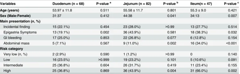
Table 1. Clinicopathologic characteristics of small bowel GISTs in 197 patients.
Variables Duodenum (n = 68) P-valuea Jejunum (n = 82) P-valueb Ileum(n = 47) P-valuec
Age (years) 53.97±11.8 0.511 55.58±11.7 0.801 55.3±9.0 0.421
Sex (Male:Female) 31:37 0.412 44:38 0.041 34:13 0.007
Main presentation (n, %)
Incidentalfinding 15 (22.1%) 0.454 23 (28.0%) >0.99 13 (27.7%) 0.514
Epigastria Symptoms 13 (19.1%) 0.002 36 (43.9%) 0.581 18 (38.3%) 0.032
GI bleeding 17 (25.0%) 0.853 22 (26.8%) 0.077 6 (12.8%) 0.154
Abdominal mass 5 (7.1%) 0.567 9 (11.0%) 0.002 16 (34.0%) <0.001
Risk category
Very low (n, %) 2 (2.9%) 0.590 1 (1.2%) >0.99 0 0.140
Low 16 (23.5%) >0.999 19 (23.2%) 0.101 5 (10.6%) 0.091
Intermediate 25 (36.8%) 0.604 26 (31.7%) 0.419 11 (23.4%) 0.155
High 25 (36.8%) 0.869 36 (43.9%) 0.004 31 (66.0%) 0.002
a,P-value between duodenum and jejunum; b,
P-value between jejunum and ileum; c,
P-value between duodenum and ileum.
doi:10.1371/journal.pone.0144277.t001
Table 2. CT findings of small bowel GISTs in 197 patients.
Variables Duodenum (n = 68) P-valuea Jejunum (n = 82) P-valueb Ileum (n = 47) P-valuec
Size (mean diameter, cm) 7.41±4.97 0.313 8.14±3.13 0.027 9.77±4.14 0.043
5 cm (n, %) 23 (33.8%) 0.480 23 (28.0%) 0.680 11 (23.4%) 0.222
5–10 cm (n, %) 24 (35.3%) >0.999 29 (35.4%) 0.437 13 (27.7%) 0.423
10 (n, %) 21 (30.9%) 0.493 30 (36.6%) 0.016 23 (48.9%) 0.003
CT enhancement (mean RTM value) 1.53±0.31 0.016 1.38±0.37 0.310 1.31±0.21 <0.001
Necrosis
25% (n, %) 33 (48.5%) 0.003 20 (24.3%) 0.521 9 (19.1%) 0.002
25–50% (n, %) 16 (23.5%) 0.544 15 (18.3%) 0.268 13 (27.7%) 0.666
50–75% (n, %) 11 (16.2%) 0.117 23 (28.0%) >0.999 13 (27.7%) 0.164
75% (n, %) 8 (11.8%) 0.010 24 (29.3%) 0.688 12 (25.5%) 0.079
Adjacent tissue involvement 14 (20.6%) 0.276 11 (13.4%) 0.098 12 (25.6%) 0.651
Lymphadenopathy 2 (2.9%) 0.204 0 <0.001 0 0.512
Metastasis 11 (16.2%) 0.208 7 (8.5%) 0.058 10 (21.3%) 0.624
a,P-value between duodenum and jejunum; b,P-value between jejunum and ileum; c,P-value between duodenum and ileum.
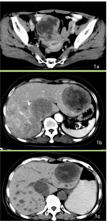
short-axis diameter), and demonstrated lymphadenopathy in CT images, but lymph node exci-sion and pathology confirmed these to be steatosis. In total, 28 cases were diagnosed with dis-tant metastases, of which the liver was the most common site (Fig 1); other sites included the peritoneum (n = 12), omentum (n = 7), and lungs (n = 1). Three patient with omental and peri-toneal metastasis presented with ascites.
One GIST was overlooked in CT images by one radiologist because of its the tiny volume (Fig 2). In some patients, invasion and adhesion was too severe to distinguish the tumor bor-der, and CT post-procedure images help to display the anatomic relationship more clearly. Despite bulky masses in some cases, intestinal obstruction rarely occurred. One case had intes-tinal obstruction, but the CT did not show corresponding features, such as intesintes-tinal dilatation and endo-luminal air-fluid levels.
Discussion
Small bowl GISTs mainly affect individuals aged from 40 to 70 years [5]. The sex distribution of small bowel stromal tumors is approximately equal. In some studies, males show a slight predominance, but this difference is not significant [3,18]. In the present series, ileal GISTs tended to occur more often in males compared with other anatomic sites. This result is interest-ing and requires confirmation with larger sample size.
The onset symptoms of GISTs mostly rely on the size and anatomic location of the tumor [6,19,20]. Small bowel stromal tumors grow more slowly than adenocarcinoma, and the clini-cal symptoms are commonly nonspecific and durative, such as digestive tract bleeding or epi-gastric symptoms. Duodenal GISTs mostly originate from postbulbar duodenal segments [21], which can be different from duodenal adenocarcinomas as the latter mostly arise from duode-nal bulbs and appear as annular lesions at the site of the periampullary [7]. In the present series, about 22.1% (15/68) of duodenal GISTs didn’t have symptoms Compared with duodenal GISTs, jejunum and ileum tumors were more prone to present with epigastric symptoms.
Anemia and periodic melena mostly arise from the ulcerated or necrotic component of tumors. Clinically, a large exoenteric mass may compress the bowel lumen, but seldom induces intestinal obstruction. In this series, only one patient presented with symptoms of an intestinal obstruction, but the CT examination excluded this suspicion.
the clinical symptoms may be more obvious or appear earlier than in the case of ileal lesions, and may thus be diagnosed at an earlier stage.
Small bowel stromal tumors mainly exhibit an exophytic growth pattern. Combined with the oral administration of an intraluminal contrast agent, CT can objectively demonstrate
Fig 1. Jejunum GISTs with liver metastasis.A 64-year-old female with jejunum GISTs with liver metastasis presented with clinical abdominal pain for 6 months, and ultrasound examination detected liver masses for 1 week. A. Enhanced CT image reveals a lobular mass with severe necrosis and periphery enhancement. B. The axial section of the liver shows low-density lesions in the liver with slight enhancement. The patient was diagnosed with a malignant stromal tumor and hepatic metastasis. C. After treatment with Gleevec, the neoplasm became cystic and reduced in size.
tumors characteristics, including CT enhancement degree, necrosis, peripheral invasion, and distant metastasis [24]. In most cases, these CT features can differentiated GISTs from other intestinal tumors [3,25,26].
Because of mass shrinking after operative excision, CT images usually appear larger than on histological examination. This study only analyzed tumor size measured on CT images. In other reports, the size of GISTs in the duodenum tended to be smaller than those in other sites of the small intestine [13]. In this study, tumor size also demonstrated a tendency of increasing from 7.41 cm in the duodenum and 8.14 cm in the jejunum to 9.77 cm in the ileum. Since large tumor size and high mitotic count are independent adverse prognostic factors [9,10,27], this tendency in tumor size suggests a higher risk category for ileal GISTs.
The degree of CT enhancement and enhancement pattern were also associated with primary site in this series. Since increased enhancement on CT scan generally indicates a greater blood supply or more active growth pattern. GISTs located in the duodenum appeared with more
Fig 2. GIST overlooked in CT images.A 64-year-old male with duodenal GISTs presented with melena for the past 20 days. A. Enhanced CT image demonstrates bowel wall thickening at the third segment on the axial section of the CT image and was overlooked. B. Post-procedural CT images (sagittal section) reveal the lesion more clearly. One radiologist had overlooked the lesion, while the other radiologist had misdiagnosed it as a heterotopic pancreas.
obvious enhancement than those in the jejunum and ileum, which may indicate a greater blood supply or a more active growth pattern. However, the enhancement pattern can also be influenced by tumor necrosis, given that CT cannot always differentiate between necrotic and tumor tissue, especially in larger lesions that are more prone to necrosis. Tumors in the duode-num were prone to be homogeneous, while in the ileum they were more prone to be heteroge-neous. It suggests that CT enhancement may not be a reliable predictor of risk category or prognosis.
Except for mitotic count and tumor size, differentiating between benign and malignant tumors also depends on neoplasm biological behavior [7], such as adjacent structure invasion and distant metastasis. GISTs are prone to appear with central liquefactive necrosis, which shows as a heterogeneous feature on CT images. Tumor necrosis can also correlate with aggres-sive behavior [28]. In this series, necrosis was more prone in GISTs located in ileum.
Hepatic metastases can appear with variable appearances on CT images. In the present report, the typical metastases presented with obvious contrast enhancement. Some lesions appeared as cyst-like lesions and might be mistaken for hepatic cysts, but the margins of the metastases were not as distinct and appeared with slight enhancement, and most of the lesions underwent an obvious response to Gleevec treatment. Local lymph metastasis is very rare in patients with GISTs [3,7]. There were two cases in this series with peritoneal lymphadenopa-thy on CT images, but the final pathologic examination confirmed that they were steatosis. No patient had confirmed local lymph nodule metastases in this study, which is consistent with previous studies [3,5].
This study had some limitations. Firstly, all patients in this series underwent enhanced CT scan only, and was lack of non-enhanced CT imaging. Thus, CT signs of hemorrhage and calci-fication can vary considerably with this method, and thus we did not analyze these indicators. secondly, it was occasionally very difficult to evaluate the tumor growth pattern (round or lob-ular) and there were disagreements between the two radiologists. As most GISTs appeared with smooth margins, these variables were not analyzed in this study.
Author Contributions
Conceived and designed the experiments: CWZ. Performed the experiments: GSX. Analyzed the data: SW YMS ZY. Wrote the paper: GSX XMZ.
References
1. Werewka-Maczuga A, Osinski T, Chrzan R, Buczek M, Urbanik A. Characteristics of computed tomog-raphy imaging of gastrointestinal stromal tumor (GIST) and related diagnostic problems. Pol J Radiol. 2011; 76(3):38–48. PMID:22802840; PubMed Central PMCID: PMC3389934.
2. Miettinen M, Lasota J. Histopathology of gastrointestinal stromal tumor. Journal of surgical oncology. 2011; 104(8):865–73. Epub 2011/11/10. doi:10.1002/jso.21945PMID:22069171.
3. Levy AD, Remotti HE, Thompson WM, Sobin LH, Miettinen M. Gastrointestinal stromal tumors: radio-logic features with pathoradio-logic correlation. Radiographics. 2003; 23(2):283–304, 456; quiz 532. Epub 2003/03/18. doi:10.1148/rg.232025146PMID:12640147.
4. DeMatteo RP. The GIST of targeted cancer therapy: a tumor (gastrointestinal stromal tumor), a mutated gene (c-kit), and a molecular inhibitor (STI571). Ann Surg Oncol. 2002; 9(9):831–9. Epub 2002/11/06. PMID:12417503.
5. Lupescu IG, Grasu M, Boros M, Gheorghe C, Ionescu M, Popescu I, et al. Gastrointestinal stromal tumors: retrospective analysis of the computer-tomographic aspects. J Gastrointestin Liver Dis. 2007; 16(2):147–51. Epub 2007/06/27. PMID:17592560.
7. Horton KM, Juluru K, Montogomery E, Fishman EK. Computed tomography imaging of gastrointestinal stromal tumors with pathology correlation. J Comput Assist Tomogr. 2004; 28(6):811–7. Epub 2004/11/ 13. PMID:15538156.
8. Yang WL, Yu JR, Wu YJ, Zhu KK, Ding W, Gao Y, et al. Duodenal gastrointestinal stromal tumor: clini-cal, pathologic, immunohistochemical characteristics, and surgical prognosis. Journal of surgical oncol-ogy. 2009; 100(7):606–10. Epub 2009/08/22. doi:10.1002/jso.21378PMID:19697360.
9. Joensuu H, Vehtari A, Riihimaki J, Nishida T, Steigen SE, Brabec P, et al. Risk of recurrence of gastro-intestinal stromal tumour after surgery: an analysis of pooled population-based cohorts. The Lancet Oncology. 2012; 13(3):265–74. Epub 2011/12/14. doi:10.1016/S1470-2045(11)70299-6PMID: 22153892.
10. Dematteo RP, Gold JS, Saran L, Gonen M, Liau KH, Maki RG, et al. Tumor mitotic rate, size, and loca-tion independently predict recurrence after resecloca-tion of primary gastrointestinal stromal tumor (GIST). Cancer. 2008; 112(3):608–15. Epub 2007/12/14. doi:10.1002/cncr.23199PMID:18076015.
11. Miettinen M, Makhlouf H, Sobin LH, Lasota J. Gastrointestinal stromal tumors of the jejunum and ileum: a clinicopathologic, immunohistochemical, and molecular genetic study of 906 cases before imatinib with long-term follow-up. The American journal of surgical pathology. 2006; 30(4):477–89. Epub 2006/ 04/21. PMID:16625094.
12. Miettinen M, Lasota J. Gastrointestinal stromal tumors: pathology and prognosis at different sites. Semin Diagn Pathol. 2006; 23(2):70–83. PMID:17193820.
13. Han IW, Jang J, Lee KB, Kang MJ, Kwon W, Park JW, et al. Clinicopathologic Analysis of Gastrointesti-nal Stromal Tumors in Duodenum and Small Intestine. World jourGastrointesti-nal of surgery. 2014. Epub 2014/10/ 02. doi:10.1007/s00268-014-2810-xPMID:25270345.
14. Edge SB, Compton CC. The American Joint Committee on Cancer: the 7th edition of the AJCC cancer staging manual and the future of TNM. Ann Surg Oncol. 2010; 17(6):1471–4. Epub 2010/02/25. doi:10. 1245/s10434-010-0985-4PMID:20180029.
15. Wong RJ, Longacre TA, Poultsides G, Park W, Rothenberg ME. Gastrointestinal stromal tumor: an unusual cause of gastrointestinal bleeding. Digestive diseases and sciences. 2013; 58(11):3112–6. Epub 2013/05/02. doi:10.1007/s10620-013-2678-xPMID:23633157.
16. Joensuu H. Risk stratification of patients diagnosed with gastrointestinal stromal tumor. Hum Pathol. 2008; 39(10):1411–9. Epub 2008/09/09. doi:10.1016/j.humpath.2008.06.025PMID:18774375.
17. Ulusan S, Koc Z, Kayaselcuk F. Gastrointestinal stromal tumours: CT findings. The British journal of radiology. 2008; 81(968):618–23. Epub 2008/07/17. doi:10.1259/bjr/90134736PMID:18628330.
18. Crosby JA, Catton CN, Davis A, Couture J, O'Sullivan B, Kandel R, et al. Malignant gastrointestinal stromal tumors of the small intestine: a review of 50 cases from a prospective database. Ann Surg Oncol. 2001; 8(1):50–9. Epub 2001/02/24. PMID:11206225.
19. Caterino S, Lorenzon L, Petrucciani N, Iannicelli E, Pilozzi E, Romiti A, et al. Gastrointestinal stromal tumors: correlation between symptoms at presentation, tumor location and prognostic factors in 47 con-secutive patients. World journal of surgical oncology. 2011; 9:13. Epub 2011/02/03. doi: 10.1186/1477-7819-9-13PMID:21284869; PubMed Central PMCID: PMC3039617.
20. Yu QX, He ZK, Wang J, Sun C, Zhao W, Wang BM. Clinical presentations of gastric small gastrointesti-nal stromal tumors mimics functiogastrointesti-nal dyspepsia symptoms. World jourgastrointesti-nal of gastroenterology: WJG. 2014; 20(33):11800–7. Epub 2014/09/11. doi:10.3748/wjg.v20.i33.11800PMID:25206285; PubMed Central PMCID: PMC4155371.
21. Xie YB, Du J, Li Q, Zhao DB, Wang CF, Cai JQ, et al. [Treatment and prognosis of patients with duode-nal gastrointestiduode-nal stromal tumors]. Zhonghua yi xue za zhi. 2012; 92(24):1694–7. Epub 2012/09/05. PMID:22944161.
22. De Vogelaere K, Van De Winkel N, Aerts M, Haentjens P, Spitali C, Van Loo I, et al. Surgical manage-ment of gastrointestinal stromal tumours: a single centre experience during the past 17 years. Acta chir-urgica Belgica. 2014; 114(3):167–73. Epub 2014/08/12. PMID:25102705.
23. Miki Y, Kurokawa Y, Hirao M, Fujitani K, Iwasa Y, Mano M, et al. Survival analysis of patients with duo-denal gastrointestinal stromal tumors. Journal of clinical gastroenterology. 2010; 44(2):97–101. Epub 2009/10/08. doi:10.1097/MCG.0b013e3181b8e754PMID:19809358.
24. Horton KM, Fishman EK. Multidetector-row computed tomography and 3-dimensional computed tomography imaging of small bowel neoplasms: current concept in diagnosis. J Comput Assist Tomogr. 2004; 28(1):106–16. Epub 2004/01/13. PMID:14716243.
26. Sandrasegaran K, Rajesh A, Rushing DA, Rydberg J, Akisik FM, Henley JD. Gastrointestinal stromal tumors: CT and MRI findings. European radiology. 2005; 15(7):1407–14. doi: 10.1007/s00330-005-2647-7PMID:15761716.
27. Gastrointestinal stromal tumours: ESMO Clinical Practice Guidelines for diagnosis, treatment and fol-low-up. Annals of oncology: official journal of the European Society for Medical Oncology / ESMO. 2014; 25 Suppl 3:iii21–6. Epub 2014/09/12. doi:10.1093/annonc/mdu255PMID:25210085.