From the Department of Gastroenterology, Coloproctology Unit, Hospital das Clínicas, Faculty of Medicine, University of São Paulo - São Paulo/SP, Brazil.
E-mail: [email protected] Received for publication on
November 14, 2003.
CASE REPORT
ANORECTAL LEIOMYOMAS: REPORT OF TWO
CASES WITH DIFFERENT ANATOMICAL PATTERNS
AND LITERATURE REVIEW
Fábio Guilherme Campos, Andrea Furlan Leite, Sérgio Eduardo Alonso Araújo, Fábio César Atuí, Vítor Seid, Angelita Habr-Gama, Desidério Roberto Kiss and Joaquim Gama-Rodrigues
CAMPOS FG et al. Anorectal leiomyomas: report of two cases with different anatomical patterns and literature review. Rev. Hosp. Clín. Fac. Med. S. Paulo 59(5):296-301, 2004.
Gastrointestinal mesenchymal tumors comprise a rare group of gastrointestinal tract wall tumors that have long been a source of confusion and controversy, especially in terms of pathological classification, preoperative diagnosis, management strategies, and prognosis. This report describes the clinical manifestations and management of 2 rectal leiomyomas and reviews the pertinent literature. Case 1: A 44-year-old woman was admitted reporting a nodule in the right para-anal region for the previous 2 years. At proctological examination, a 4-cm diameter fibrous mass situated in the para-anal region that produced an arch under the smooth muscle on the right rectal wall just above the anorectal ring was noted. Computed tomography and magnetic resonance imaging of the abdomen and pelvis showed the lesion and detected no other abnormalities. Surgical treatment consisted of wide local resection of the tumor through a para-anal incision, with no attempts to perform lymphadenectomy. Case 2: A 40-year-old male patient was admitted reporting constant anal pain for 4 months. He presented a 3-cm submucosal nodule at the anterior rectal wall just above the dentate line. After 2 inconclusive preoperative biopsies, transanal resection of the tumor was performed. Histological analysis of the specimen showed a benign leiomyoma. A review of the literature is presented, emphasizing some clinical and therapeutic aspects of this unusual rectal tumor.
KEY WORDS: Gastrointestinal stromal tumors. Leiomyoma. Leiomyosarcoma. Rectum. Literature.
Gastrointestinal mesenchymal tumors are nonepithelial lesions that exhibit an immature proliferation of epithelioid or spindle cells from the gastrointestinal tract muscle layer. Historically, these tumors have been called benign (leiomyoma) or malig-nant (leiomyosarcomas). More re-cently, pathologists have begun to shift from these terms to gastrointestinal stromal tumors (GIST).
Stromal tumors may occur in any muscle layer segment of the digestive tract, such as smooth muscle tissue (muscularis mucosa or muscularis
pro-pria), nervous tissue originating in the myenteric plexus, or mesenchymal primitive cells. Thus, GIST are histologically classified into 4 types: smooth muscle, neural, mixed, and un-differentiated. Additionally, the smooth muscle terminology may still be used when cellular differentiation is clearly evidenced.5
Pathological and prognostic clas-sifications separate leiomyomas, low-grade leiomyosarcomas, and high-grade leiomyosarcomas.3 Histological
guidelines for highly malignant tumors do exist, including size (greater than 5 cm), mitotic rate (greater than 10 per 10 high-powered fields), necrosis number, increased vascularity, and cel-lular atypia. Low-grade lesions may have only 1 mitosis per 10 high-pow-ered fields and be smaller than 5 cm in size.18,24
gastrointestinal regions. The stomach and small bowel are more frequently af-fected by these tumors, and the rectum is estimated to account for 7% to 11% of all gastrointestinal smooth muscle tumors.18,21 Kim et al.8 reported 19
pa-tients with GIST in Georgia (USA) as follows: 12 gastric, 2 duodenal, 3 jejunal, and 2 rectal. Among 24 surgi-cally treated patients during the last 10 years in Milano (Italy), Chiara et al.2
observed 6 gastric leiomyomas, 1 ileal leiomyoma, 4 gastric leiomyosarcomas, 1 esophageal leiomyosarcoma, 4 ileal leiomyosarcomas, 2 rectal leiomyosar-comas, and 6 gastric leiomyoblastomas. In Taiwan, Chou et al.3 reported 80
gastrointestinal smooth muscle tumors that were surgically removed between 1986 and 1992 as follows: 1 esophageal, 32 gastric, 33 intestinal, 2 colonic, and 12 rectal.
The incidence of rectal GIST is very low. Many years ago, it was esti-mated that 1 leiomyoma may be found in every 2000 or more rectal tumors.10
Ten years later, Zerilli et al.25 estimated
this incidence to be around 0.15 and 0.3% of colorectal malignant neo-plasms. Leiomyomas located in the anal canal and sphincter are the rarest ones. In Brazil, papers focusing on smooth muscle tumors of the rectum have been rarely published.9,16,18,20
Leiomyomas have a highly variable clinical course, and the therapeutic strategy is still controversial.8 Due to
their submucosal origin, these tumors are often asymptomatic at initial stages. When present, symptoms are similar to those observed in common anorectal diseases, namely, local dis-comfort or pain (related or not to def-ecation), sensation of a foreign body, change in bowel habits, and rectal bleeding.25
Leiomyomas occur mainly in pa-tients between 40 and 50 years. Clini-cal diagnosis depends on awareness of these lesions, digital rectal examina-tion, proctoscopy, and tissue biopsy. A
great majority of rectal smooth muscle and stromal tumors are GIST with vari-ations ranging from minimal indolent tumors to overt sarcomas.12 While
dif-ferential histological diagnosis be-tween benign and malignant forms is a dilemma, treatment should always be surgical.
Preoperative diagnosis is difficult to achieve because a biopsy is often valueless, since it does not involve the entire tumor mass.4,14,19 When
diagno-sis provided by biopsy does not show the malignant nature of the lesion, its histological features can be assessed only after complete local excision.17
The present paper reports 2 cases of rectal leiomyoma, giving emphasis to their clinical manifestations, diag-nosis, and management; a review of the pertinent literature is also presented.
CASE REPORTS
Case 1
A 44-year-old-woman was admit-ted reporting a slow-growing nodule at the right para-anal region for the pre-vious 2 years. The tumor was painless, and the patient had no bleeding, change in bowel habits, or weight loss. She reported a family history of colon (aunt) and breast cancer (mother). Gen-eral physical examination was unre-markable. Upon proctologic assess-ment, a delimited mobile fibrous mass situated at the right para-anal region was noted. Although this mass did not invade the rectum, it was palpable just contiguously to the right rectal wall; on digital examination, it was felt like a little bulge just above the anorectal ring.
Magnetic resonance images showed a slight parietal thickening of the right distal rectum and a 14.5 x 7.0 x 6.0 cm solid, well-defined mass in the ischioanal space extending up to the gluteus region. Other
abdomi-nal structures had no changes (Figure 1). Computed tomography (CT) find-ings were absolutely similar to those already described by magnetic reso-nance.
Under epidural anesthesia, surgery was performed in the lithotomy posi-tion. Considering the location of the mass, a shallow para-anal incision was made radially on the right side of the anal margin. Dissection of the subcu-taneous fat provided tumor visualiza-tion, and progressive liberation of sur-rounding tissues was made in order to achieve the superior margins of the tumor close to the sciatic tuberosity. After resection, the wound was re-paired by primary closure (Figure 2).
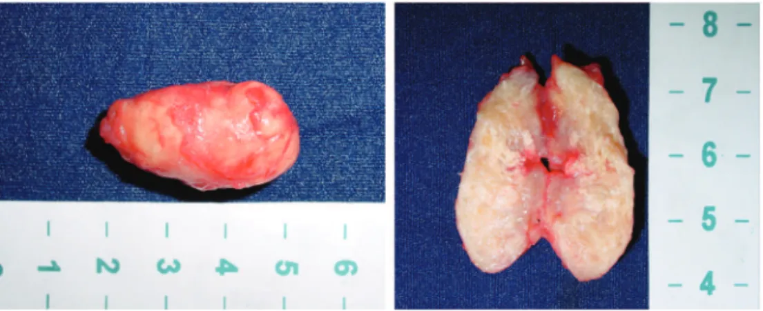
The patient was released on the second postoperative day. Macro-scopic and histological analysis re-vealed features of a benign leiomyoma (smooth muscle GIST) (Figure 3).
Case 2
The second patient was a 40-year-old man whose complaint was con-stant anal pain during the previous 4
Figure 2 - Perineal wound closed after excision of the tumor (case 1).
months. At proctologic examination, a submucosal round nodule situated at the anterior rectal wall just above the dentate line was noted. The lesion had
an approximately 3-cm diameter, and there was no mucosal ulceration. Two preoperative biopsies were inconclu-sive.
Tumor resection was performed un-der regional anesthesia with the pa-tient positioned in the jack-knife po-sition (Figure 4). A transanal excision was made with the aid of an anal de-vice used for stapled hemorrhoidec-tomy. After a midline incision over the mucosa covering the tumor, 3 stitches were placed in the surrounding mucosa to facilitate the access to the tumor. The tumor was then carefully dis-sected and enucleated after liberation from the inner planes. The mucosa was then closed using absorbable and in-terrupted suture (Figure 5). Histologi-cal analysis of the specimen revealed a benign leiomyoma (smooth muscle GIST) (Figure 6).
DISCUSSION
Despite its low incidence and prevalence, rectal and anal leiomyo-mas have been discussed in case
re-ports and review papers. These publi-cations have usually dealt with the ap-proaches of diagnostic tools and sur-gical management.
In a recent publication, Hatch and coworkers5 reviewed all case reports
about rectal and anal canal stromal tumors described in the world litera-ture between 1881 and 1996. This re-view included 432 leiomyomas and 480 leiomyosarcomas. The review re-vealed that that leiomyomas predomi-nately occur between 40 and 59 years of age. Our 2 patients were 40 and 44 years of age. In another review,12 GIST
were found to occur in adults with a median age of 60 years (range, 17-90 years) with a significant male predomi-nance (71%).
Leiomyomas often remain asymp-tomatic until they have reached a fairly large size. The most common symptoms are bleeding, palpable mass, and rectal pain.5 Patients usually
sented clinical complaints for the pre-vious 12 months, and those with leio-myomas tended to tolerate symptoms longer before attaining medical inter-vention.4 Regarding the cases reported
here, the woman presented no local pain or bleeding. Despite the long du-ration of symptoms, she only reported a slow-growing mass. This clinical pic-ture of an asymptomatic mass has also been commonly observed in other re-ports.10,19 Occasionally, patients will
report bleeding (if the overlying mu-cosa ulcerates), constipation, pain, or a sense of fullness. Almost always, the chain of events leading to diagnosis starts when the tumor is discovered in-cidentally by digital examination or as a submucosal mass at rectoscopy.10,25
Our male patient reported rectal pain and a sense of fullness.
Tumors may vary from small asymptomatic intramural nodules to large masses that bulge into pelvis, causing pain, rectal bleeding, or ob-struction. Colonic and rectal leiomyo-mas often present as intraluminal
Figure 3 - Macroscopic vision of the opened mass (case 1).
Figure 6 - Macroscopic aspects of the resected lesion (case 2). Figure 4 - Patient in Jack-knife position (case 2).
polypoid masses.12 In a collective
re-view of smooth muscle tumors of the rectum and anal canal, Hatch et al.5
re-ported that intraluminal growth of both leiomyomas and leiomyosarcomas was more frequently seen than extraluminal or intramural patterns, and tumors were more likely to be found in the rectum than in the anus.
The 2 cases presented here had tumors in a close proximity to the rec-tum, and while the tumor in the female patient (case 1) presented as an extra-mural lesion, the man (case 2) had a lesion that could be characterized as an intramural tumor.
The majority of smooth muscle tumors appear as submucosal nodules, although a few of them have been de-scribed as polypoid. Ulceration of the overlying mucosa may occur in both leiomyomas and leiomyosarcomas. Stromal tumor dissemination occurs primarily by direct extension to adja-cent organs. Hematogenous metastasis can reach the liver, lung, bones, and
brain.12 Although involvement of
lymph nodes rarely occurs, it is asso-ciated with poor survival rates.1
Therefore, imaging techniques are useful for preoperative staging, since they can describe the relationships with the sphincters and urogenital tract, and they can detect metastatic
spread to regional lymph nodes.25
Complementary investigation, such as with CT, endorectal ultrasonography, and magnetic resonance imaging scan, strongly corroborates the diagnosis. Endorectal ultrasound can help to de-fine the extent of disease and may be a useful adjunct in deciding about the appropriate surgical procedure.6
Dur-ing the treatment of our female patient, information obtained from physical assessment, CT scan, and magnetic resonance were sufficient and ruled out the need of endorectal ultrasonog-raphy.
Furthermore, radiological evalua-tion was very useful in defining
opera-tive strategy. In the first patient, the para-anal location of the tumor, as sug-gested by physical examination and confirmed by the magnetic resonance, allowed us to excise the tumor through a radial incision starting at the right anal margin. Thus, the tumor mass was easily found, dissected, and excised. During the treatment of the second pa-tient, rectal assessment clearly showed a small submucosal and mobile tumor. These anatomical features suggested that surgery could be safely accom-plished through a transanal approach.
The lack of reliable criteria for ma-lignancy is the main problem the sur-geon faces when selecting the opera-tive procedure. Although most of the 150 leiomyomas of the rectum re-ported since 1872 were not larger than 5 cm, Le Borgne et al.11 described 3
rectal leiomyomas measuring more than 5 cm. Hatch et al.5 found that
rec-tal leiomyosarcomas tended to be larger than leiomyomas, as was also the case for these neoplasms in other gastrointestinal locations. Tumors with an original size larger than 5 cm are those that have shown the highest ten-dency to recur, mostly as sarcomas. Therefore, recurrent lesions should be treated radically from the beginning.10
Additionally, smooth-muscle rectal tumors should be considered more dangerous than those in other loca-tions in the gastrointestinal tract, since half are malignant and only one-fifth of patients who have sarcomas survive 5 years.
When evaluating the clinical symp-toms of a patient, one must have in mind that bleeding, constipation, and weight loss are associated with a higher risk of malignancy.5
Preoperative diagnosis can be dif-ficult, and the final diagnosis is often made at the time of surgical treatment, such as with the second patient re-ported here. Preoperative histological diagnosis is adequate in only 29% of cases.2 Microscopic diagnosis and
dif-ferentiation of malignant from benign features require a pathologist with spe-cial interest and expertise with these lesions. The ultimate proof of malig-nancy is therefore determined by re-currence of the tumor or metastasis. Since they grow within the intestinal wall, symptoms are usually few or late, leading to delay in diagnosis.17
Leiomyomas are relatively insensi-tive to adjuvant therapy. Therefore, their treatment is primarily surgical and should guarantee complete clear-ance of the tumor.1
The choice of surgical approach for a rectal lesion depends mainly on clinical and histopathological find-ings. Small and benign-appearing le-sions for which histology has ex-cluded malignancy should be treated by local excision with adequate mar-gins, followed by periodic surveil-lance. With complete resection of the tumor, the clinical course is favorable, with very few local recurrences. Local excision of low rectal lesions may be accomplished by a conventional transanal excision, while upper tumors may be excised using either transanal endoscopic microsurgery or a posterior approach.15,25
Since our 2 patients had tumors with no gross or histological features of malignancy, their management through a local excision was consid-ered adequate, and the patients were assigned to a follow-up program.
True rectal leiomyosarcomas are rare and account for less than 0.1% of all malignant tumors of the rectum. One estimate is that less than 300 cases have been reported so far, and anal le-sions are even rarer.23 They tend to
oc-cur between 50 to 69 years of age, and approximately 20% of rectal leiomy-osarcomas reported from 1881 to 1996 had metastasized at diagnosis.
even nonoperative therapy.7, 13
Chemo-therapy and radioChemo-therapy are generally not effective.24 Although there is no
clear evidence that adjuvant therapy influences overall survival, further tri-als are needed to establish its exact role, since good results have been re-ported in selected patients, with dis-ease-free interval prolongation.17, 23
Radical surgery is indicated for lo-cal recurrence of benign tumors, and treatment of larger (greater than 5 cm) and malignant rectal myomas should be very aggressive from the onset (low an-terior resection or abdominoperineal re-section), although there is a question-able rate of survival improvement.11,14
According to Chou et al.,3
leiomyosar-comas usually measure more than 10 cm, and the significant factors affecting survival rates at univariate analysis are maleness, size greater than 5 cm, inad-equate resection, and advanced-stage and high-grade disease.
Although the most common therapy for large and low differentiated leiomyosarcomas of the rectum is radi-cal resection through abdominoperi-neal resection or low anterior resection, the need for radical resection of this extent has been questioned with regard to treatment of high-graded tumors smaller than 2.5 cm. Some authors rec-ommend a wide local excision;
how-ever, in this case a high local-recur-rence rate has to be expected. This is the reason for a more radical treatment of all leiomyosarcomas of the rectum.
Vorobyov et al.22 reported their
ex-perience with the treatment of 36 rec-tal leiomyomas from 1972 to 1990 in Moscow. There were 13 male (36 %) and 23 female (64 %) patients, and median age was 52.1 years. Electroexcision of tumors measuring less than 1 cm was performed endo-scopically in 12 patients. Leiomyomas measuring 2.5 to 5 cm were removed through the transanal approach in 10 patients. Six patients underwent exci-sion of the tumor through the pararec-tal approach, whereas leiomyomas lo-cated in the rectovaginal wall were re-moved through the vagina in 1 patient. Abdominoperineal extirpation and abdominoanal resection of the rectum was performed in 7 patients with tumors measuring 8 to 20 cm. Recur-rences were noted in 9 patients after transanal, pararectal, or transvaginal excision of leiomyomas. In 7 of them, malignant transformation of the tumor occurred at terms ranging from 9 months to 9.5 years.
The local recurrence rate for resect-able leiomyosarcomas was more than 80%, exceeding the propensity of leio-myosarcomas in other areas of the
gastrointestinal tract to recur.5 In the
study performed by Miettinen et al.,12
70% of patients with tumors > 5 cm with more than 5 mitosis/50 high power fields (HPF) (n = 31) died of dis-ease, whereas only 1 tumor <2 cm with < 5 mitosis/50 HPF (n = 21) recurred, and none caused death. Long latency was common between the primary op-eration and recurrences and metastases; either one occurring in 60 of 111 patients with follow-up (54%).
Therefore, extended follow-up is required, because long-term recur-rences seem to be also possible in cases involving low-grade lesions.2 Biologic
behavior also varies with location; co-lonic tumors are generally less aggres-sive when compared to rectal ones. Rectal tumors manifest greater recur-rence and dissemination rates, even af-ter wide resections with curative pur-poses.5
According to Witzigmann et al.,24
the prognosis for rectal leiomyosarco-mas is generally poor. Survival rates vary from 20% to 25% in 5 years.1
Al-though size, histological grade, and local staging play an important role, complete resection is considered the most significant favorable prognostic factor. Regarding anal leiomyosarco-mas, the evaluation of prognosis is hampered because its rarity.
RESUMO
Campos FG e col. Leiomiomas ano-retais: descrição de dois casos com características anatômicas diferen-tes e revisão da literatura. Rev. Hosp. Clín. Fac. Med. S. Paulo 59(5):296-301, 2004.
Os tumores mesenquimais gastro-intestinais constituem um grupo raro de neoplasias que têm sido fonte de confusão e controvérsia, especialmen-te quanto à classificação patológica,
diagnóstico pré-operatório, manuseio e prognóstico. O presente artigo des-creve as manifestações clínicas e o tra-tamento de dois pacientes com leio-mioma retal e revê a literatura perti-nente. Caso 1: Uma mulher de 44 anos foi admitida referindo um nódulo na região paranal direita nos últimos 2 anos. Ao exame físico notou-se uma massa fibrosa de 4 centímetros de diâ-metro situada na região paranal que produzia um discreto abaulamento na
apresentava nódulo submucoso de 3 cm na parede retal anterior, logo aci-ma da linha pectínea. Após duas biópsias inconclusivas, realizou-se a ressecção transanal do tumor. A
análi-se histológica do espécime demonstrou tratar-se de um leiomioma benigno. Uma breve revisão da literatura é apre-sentada, enfatizando alguns aspectos clínicos e terapêuticos deste tumor
retal pouco comum.
UNITERMOS: Tumores estromais
gastrointestinais. Leiomioma. Leio-miossarcoma. Reto. Literatura.
REFERENCES
1 . Brand MI, Saclarides TJ. Lymphoma, neuroendocrine and soft tissue tumors of the rectum. Clinics Colon Rectal Surg 2002;15:71-9.
2 . Chiara O, Canini T, Segala M, Tiberio GA, Giulini SM, Tiberio G. Smooth-muscle-cell tumors of the gastroenteric tract. A review of cases. Minerva Chir 1997;52(10):1147-55.
3 . Chou FF, Eng HL, Sheen-Chen SM. Smooth muscle tumors of the gastrointestinal tract: analysis of prognostic factors. Surgery 1996;119(2):171-7.
4 . Haque S, Dean PJ. Stromal neoplasms of the rectum and the anal canal. Hum Pathol 1992;23:762-7.
5 . Hatch KF, Blanchard DK, Hatch GF 3rd, Wertheimer-Hatch L, Davis GB, Foster RS Jr, et al. Tumors of the rectum and anal canal. World J Surg 2000;24 (4):437-43.
6 . Hsieh JS, Huang CJ, Wang JY, Huang TJ. Benefits of endorectal ultrasound for management of smooth-muscle tumor of the rectum: report of three cases. Dis Colon Rectum 1999;42(8):1085-8.
7 . Zbar AP, Sokolowsky N, Sandiford N, Prussia PR. Leiomyosarcoma of the rectum: report a of a case and review of the literature. Dis Colon Rectum 1986; 29: 427-32. 8 . Kim CJ, Day S, Yeh KA. Gastrointestinal stromal tumors: analysis
of clinical and pathologic factors. Am Surg 2001;67(2):135-7 .
9 . Kiss DR, Iwasso S, Tessler S et al. Leiomyosarcoma of the rectum. Report of a case. AMB Rev Assoc Med Bras 1979;25(2):59-60.
10. Kusminsky RE, Bailey W. Leiomyomas of the rectum and anal canal: report of six cases and review of the literature. Dis Colon Rectum 1977;20(7):580-99.
11. Le Borgne J, Guiberteau-Canfrere V, Lehur Pa, et al. Leiomyoma of the rectum. Chirurgie 1993-94;119(4):212-5.
12. Miettinen M, Furlong M, Sarlomo-Rikala M, Burke A, Sobin LH, Lasota J. Gastrointestinal stromal tumors, intramural leiomyomas, and leiomyosarcomas in the rectum and anus: a clinicopathologic, immunohistochemical, and molecular genetic study of 144 cases. Am J Surg Pathol 2001; 25 (9): 1121-33.
13. Minsky Bd, Cohen AM, Hajdu SI - Conservative management of anal leiomyosarcoma. Cancer 1991;68:1640-3.
14. Nemer FD, Stoeckinger JM, Evans OT. Smooth-muscle rectal tumors: a therapeutic dilemma. Dis Colon Rectum 1977;20(5):405-13.
15. Piccinini EE, Ugolini G, Rosati G, Conti A. Transanal local resection for benign and malignant rectal tumours. : Int J Colorectal Dis 1995;10(2):112-6.
16. Ramos JR, Pinho M, Ramos RP, et al. Leiomiossarcoma do reto – Relato de um caso. Rev bras Colo-Proct 1987;7:107-9. 17. Ricca L, Ferri M, De Siena T, Ricci F, Laghi A, Ziparo V. Stromal
tumors of the rectum: a case report and review of the literature. Chir Ital 2002; 54 (5): 709-16.
18. Sakano AI, Bresciani CJC, Habr-Gama A, Alves VAF, Gama-Rodrigues JJ. Gastrointestinal stromal tumors: anatomopathologic characterization and correlation with prognostic factors. Arq Bras Cir Dig 2003;16(1):10-3. 19. Serra J, Garriga J, Escuder J, Alonso M, Piera J, Puig la Calle J.
Leiomyoma of the rectum. A diagnostic and therapeutic dilemma. J Chir (Paris) 1987;124(8-9):450-3.
20. Souza GA. Leiomiossarcoma perianorretal. Relato de um caso operado. Rev Bras Colo-Proct 1983;3:143-5.
21. Vandoni RE, Givel JC, Essinger AR. Rectal leiomyosarcoma: acute presentation after local injury. Eur J Surg 1992;158:383. 22. Vorobyov GI, Odaryuk TS, Kapuller LL, Shelygin YA, Kornyak
BS. Surgical treatment of benign, myomatous rectal tumors. Dis Colon Rectum 1992;35(4):328-31.
23. Wang TK, Chung MT. Anorectal leiomyosarcomas. J Gastroenterol 1998;33(3):402-7
24. Witzigmann H, Sagasser J, Leipprandt E, et al. Leiomyosarcoma of the rectum. Zentralbl Chir 1995;120(1):69-72.