Kadow JS et al. / Encapsulating peritonitis: CT and surgical correlation
Radiol Bras. 2014 Jul/Ago;47(4):262–264 262
0100-3984 © Colégio Brasileiro de Radiologia e Diagnóstico por Imagem
Case report
Encapsulating peritonitis: computed tomography and surgical
correlation
*
Peritonite encapsulante: tomografia computadorizada e correlação cirúrgica
Kadow JS, Fingerhut CJP, Fernandes VB, Coradazzi KRS, Silva LMS, Penachim TJ. Encapsulating peritonitis: computed tomography and surgical corre-lation. Radiol Bras. 2014 Jul/Ago;47(4):262–264.
Abstract
R e s u m o
Sclerosing encapsulating peritonitis is a rare and frequently severe entity characterized by total or partial involvement of small bowel loops by a membrane of fibrous tissue. The disease presents with nonspecific clinical features of intestinal obstruction, requiring precise imaging diagnosis to guide the treatment. The present report emphasizes the importance of computed tomography in the diagnosis of this condition and its confirmation by surgical correlation.
Keywords: Peritonitis; Peritoneal fibrosis; Sclerosing encapsulating peritonitis; Computed tomography.
A peritonite esclerosante encapsulante é uma entidade rara, muitas vezes grave, caracterizada pelo envolvimento total ou parcial de alças do intestino delgado por uma membrana de tecido fibroso. Apresenta quadro clínico inespecífico de obstrução intestinal, exigindo diagnóstico por imagem preciso para orientação do tratamento. O presente relato enfatiza a importância da tomografia computadorizada no diagnóstico desta doença, com confirmação por correlação cirúrgica.
Unitermos: Peritonite; Fibrose peritoneal; Esclerose peritoneal encapsulante; Tomografia computadorizada.
* Study developed at Hospital e Maternidade Celso Pierro – Pontifícia Universi-dade Católica de Campinas (PUC-Campinas), Campinas, SP, Brazil.
1. MDs, Residents, Hospital e Maternidade Celso Pierro – Pontifícia Universidade Católica de Campinas (PUC-Campinas), Campinas, SP, Brazil.
2. MD, Radiologist, Chief of Hospital e Maternidade Celso Pierro – Pontifícia Universidade Católica de Campinas (PUC-Campinas), Campinas, SP, Brazil.
Mailing Address: Dr. Vinicius de Barros Fernandes. Avenida Onze de Junho, 970, ap. 71, Vila Clementino. São Paulo, SP, Brazil, 04041-003. E-mail: vinicius.barros. [email protected].
Received April 19, 2013. Accepted after revision October 17, 2013.
means of exploratory laparotomy, with emphasis on some clinical and imaging signs which contribute to increase the suspicion level, allowing for the planning of appropriate man-agement and avoiding unnecessary surgical approach.
CASE REPORT
A male, brown, 37-year-old patient was referred by other service, complaining of diffuse abdominal pain and palpable abdominal mass for about one year, with probable diagno-sis of umbilical hernia. The patient reported a previous his-tory of paracoccidioidomycosis diagnosed for about 20 years ago, and over this period the condition progressed with vo-luminous ascites. Also, the patient reported a history of smoking and hepatic cirrhosis secondary to alcohol use with portal hypertension. At admission, clinical examination dem-onstrated a good general condition, with abdominal disten-sion and presence of hydroaerial noise, besides a large, pal-pable, mobile and painful mass in the right hypochondrium and a small-sized umbilical hernia.
Abdominal computed tomography (CT) demonstrated agglomerated small bowel loops with moderate distension at the level of the mesogastrium involved by a thick and regu-lar membrane and a moderate amount of loculated ascitic fluid in the pelvis (Figure 1).
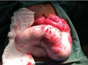
At the third day after the admission, the patient presented obstructive acute abdomen and was submitted to exploratory laparotomy. A thick, fibrous, grayish-white membrane was intraoperatively observed, involving small bowel loops in continuation to the visceral peritoneum, similar to a cocoon
Juliana Santos Kadow1, Carla Jeronimo Peres Fingerhut1, Vinicius de Barros Fernandes1, Klaus
Rizk Stuhr Coradazzi1, Lucas Marciel Soares Silva1, Thiago José Penachim2
http://dx.doi.org/10.1590/0100-3984.2013.1780
INTRODUCTION
Sclerosing encapsulating peritonitis (SEP) is a rare and curious entity of unknown etiology. Such a condition is char-acterized by partial or total encasement of the small bowel by a thick membrane of fibrotic connective tissue resembling a cocoon, that may extend and involve other organs such as the large bowel, liver and stomach(1). SEP presents with
nonspecific clinical manifestations such as insidious abdomi-nal pain and weight loss in addition to recurrent episodes of acute or subacute intestinal obstruction, either with or with-out the presence of an associated abdominal mass(2,3).
This condition has been described with different names including “abdominal cocoon” by Foo in 1978(3),
“peritoni-tis chronica fibrosa incapsulata” by Owtschinnikow in 1907(4),
and “sclerosing encapsulating peritonitis” by Deeb et al. in 1998(4), many times in association with conditions which lead
to recurrent peritonitis and peritoneal dialysis.
Kadow JS et al. / Encapsulating peritonitis: CT and surgical correlation
Radiol Bras. 2014 Jul/Ago;47(4):262–264 263
(Figure 2). Removal of the capsule and adhesionlysis were performed in addition to segmental enterectomy.
Histological analysis of the membrane that involved the small bowel loops revealed the presence of a chronic nongranulomatous inflammatory process in association with interstitial fibrosis, with vascular ectasia on a dense connec-tive tissue, besides ischemic necrosis and acute serositis of the resected segment of small bowel.
The patient’s evolution was unsatisfactory, with diagno-sis of entero-enteral fistula and at the 17th postoperative day further surgical procedure was required for enterocutaneous fistulotomy. After the procedure, the patient evolved with recurrent infections, severe denutrition and death 46 days after admission.
DISCUSSION
SEP may be classified into primary or idiopathic and secondary(1–4). Primary SEP has already been associated with
retrograde menstruation in women and with abnormality in the embryonic development of the peritoneum, with possi-bility of concomitant greater omentum hypoplasia and me-senteric vessels malformation(1,3). Secondary SEP is
associ-ated with predisposing factors such as peritoneal dialysis, recurrent peritonitis, infectious and noninfectious granulo-matous diseases, autoimmune diseases (systemic lupus erythematosus), long term practolol therapy, abdominal catheters (Le Veen shunts), intraperitoneal chemotherapy, liver transplant, cirrhosis, endometriosis, ovarian luteinized thecoma, S-protein deficiency, dermoid cyst rupture, expo-sure to asbestos and to fibrogenic materials(1).
Almost all the cases of SEP described in the literature were intraoperatively diagnosed. A preoperative diagnosis re-quires a high level of clinical suspicion. Usually, the first clinical signs are nonspecific and frequently the condition cannot be recognized until the patient develops partial or total small bowel obstruction. Symptoms include pain and recur-Figure 1. Non-contrast-enhanced (A) and contrast-enhanced (B,C) abdominal
CT, axial sections, identifying conglomerate of small bowel loops with moderate air-fluid distension (white arrow) concentrated principally in the region of the me-sogastrium and involved by a thick and regular membrane (black arrow), in asso-ciation with thickening and greater enhancement of peritoneal reflections, as well as moderate amount of fluid with loculated aspect (arrow head) most concen-trated in the pelvis.
A
B
C
Kadow JS et al. / Encapsulating peritonitis: CT and surgical correlation
Radiol Bras. 2014 Jul/Ago;47(4):262–264 264
rent abdominal distension, nausea, vomiting, anorexia, weight loss, denutrition, recurrent episodes of acute, sub-acute or chronic intestinal obstruction, besides abdominal mass(1,4).
Considering the nonspecificity of clinical findings of SEP, imaging methods become a useful tool for an early di-agnosis, directly contributing in de adoption of an appro-priate treatment. In patients with SEP, abdominal CT dem-onstrates agglomerated and distended small bowel loops con-centrated in an abdominal segment, involved by a thick membrane, with peritoneal thickening, ascites and loculated fluid collections and possible peritoneal calcifications. Ad-ditionally, fibrosis leads to retraction of the mesenteric root, causing adhesions and loops conglomerate, leading to in-testinal obstruction and dysfunction. Therefore, as compared with other imaging techniques, CT provides a comprehen-sive view of the condition as well as of any associated com-plication, besides helping to rule out other possible causes of intestinal obstruction(5).
The treatment for SEP consists in surgical excision of the fibrotic membrane, intestinal loops adhesionlysis and re-section in case of inviability of the affected intestinal seg-ment. After appropriate surgical management, the
progno-sis is good, but it depends on the coexistence or not with other diseases(6).
Finally, the present report highlights SEP as a rare dis-ease, whose preoperative diagnosis depends on imaging evaluation, hence the great relevance of CT.
Considering the relevance of the preoperative imaging diagnosis of SEP, it is necessary for the radiologist to be aware and attentive to the tomographic findings suggestive of the diagnosis.
REFERENCES
1. Tannoury JN, Abboud BN. Idiopathic sclerosing encapsulating peri-tonitis: abdominal cocoon. World J Gastroenterol. 2012;18:1999– 2004.
2. Hosein HH, Quane LK, Cohen AJ. Abdominal cocoon. Appl Radiol. 2003;32(10).
3. Altinli E, Sumer A, Celik A. Abdominal cocoon: a rare cause of in-testinal obstruction. Israeli Journal of Emergency Medicine. 2007;7: 42–4.
4. Ranganathan S, Abdullah BJJ, Sivanesaratnam V. Abdominal cocoon syndrome. J HK Coll Radiol. 2003;6:201–3.
5. Gupta S, Shirahatti RG, Anand J. CT findings of an abdominal co-coon. AJR Am J Roentgenol. 2004;183:1658–60.