Infants' Peripheral Blood Lymphocyte
Composition Reflects Both Maternal and
Post-Natal Infection with
Plasmodium
falciparum
Odilon Nouatin1,2☯, Komi Gbédandé1,2☯, Samad Ibitokou1,2, Bertin Vianou1,2,
Parfait Houngbegnon1,2, Sem Ezinmegnon1,2, Sophie Borgella1,3, Carine Akplogan1,2, Gilles Cottrell1,3,4, Stefania Varani5, Achille Massougbodji1, Kabirou Moutairou2,
Marita Troye-Blomberg6, Philippe Deloron3,4, Adrian J. F. Luty7¤‡, Nadine Fievet1,3,4‡
*
1Centre d’Etude et de Recherche sur le Paludisme Associéàla Grossesse etàl’Enfance (CERPAGE), Faculté des Sciences de la Santé, Université d’Abomey-Calavi, Cotonou, Benin,2Département de Biochimie et de Biologie Cellulaire, Faculté des Sciences et Techniques, Université d’Abomey-Calavi, Cotonou, Bénin,3Institut de Recherche pour le Développement, MERIT UMR D216 Mère et enfant face aux
infections tropicales, Paris, France,4PRES Sorbonne Paris Cité, Université Paris Descartes, Faculté de Pharmacie, Paris, France,5Unit of Microbiology, Department of Diagnostic, Experimental and Specialty Medicine, University of Bologna, Bologna, Italy,6Department of Molecular Biosciences, the Wenner-Gren Institute, Stockholm University, Stockholm, Sweden,7Department of Medical Microbiology, Radboud University Nijmegen Medical Centre, Nijmegen, The Netherlands
☯These authors contributed equally to this work.
¤ Current address: IRD MERIT UMR D216 Mère et enfant face aux infections tropicales, Université Paris
Descartes, Faculté de Pharmacie, 4 avenue de l’Observatoire, 75006 Paris, France ‡These authors also contributed equally to this work.
*fievet.nadine@ird.fr
Abstract
Maternal parasitoses modulate fetal immune development, manifesting as altered cellular immunological activity in cord blood that may be linked to enhanced susceptibility to infec-tions in early life.Plasmodium falciparumtypifies such infections, with distinct placental infection-related changes in cord blood exemplified by expanded populations of parasite antigen-specific regulatory T cells. Here we addressed whether such early-onset cellular immunological alterations persist through infancy. Specifically, in order to assess the poten-tial impacts ofP.falciparuminfections either during pregnancy or during infancy, we quanti-fied lymphocyte subsets in cord blood and in infants' peripheral blood during the first year of life. The principal age-related changes observed, independent of infection status, con-cerned decreases in the frequencies of CD4+, NKdimand NKT cells, whilst CD8+, Treg and Teff cells' frequencies increased from birth to 12 months of age.P.falciparuminfections present at delivery, but not those earlier in gestation, were associated with increased fre-quencies of Treg and CD8+T cells but fewer CD4+and NKT cells during infancy, thus accentuating the observed age-related patterns. Overall,P.falciparuminfections arising during infancy were associated with a reversal of the trends associated with maternal infec-tion i.e. with more CD4+cells, with fewer Treg and CD8+cells. We conclude that maternal P.falciparuminfection at delivery has significant and, in some cases, year-long effects on OPEN ACCESS
Citation:Nouatin O, Gbédandé K, Ibitokou S, Vianou
B, Houngbegnon P, Ezinmegnon S, et al. (2015) Infants' Peripheral Blood Lymphocyte Composition Reflects Both Maternal and Post-Natal Infection with
Plasmodium falciparum. PLoS ONE 10(11): e0139606. doi:10.1371/journal.pone.0139606
Editor:Georges Snounou, Université Pierre et Marie
Curie, FRANCE
Received:April 22, 2015
Accepted:September 14, 2015
Published:November 18, 2015
Copyright:© 2015 Nouatin et al. This is an open
access article distributed under the terms of the
Creative Commons Attribution License, which permits unrestricted use, distribution, and reproduction in any medium, provided the original author and source are credited.
Data Availability Statement:Ethical approval of this
study does not allow for public deposition of the individual-level data. Due to these ethical restrictions, the data are available upon request from the corresponding author pending ethical approval.
Funding:This paper describes work undertaken in
the composition of infants' peripheral blood lymphocyte populations. Those effects are superimposed on separate and independent age- as well as infant infection-related alter-ations that, respectively, either match or run counter to them.
Introduction
Infectious diseases during pregnancy affect infants’responses to vaccination [1,2] their suscep-tibility to postnatal infection [3] and their development of immunopathological disorders such as allergy [4]. These detrimental and long-lasting outcomes are the result of exposuresin utero
that alter foetal immune responses. Examples of diseases known to have such effects include the malaria parasite,Plasmodium falciparum,Trypanosoma cruzi, the cause of Chagas’disease,
Schistosomaspp that cause bilharzia, andWuchereria bancrofti, one of the species of nematode worm responsible for filariasis [5–8].
Malaria due toP.falciparumcauses an estimated 584 000 deaths per year in African children and is also responsible for severe morbidity in pregnant women (WHO 2014). Sequestration and massive accumulation of infected erythrocytes in the placenta is the primary pathological phenomenon that characterizes pregnancy-associated malaria (PAM) [9]. PAM leads to a higher risk of maternal and foetal anemia, intra-uterine growth retardation, low birth weight and prematurity [10].
It is now well-established thatP.falciparuminfection of the mother during pregnancy will also impact subsequent weight and growth development in early life [11,12]. PAM also influ-ences susceptibility to malaria [13,14] and to other infections [15] in early life, suggesting long-lasting post-natal effects of PAM. The immunological mechanisms that establish and maintain what has been termed the‘tolerant phenotype’phenomenon with respect to PAM and malaria in infancy are not well understood [1]. That PAM does demonstrably affect immune functions in the newborn has nevertheless been reported in several studies, character-ized by
1. modulation of immunological responses at birth (in cord blood) as well as during infancy and childhood in those born to mothers with placental infection at delivery [16–18];
2. the presence of parasite antigen-specific memory and regulatory T cells in cord blood from exposed children [19–21],
3. larger populations of T regulatory CD4+cells (Treg) in cord blood from offspring of women with PAM when compared with those of uninfected women [19,22].
During childhood, the proportion of CD4+and CD8+T cells increases, attaining a profile resembling that of adults by 5 years of age [23]). CD4+T helper (Th) cells are mainly involved in the activation of other immune cells to construct an immune response, while CD8+T cells actively eliminate infected cells through their cytotoxic properties. An increased proportion of CD4-T cells (comprising mostly CD8+cells) before the age of 5 years, whilst the proportion of CD4+T cells was stable during this period, has also been reported [24]. These fluctuations in the relative numbers of CD4+and CD8+cells during early life may be a reflection of a continu-ous redistribution among these subsets throughout life [25].
Treg suppress antigen-specific responsesin vivoandin vitroand are involved in the control of autoimmune responses. The frequency of Treg has been reported to increase inP. falcipa-rum-infected adults with a dose-dependent effect of parasite load [26–29]. During foetal and
Malaria Modifies Early-Life Lymphocyte Composition
PLOS ONE | DOI:10.1371/journal.pone.0139606 November 18, 2015 2 / 23
Affaires Etrangères of France (project REFS No. 2006-22), the Institut de Recherche pour le Développement, which contributed to the study financially and with research material. Financial support was also provided by the AIRD-DPF to SI and KG (PhD research scholarship) and the French Ambassy to KG (PhD research scholarship). The funders had no role in study design, data collection and analysis, decision to publish, or preparation of the manuscript.
Competing Interests:The authors have declared
that no competing interests exist.
Abbreviations:ANV, antenatal visit; CERPAGE,
early life, the development of Treg is also crucial to suppress foetal anti-maternal immunity and in the maintenance of foetal self-tolerance [30]. These cells produce large amounts of IL-10 [31] [32], and inhibit the production of Th1 cytokines in monocytes, macrophages, T cells and natural killer (NK) cells. Treg have also been shown to play a role in the downmodulation of NK cells’activities [33].
In humans, two distinct NK cell populations have been identified based on differential CD16 and CD56 expression. The majority of NK cells in peripheral blood are NKdim(CD56low CD16high) with cytotoxicity as their main effector function. The minor population is defined as NKbright(CD56highCD16low) and favours cytokine production [34]. It has been shown that NK cells from cord blood are phenotypically and functionally mature [35]’and able to produce interferon-γ(IFN-γ) and proliferate in response to IL-2, in a manner comparable to those of adults. NK cells purportedly play an important role in early immunity against malaria, being among the first cells in peripheral blood to produce protective IFN-γin response toP. falcipa-rum-infected erythrocytes [36,37]. Further, IFN-γ-producing NK cells in the placenta have been reported to exert a protective role against PAM [38]. The expansion of a subset of T cells expressing NK cell markers, NKT cells [39], has been reported in peripheral blood during severeP.falciparuminfection [40] and in the livers of mice in a rodent model of malaria infec-tion, where these cells were also associated with anti-parasitic activity [41]. NKT cells are nor-mally present in very low numbers in peripheral blood, but they are activatedin utero[42], likely contributing to the control of foetal Th2 responses [43].
Thus, different cellular components of the innate immune system, including Treg, NK and NKT cells, are known to be associated with putatively protective responses againstP. falcipa-rum. However, prospective studies of the impact of PAM on the profile and function of these cells in infants have yet to be reported. The aim of the study described here was thus to deter-mine the phenotypic profiles of different circulating lymphocyte populations of infants born to mothers with or without PAM and to evaluate prospectively whetherP.falciparuminfection of the mother during pregnancy or of the infant would affect the profiles of these cells during the first year of life.
Materials and Methods
Ethics statement
The study was approved by the ethics committees of the Faculty of Health Sciences (University of Abomey-Calavi, Benin) and of the Research Institute for Development (IRD) in France. Written informed consent was obtained from all women or guardians on behalf of the minors/ children included in the study.
Study design
rapid diagnostic test, RDT) received a treatment regimen of quinine. In case of clinical symp-toms between antenatal visits (ANV), women were encouraged to attend the maternity clinic to receive care.
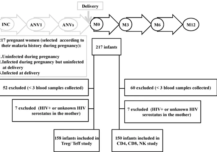
As part of the STOPPAM project, 217 pregnant women were selected on the basis of theirP.
falciparuminfection histories (uninfected during pregnancy, n = 99; infected during pregnancy but uninfected at delivery, n = 71; infected at delivery, n = 47); following delivery, the infants of these mothers were followed-up every two weeks from birth to 12 months of age. The follow-up of infants included routine tests to detectP.falciparum, clinical assessments and growth measurements, details of all of which have been published previously (Fig 1) [46]. To perform immunological studies, 10 ml of cord blood were collected at delivery, while 2 ml peripheral blood samples were obtained at 3, 6 and 12 months of age. Samples were processed at the labo-ratory of the Research Center for Malaria in Pregnancy and Infancy (CERPAGE). Infants for whom fewer than 3 blood samples were collected during the follow-up period were excluded from the study as well as infants whose mothers tested positive for HIV or had an unknown HIV serostatus.
P
.
falciparum
infection status
Clinical and parasitological assessments were performed at each ANV in mothers and during the follow-up of infants. Parasites were detected by thick blood smear (TBS). Briefly, smears were prepared, stained with Giemsa and examined by two experienced laboratory technicians for the presence and density of parasites. Smears were considered negative if no asexual stage
Plasmodiumparasite was detected by counting 500 leucocytes. Parasites were counted against 200 leukocytes and parasite density was estimated assuming 8000 leukocytes/μl of blood. At delivery, TBS were made from peripheral, placental and cord blood samples. Infection withP.
falciparumin the mothers at delivery was defined by the presence of parasites in placental and/ or maternal peripheral blood.
Immunophenotyping
The monoclonal antibodies (mAb) used for T- and NK-cell labeling were the following: anti-CD8-FITC, anti-CD4-PerCP, anti-CD3-APC, anti-CD56-PE antibodies (all BD Biosciences). For Treg and CD4+T effector cell (Teff) labeling CD4-PerCP, CD25-FITC, anti-CD127-PE and anti-Foxp3-APC monoclonal antibodies were used (BD Biosciences and eBioscience).
In a first step, 5μl of FcR blocking reagent (Miltenyi Biotec, Cologne, Germany) were added to 100μl of whole bloodex vivoand incubated for 15 min at 4°C. Cells were then specifically labeled by adding 5μl of each mAb, lightly vortexed and then incubated for 30 min at 4°C in the dark. Two ml of Facs Lysing buffer (BD Biosciences) were then added and cells incubated for a further 15 min at room temperature, in the dark. After spin-washing twice with PBS 3% FCS, at least 200,000 cells were then acquired on a flow cytometer (BD FacsCalibur).
Treg and Teff labeling was performed according to the manufacterer’s recommendation (eBioscience). Briefly, 1 ml whole bloodex vivowas washed with PBS-3% Foetal Bovin Serum (FBS). After washing, 8μl of anti-CD25-FITC, 6μl of CD127-PE and 6μl of anti-CD4-PerCP antibodies were added. Cells were incubated for 25 min at 4°C. Red blood cells were then lysed by adding Facs Lysing buffer and cells incubated for 15 min in the dark. After spin-washing with PBS, cells were incubated overnight with Fix-Perm buffer (eBioscience), then washed with Perm-Wash Buffer (eBioscience). Cells were then incubated in Perm-Wash Buffer with 2% rat serum (eBioscience) for 15 minutes in the dark, followed by the addition of
Malaria Modifies Early-Life Lymphocyte Composition
10μl of anti-Foxp3-APC and incubated for a further 15 minutes in the dark. After two spin-washes with Perm-Wash Buffer and PBS 3% FCS, at least 1,000,000 cells were acquired.
Treg (CD4+CD25+CD127-) and Teff (CD4+CD25+CD127+) cells were gated from the whole lymphocyte population (S1 Fig). CD4+and CD8+were gated on CD3+cells, whilst NK cells were characterized as CD3-CD56+cells and NKT cells were defined as CD3+CD56+cells. For the purposes of our study, NKdimcells were defined as CD3-cells with weak expression of CD56, whereas NKbrightcells were also CD3-but with strong expression of CD56. Levels of expression of Foxp3 were evaluated with a relative value (RV) corresponding to the MFI of Foxp3 in the Treg (or Teff) divided by the MFI of FoxP3 in the naïve T cell population (CD4+CD25-CD127+)
Statistical analysis
The association between T- and NK-cell frequencies and infant age was investigated by using analyses of variances for repeated measures.
To examine the contribution of PAM and/or infant plasmodial infection to T- and NK-cell frequencies, the first set of analyses proceeded as follows: for each phenotype frequency we Fig 1. Flow diagram of birth cohort study.217 pregnant women were enrolled under 24 weeks of gestation and their infants were longitudinally followed-up from birth to 12 months of age. For the Treg/Teff part: 59 newborns were excluded (52 newborns<3 blood samples collected and 7 newborns HIV+ or unknown HIV serostatus in the mother). Data from 158 infants were included for analyses. For the CD4, CD8, NK part: 67 newborns were excluded (60 newborns<3 blood samples collected and 7 newborns HIV+or unknown HIV serostatus in the mother). Data from 150 infants were included for analyses.
built a multivariate linear mixed model (LMM) that takes into account the correlation between repeated measurements as well as other potential confounders in the relationship between infection and the phenotype frequency. We proceeded in two steps; first a univariate model investigated the association between baseline characteristics and T-/NK-cell frequencies. Base-line characteristics were gravidity, maternal anaemia (Hb<11 g/dl), prematurity (<37 weeks),
low birth weight (<2500 g) and infant gender. Variables showing a p-value<0.2 were selected
for the multivariate model (second step).
We built 3 variables related to maternal plasmodial infections as follows: (i) infection before the third trimester of pregnancy, (ii) infection during the third trimester of pregnancy but more than 10 days before delivery, (iii) infection from 10 days prior to delivery up to and including delivery. Designation of the latter group was based on the premise that infections detected during an emergency visit occurring 10 days or less before delivery, and therefore treated, were too close in time to delivery to be separable from it, and because most of those concerned were also found to be infected at delivery.
We also built 3 variables related to infections during infancy as follows: (i) those occurring before three months of age, (ii) those occurring between 3 and 6 months of age, (iii) those occurring between 6 and 12 months of age. All the variables related toP.falciparuminfection were included in the multivariate model with the baseline characteristics selected at the univar-iate step. The final multivarunivar-iate model retained all the variables related toP.falciparum infec-tion along with the baseline characteristics showing significance at the 0.05 level.
To graphically illustrate the predicted effect of maternal infection on the phenotype fre-quencies of infants, we then computed the mean predicted phenotype frefre-quencies of infants born from uninfected mothers at each time-point, as well as those of the same infants if they were born to an infected mother.
In a second set of analyses, the impact of infantP.falciparuminfection on their T-/NK-cell frequencies was examined through three questions. Does infantP.falciparuminfection before 3 months of age have a significant effect on the T-/NK-cell responses at 3 months? Does infant
P.falciparuminfection between 3 and 6 months of age have a significant effect on their T-/NK-cell responses at 6 months? Does infantP.falciparuminfection between 6 and 12 months of age have a significant effect on the T-cell/NK-cell responses at 12 months? To answer each question we employed three linear regression models (at 3 months, 6 months and 12 months). The final models were obtained after adjusting for maternalP.falciparuminfection, gravidity, sex, prematurity and LBW.
In a third set of analyses we aimed to identify whether T-/NK-cell frequencies could affect the occurrence of infection during the ensuing months. To achieve this aim we employed a logistic mixed model where the dependent variable was the time-dependent binary variable
“occurrence of infection between 2 consecutive measurement of the T-/NK-cell frequencies”
(i.e. infection arising before 3 months of age, infection occurring between 3 and 6 months of age, and infection occurring between 7 and 12 months of age). This dependent variable was regressed on the T-/NK-cell frequencies in 2 different ways: in a first model we determined whether phenotype frequencies at birth were associated with the occurrence of infection during the first 12 months of age; in this model the dependent“infection occurence”variable was regressed on the T-/NK-cell frequencies in cord blood. In a second model, we built a time-dependent covariate“T-cell/NK-cell frequencies at the previous measurement”, so that, for infection occurring prior to 3 months of age, we considered the T-/NK-cell frequencies in cord blood cells, for occurrence of infection between 3 and 6 months of age we considered T-/NK-cell frequencies in blood T-/NK-cells obtained at 3 months of age and for occurrence of infection between 7 and 12 months of age we considered T-/NK-cell frequencies in blood cells obtained at 6 months of age. As in the previous analyses, potential confounders were first analyzed in a
Malaria Modifies Early-Life Lymphocyte Composition
univariate model and then in a multivariate model. The models allowed estimation of the adjusted values of the phenotype frequencies (for any set of covariates) of all infants at each time-point.
Statistical significance in all multivariate analyses was considered ifp<0.05. All analyses
were performed using the R statistical package (R Development Core Team; R Foundation for Statistical Computing, Vienna, Austria;http://www.R-project.org) and graphs made with graphPad (Prism 5.0).
Results
Characteristics of the study population
Between November 2008 and April 2011, 217 mother/infant pairs were enrolled in the study
(Fig 1) but of these 59 were excluded due to insufficient numbers of blood samples (52) and
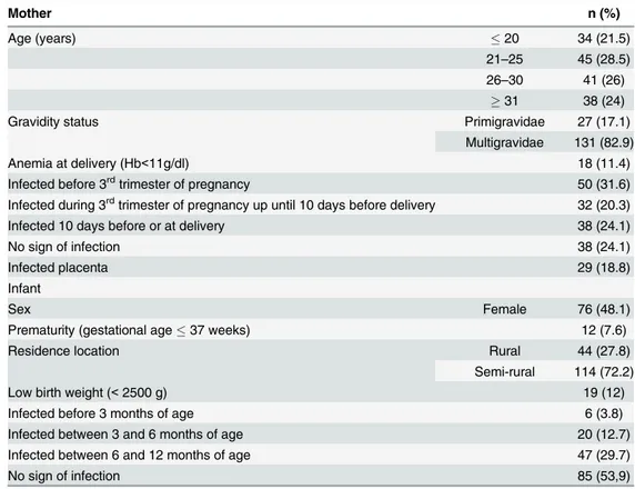
HIV sero-status (7 either HIV+ or unknown). The characteristics of the 158 pairs retained for the analysis presented here are presented inTable 1. Women had a mean age of 26.8 years (95% CI 25.9–27.7), whilst 27 (17.1%) were primigravidae and 18 (11.4%) had anemia (Hb<11g/dL) at delivery.
Infections before the third trimester of pregnancy were detected in 50 (32%) women, whilst 32 (20%) were infected during the third trimester of pregnancy, and in 38 (24%) women infec-tions occurred within 10 days of or at delivery. The latter group of women infected 10 days before or at delivery comprised women infected at delivery and women shown to be infected close to delivery when attending an emergency visit; most of the women belonging to the
Table 1. Characteristics of the study population (n = 158 mother/infant pairs).
Mother n (%)
Age (years) 20 34 (21.5)
21–25 45 (28.5) 26–30 41 (26)
31 38 (24)
Gravidity status Primigravidae 27 (17.1)
Multigravidae 131 (82.9)
Anemia at delivery (Hb<11g/dl) 18 (11.4)
Infected before 3rdtrimester of pregnancy 50 (31.6)
Infected during 3rdtrimester of pregnancy up until 10 days before delivery 32 (20.3)
Infected 10 days before or at delivery 38 (24.1)
No sign of infection 38 (24.1)
Infected placenta 29 (18.8)
Infant
Sex Female 76 (48.1)
Prematurity (gestational age37 weeks) 12 (7.6)
Residence location Rural 44 (27.8)
Semi-rural 114 (72.2)
Low birth weight (<2500 g) 19 (12)
Infected before 3 months of age 6 (3.8)
Infected between 3 and 6 months of age 20 (12.7)
Infected between 6 and 12 months of age 47 (29.7)
No sign of infection 85 (53,9)
latter group were also found to be infected at delivery. Babies' mean birth weight was 3053g (2987–3119).
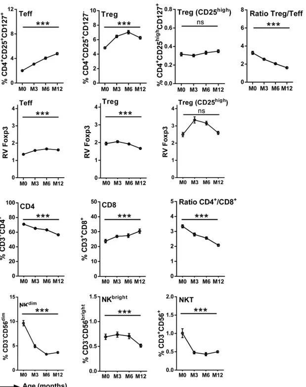
Variations in T- and NK cell frequencies according to age in the first year
of life
There was a significant effect of infant age on the T lymphocyte profiles during the first year of life. Thus the frequency of CD4+cells was higher at birth and decreased with age, while the fre-quency of CD8+cells was lower at birth and increased with age; the CD4+/CD8+ratio conse-quently decreased with age (Fig 2). Despite the overall decline in CD4+cells, the frequencies of Treg and of Teff increased between 0 and 12 months of age, although the Treg/Teff ratio decreased with infant’s age, suggesting that Teff increased more rapidly than Treg (Fig 2). Foxp3 was detected in Treg and to a lesser extent in Teff, with a clearly higher level of expres-sion in Treg. The subset of Treg with the highest levels of expresexpres-sion of CD25 expressed the highest levels of Foxp3 (Fig 2), a key transcription factor that is required for development, maintenance, and function of these cells [47], suggesting a Treg cell subpopulation with specific immune function. With increasing age, the relative expression of Foxp3 increased in Teff and decreased Treg. In the CD25highsubpopulation of Treg the relative level of Foxp3 expression increased from birth to 6 months of age but declined thereafter (Fig 2). The frequencies of all NK cell subpopulations decreased with age (Fig 2). With the exception of the CD25high subpop-ulation of Treg all of these age-dependent variations were shown to be statistically significant (p<0.001) firstly in analyses of variance for repeated measures (Fig 2), and subsequently in
multivariate analyses (Tables2and3).
Thus, we observed variations in the frequency of T- and NK-cell subsets within the first year of life, with an overall decrease of CD4, NKdimand NKT cells and an increase of CD8, Treg and Teff over time.
Maternal
P
.
falciparum
infection at delivery influences the immune cell
profile of infants
Next, we examined whether maternalP.falciparuminfection could influence the frequencies of lymphocytes in the offspring.
Univariate analyses showed thatP.falciparuminfection before the third trimester of preg-nancy was associated only with a decreased frequency of NKdimin infants (data not shown), whilst infection during the third trimester of pregnancy had no effect on any parameter (data not shown). In contrast, maternalP.falciparuminfection at delivery was associated with sev-eral alterations in infant’s lymphocyte populations (S1 Table,Fig 3). Most notable were the increased frequencies of both Treg and of CD8+cells throughout infancy but, conversely, reduced frequencies of both CD4+and NKT cells (Fig 3). Further, maternal infection at deliv-ery was associated with an increased frequency of infant NKdimcells at birth, but no effect was seen on NKbrightfrequencies.
The results of multivariate analyses of the effects of maternal infection at delivery are illus-trated in Tables2and3with a graphical depiction inFig 4of an adjusted model, based on the multivariate LMM analysis. The significantly increased frequencies of CD8+cells in infants at birth, 3 and 6 months of age as well as increased proportions of Treg in infants at 3 months of age were confirmed in the multivariate analyses (Fig 4). In addition, the significantly reduced frequencies of CD4+cells in infants at 3 and 6 months of age and lower NKT cell frequencies at birth, 6 and 12 months of age (Fig 4) were also confirmed. A significant increase in the fre-quency of infant NKdimcells was shown at birth, but significantly lower frequencies were pres-ent at 3 months of age (Fig 4).
Malaria Modifies Early-Life Lymphocyte Composition
In summary, maternal infection at delivery had a significant effect on the profile of immune cells of the infant, with increased frequencies of Treg and CD8+cells and a reduced percentage of CD4+and NKT cells. Accordingly, the CD4/CD8 ratio was also reduced over time in infants born to mothers that were infected close to or at delivery.
Fig 2. Frequencies of cell subsets in infants' peripheral blood during the first year of life.Curves include dots representing mean with SEM (standard error of the mean). The statistical significance of differences indicated refers to analyses of variances for repeated measures.***, p<0.001.
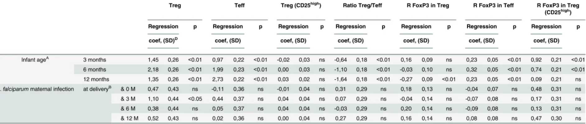
Table 2. Multivariate (LMM) analyses of alterations in circulating T- and NK-cell subset frequencies in peripheral blood as a function of infants’age and ofP.falciparum
infection at delivery.
Treg Teff Treg (CD25high) Ratio Treg/Teff R FoxP3 in Treg R FoxP3 in Teff R FoxP3 in Treg
(CD25high)
Regression p Regression p Regression p Regression p Regression p Regression p Regression p
coef, (SD)D coef, (SD) coef, (SD) coef, (SD) coef, (SD) coef, (SD) coef, (SD)
Infant ageA 3 months 1,45 0,26 <0.01 0,97 0,22 <0.01 -0,02 0,03 ns -0,64 0,18 <0.01 0,16 0,09 ns 0,23 0,05 <0.01 0,92 0,21 <0.01
6 months 2,18 0,26 <0.01 1,99 0,23 <0.01 0,00 0,03 ns -1,10 0,18 <0.01 -0,03 0,10 ns 0,32 0,05 <0.01 0,74 0,21 <0.01
12 months 1,35 0,26 <0.01 2,73 0,22 <0.01 0,03 0,02 ns -1,64 0,18 <0.01 -0,27 0,09 <0.01 0,23 0,05 <0.01 0,09 0,21 ns
P.falciparummaternal infection at deliveryB & 0 M 0,47 0,43 ns -0,11 0,36 ns -0,01 0,04 ns 0,31 0,29 ns 0,18 0,13 ns -0,04 0,07 ns 0,48 0,31 ns
& 3 M 1,10 0,44 <0.05 0,44 0,37 ns 0,04 0,04 ns 0,07 0,29 ns -0,04 0,14 ns -0,07 0,08 ns 0,17 0,31 ns
& 6 M 0,38 0,44 ns 0,05 0,37 ns 0,04 0,04 ns -0,03 0,29 ns 0,20 0,14 ns -0,09 0,08 ns 0,13 0,31 ns
& 12 M 0,52 0,43 ns 0,02 0,36 ns 0,00 0,04 ns 0,27 0,29 ns 0,16 0,14 ns 0,08 0,08 ns 0,47 0,30 ns
AThe reference values used for comparison are those recorded in cord blood (M0);
Bdenotes the in
fluence of infection at delivery/10 days prior to delivery on neonatal/infant responses measured at designated time-points; M0: cord blood, M3, M6, M12: blood drawn at 3, 6 & 12 months of age;
Cdenotes the in
fluence of infection at time-points in the infant on the compostion of neonatal/infant peripheral blood lymphocyte subsets. DPositive/negative coef
ficients indicate cell subset frequencies above/below control (uninfected) levels; SD: standard deviation. All the data were adjusted onP.falciparuminfection history of mother, gravidity, infant age, low birth weight
doi:10.1371/journal.pone.0139606.t002
Malaria
Modifies
Early-Life
Lympho
cyte
Compositio
n
PLOS
ONE
|DOI:10.137
1/journal.p
one.0139606
November
18,
2015
10
CD4 CD8 Ratio CD4/CD8 NKT NKdim
NKbright
Regression coef, (SD)
p Regression coef, (SD)
p Regression coef, (SD)
p Regression coef, (SD)
p Regression coef, (SD)
p Regression coef, (SD)
p
Infant ageA 3 months -0,08 0,02 <0.01 0,11 0,03 <0.01 -0,55 0,12 <0.01 -0,70 0,09 <0.01 -0,48 0,09 <0.01 0,36 0,15 <0.05
6 months -0,11 0,02 <0.01 0,15 0,03 <0.01 -0,79 0,12 <0.01 -0,79 0,09 <0.01 -0,84 0,09 <0.01 0,34 0,15 <0.05
12 months -0,25 0,02 <0.01 0,28 0,03 <0.01 -1,34 0,12 <0.01 -0,54 0,09 <0.01 -0,75 0,10 <0.01 0,01 0,15 ns
P.falciparummaternal infection at deliveryB & 0 M -0,02 0,03 ns 0,15 0,06 <0.05 -0,61 0,21 <0.01 -0,54 0,14 <0.01 0,44 0,14 <0.01 0,31 0,22 ns
& 3 M -0,08 0,03 <0.05 0,17 0,06 <0.05 -0,65 0,22 <0.01 -0,18 0,14 ns -0,28 0,14 <0.05 -0,24 0,23 ns
& 6 M -0,07 0,03 <0.05 0,14 0,06 <0.05 -0,55 0,22 <0.05 -0,28 0,14 <0.05 0,00 0,14 ns -0,11 0,22 ns
& 12 M -0,01 0,04 ns 0,05 0,06 ns -0,25 0,22 ns -0,31 0,14 <0.05 -0,09 0,15 ns 0,30 0,23 ns
AThe reference values used for comparison are those recorded in cord blood (M0);
Bdenotes the in
fluence of infection at delivery/10 days prior to delivery on neonatal/infant responses measured at designated time-points; M0: cord blood, M3, M6, M12: blood drawn at 3, 6 & 12 months of age;
Cdenotes the in
fluence of infection at time-points in the infant on the compostion of neonatal/infant peripheral blood lymphocyte subsets. DPositive/negative coef
ficients indicate cell subset frequencies above/below control (uninfected) levels; SD: standard deviation. All the data were adjusted onP.falciparuminfection history of mother, gravidity, infant age, low birth weight
doi:10.1371/journal.pone.0139606.t003
Malaria
Modifies
Early-Life
Lympho
cyte
Compositio
|DOI:10.137
1/journal.p
one.0139606
November
18,
P
.
falciparum
infection in infants impacts on the immune cell profile
We next evaluated the effect ofP.falciparuminfections in infants on their immune cell profile during the first year of life. (S1 Table&Table 4). We observed that infants infected before 3 months of age had higher frequencies of CD4+and lower frequencies of CD8+cells than those not infected in the same period. In addition, infants infected between 3 and 12 months of age exibited a lower frequency of Treg than those uninfected in the same period as well as a lower frequency of Treg expressing CD25highin infants infected between 3 and 6 months of age by univariate analysis (S1 Table). In the logistic mixed model (Table 4), the impact of infantP. fal-ciparuminfection on their T-/NK-cell frequencies was examined prospectively according to their infection histories 3 months prior to blood sampling at 3, 6 and 12 months. At 3 months, those infected previously had more CD4+and fewer CD8+cells. At 6 months, infants infected previously had lower relative values of FoxP3 in Treg and in TregCD25high. At 12 months infants infected previously had a tendency towards higher frequencies of NKT cells but lower frequencies of Treg (Table 4).
Fig 3. Frequencies of cell subsets in infants' peripheral blood over time segregated on the basis of presence or absence of maternal infection at delivery.(white for negative and hatched for maternal infection). Curves include dots representing mean with SEM (standard error of the mean).
doi:10.1371/journal.pone.0139606.g003
Malaria Modifies Early-Life Lymphocyte Composition
Immunological predictors of the occurrence of
P
.
falciparum
in infants
Finally, we wanted to determine whether the varying frequencies of the cell populations we investigated were associated with a higher probability ofP.falciparuminfection in infancy. Firstly, we assessed whether cord blood cell frequencies were predictive ofP.falciparum
infection during infancy. In univariate analysis, lower cord blood Teff but higher CD4+cell Fig 4. Adjusted profiles of lymphocyte frequencies in infants during the first year of life according toP.falciparumexposurein utero.Values are derived from residuals in the multivariate LMM model. The statistical significance of differences indicated refers to those presented in Tables2and3.*, p
<0.05.
doi:10.1371/journal.pone.0139606.g004
Table 4. A logistic mixed model to evaluate the impact of infantP.falciparuminfection on the frequen-cies of circulating lymphocyte sub-types.
Impact of infant malaria between
0-3M on cellular responses at 3M
Impact of infant malaria between
3-6M on cellular responses at 6M
Impact of infant malaria between 6–12 M on cellular
responses at 12M
Estimate (SD)
p-value
Estimate (SD)
p-value
Estimate (SD)
p-value
Treg Infected -0.88 (0.9) 0.33 -0.59 (0.73) 0.422 -0.91 (0.4) 0.024 Teff Infected 0.26 (0.65) 0.685 -0.38 (0.55) 0.491 -0.44 (0.42) 0.295 Treg CD25high Infected 0.03 (0.07) 0.637 -0.07 (0.05) 0.198 -0.02 (0.03) 0.576 Treg/Teff ratio Infected -0.85 (0.57) 0.136 0.03 (0.25) 0.896 -0.31 (0.2) 0.127 FoxP3 RV in Treg Infected -0.18 (0.31) 0.575 -0.34 (0.12) 0.006 0.06 (0.07) 0.402 FoxP3 RV in Teff Infected -0.04 (0.15) 0.765 -0.13 (0.13) 0.299 -0.08 (0.08) 0.325 FoxP3 RV in Treg
CD25high Infected -0.21 (0.91) 0.819 -0.89 (0.32) 0.006 0.14 (0.20) 0.482
CD4+ Infected 9.66 (4.12) 0.02 3.60 (2.83) 0.21 -1.91 (2.27) 0.4 CD8+ Infected -9.58 (3.66) 0.01 -3.34 (2.24) 0.14 1.37 (1.89) 0.47 NKdim Infected -1.92 (1.56) 0.22 0.26 (0.55) 0.64 0.33 (0.51) 0.52
NKbright Infected 0.37 (0.25) 0.15 -0.03 (0.19) 0.87 0.01 (0.08) 0.91
NKT Infected 0.00 (0.18) 0.99 0.08 (0.16) 0.61 0.14 (0.07) 0.06 Estimate: Positive/negative coefficients indicate cell subsets frequencies above/below control (uninfected) levels. All the data were adjusted onP.falciparuminfection history of mother, gravidity, infant age, low birth weight.SD: standard deviation
frequencies were associated with an increased risk ofP.falciparuminfection during the first year of life (Table 5). In multivariate analysis, only the association with a significantly lower fre-quency of Teff remained.
We next examined the impact of cell frequencies at specific time-points on the occurrence ofP.falciparuminfection in the ensuing months of infancy. Univariate analysis showed that higher proportions of Treg, Teff and CD8+cells and lower proportions of CD4+and NK cells were significantly associated with an increased risk ofP.falciparuminfection in the first year of life. Multivariate analysis showed that lower proportions of Teff, CD4+, NK cells and higher proportions of CD8+cells were significantly associated with an increased risk of developingP.
falciparuminfection during the first year of life (Table 6).
Discussion
The characterization of factors associated with increased susceptibility of infants toP. falcipa-ruminfection during the first year of life is a priority in the development of a vaccine against malaria. Here, we performed a longitudinal study of immune cell profiles in the first year of life of a large cohort of infants born to mothers with varying histories of infection during pregnancy.
Table 5. T and NK cell cord blood frequencies as immunological predictors of malaria in infants.
Univariate Multivariate
OR IC 95% p-value Ajusted OR IC 95% p-value
Treg 0.90 [0.66; 1.22] ns 1.14 [0.80; 1.61] 0.47
Teff 0.55 [0.31; 1.00] 0.04 0.49 [0.26; 0.92] 0.02
CD4+ 1.07 [1.00; 1.15] 0.04 1.03 [0.89; 1.18] 0.69
CD8+ 0.94 [0.87; 1.01] 0.09 0.97 [0.83; 1.13] 0.68
NK (NKdim+ NKbright)
* 0.98 [0.92; 1.05] ns 0.99 [0.92; 1.06] 0.73
Data were adjusted on theP.falciparuminfection history of the mother, gravidity, infant age, low birth weight.
*Results cumulate both NKdimand NKbrightto avoid introducing two variables that are highly correlated into
the model.
doi:10.1371/journal.pone.0139606.t005
Table 6. T-/NK-cell frequencies and the occurrence of malaria during the first 12 months of life.
Univariate Multivariate Model
OR IC 95% p-value Adjusted OR IC 95% p-value
Treg 1.20 [1.08; 1.33] <0.001
Teff 1.29 [1.18; 1.42] <0.001 0.85 [0.74; 0.97] <0.001 CD4+ 0.95 [0.93; 0.97] <0.001
CD8+ 1.05 [1.02; 1.08] <0.001 1.07 [1.02; 1.11] <0.01
NK (NKdim+ NKbright)* 0.87 [0.83; 0.91] <0.001 0.79 [0.73; 0.87] 0.02
Data were adjusted on theP.falciparuminfection history of the mother, gravidity, infant age, low birth weight
*Results cumulate both NKdimand NKbrightto avoid introducing two variables that are highly correlated into
the model.
doi:10.1371/journal.pone.0139606.t006
Malaria Modifies Early-Life Lymphocyte Composition
We observed significant age-related changes in the frequencies of CD4+, CD8+, Treg, Teff, NK and NKT cells from birth to 12 months of age. Those age-related changes were thus taken into consideration in multivariate analyses assessing the impact ofP.falciparumexposurein uteroor infection during the first year of life on infants' immune cell frequencies.
It was, firstly, notable that the CD4+cell frequency was highest at birth and then declined over the ensuing 12 months whilst the reverse was seen for CD8+cell frequencies. The decline of CD4+cell frequency in relation to age contrasts with the stable numbers reported by some [24], but is consistent with the decline seen in older-age children (1–13 years) reported by oth-ers [48,49]. Similarly, the increasing CD8+cell frequency during the first 12 months of life is consistent with some but not all previous reports [24,49]. The disparities in reported findings may simply reflect heterogeneity of the different study populations. Mechnistically, declining numbers of CD4+T cells during the first year of life may be explained by apoptosis [50] an age-dependent phenomenon, as cord blood lymphocytes are less sensitive to TNF-α-induced apo-ptosis than aged T cells [51]. Alternatively or additionally, it could reflect migration of Treg out of the circulation to the gut, which is the primary site of Treg stimulation in responses to exoge-nous antigens during the first 18 month of life through specific homing receptors expressed specifically at birth [52]. Also, it has been reported that neonatal CD8+T cells proliferate more than CD4+T cells as CD8+T cells are more sensitive to IL-7 than CD4+T cells, a process that decreases during the first years of life [53–55].
The fact that the frequencies of Treg and Teff increased from birth to 12 months of age—in marked contrast to the overall decline of CD4+T cells, it should be noted—is likely an indica-tion of the development of infant immunity through the first year of life. The increased Treg frequency and increased relative expression of FoxP3 in the CD25highsubpopulation of Treg during the first year of life are in line with studies reporting increased proportions of FoxP3 T cells in peripheral blood soon after birth compared to cord blood [52,56]. Treg maturation and their suppressive function is FoxP3-dependent. It has been demonstrated that cord blood cells express lower levels of Foxp3 compared to adult cells and that this contributes to the immature nature of neonatal immunity [57–59].
The two functionally distinct NK-cell subpopulations, cytotoxic CD56dimNK cells and cyto-kine-producing CD56brightNK cells, showed contrasting profiles, with a gradual decrease of the NK CD56dimand increasing NK CD56brightduring the first year of life. Others have reported similar findings [24]. NK cells play a major role in innate immune responses that are relatively more important in early life when adaptive immunity has yet to develop, possibly explaining the high frequency of cytotoxic NKdimcells present at birth. NKT cells are present at comparable frequencies in cord blood and adult peripheral blood mononuclear cells [60]. We have not found data other than our own concerning the gradual decrease in NKT frequen-cies from birth to 6 months of age. NKT cellsin uteroare unique in acquiring a memory-acti-vated phenotype through a contact with a natural ligand [42], suggesting that these NKT cells already exert an important immune regulatory function before birth and in self tolerance. Decreased post-natal NKT frequencies may be related to the overall change in the frequency distibution of different lymphocyte phenotypes.
considered essential triggers of early and effective NK-cell IFN-γresponses upon contact with
P.falciparum-infected erythrocytes [61]. They are, furthermore, required for generation and maintenance of protective antibody-mediated responses toP.falciparumand other pathogens [62]. Excessive loss of such cells during infancy may therefore play a pivotal role in the enhanced susceptibility to infection suffered by those born to mothers with placental infection.
The Treg and Teff frequencies were similar in cord blood of newborns born to mothers with
P.falciparuminfection compared to those born to uninfected mothers, again consistent with some previous studies [22,63] but not with others [20,64,65].In uteroexposure toP. falcipa-rumat delivery was nevertheless associated with significantly higher frequencies of Treg at three months of age. Persistence of Treg followingin uterocontact with non-self antigens is a reported phenomenon [30,66]. Several studies have shown thatex vivostimulation of cord blood cells withP.falciparumblood stage antigens reveals populations of CD4+T cells with a Treg phenotype [67]. Such cells, in cord blood of offspring born to women with placental malaria, have been shown to produce more IL-10 compared to cells of those born to mothers without placental malaria [64,65]. Compatible with these findings, others have shown that a soluble extract ofP.falciparum-infected red blood cells induces the differentiation of polyclon-ally-activated Treg CD4+T cells with strong suppressive activity, and that this activation was mediated by membrane bound TGF-βon the Treg cells leading to immune evasion and reduced pro-inflammatory responses [29].
Resource constraints meant that we were unable to assess the functional characteristics of the different T cell subsets that we quantified, but we hypothesize that higher numbers of Treg in infants born to mothers infected at delivery would be associated with enhanced immunoreg-ulatory activity and with concomitantly reduced pro-inflammatory responses. Such a profile would likely contribute to the increased susceptibility to infections observed in children born to infected mothers [13–15,20]. Treg induced during infection are thought to limit the magni-tude of subsequent parasite-specific IFN-γresponses [68]. Prenatal exposure to plasmodial blood-stage antigens induces Treg that, in some newborns, primarily suppress Th1- type recall responses [2,20]. Thus, the persistence of such Treg post-natally may affect the susceptibility of children both toP.falciparumand to other infections. We did not observe any alteration of the relative expression of FoxP3 in Treg or Teff. It could have been more informative to deter-mine the expression of other functional markers such as CTLA-4 and/or TNFRII. It is known that Treg can dampen NK-cell activation/ proliferation and cytotoxic activity [69,70] as well as NKT cell proliferation and cytokine production [39]. In line with these observations, and in association with the increased Treg frequency in infants born to mothers infected at delivery, we found a reduced frequency of circulating NKT cells in infants of mothers infected withP.
falciparumclose to or at delivery as compared to the other group at birth. Further, maternal infection close to or at delivery was associated with an increased frequency of infant NKdim cells at birth, and to lower frequencies of these cells at 3 months of age. These observations are suggestive of an early inflammatory response being needed to protect against plasmodial para-sites. Relatively diminished pro-inflammatory activity—as reflected by cord plasma cytokine profiles—is associated with more severe clinical outcomes of infection withP.falciparum dur-ing infancy [71,72]. Assessing plasma cytokine profiles in our cohort of infants is thus of obvi-ous interest in the context of the findings we present here as well as those we have previobvi-ously reported [73].
Infection withP.falciparumin infancy also altered the immune cell profile, with infants infected before 3 months of age having a higher frequency of CD4+T cells and a lower fre-quency of CD8+T cells. It should be stressed that this, again, is a pattern superimposed on what is, in this case, the markedly contrasting background of an overall age-related decline in CD4+cells and an increase in CD8+cells, a pattern that, as was noted earlier, placental
Malaria Modifies Early-Life Lymphocyte Composition
infection at delivery further exacerbated. Again, we have no specific functional data on which to base any interpretation of this observation. It is nevertheless plausible to suppose that any infection-induced expansion of a population of CD4+T cells in such infants will primarily comprise atypical, transitional cells that have not undergone full maturation. Theory suggests that such effector-type cells induced in infancy remain in a state of anergy with little if any functional anti-pathogen activity [74].
Reduced Treg frequencies were observed in infants who were infected between 6 and 12 months of age, and the relative expression of FoxP3 in the CD4+CD25+and CD25highCD4+ subpopulation was reduced in infants who were infected between 3 and 6 months of age. These findings contrast with the increase in number of Treg in the peripheral blood of older children and adults duringP.falciparuminfections [26–29]. The data we present here comprise obser-vations after the infection event, however, and may thus simply reflect the physiological con-traction of pathogen-induced responses following clinical recovery and clearance of the parasite.
One of the major limitations of our study concerns the absence of data with respect to co-infections such as, for example, chronic maternal helminth co-infections. Such co-infections are known to affect anti-plasmodial immune responses [75–77]. Furthermore, contactin utero
with maternally-derived filariasis antigens reportedly modifies cord blood cell responses such that they display a predominantly anti-inflammatory profile [78]. On the other hand, the prev-alence of (soil-transmitted) helminth infections in the first 12 months of life is likely less than 10% [79], and thus any possible influence on the lymphocyte profiles we observed here is likely to have been minimal.
In conclusion, we have found that maternal infection withP.falciparumat delivery, but not earlier in pregnancy profoundly affects the immune cell profile of their babies, with, in some cases, effects lasting at least 1 year. Cells of both the innate and adaptive arms of the immune system are affected. These findings echo our observations on TLR-mediated innate immune responses of the same group of infants [73]. Infection at delivery, but not earlier in pregnancy, was associated with significantly higher TLR3-mediated IL-6 and IL-10 responses in the first 3 months of life, and with significantly higher TLR3-/TLR7/8-/TLR9-mediated TNF-αand TLR9-mediated IL-10 responses at 6 or 12 months of age. Whether those alterations, along with the maternal infection-related relative increase in CD8+cells and relative decrease in CD4+cells in the post-natal period that we report here, have a direct bearing on infants' immune response to infection remains an open question. Plausibly, such changes could form the basis for an explanation of the increased susceptibility to malaria of infants born to mothers with infections at delivery, but proof of causality awaits further study.
Supporting Information
S1 Fig. The gating strategies for Treg (CD4+CD25+CD127-) and Teff (CD4+CD25+CD127+). Cell frequencies were determined as a percentage from the whole lymphocyte population, and relative FoxP3 expression level determined as a function of FoxP3 expresssion by naïve CD4+T cells (CD4+CD25-).
(TIFF)
S1 Table. Univariate analysis of alterations in circulating T- and NK-cell subset frequencies in cord/infant blood as a function ofP.falciparuminfection detected either in the mother at delivery or during infancy
Acknowledgments
We are grateful to all women and infant who participated in the study. We thank all the field and administration staffs of Akodeha, Comé central and Ouedèmé Pedah health centers for their valuable contribution. We particularly thank Jacqueline Affedjou, Jean-Claude Sagbo, Bernadette Gandounou, Gildas Gbaguidi and all field staff in the site of STOPPAM for their hard work and dedication to this study. We would like to thank Bich-Tram Huynh, Sebastien Dechavanne and Valérie Briand for database management, Aurax Fernando, Charles Ahouan-sou, Pépin Kounou, Honoré Kounou, and Darius Sossou for their lab contribution; Thor Theander and Nicaise Ndam for their contribution to the design of the study.
Author Contributions
Conceived and designed the experiments: ON KG SI AM PD KM MTB AJFL NF. Performed the experiments: ON KG SI BV SE SB CA NF. Analyzed the data: PH GC. Contributed reagents/materials/analysis tools: ON KG SI BV SE CA NF. Wrote the paper: ON KG SI SV MTB PD AJFL NF. Designed and supervised the immunoassays: ON KG SI AJFL NF. Carried out the immunoassays: ON KG SI BV SE CA NF.
References
1. Labeaud AD, Malhotra I, King MJ, King CL, King CH. Do antenatal parasite infections devalue child-hood vaccination? PLoS neglected tropical diseases. 2009; 3(5):e442. Epub 2009/05/30. doi:10.1371/ journal.pntd.0000442PMID:19478847; PubMed Central PMCID: PMC2682196.
2. Malhotra I, Dent A, Mungai P, Wamachi A, Ouma JH, Narum DL, et al. Can prenatal malaria exposure produce an immune tolerant phenotype? A prospective birth cohort study in Kenya. PLoS Med. 2009; 6 (7):e1000116. Epub 2009/07/29. doi:10.1371/journal.pmed.1000116PMID:19636353; PubMed Cen-tral PMCID: PMC2707618.
3. Dauby N, Goetghebuer T, Kollmann TR, Levy J, Marchant A. Uninfected but not unaffected: chronic maternal infections during pregnancy, fetal immunity, and susceptibility to postnatal infections. The Lancet infectious diseases. 2012; 12(4):330–40. Epub 2012/03/01. S1473-3099(11)70341-3 [pii] doi: 10.1016/S1473-3099(11)70341-3PMID:22364680.
4. Thornton CA, Macfarlane TV, Holt PG. The hygiene hypothesis revisited: role of materno-fetal interac-tions. Current allergy and asthma reports. 2010; 10(6):444–52. Epub 2010/09/03. doi: 10.1007/s11882-010-0148-5PMID:20809222.
5. Cuna WR, Choque AG, Passera R, Rodriguez C. Pro-inflammatory cytokine production in chagasic mothers and their uninfected newborns. J Parasitol. 2009; 95(4):891–4. Epub 2009/01/24. GE-1927 [pii] doi:10.1645/GE-1927.1PMID:19161249.
6. Kurtis JD, Higashi A, Wu HW, Gundogan F, McDonald EA, Sharma S, et al. Maternal Schistosomiasis japonica is associated with maternal, placental, and fetal inflammation. Infection and immunity. 2011; 79(3):1254–61. Epub 2010/12/15. IAI.01072-10 [pii] doi:10.1128/IAI.01072-10PMID:21149589; PubMed Central PMCID: PMC3067505.
7. Malhotra I, Mungai PL, Wamachi AN, Tisch D, Kioko JM, Ouma JH, et al. Prenatal T cell immunity to Wuchereria bancrofti and its effect on filarial immunity and infection susceptibility during childhood. The Journal of infectious diseases. 2006; 193(7):1005–13. Epub 2006/03/07. doi:10.1086/500472PMID: 16518763.
8. Vekemans J, Truyens C, Torrico F, Solano M, Torrico MC, Rodriguez P, et al. Maternal Trypanosoma cruzi infection upregulates capacity of uninfected neonate cells To produce pro- and anti-inflammatory cytokines. Infection and immunity. 2000; 68(9):5430–4. Epub 2000/08/19. PMID:10948177; PubMed Central PMCID: PMC101811.
9. Brabin BJ, Romagosa C, Abdelgalil S, Menendez C, Verhoeff FH, McGready R, et al. The sick pla-centa-the role of malaria. Placenta. 2004; 25(5):359–78. Epub 2004/04/15. S0143400403003072 [pii]. PMID:15081631.
10. Desai M, ter Kuile FO, Nosten F, McGready R, Asamoa K, Brabin B, et al. Epidemiology and burden of malaria in pregnancy. The Lancet infectious diseases. 2007; 7(2):93–104. Epub 2007/01/26. S1473-3099(07)70021-X [pii] PMID:17251080.
11. Kalanda BF, van Buuren S, Verhoeff FH, Brabin BJ. Catch-up growth in Malawian babies, a longitudinal study of normal and low birthweight babies born in a malarious endemic area. Early human
Malaria Modifies Early-Life Lymphocyte Composition
development. 2005; 81(10):841–50. Epub 2005/08/20. doi:10.1016/j.earlhumdev.2005.06.006PMID: 16109465.
12. Walther B, Miles DJ, Crozier S, Waight P, Palmero MS, Ojuola O, et al. Placental malaria is associated with reduced early life weight development of affected children independent of low birth weight. Malar J. 2010; 9:16. Epub 2010/01/16. doi:10.1186/1475-2875-9-16PMID:20074331; PubMed Central PMCID: PMC2841609.
13. Le Hesran JY, Cot M, Personne P, Fievet N, Dubois B, Beyeme M, et al. Maternal placental infection with Plasmodium falciparum and malaria morbidity during the first 2 years of life. Am J Epidemiol. 1997; 146(10):826–31. Epub 1997/12/31. PMID:9384203.
14. Mutabingwa TK, Bolla MC, Li JL, Domingo GJ, Li X, Fried M, et al. Maternal malaria and gravidity inter-act to modify infant susceptibility to malaria. PLoS Med. 2005; 2(12):e407. Epub 2005/11/02. 05-PLME-RA-0127R3 [pii] doi:10.1371/journal.pmed.0020407PMID:16259531; PubMed Central PMCID: PMC1277932.
15. Rachas A, Le Port A, Cottrell G, Guerra J, Choudat I, Bouscaillou J, et al. Placental Malaria is Associ-ated With Increased Risk of Nonmalaria Infection During the First 18 Months of Life in a Beninese Popu-lation. Clin Infect Dis. 2012. Epub 2012/05/23. cis490 [pii] doi:10.1093/cid/cis490PMID:22610927.
16. Adegnika AA, Kohler C, Agnandji ST, Chai SK, Labuda L, Breitling LP, et al. Pregnancy-associated malaria affects toll-like receptor ligand-induced cytokine responses in cord blood. The Journal of infec-tious diseases. 2008; 198(6):928–36. Epub 2008/08/08. doi:10.1086/591057PMID:18684097.
17. Fievet N, Varani S, Ibitokou S, Briand V, Louis S, Perrin RX, et al. Plasmodium falciparum exposure in utero, maternal age and parity influence the innate activation of foetal antigen presenting cells. Malar J. 2009; 8:251. Epub 2009/11/06. 1475-2875-8-251 [pii] doi:10.1186/1475-2875-8-251PMID:19889240; PubMed Central PMCID: PMC2780449.
18. Walther B, Miles DJ, Waight P, Palmero MS, Ojuola O, Touray ES, et al. Placental malaria is associated with attenuated CD4 T-cell responses to tuberculin PPD 12 months after BCG vaccination. BMC infec-tious diseases. 2012; 12:6. Epub 2012/01/17. doi:10.1186/1471-2334-12-6PMID:22243970; PubMed Central PMCID: PMC3274427.
19. Broen K, Brustoski K, Engelmann I, Luty AJ. Placental Plasmodium falciparum infection: causes and consequences of in utero sensitization to parasite antigens. Mol Biochem Parasitol. 2007; 151(1):1–8. Epub 2006/11/04. S0166-6851(06)00286-6 [pii] doi:10.1016/j.molbiopara.2006.10.001PMID: 17081634.
20. Mackroth MS, Malhotra I, Mungai P, Koech D, Muchiri E, King CL. Human cord blood CD4+CD25hi reg-ulatory T cells suppress prenatally acquired T cell responses to Plasmodium falciparum antigens. J Immunol. 2011; 186(5):2780–91. Epub 2011/02/01. jimmunol.1001188 [pii] doi:10.4049/jimmunol. 1001188PMID:21278348.
21. Malhotra I, Mungai P, Muchiri E, Ouma J, Sharma S, Kazura JW, et al. Distinct Th1- and Th2-Type pre-natal cytokine responses to Plasmodium falciparum erythrocyte invasion ligands. Infection and immu-nity. 2005; 73(6):3462–70. Epub 2005/05/24. PMID:15908375; PubMed Central PMCID:
PMC1111871.
22. Flanagan KL, Halliday A, Burl S, Landgraf K, Jagne YJ, Noho-Konteh F, et al. The effect of placental malaria infection on cord blood and maternal immunoregulatory responses at birth. European journal of immunology. 2010; 40(4):1062–72. Epub 2009/12/30. doi:10.1002/eji.200939638PMID:20039298.
23. Reichert T, DeBruyere M, Deneys V, Totterman T, Lydyard P, Yuksel F, et al. Lymphocyte subset refer-ence ranges in adult Caucasians. Clin Immunol Immunopathol. 1991; 60(2):190–208. Epub 1991/08/ 01. PMID:1712687.
24. Sundstrom Y, Nilsson C, Lilja G, Karre K, Troye-Blomberg M, Berg L. The expression of human natural killer cell receptors in early life. Scandinavian journal of immunology. 2007; 66(2–3):335–44. Epub 2007/07/20. SJI1980 [pii] doi:10.1111/j.1365-3083.2007.01980.xPMID:17635811.
25. Saule P, Trauet J, Dutriez V, Lekeux V, Dessaint JP, Labalette M. Accumulation of memory T cells from childhood to old age: central and effector memory cells in CD4(+) versus effector memory and termi-nally differentiated memory cells in CD8(+) compartment. Mechanisms of ageing and development. 2006; 127(3):274–81. Epub 2005/12/15. S0047-6374(05)00275-7 [pii] PMID:16352331.
26. Minigo G, Woodberry T, Piera KA, Salwati E, Tjitra E, Kenangalem E, et al. Parasite-dependent expan-sion of TNF receptor II-positive regulatory T cells with enhanced suppressive activity in adults with severe malaria. PLoS pathogens. 2009; 5(4):e1000402. Epub 2009/04/25. doi:10.1371/journal.ppat. 1000402PMID:19390618; PubMed Central PMCID: PMC2668192.
28. Walther M, Tongren JE, Andrews L, Korbel D, King E, Fletcher H, et al. Upregulation of TGF-beta, FOXP3, and CD4+CD25+ regulatory T cells correlates with more rapid parasite growth in human malaria infection. Immunity. 2005; 23(3):287–96. Epub 2005/09/20. S1074-7613(05)00245-1 [pii] PMID:16169501.
29. Clemente A, Caporale R, Sannella AR, Majori G, Severini C, Fadigati G, et al. Plasmodium falciparum soluble extracts potentiate the suppressive function of polyclonal T regulatory cells through activation of TGFbeta-mediated signals. Cell Microbiol. 2011; 13(9):1328–38. Epub 2011/06/28. doi:10.1111/j. 1462-5822.2011.01622.xPMID:21699642.
30. Mold JE, Michaelsson J, Burt TD, Muench MO, Beckerman KP, Busch MP, et al. Maternal alloantigens promote the development of tolerogenic fetal regulatory T cells in utero. Science. 2008; 322
(5907):1562–5. Epub 2008/12/06. doi:10.1126/science.1164511PMID:19056990; PubMed Central PMCID: PMC2648820.
31. Moore KW, de Waal Malefyt R, Coffman RL, O'Garra A. Interleukin-10 and the interleukin-10 receptor. Annu Rev Immunol. 2001; 19:683–765. Epub 2001/03/13. 19/1/683 [pii] doi:10.1146/annurev. immunol.19.1.683PMID:11244051.
32. Levings MK, Sangregorio R, Galbiati F, Squadrone S, de Waal Malefyt R, Roncarolo MG. IFN-alpha and IL-10 induce the differentiation of human type 1 T regulatory cells. J Immunol. 2001; 166(9):5530– 9. Epub 2001/04/21. PMID:11313392.
33. Ralainirina N, Poli A, Michel T, Poos L, Andres E, Hentges F, et al. Control of NK cell functions by CD4 +CD25+ regulatory T cells. Journal of leukocyte biology. 2007; 81(1):144–53. Epub 2006/09/09. PMID: 16959895.
34. Farag SS, Caligiuri MA. Human natural killer cell development and biology. Blood Rev. 2006; 20 (3):123–37. Epub 2005/12/21. S0268-960X(05)00055-X [pii] PMID:16364519.
35. Dalle JH, Menezes J, Wagner E, Blagdon M, Champagne J, Champagne MA, et al. Characterization of cord blood natural killer cells: implications for transplantation and neonatal infections. Pediatr Res. 2005; 57(5 Pt 1):649–55. Epub 2005/02/19. 01.PDR.0000156501.55431.20 [pii] doi:10.1203/01.PDR. 0000156501.55431.20PMID:15718362.
36. Horowitz A, Newman KC, Evans JH, Korbel DS, Davis DM, Riley EM. Cross-talk between T cells and NK cells generates rapid effector responses to Plasmodium falciparum-infected erythrocytes. J Immu-nol. 2010; 184(11):6043–52. Epub 2010/04/30. doi:10.4049/jimmunol.1000106PMID:20427769.
37. Newman KC, Korbel DS, Hafalla JC, Riley EM. Cross-talk with myeloid accessory cells regulates human natural killer cell interferon-gamma responses to malaria. PLoS pathogens. 2006; 2(12):e118. Epub 2006/12/13. PMID:17154717; PubMed Central PMCID: PMC1687207.
38. Othoro C, Moore JM, Wannemuehler KA, Moses S, Lal A, Otieno J, et al. Elevated gamma interferon-producing NK cells, CD45RO memory-like T cells, and CD4 T cells are associated with protection against malaria infection in pregnancy. Infection and immunity. 2008; 76(4):1678–85. Epub 2008/02/ 06. doi:10.1128/IAI.01420-07PMID:18250175; PubMed Central PMCID: PMC2292852.
39. Azuma T, Takahashi T, Kunisato A, Kitamura T, Hirai H. Human CD4+ CD25+ regulatory T cells sup-press NKT cell functions. Cancer research. 2003; 63(15):4516–20. Epub 2003/08/09. PMID: 12907625.
40. Watanabe H, Weerasinghe A, Miyaji C, Sekikawa H, Toyabe S, Mannor MK, et al. Expansion of uncon-ventional T cells with natural killer markers in malaria patients. Parasitology international. 2003; 52 (1):61–70. Epub 2003/01/25. PMID:12543148.
41. Pied S, Roland J, Louise A, Voegtle D, Soulard V, Mazier D, et al. Liver CD4-CD8- NK1.1+ TCR alpha beta intermediate cells increase during experimental malaria infection and are able to exhibit inhibitory activity against the parasite liver stage in vitro. J Immunol. 2000; 164(3):1463–9. Epub 2000/01/21. PMID:10640763.
42. van Der Vliet HJ, Nishi N, de Gruijl TD, von Blomberg BM, van den Eertwegh AJ, Pinedo HM, et al. Human natural killer T cells acquire a memory-activated phenotype before birth. Blood. 2000; 95 (7):2440–2. Epub 2000/03/25. PMID:10733519.
43. Harner S, Noessner E, Nadas K, Leumann-Runge A, Schiemann M, Faber FL, et al. Cord blood Val-pha24-Vbeta11 natural killer T cells display a Th2-chemokine receptor profile and cytokine responses. PloS one. 2011; 6(1):e15714. Epub 2011/02/10. doi:10.1371/journal.pone.0015714PMID:21305060; PubMed Central PMCID: PMC3031538.
44. Huynh BT, Fievet N, Gbaguidi G, Borgella S, Mevo BG, Massougbodji A, et al. Malaria associated symptoms in pregnant women followed-up in Benin. Malar J. 2011; 10:72. Epub 2011/04/02. 1475-2875-10-72 [pii] doi:10.1186/1475-2875-10-72PMID:21453493; PubMed Central PMCID: PMC3076273.
45. Le Port A, Watier L, Cottrell G, Ouedraogo S, Dechavanne C, Pierrat C, et al. Infections in infants during the first 12 months of life: role of placental malaria and environmental factors. PloS one. 2011; 6(11):
Malaria Modifies Early-Life Lymphocyte Composition
e27516. Epub 2011/11/19. doi:10.1371/journal.pone.0027516PONE-D-11-02896 [pii]. PMID: 22096588; PubMed Central PMCID: PMC3214070.
46. Borgella S, Fievet N, Huynh BT, Ibitokou S, Hounguevou G, Affedjou J, et al. Impact of pregnancy-associated malaria on infant malaria infection in southern Benin. PloS one. 2013; 8(11):e80624. Epub 2013/11/16. doi:10.1371/journal.pone.0080624PMID:24236190; PubMed Central PMCID: PMC3827421.
47. Hori S, Takahashi T, Sakaguchi S. Control of autoimmunity by naturally arising regulatory CD4+ T cells. Advances in immunology. 2003; 81:331–71. Epub 2004/01/09. PMID:14711059.
48. Comans-Bitter WM, de Groot R, van den Beemd R, Neijens HJ, Hop WC, Groeneveld K, et al. Immuno-phenotyping of blood lymphocytes in childhood. Reference values for lymphocyte subpopulations. The Journal of pediatrics. 1997; 130(3):388–93. Epub 1997/03/01. S0022-3476(97)70200-2 [pii]. PMID: 9063413.
49. Shahabuddin S, Al-Ayed I, Gad El-Rab MO, Qureshi MI. Age-related changes in blood lymphocyte sub-sets of Saudi Arabian healthy children. Clinical and diagnostic laboratory immunology. 1998; 5(5):632– 5. Epub 1998/09/08. PMID:9729529; PubMed Central PMCID: PMC95633.
50. Gupta S, Gollapudi S. Molecular mechanisms of TNF-alpha-induced apoptosis in aging human T cell subsets. Int J Biochem Cell Biol. 2005; 37(5):1034–42. Epub 2005/03/04. S1357-2725(04)00340-1 [pii] PMID:15743676.
51. Aggarwal S, Gollapudi S, Yel L, Gupta AS, Gupta S. TNF-alpha-induced apoptosis in neonatal lympho-cytes: TNFRp55 expression and downstream pathways of apoptosis. Genes and immunity. 2000; 1 (4):271–9. Epub 2001/02/24. PMID:11196704.
52. Grindebacke H, Stenstad H, Quiding-Jarbrink M, Waldenstrom J, Adlerberth I, Wold AE, et al. Dynamic development of homing receptor expression and memory cell differentiation of infant CD4+CD25high regulatory T cells. J Immunol. 2009; 183(7):4360–70. Epub 2009/09/08. jimmunol.0901091 [pii] doi:10. 4049/jimmunol.0901091PMID:19734224.
53. Fry TJ, Mackall CL. The many faces of IL-7: from lymphopoiesis to peripheral T cell maintenance. J Immunol. 2005; 174(11):6571–6. Epub 2005/05/21. PMID:15905493.
54. Schonland SO, Zimmer JK, Lopez-Benitez CM, Widmann T, Ramin KD, Goronzy JJ, et al. Homeostatic control of T-cell generation in neonates. Blood. 2003; 102(4):1428–34. Epub 2003/04/26. PMID: 12714521.
55. Smith NL, Wissink E, Wang J, Pinello JF, Davenport MP, Grimson A, et al. Rapid proliferation and dif-ferentiation impairs the development of memory CD8+ T cells in early life. J Immunol. 2014; 193 (1):177–84. Epub 2014/05/23. doi:10.4049/jimmunol.1400553PMID:24850719; PubMed Central PMCID: PMC4065808.
56. Fuchizawa T, Adachi Y, Ito Y, Higashiyama H, Kanegane H, Futatani T, et al. Developmental changes of FOXP3-expressing CD4+CD25+ regulatory T cells and their impairment in patients with FOXP3 gene mutations. Clin Immunol. 2007; 125(3):237–46. Epub 2007/10/06. PMID:17916446.
57. Fujimaki W, Takahashi N, Ohnuma K, Nagatsu M, Kurosawa H, Yoshida S, et al. Comparative study of regulatory T cell function of human CD25CD4 T cells from thymocytes, cord blood, and adult peripheral blood. Clinical & developmental immunology. 2008; 2008:305859. Epub 2008/09/26. doi:10.1155/ 2008/305859PMID:18815628; PubMed Central PMCID: PMC2547481.
58. Tang J, Li BQ, Ruan J, Zhang LJ. [Expression of Foxp3 in CD4+CD25high Treg cells of neonatal cord blood]. Xi Bao Yu Fen Zi Mian Yi Xue Za Zhi. 2008; 24(4):365–7. Epub 2008/04/09. PMID:18394345.
59. Hrdy J, Kocourkova I, Prokesova L. Impaired function of regulatory T cells in cord blood of children of allergic mothers. Clinical and experimental immunology. 2012; 170(1):10–7. Epub 2012/09/05. doi:10. 1111/j.1365-2249.2012.04630.xPMID:22943196; PubMed Central PMCID: PMC3444712.
60. D'Andrea A, Goux D, De Lalla C, Koezuka Y, Montagna D, Moretta A, et al. Neonatal invariant Val-pha24+ NKT lymphocytes are activated memory cells. European journal of immunology. 2000; 30 (6):1544–50. Epub 2000/07/18. PMID:10898489.
61. Horowitz A, Riley EM. Activation of human NK cells by malaria-infected red blood cells. Methods Mol Biol. 2010; 612:429–46. Epub 2009/12/25. doi:10.1007/978-1-60761-362-6_29PMID:20033658.
62. Bisseye C, Yindom LM, Simpore J, Morgan WD, Holder AA, Ismaili J. An engineered Plasmodium fal-ciparum C-terminal 19-kilodalton merozoite surface protein 1 vaccine candidate induces high levels of interferon-gamma production associated with cellular immune responses to specific peptide sequences in Gambian adults naturally exposed to malaria. Clinical and experimental immunology. 2011; 166 (3):366–73. Epub 2011/11/09. doi:10.1111/j.1365-2249.2011.04467.xPMID:22059995; PubMed Central PMCID: PMC3232385.