Inflammation by Bifidobacterium Breve Soluble Factors
Elise Heuvelin1,2, Corinne Lebreton1,2, Corinne Grangette3, Bruno Pot3, Nadine Cerf-Bensussan1,2, Martine Heyman1,2*
1INSERM U793, Paris, France,2Universite´ Paris Descartes, Faculte´ de Me´decine Rene´ Descartes, IFR94, Paris, France,3Laboratoire des Bacte´ries Lactiques et Immunite´ des Muqueuses, Institut Pasteur de Lille, IFR 142, Lille, France
Abstract
Objectives:Soluble factors released byBifidobacterium breveC50 (Bb) alleviate the secretion of pro-inflammatory cytokines by immune cells, but their effect on intestinal epithelium remains elusive. To decipher the mechanisms accounting for the cross-talk between bacteria/soluble factors and intestinal epithelium, we measured the capacity of the bacteria, its conditioned medium (Bb-CM) and other Gram(+) commensal bacteria to dampen inflammatory chemokine secretion.
Methods:TNFa-induced chemokine (CXCL8) secretion and alteration of NF-kB and AP-1 signalling pathways byBbwere studied by EMSA, confocal microscopy and western blotting. Anti-inflammatory capacity was also testedin vivoin a model of TNBS-induced colitis in mice.
Results: Bb and Bb-CM, but not other commensal bacteria, induced a time and dose-dependent inhibition of CXCL8 secretion by epithelial cells driven by both AP-1 and NF-kB transcription pathways and implying decreased phosphorylation of p38-MAPK and IkB-amolecules. In TNBS-induced colitis in mice,Bb-CM decreased the colitis score and inflammatory cytokine expression, an effect reproduced by dendritic cell conditioning withBb-CM.
Conclusions: Bband secreted soluble factors contribute positively to intestinal homeostasis by attenuating chemokine production. The results indicate thatBbdown regulate inflammation at the epithelial level by inhibiting phosphorylations involved in inflammatory processes and by protective conditioning of dendritic cells.
Citation:Heuvelin E, Lebreton C, Grangette C, Pot B, Cerf-Bensussan N, et al. (2009) Mechanisms Involved in Alleviation of Intestinal Inflammation by Bifidobacterium Breve Soluble Factors. PLoS ONE 4(4): e5184. doi:10.1371/journal.pone.0005184
Editor:Stefan Bereswill, Charite´-Universita¨tsmedizin Berlin, Germany
ReceivedFebruary 12, 2009;AcceptedMarch 11, 2009;PublishedApril 17, 2009
Copyright:ß2009 Heuvelin et al. This is an open-access article distributed under the terms of the Creative Commons Attribution License, which permits unrestricted use, distribution, and reproduction in any medium, provided the original author and source are credited.
Funding:This work was funded by INSERM, the Princess Grace Foundation, Bledina-SA and the Fondation pour la Recherche Medicale. The funders had no role in study design, data collection and analysis, decision to publish, or preparation of the manuscript.
Competing Interests:The authors have declared that no competing interests exist. * E-mail: martine.heyman@inserm.fr
Introduction
Intestinal homeostasis depends in part on the equilibrium between absorption (nutrients, Na+
ions), secretion (chloride ions, IgA, mucus, cytokines) and barrier function of the digestive epithelium. Disturbance of intestinal homeostasis results in chronic inflammation and release of pro-inflammatory cytokines resulting in diarrhoea and injury to the gut. The intestinal epithelium constitutes a major interface between intestinal contents, including bacteria, and the internal milieu, providing an important contribution to the regulation of intestinal homeostasis. Probiotics are specific bacterial strains capable of stimulating protective immune responses in physiological conditions[1] but which also dampen inflammation in some inflammatory bowel diseases [2].
One of the main functions of the intestinal epithelium is to regulate the ion transport controlling the balance between absorption and secretion of fluid. Intestinal epithelial cells (IECs) are considered an integral part of the innate immune system, constituting important targets for bacteria and cytokines. Their polarization contributes to the fine regulation of intestinal homeostasis [3–5]. In the physiological steady-state, minimal
stimulation of IECs by luminal bacteria occurs at the apical pole. Indeed, pathogen recognition receptors (PRRs) are functionally silenced in the healthy intestine due to their redistribution to internal or basolateral compartments or to the delivery of inhibitory signals [6,7]. In contrast, in pathology, recognition of invading bacteria promotes signalling cascades of pro-inflamma-tory cytokines and chemokines[8] and the recruitment/activation of mucosal immune cells. In this context, selected strains of probiotic bacteria have been proposed as tools in the prevention or treatment of inflammatory bowel diseases, especially in ulcerative colitis [2]. Mechanisms sustaining such beneficial effects have been partially identified in vitro[9,10] and depend mainly upon alleviation of NF-kB-dependent transcriptional activity. We have already demonstrated that soluble factors released by the probiotic bacteria Bifidobacterium breve (Bb) C50 can down-regulate the production of inflammatory cytokines by immune cells [11], with these factors maintaining their inhibitory activity after crossing an epithelial barrier. Herein, we demonstrated the inhibitory effect of
Methods
Bacteria and conditioned media
Bifidobacterium breve (Bb) C50 was purchased from Bledina-SA (Steenvoorde, France).Bifidobacterium breve 15698 andLactobacillus rhamnosus 10893 were purchased from ATCC, and Eubacterium rectale L15, isolated from a dominant human microflora, was provided by G. Corthier (INRA, Jouy en Josas, France). Bacteria cultured in BH medium were seeded for 24 hours in DMEM (Gibco) containing 10% fetal calf serum (FCS) and 2% inulin.Bb -conditioned-media corresponding to 36108 CFU/ml were prepared by ultracentrifugation (100,000g, 1 h) and adjusted to pH 7.0. As demonstrated with immune cells, active soluble factors in Bb-conditioned media are less than 3 kD [11]. Conditioned media were thus ultrafiltrated on CentriplusH YM-3 (Millipore) and sterilized. This fraction constituted the Bb -conditioned medium (Bb-CM), also noted as ‘‘soluble factors’’ in this study.
Effect ofBb,Bb-CM or commensal bacteria on chemokine secretion pathways in intestinal epithelial cells
HT29-19A cells were seeded on 24-well plates (FalconH) at 26105 cells/well for 3 days. After overnight starving of FCS, monolayers were stimulated either basolaterally with TNFa
(10 ng/ml) and/or apically with Bb-CM or Bb at increasing multiplicity of infection (MOI: 10, 50 or 100), i.e. number of bacteria per epithelial cell. Conditioned media were collected after 4 hours for cytokine assays.
Human cytokine antibody array and CXCL8 assay
RayBioH Human Cytokine Antibody Array V membranes (Raybiotech Inc) were incubated with HT29-19A conditioned media and processed according to the manufacturer’s instructions (Scheme of the various cytokines/chemokines detected can be found at http://www.raybiotech.com). Positive spots were ana-lyzed with a CCD camera (Fuji LAS-1000 plus) and semi-quantified with the Image Gauge software (Molecular Dynamics).
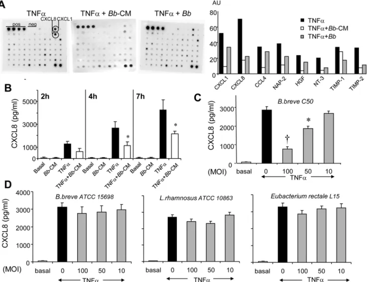
Figure 1.Bband its soluble factors dampen TNFa-induced cytokine secretion in epithelial cells. A.After 4 hour-incubation of HT29-19A cells with TNFa6Bb(MOI 100) orBb-CM, epithelial cell supernatants were tested by the RaybioHhuman cytokine array V (One representative array of two experiments). Secretion of chemokines (CXCL1, CXCL8, CCL4) and of other inflammatory molecules was inhibited in the presence ofBbandBb -CM as judged by densitometric analysis.B.Long lasting inhibition (up to 7 hours) of CXCL8 secretion byBb-CM (,50%) was observed using ELISA.
Secretion of CXCL8 was assayed using the Duoset enzyme-linked immunosorbent assay (ELISA) (R&D Systems).
Epithelial cell viability
As indexes of epithelial viability, we measured transepithelial electrical resistance (R), early apoptosis and zonula occludens 1 (ZO-1) distribution.
Filter-grown HT29-19A cell monolayers were cut-out from the insert and mounted in Ussing chambers. Potential difference (PD) and short-circuit current (Isc) were recorded and R was calculated
according the ohm’s law.
Epithelial cell apoptosis was assessed with the monoclonal antibody M30 CytoDEATH (Roche Diagnostics, Meylan, France) which recognizes cleaved cytokeratin 18, a marker of early apoptosis. A 10 min-treatment of epithelial cells with H2O2
(100mM) was used as a positive control. Filter-grown HT29-19A cell monolayers (CostarHclear 3460) were incubated for 4 hours withBb, and treated according to the manufacturer. Cells were incubated successively with mouse monoclonal IgG2b anti-M30
(1:10) and rabbit polyclonal anti-ZO-1 (24mg/ml, Zymed, Clinisciences) for 60 and 30 min respectively. After washes, Cy5-goat anti-mouse IgG (H+L) and FITC-goat anti-rabbit (Jackson Laboratories, Immunotech, 15mg/ml) were added for 30 min. Cell monolayers were observed by confocal microscopy.
Activation of NF-kB and AP-1
Immunofluorescence analysis of nuclear translocation of p65-NF-kB. HT29-19A cells were grown for 3 days in
eight-chamber slides (Lab-tek, Nunc), starved of FCS overnight and treated for 30 min with TNFa with or without Bb-CM. After fixation in formaldehyde 4%, cells were incubated for 90 min with a mouse monoclonal anti-p65 antibody (2mg/ml, Santa Cruz) and FITC-goat anti-mouse IgG (Jackson laboratories) for 30 min. Nuclei were labelled using TOPRO-3 (0.04mg/ml) and cell preparations observed using confocal microscopy.
Electrophoretic Migration Shift Assay (EMSA). HT29-19A monolayers were treated with TNFa6bacteria for 4 hours and nuclear extracts (3mg) were prepared and incubated in binding buffer with 0.35 pmol of 32P-labeled DNA probes
corresponding to the kB or AP-1 binding sites (Promega) as previously described [12]. After 1 hour incubation, protein-DNA complexes were resolved in a 5% polyacrylamide gel in Tris-borate-EDTA buffer. The gel was exposed to a PhosphorImager screen (Molecular Dynamics) and quantified with ImageQuant software (Molecular Dynamics).
Western Blot analysis of p38 MAPK phosphorylation and IkB-a complex. HT29-19A cell lysates (30mg) were
fractionated on SDS-PAGE and transferred onto PVDF membrane. Membranes were blocked and incubated with mouse anti-phospho-p38 (2mg/ml; Sigma-Aldrich), mouse anti-phospho-IkB-a (Ser32/36) (1:200; Cell Signaling), rabbit polyclonal anti-p38 (12mg/ml; Sigma-Aldrich), rabbit polyclonal anti IkB-a
(1mg/ml; Santa Cruz) or mouse anti-b-actin (2mg/ml; Santa Cruz). Appropriate HRP-conjugated secondary antibodies were used and membranes were revealed with a CCD camera (Fuji LAS-1000 plus).
Trinitrobenzene sulfonic acid (TNBS)-induced colitis in mice
Animal experiments were performed in an accredited estab-lishment (nuA59107, animal facility of the Institut Pasteur de Lille, France) and carried out in accordance with the guidelines of laboratory animal care published by the French Ethical committee and the rules of the European Union Normatives (number 86/ 609/EEC). Colitis was induced by intra-rectal administration of 50ml TNBS (110 mg/kg) in 0.9% (w/v) NaCl/ethanol (50:50 v/v) in anesthetized BALB/c mice (female, 7–8-weeks-old) as described [13]. The colitis control group received TNBS only. TheBbi.g. group received a daily intragastric administration of 108CFU of
Bbin 100ml gavage buffer 5 days before colitis induction. TheBb -CM i.p. group received two intra-peritoneal (i.p. 200ml) injections of a 5-fold concentratedBb-CM at one day intervals before colitis induction. Other groups received a single i.p. administration of 26106murine dendritic cells in 100ml PBS, either untreated (DC group) or pre-conditioned withBb-CM (DCBb-CMgroup)
simulta-neously to TNBS administration. DCs were prepared from bone marrow dendritic cells (BMDCs) as previously described [14] and incubated with or withoutBb-CM for 18 hours.
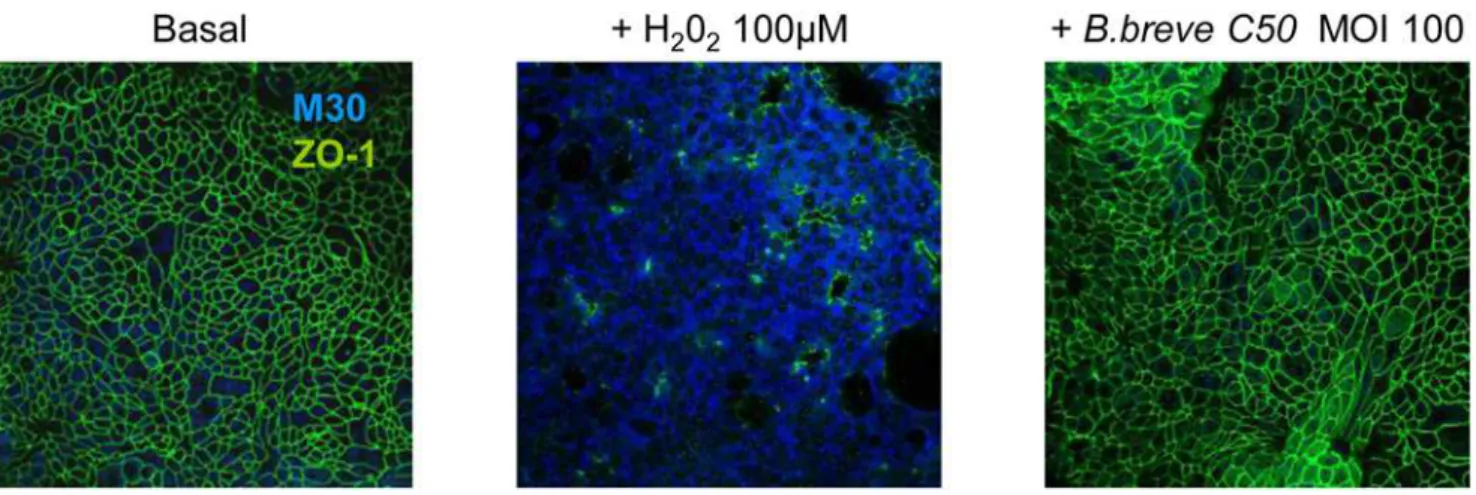
Figure 2.Bbpreserves the integrity of HT29-19A epithelial cells.Filter-grown HT29-19A monolayers were treated with 10 ng/ml TNFawith or without liveBbplaced in the apical compartment for 4 h. As index of epithelial viability, we measured markers of apoptosis (M30) and epithelial tight junction integrity (ZO-1). After 4 hour-treatment with Bb, HT29-19A monolayers (CostarHclear 3460) were labelled with the monoclonal antibody M30 CytoDEATH recognizing caspase-cleaved cytokeratin 18 (1:10, Roche Diagnostics) and rabbit polyclonal anti-ZO-1 (24mg/ml, Zymed). Cells were observed with the laser scanning confocal microscope LSM 510 Carl Zeiss. Treatment with H2O2(100mM), used as a positive control, induced apoptosis (blue M30 labeling) and alteration of ZO-1 distribution (green). In contrast, in basal condition or after treatment withBbat MOI 100, no M30 labeling was observed and ZO-1 distribution was preserved. Results are representative of three independent experiments.
Animals were sacrificed 48 h after TNBS administration and macroscopic colonic damage was analyzed using the Wallace scoring method [15]. Protection observed with the various treatments was calculated taking the Wallace score of TNBS mice (control) as the higest (100%) colonic damage. Total RNA was isolated from colon tissue using the RNeasy kit (Qiagen). Reverse transcription was performed on 2mg, followed by real-time PCR (7300 PCR system from Applied Biosystems) using the Taq-Man PCR Master Mix (Applied Biosystems) and primers and probes designed by Applied Biosystems for murine IL-1b, TNF-a, CXCL1 (KC), COX-2, IL-6, IL-23 and IL-17 and TATA-box binding protein (Tbp). Data were normalized to expression of Tbp.
Statistical analysis
Statistical analysis was performed using the SAS package. The results are expressed as scatter plots with medians, or as
means6SEM. Differences between groups were compared by paired t-test or non-parametric tests (Wilcoxon) and were considered significant for values of p,0.05.
Results
Bbsoluble factors inhibit secretion of inflammatory cytokines by intestinal epithelial cells
Bb soluble factors inhibit LPS-induced TNFa secretion in immune cells [11]. Their effect on TNFa-induced cytokine secretion in HT29-19A epithelial cells was tested using RaybioH
membranes that allow screening of a large panel of inflammatory factors. Bb and Bb-CM inhibited TNFa-induced secretion of CXCL8 (IL-8), CXCL1 (GRO) and CCL4 (MIP-1b) and to a lesser extent of tissue inhibitor of metalloproteinases (TIMP-1, TIMP-2), neutrophil activating factor-2 (NAP-2), neurotrophin
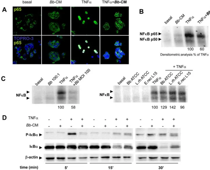
Figure 3.Bband its soluble factors inhibit TNFa-induced NF-kB in epithelial cells. A.HT29-19A cells were incubated for 30 min with TNFa6Bb-CM and localization of the NF-kB p65 subunit was observed by confocal microscopy. Following exposure to TNFa, p65 (green) was translocated into the nuclei (blue) and co-localization was observed (white spots).Bb-CM inhibited TNFa-induced p65 subunit translocation. n = 3 independent experiments. B.HT29-19A cells were incubated for 4 hours with TNFa6Bb-CM. Electrophoretic migration shift assay (EMSA) was performed following extraction of nuclear proteins. TNFacondition was taken as 100% activation andBb-CM induced a 40% inhibition of NF-kB activation (n = 2 independent experiments).C.Epithelial cells were incubated with TNFa6Bbat MOI 100. Activation of NF-kB was inhibited by,40%
NT-3 and growth factors such as hepatocyte growth factor (HGF) (Fig. 1A). Importantly,BbandBb-CM alone did not modify the cytokine profile observed under basal conditions (data not shown). TNFa-induced secretion of CXCL8, tested by ELISA, increased with time but a constant fraction (50 to 58%) was inhibited byBb -CM (1282.56211vs611.56282 pg/ml after 2 hours, 26956555
vs 11346342 pg/ml after 4 hours, p,0.01; and 42546922 vs
21596227 pg/ml after 7 hours; p,0.01) (Fig. 1B). Notably,Bb -CM also inhibited IL-1b-induced CXCL8 secretion (data not shown).
We next evaluated the capacity of apically appliedBbto inhibit TNFa-induced CXCL8 production in the basolateral compart-ment of polarized HT29-19A epithelial cells. Basal secretion of CXCL8 was not modified byBb(data not shown), butBbinduced a dose-dependent inhibition of TNFa-induced CXCL8 secretion that was maximal at MOI 100 (73% inhibition, p,0.0001) (Fig. 1C). In contrast, no inhibition was observed with the other tested Gram (+) bacteria (Fig. 1D).
The inhibitory effects ofBbon chemokine secretion were not due to the alteration of HT29-19A epithelial integrity as attested by stable electrical resistance of epithelial monolayers in the presence of bacteria at high concentration (MOI 100, R = 98611 ohms.cm2) as compared to control epithelial cells (R = 11669V.cm2). In addition, integrity of tight junctions was also attested by a normal distribution of ZO-1 protein and by the lack of bacteria-induced apoptosis of epithelial cells (Fig. 2).
Bbsoluble factors inhibit the NF-kB pathway
Bb-CM inhibited TNFa-induced nuclear translocation of the p65 NF-kB subunit. Nuclear translocation was observed in 83% of cells activated by TNFa alone, but in only 30% of cells preincubated withBb-CM (Fig. 3A). AccordinglyBbsoluble factors impaired the formation of p65- and p50-DNA complexes in response to a 4-hour stimulation with TNFa (Fig. 3B). In contrast to Bb (MOI 100), which inhibited by 40% the formation of the DNA complexes (Fig. 3C), other Gram (+) bacteria had no effect. NeitherBbnor other Gram (+) bacteriaper seinduced the binding of p65- and p50-NF-kB on epithelial DNA. As shown in Fig. 3D,Bb-CM also reduced the early phosphor-ylation of IkB-a(5 min) induced by TNFa2a step that otherwise commits the molecule to subsequent ubiquitination and degrada-tion2and accordingly promoted stabilization of IkB-afrom 15 to 30 min.
Bbsoluble factors inhibit the AP-1 pathway
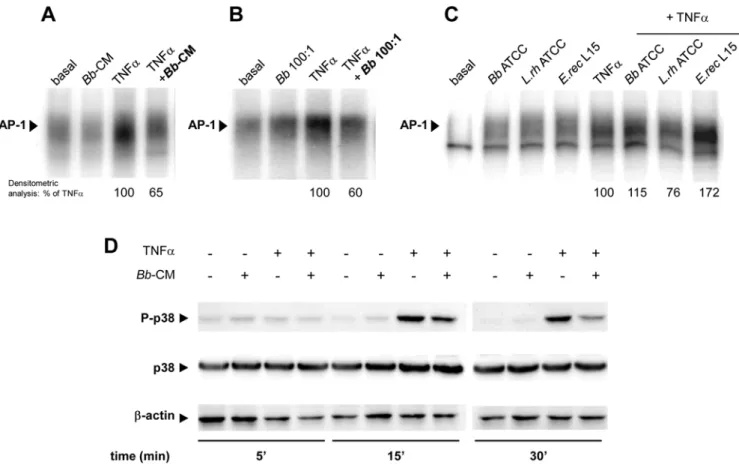
DNA binding of AP-1 induced by TNFawas inhibited by 35% in the presence ofBb-CM (Fig. 4A) and by 40% byBbat MOI 100 (Fig. 4B). In contrast toBb, none of the tested Gram (+) bacteria had any inhibitory effect (Fig. 4C). Furthermore,Bb-CM inhibited p38-MAPK phosphorylation, an important step in AP-1 activation (Fig. 4D). This inhibition, observed at 15 min (- 44%), was still visible after 30 min (- 63%).
Figure 4.Bband its soluble factors inhibit TNFa-induced phosphorylation of p38-MAPK in epithelial cells. A–C.HT29-19A cells were incubated for 4 hours on the apical surface with liveBbor commensal bacteriaB.breveATCC 15698,L.rhamnosus10863,E.rectaleL15 at MOI 100, together with basolateral TNFa. Activation of AP-1 assessed by EMSA was inhibited by,35% in the presence ofBb-CM and by,40% in the presence
ofBb, but not in the presence of other bacteria. n = 2 independent experiments.D.HT29-19A cells were treated with TNFa6Bb-CM. Total proteins were analysed by SDS-PAGE and revealed with antibodies to phospho-p38-MAPK, p38-MAPK and b-actin. Bb-CM inhibited TNFa-induced phosphorylation of p38-MAPK from 15 to 30 min of incubation. n = 3 independent experiments.
Bbsoluble factors alleviate inflammation in a murine model of TNBS colitis
We next investigated the capacity of Bb soluble factors to dampen the colonic inflammatory response in TNBS-induced colitis in mice. The high inflammatory score observed in control TNBS mice, (Wallace score WS = 4.560.4) was decreased inBb
i.g. mice (WS to 3.260.8), indicating a mild protection (30%) (Fig. 5A, 5B). Accordingly, high mRNA expression of pro-inflammatory cytokines/mediators observed in control TNBS mice slightly decreased inBbi.g. mice (Fig. 5C). In contrast, pre-treatment with Bb-CM by i.p. route induced a significant protection against colitis (WS = 1.760.8, 62% protection) and a statistically significant decrease in pro-inflammatory cytokine expression [IL-1b, CXCL1 (equivalent to human IL-8), COX2, IL-23, IL-6]. Since i.p. administration ofBb-CM was efficient in
dampening inflammation, the effect of soluble factors on mucosal dendritic cells (DC) was tested. DC pre-conditioned withBb-CM (DCBb-CM) were injected i.p. into mice at the same time as TNBS
challenge. In contrast to non-treated DC (WS = 4.460.4), DC Bb-CM conferred a significant protection against colitis
(WS = 2.360.4, 49% protection, p,0.01) and reduced the expression of all pro-inflammatory cytokines/mediators. These results indicate a protective effect ofBbsoluble factorsin vivoand suggest that this protective effect relies primarily on their capacity to condition regulatory dendritic cells.
Discussion
Selected strains of probiotic micro-organisms participate in the control of intestinal homeostasis and help preventing intestinal
Figure 5. Protective effect ofBbandBb-CM in TNBS-induced colitis in mice. A.Wallace scores were used to semi-quantify colitis activity in BALB/c mice after colonic instillation of TNBS (control group) with or without intragastric pre-treatment with 108CFUBb(Bbi.g.), intraperitoneal injection ofBb-CM (Bb-CM i.p.), intraperitoneal injection of non treated-DC (DC) orBb-CM conditioned-DC (DCBb-CM). The mild inhibition of colitis observed withBbi.g. became significant withBb-CMand DCBb-CM.B.Percentage protection in mice of the different groups compared to control (TNBS only).C.Quantitative RT-PCR analysis of mRNA expression of pro-inflammatory cytokines/mediators in the colon, 48 hours after TNBS-induced colitis with or without treatment. A significant inhibition of pro-inflammatory molecules in colonic mucosa (IL-1b, CXCL-1, IL-23, IL-6, COX2) was observed in the presence ofBb-CM orBb-CM pre-conditioned DC. Scatter plots are presented with medians. *p,0.05,{p,0.01vsTNBS; n = 7 to 8 mice per group.
disorders. In the intestinal microflora, Bifidobacterium species are generally considered beneficial to the host and various strains of bifidobacteria are used as probiotics. The mechanisms by which these bacteria can modulate the function of epithelial and immune cells remain incompletely elucidated. We have previously shown that soluble factors fromBifidobacterium breveC50 decrease pro-inflammatory cytokine secretion by immune cells [11]. We now show that these soluble factors effectively dampen intestinal inflammation by targeting both epithelial and local dendritic cells.
In inflammatory conditions, an important function of intestinal epithelial cells is to control the influx and activation of immune cells into thelamina propriathrough the production of chemokines and cytokines. Previous studies have supported the view that selected commensal or probiotic bacteria can alleviate inflamma-tion [16]. Along this line, we have already demonstrated that the low molecular weight soluble factors (,3 kD) produced by Bb
inhibit LPS-induced TNFasecretion by immune cells [11]. In this study,BbandBb-CM, unlike other tested Gram (+) bacteria, were able to inhibit the release of chemokines and various inflammatory molecules in epithelial cells. The most prominent effect ofBb-CM was observed on CXCL8 secretion. Inhibition of CXCL8 secretion by probiotics has been reported in epithelial cells but the relative importance of whole bacteriaversussoluble factors have remained elusive [17,18]. Different steps along the NF-kB pathway were targeted according to bacterial strains tested. Commensal Salmonella[9] or Lactobacillus casei[10] could inhibit p65 nuclear translocation through a decrease in IkB-a ubiquitina-tion and degradaubiquitina-tion, B. thetaiotaomicron commensal bacteria promoted the nuclear export of RelA and PPAR-c[19] while VSL#3 inhibited proteasome degrading activity[20] without altering IkB-a phosphorylation. Our study indicates that both pathways implicated in CXCL8 expression, NF-kB and AP-1 [21], are targeted byBbsoluble factors. Interestingly,Bbinhibited key early phosphorylation steps in these two distinct signaling cascades. These results suggest that as yet unknown soluble factor(s) present in the low molecular weight active fraction ofBb -CM interfere with serine-threonine kinases and cellular phosphor-ylation. The nature of this(ese) factor(s) is not yet elucidated. Previous studies [11] underlined that inhibition of LPS-induced TNFa-release in immune cells byBbC50 was heat-resistant, was not related to short-chain fatty acids (butyrate or lactate) and that pepsin-trypsin- or proteinase K- treated CM still retained an inhibitory effect ([11] and unpublished result). Additional unpublished studies indicated that DNAse I treatment of CM or the use of immune cells from TLR92/2mice[22] did not point to the role of TLR9 ligands as active component(s) ofBb-CM. Studies
aiming to characterize the active factors by differential mass spectrometry of active and inactive Bifidobacterium strains (Bb
C50 andBbATCC 15698) are in progress and may help gaining further mechanistic insight.
To assess whetherin vitroobservations could be validatedin vivo, we used a mouse model of colitis. Significant protection was observed after pre-treatment by i.p. injection of Bb soluble factors. Similar protection was obtained after adoptive transfer of dendritic cells pre-treated withBb-conditioned medium. These results are coherent with our previousin vitro results in immune cells[11] and indicate thatBb-derived soluble factors can exertin vivo anti-inflammatory effects through interaction with local immune cells, as also reported with selected strains of lactic acid bacteria [14]. WhenBbwas administered intragastrically, a milder protection was observed, suggesting that the anti-inflammatory properties ofBbrequire the release of sufficient amounts of soluble factors in situ, in the vicinity of colonic mucosa. In our previous study, the inhibitory capacity of soluble factors on immune cells was maintained after crossing an epithelial cell monolayer. However the stability of these factors in the intestinal lumen may be insufficient to allow their delivery to the colonic mucosa. This hypothesis is in keeping with the observation that protection by probiotics is often more effective in the proximal intestine (rotavirus diarrhea) than in the colon (inflammatory bowel diseases). In this respect, it is interesting thatFecalibacterium prausnitzii, a commensal strain poorly represented in the microbi-ota of patients with Crohn’s disease, can produce soluble anti-inflammatory factors which may thus be delivered directly to the adjacent mucosa [23].
Taken together, our results indicate that small soluble factors released byBifidobacterium breveC50 might help maintain intestinal homeostasis by targeting cells of the innate immune system such as epithelial and dendritic cells. Studies are needed to further characterize the Bb soluble factor(s) responsible for the inhibition of kinases involved in multiple steps of intestinal inflammation.
Acknowledgments
We thank Ve´ronique Peucelle-Dennin for her skilful help with animal experiments.
Author Contributions
Conceived and designed the experiments: EH CL CG BP MH. Performed the experiments: EH CL CG. Analyzed the data: EH CL CG MH. Wrote the paper: EH NCB MH.
References
1. Isolauri E, Joensuu J, Suomalainen H, Luomala M, Vesikari T (1995) Improved immunogenicity of oral D x RRV reassortant rotavirus vaccine by Lactobacillus casei GG. Vaccine 13: 310–312.
2. Gionchetti P, Rizzello F, Venturi A, Brigidi P, Matteuzzi D, et al. (2000) Oral bacteriotherapy as maintenance treatment in patients with chronic pouchitis: a double-blind, placebo-controlled trial. Gastroenterology 119: 305–309.
3. McCole DF, Barrett KE (2007) Varied role of the gut epithelium in mucosal homeostasis. Curr Opin Gastroenterol 23: 647–654.
4. Lee J, Rachmilewitz D, Raz E (2006) Homeostatic effects of TLR9 signaling in experimental colitis. Ann N Y Acad Sci 1072: 351–355.
5. Abreu MT, Fukata M, Arditi M (2005) TLR signaling in the gut in health and disease. J Immunol 174: 4453–4460.
6. Shibolet O, Podolsky DK (2007) TLRs in the Gut. IV. Negative regulation of Toll-like receptors and intestinal homeostasis: addition by subtraction. Am J Physiol Gastrointest Liver Physiol 292: G1469–G1473.
7. Lee J, Mo JH, Katakura K, Alkalay I, Rucker AN, Liu YT, et al. (2006) Maintenance of colonic homeostasis by distinctive apical TLR9 signalling in intestinal epithelial cells. Nat Cell Biol 8: 1327–1336.
8. Gribar SC, Anand RJ, Sodhi CP, Hackam DJ (2008) The role of epithelial Toll-like receptor signaling in the pathogenesis of intestinal inflammation. J Leukoc Biol 83: 493–498.
9. Neish AS, Gewirtz AT, Zeng H, Young AN, Hobert ME, et al. (2000) Prokaryotic regulation of epithelial responses by inhibition of IkappaB- alpha ubiquitination. Science 289: 1560–1563.
10. Tien MT, Girardin SE, Regnault B, Le BL, Dillies MA, Coppee JY, et al. (2006) Anti-inflammatory effect of Lactobacillus casei on Shigella-infected human intestinal epithelial cells. J Immunol 176: 1228–1237.
11. Menard S, Candalh C, Bambou JC, Terpend K, Cerf-Bensussan N, et al. (2004) Lactic acid bacteria secrete metabolites retaining anti-inflammatory properties after intestinal transport. Gut 53: 821–828.
12. Ten RM, Paya CV, Israel N, Le BO, Mattei MG, et al. (1992) The characterization of the promoter of the gene encoding the p50 subunit of NF-kappa B indicates that it participates in its own regulation. EMBO J 11: 195–203.
14. Foligne B, Zoumpopoulou G, Dewulf J, Ben YA, Chareyre F, et al. (2007) A key role of dendritic cells in probiotic functionality. PLoS ONE 2: e313. 15. Morris GP, Beck PL, Herridge MS, Depew WT, Szewczuk MR, et al. (1989)
Hapten-induced model of chronic inflammation and ulceration in the rat colon. Gastroenterology 96: 795–803.
16. Borruel N, Carol M, Casellas F, Antolin M, de Lara F, et al. (2002) Increased mucosal tumour necrosis factor alpha production in Crohn’s disease can be downregulated ex vivo by probiotic bacteria. Gut 51: 659–664.
17. Ko JS, Yang HR, Chang JY, Seo JK (2007) Lactobacillus plantarum inhibits epithelial barrier dysfunction and interleukin-8 secretion induced by tumor necrosis factor-alpha. World J Gastroenterol 13: 1962–1965.
18. Ma D, Forsythe P, Bienenstock J (2004) Live Lactobacillus reuteri is essential for the inhibitory effect on tumor necrosis factor alpha-induced interleukin-8 expression. Infect Immun 72: 5308–5314.
19. Kelly D, Campbell JI, King TP, Grant G, Jansson EA, et al. (2004) Commensal anaerobic gut bacteria attenuate inflammation by regulating
nuclear-cytoplasmic shuttling of PPAR-gamma and RelA. Nat Immunol 5: 104–112.
20. Petrof EO, Kojima K, Ropeleski MJ, Musch MW, Tao Y, et al. (2004) Probiotics inhibit nuclear factor-kappaB and induce heat shock proteins in colonic epithelial cells through proteasome inhibition. Gastroenterology 127: 1474–1487.
21. Hoffmann E, Dittrich-Breiholz O, Holtmann H, Kracht M (2002) Multiple control of interleukin-8 gene expression. J Leukoc Biol 72: 847–855. 22. Rachmilewitz D, Katakura K, Karmeli F, Hayashi T, Reinus C, et al. (2004)
Toll-like receptor 9 signaling mediates the anti-inflammatory effects of probiotics in murine experimental colitis. Gastroenterology 126: 520–528.