Associated Responses: A Mechanism for Metal-Specific
Orthopedic Implant Failure
Lauryn Samelko1,2, Marco S. Caicedo1, Seung-Jae Lim3, Craig Della-Valle1, Joshua Jacobs1, Nadim J. Hallab1*
1Department of Orthopedic Surgery, Rush University Medical Center, Chicago, Illinois, United States of America,2Department of Immunology, Rush University Medical Center, Chicago, Illinois, United States of America,3Department of Orthopedic Surgery, Samsung Medical Center, Seoul, Korea
Abstract
The historical success of orthopedic implants has been recently tempered by unexpected pathologies and early failures of some types of Cobalt-Chromium-Molybdenum alloy containing artificial hip implants. Hypoxia-associated responses to Cobalt-alloy metal debris were suspected as mediating this untoward reactivity at least in part. Hypoxia Inducible Factor-1a is a major transcription factor involved in hypoxia, and is a potent coping mechanism for cells to rapidly respond to changing metabolic demands. We measured signature hypoxia associated responses (i.e. HIF-1a, VEGF and TNF-a) to Cobalt-alloy implant debris both in vitro (using a human THP-1 macrophage cell line and primary human monocytes/ macrophages) and in vivo. HIF-1ain peri-implant tissues of failed metal-on-metal implants were compared to similar tissues from people with metal-on-polymer hip arthroplasties, immunohistochemically. Increasing concentrations of cobalt ions significantly up-regulated HIF-1a with a maximal response at 0.3 mM. Cobalt-alloy particles (1 um-diameter, 10 particles/ cell) induced significantly elevated HIF-1a, VEGF, TNF-a and ROS expression in human primary macrophages whereas Titanium-alloy particles did not. Elevated expression of HIF-1awas found in peri-implant tissues and synovial fluid of people with failing Metal-on-Metal hips (n = 5) compared to failed Metal-on-Polymer articulating hip arthroplasties (n = 10). This evidence suggests that Cobalt-alloy, more than other metal implant debris (e.g. Titanium alloy), can elicit hypoxia-like responses that if unchecked can lead to unusual peri-implant pathologies, such as lymphocyte infiltration, necrosis and excessive fibrous tissue growths.
Citation:Samelko L, Caicedo MS, Lim S-J, Della-Valle C, Jacobs J, et al. (2013) Cobalt-Alloy Implant Debris Induce HIF-1aHypoxia Associated Responses: A Mechanism for Metal-Specific Orthopedic Implant Failure. PLoS ONE 8(6): e67127. doi:10.1371/journal.pone.0067127
Editor:Partha Mukhopadhyay, National Institutes of Health, United States of America
ReceivedMarch 19, 2013;AcceptedMay 14, 2013;PublishedJune 20, 2013
Copyright:ß2013 Samelko et al. This is an open-access article distributed under the terms of the Creative Commons Attribution License, which permits unrestricted use, distribution, and reproduction in any medium, provided the original author and source are credited.
Funding:Research reported in this publication was supported in part by Crown Family Chair of Orthopedics and the National Institute of Arthritis and Musculoskeletal and Skin Diseases of the National Institutes of Health under Award Number R01AR060782. The funders had no role in study design, data collection and analysis, decision to publish, or preparation of the manuscript.
Competing Interests:The authors have declared that no competing interests exist.
* E-mail: [email protected]
Introduction
The well established ability of total joint implants to restore function and mobility has been recently shocked by the unexpected early failures of some artificial hip implants, (up to 5% failure as early as 6 years post-operatively of some metal on metal designs) [1–3]. These failures are attributed to excessive exposure to metal debris, e.g. Cobalt-Chromium-Molybdenum-alloy (Co-Cobalt-Chromium-Molybdenum-alloy) metal debris. The pathophysiological reasons for this remain largely unknown. But they are different than the well established long term (.15 years), mechanisms by which particulate plastic and metal debris cause failure by inducing a subtle but persistent innate macrophage inflammatory responses (i.e. granulomas) that invades the bone-implant interface and leads to bone resorption and eventual aseptic loosening of the implant [4–7].
Sites of inflammation or tissue damage are generally character-ized by decreased availability of oxygen, leading to a hypoxic state, which in turn further promotes inflammation and tissue damage at the affected area [8]. Myeloid cells (e.g. macrophages) of innate immunity have the capability to respond and function in a hypoxic
by increasing the secretion of pro-inflammatory cytokines [10]. In the context of orthopedics, recent studies have linked VEGF to the development of aseptic loosening and bone resorption [14,15]. In this study we examined the expression of HIF-1a protein, a signature hypoxia-related transcription factor upstream of VEGF [13]. We investigated if HIF-1aexpression contributes to metal-implant debris induced aseptic inflammation, particularly in cases of elevated exposure to metal implant debris (e.g. in metal-on-metal, MoM hip replacement implants). Bothin vitroandin vivo, we examined whether MoM arthroplasty debris (Cobalt-alloy) pref-erentially to other metals (e.g. Titanium alloy) induces local pathology responses by creating cobalt-induced hypoxic-like responses as evidenced by the production and accumulation of HIF-1aprotein and known associated reactions (i.e. inflammatory and angiogenic).
Materials and Methods
Ethics Statement
This study and consent process received approval from the Rush University Institutional Review Board (10052606-IRB01-CR02). Participants provided their written consent to participate in this study, and participation was limited to a one time blood draw or banked tissue specimens. All participants were 18 years of age or older.
Media and Challenge Agents
Growth medium for the human monocyte cell line THP-1 (ATCC) was RPMI 1640 supplemented with L-Glutamine, Penicillin, Streptomycin, 25 mM Hepes (Lonza, Walkersville, MD USA) and 10% heat inactivated fetal bovine serum (FBS; Hyclone Laboratories, Logan, UT). Human primary monocytes were cultured in RPMI 1640 supplemented with L-Glutamine, Penicillin, Streptomycin, 25 mM Hepes and 10% heat inactivated autologous serum.
The mean particle sizes of Cobalt-alloy (Co-alloy, approx 60%Co, 28%Cr, ,6%Molybdenum, ,1%Nickel, ASTM F75) and Titanium-alloy (Ti-alloy, 90%Ti, 6%Al, 4%V, ASTM F136) particles were characterized by using laser diffraction and Scanning Electron Microscopy. Particles have a mean diameter of 1.2mm (.95%,3 um) for Ti alloy and 0.9mm (.95%,3 um) for Co-alloy particles. Subsequent to characterization, particles were cleaned, sterilized, and tested for endotoxin levels before use in experiments (,0.01 eU, Kinetic QCL, Bioengineering Solu-tions Inc, Oak Park, IL). Cobalt ions were produced using CoCl2 (a known hypoxic mimetic) at a final concentration of 0.1 mM working solution in sterile H2O (Sigma-Aldrich, St. Louis, MO). Challenge concentrations were previously determined to be non-toxic after 24 hours challenge [6,16,17].
Monocyte/macrophage Cell Line
Human THP-1 monocytes were differentiated into macrophag-es by culturing 2.06105monocytes in 48 well plates or 1.06105in
96 well plates with phorbol ester (TPA) for 18–24 hours prior to metal challenge. Newly differentiated macrophages were chal-lenged with particulate and soluble metals for 6 hours or 16 hours at 37uC 5% CO2and supernatants were subsequently analyzed for mature Il-1b, VEGF, and TNF-aproduction.
Primary Monocyte/Macrophage Cells
Peripheral blood monocytes were obtained from healthy donors (n = 4) by venipuncture with IRB-approved informed consent. Peripheral blood mononuclear cells (PBMCs) were isolated by Ficol gradient separation and collected for further purification.
Peripheral blood CD14+
monocytes were isolated from PBMCs by negative selection with a specific magnetic bead antibody cocktail to CD3, CD7, CD16, CD19, CD56, CD123 and Glycophorin A (Miltenyi Biotec). Isolated human primary monocytes were assessed for .90% purity using FACS. Freshly isolated human primary macrophages were challenged with: Ti alloy particles of 1.2mm at 1:1, 5:1, 10:1, and 20:1(particles:cell) ratio, Co- alloy particles of 0.9mm at 1:1, 5:1, 10:1, and 20:1(particles:cell) ratio, and with 0.001 mM, 0.01 mM, 0.1 mM, and 0.3 mM CoCl2.
Cytokine Analysis
Sandwich ELISAs for VEGF (R&D Systems) were used to detect metal-induced THP-1 and human primary macrophages VEGF secretion following manufacturer’s instructions. Superna-tants from particle challenged macrophages were collected at 6 hours and 16 hours of challenge for VEGF and stored at280uC prior to analysis.
Total HIF-1aAssay
Cell Based ELISA for HIF-1a (R&D Systems) was used to detect metal induced THP-1 and human primary macrophage total HIF-1asecretion in the context of the whole cell following the manufacturer’s instructions. After fixation of the cells in the wells, the plate was read using a fluorescence plate reader with excitation at 540 nm and emission at 600 nm and a second reading was performed with excitation at 360 nm and emission at 450 nm. The total amount of HIF-1ain the cells is represented by the readings at 600 nm and the readings at 450 nm represented the amount of total Cytochrome C (housekeeping protein) in the cells. To determine the total amount of HIF-1a activated in the cells, the total HIF-1a fluorescence at 600 nm in each well was normalized to that of the total Cytochrome C fluorescence at 450 nm. The normalized triplicate readings for each sample are then averaged. The normalized averages of challenged cells were normalized to unchallenged control cells and represented as the percentage increase of HIF-1a expression in comparison to unchallenged control cells.
Detection of HIF-1ain Human Tissue and Synovial Fluid Samples
Human/Mouse Total HIF-1a DuoSet intracellular ELISA (R&D Systems) was used to measure HIF-1a in homogenized peri-implant tissue samples from patients with MoM (n = 5) or MoP (n = 10) implants and in synovial fluid samples from patients with MoM (n = 4) or MoP (n = 5) implants following the manufacturer’s instructions.
Collection of Human Tissue Samples
Periprosthetic tissues were obtained from 5 patients with Metal (MoM) implant and from 10 patients with Metal-on-Polyethylene (MoP) implant undergoing primary revision surgery. All samples were freshly frozen in liquid nitrogen and stored at
280uC until time of analysis. Synovial fluid samples were also obtained from 4 patients with MoM implants and from 5 patients with MoP implants at the time of primary revision surgery. All synovial fluid samples were stored at280uC
Analysis of Human Tissue Samples
centrifuged at 2000 rpm for 10 min in a microcentrifuge. Supernatants containing cellular proteins were subsequently collected and stored at 280uC. HIF-1a protein concentrations in the supernatants of periprosthetic tissue and synovial fluid samples were quantified using Human Mouse Total HIF-1a ELISA.
Immunohistochemistry
Peri-implant tissues were used to detect the presence of HIF-1a using standard immunohistochemistry techniques. Briefly, the specimens were trimmed and excess bone fragments were removed to produce a 2 cm2 piece of tissue. Tissues were then placed in cassettes and covered with OCT embedding compound, placed in cooled isopentane, sectioned with a cryostat, fixed in anhydrous acetone for 20 seconds and stored at 270u C. The endogenous peroxides within the samples were blocked with peroxide (H202). Using primary and secondary antibodies combined with the Avitan-Biotin Complex (ABC)-horseradish peroxidase technique, macrophage intracellular and membrane bound cytokines were stained for HIF-1a.
Cytokine Analysis
Supernatants from particle challenged THP-1 and human primary macrophages were collected after 6 hours and 16 hours of challenge for VEGF and TNF-a by ELISA (R&D Systems) following manufacturer’s instructions.
Reactive Oxygen Species (ROS) Assay
Intracellular generation of ROS was determined using oxida-tion of 29,79-dichlorodihydrofluorescein diacetate (H2DCFDA; Invitrogen). All THP-1 cells were plated at 1.56105 cells per well in a black-clear bottomed 96-well plate (BD Falcon) in RPMI with 10% fetal bovine serum (FBS) with phorbol ester (TPA) for 18– 24 h. Differentiated macrophages were challenged for 2.5 hours with: Ti alloy particles and Co-alloy particles at a ratio of 5:1 (particles:cell) ratio, 0.001 mM, 0.01 mM, and 0.1 mM CoCl2 (Cobalt ions). Culture medium was removed and followed by two washes 1X phosphate-buffered saline (PBS) to remove unbound particulate and soluble metals. The cells were then incubated with ROS fluorophore H2DCFDA at 10mM for 30 min followed by two washes of 1X PBS. Fluorescence was measured (485 nm detection and 520 nm emission wavelengths). The fluorescence intensities of challenged cells were normalized to untreated controls for comparison.
Statistical Analysis
One-way ANOVA with Bonferroni post test was used to determine significance for intragroup comparison of metal-treated macrophage cell line and human primary macrophages, where p value,0.05 was considered significant. Student’s t-test was used for cytokine comparison to associated untreated controls.
Results
To determine if particulate and soluble metal implant debris induce a hypoxic (or hypoxic-like) micro-environment and activate
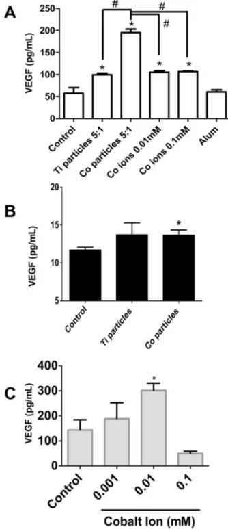
Figure 1. Total HIF-1a protein expression in vitro. (A) THP-1 macrophages (80,000cells/well) were cultured with increasing ratios of Ti-alloy and Co-alloy particles to cells (from 1/1, 5/1, 10/1, 20/1 particles/ cell) or to increasing concentrations of CoCl2for 6 hrs (0.01–0.3 mM). HIF-1aprotein levels were determined using Total HIF-1aCell-Based ELISA kit. Averages of challenged cells were normalized to unchal-lenged control cells and represented as the percentage increase of HIF-1a expression in comparison to unchallenged control cells. (B–C) Human primary macrophages (n = 4) were cultured with Ti-alloy and
Co-alloy particles at a 10:1 (particles:cell) ratio for 6 hours (B) and with increasing concentrations of CoCl2for 16 hours at 0.001–0.1 mM CoCl2 (C). HIF-1a protein levels were determined using Total HIF-1a Cell-Based ELISA kit. Averages of challenged cells were normalized to unchallenged control cells and represented as the percentage increase of HIF-1aexpression in comparison to unchallenged control cells. Note: *p,0.05, determined by Student’s t-test,#p,0.05ANOVA.
the key transcription factor HIF-1a, we evaluated the response of differentiated THP-1 macrophages with increasing concentrations of Titanium-alloy (Ti-alloy) particles, Co-alloy particles and Cobalt-chloride ions, and measured THP-1 up-regulation of HIF-1a protein expression at 6 hours challenge (Fig. 1A). Increasing doses of Ti-alloy, Co-alloy particles and CoCl2induced increased expression of HIF-1ain THP-1 macrophages compared to untreated controls. Moreover, increasing concentrations of cobalt ions significantly up-regulated HIF-1awith a peak response at 0.3 mM, likely due to other toxic effects of cobalt at concentrations higher than 0.3 mM. This type of reactivity dissipated by 16 hours in primary human macrophages indicating that HIF-1a protein half-life is likely relatively short (Fig. 1C). Primary human macrophages (n = 4 subjects) demonstrated comparable results to THP-1 macrophages when treated with Co-alloy particles at a ratio of 10:1 (particles: cell). However, Ti-alloy particles only modestly induced HIF-1aprotein expression in human primary macrophages (Fig 1B), significantly less than Co-alloy particles. Therefore, this in vitro data demonstrates that particulate Co-alloy debris derived from orthopedic implants can induce the stabilization of HIF-1a expression and suggests that Co-alloy particles are likely more bioreactive than Ti-alloy particles due to significant expression of HIF-1a in human macrophages.
HIF-1a protein is a major transcription factor central in regulating hypoxia mediated gene expression [18] and the regulation of angiogenesis by HIF-1ais an important homeostatic mechanism to increase oxygen supply to meet metabolic demand [19]. Therefore, if HIF-1awas indeed up-regulated as a result of metal implant debris challenge, then downstream secretion of VEGF would also occur (Fig 2A). Thus as expected, Ti-alloy, Co-alloy particles and Co ions induced significant increases in VEGF secretion at 16 hours in the human macrophage cell line (THP-1). In proportion to HIF-1a, Co-alloy particles significantly increased VEGF by more than double that of Ti-alloy particles and cobalt ions. However, VEGF responses in primary human macrophages (n = 4) were not consistent in response to particle and ion challenge, compared to THP-1 macrophages, where only 2 of 4 subjects demonstrated a significant VEGF increase to either particles or ion Cobalt-based metal challenge as early as 6 hrs (Co-alloy particles at 6 hrs,Fig. 2B) and that this response increases over time (CoCl2 at 16 hrs, Fig. 2C). These data show that particulate and soluble Co-alloy implant debris can induce the secretion of VEGF in THP-1 macrophages in response to MoM implant debris due to HIF-1a upregulation; however, VEGF secretion by primary human macrophages appears to be more subject dependent, which may explain why metal-on-metal hip implant pathologies (e.g. pseudotumor formations) are not clinically found in all patients with MoM hip replacements with elevated levels of Co-alloy metal debris.
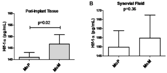
To determine if hypoxia induced by implant metals could be identified clinically in vivo, peri-implant tissues and hip joint synovial fluids of people with failing MoM total hip replacements were evaluated for HIF-1a expression using tissue homogenates and immunohistochemical analysis. We quantitatively analyzed HIF-1a protein expression in revision sample tissue sections of MoM and metal-on-polyethylene (MoP) implants to determine the differential expression of HIF-1a in implants that have an abundance of metal vs. polymer implant debris. We detected a significantly higher mean expression of HIF-1a in patients with failing MoM hips (n = 5) compared to MoP (n = 10) patient tissue samples (Fig 3A). This was supported by analysis of increased HIF-1a expression observed in the synovial fluid samples from MoM patients (n = 4) as compared to MoP (n = 5) patients
Figure 2. VEGF secretion due to particulate and soluble implant debris. (A) VEGF production was assessed after THP-1 differentiated macrophages were challenged with Ti-alloy (1.2mm) and
Co-alloy (0.9mm) particles at 5:1 (particles:cell) ratio and with 0.01 mM
(Fig 3B). To further characterize HIF-1aexpression, we evaluated if HIF-1a protein was differentially present in MoM and MoP peri-implant tissues (Fig. 4). Immunohistochemistry analyses of HIF-1a expression was detected in both MoM and MoP peri-implant tissue samples; however, MoM peri-peri-implant tissue samples expressed increased HIF-1a protein expression as compared to MoP tissue samples.
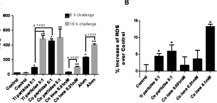
Ti-alloy, Co-alloy and Cobalt ion challenged THP-1 macro-phages (5:1 ratio of particles per cell at 6 hours and 16 hours) induced significantly elevated TNF-a production (Fig 5A). And Co-alloy particles induced significant increase in TNF-a produc-tion when compared to Ti-alloy particles at 6 hours of challenge (p,0.05). These combined results demonstrate that different metals behave differently where preferentially Co-alloy particles
induce a hypoxic or hypoxic-like micro-environment that results in HIF-1a accumulation that in turn augments NFkB signaling pathway and the downstream release of TNF-ain macrophages. To determine if a possible mechanism by which particulate and soluble implant debris can result in the induction of HIF-1a activity, we evaluated intracellular production of ROS in THP-1 macrophages in response to Ti-alloy, Co-alloy particles and CoCl2 for 2.5 hours (Fig. 5B). Ti-alloy, Co-alloy particles and Co ions significantly increased ROS production in THP-1 cells compared to untreated controls (p,0.05). Moreover, ROS production correlates with HIF-1a protein expression in response to metal implant debris, with Co-alloy particles inducing greater produc-tion of both when compared to Ti-alloy particles. These results reiterate that not all metal implant debris induces the same biologic responses despite similarity in dose and size and shape. Thus debris from particular types of implants (modern MoM hip arthroplasty implants) can result in ROS generation that in turn could potentially lead to HIF-1asignaling in THP-1 macrophages.
Discussion
Collectively, ourin vitroandin vivodata support the contention that metal particulate and soluble degradation products can effect local innate immune responses and tissues in a specific pathophysiologic manner where hypoxic (or hypoxic-like) micro-environment is produced, evidenced by accumulation of HIF-1a protein. High numbers of macrophages in peri-implant tissues are indicative of aseptic loosening and periprosthetic osteolysis [14]. VEGF is integral to this process [14,15] and is known to be a downstream target of HIF-1aprotein production and is a potent angiogenic factor that is up-regulated by macrophages in a hypoxic environment [11]. Until now however, the ability of implant debris to induce HIF-1a reactivity in the context of an innate macrophage response remained unknown. HIF-1a is a potent coping mechanism utilized by macrophages in response to a hypoxic microenvironment used to help mediate the necessary cellular mechanisms in order to maintain activity and viability [8]. Moreover, hypoxia can result in the activation of the NFkB pathway where it has been shown that downstream signaling of TNF-a has a critical role in the biologic reactivity to implants,
Figure 3. Total HIF-1aprotein expressionin vivo.(A) The expression level of total HIF-1aprotein in peri-implant patient homogenized tissue samples of metal-on-metal (MoM) (n = 5) and metal-on-polythetheylene (MoP) (n = 10) implants and quantified with the Human/Mouse Total HIF-1a DuoSet IC ELISA. (B) The expression level of total HIF-1aprotein in synovial fluid patient samples of metal (MoM) (n = 4) and metal-on-polythetheylene (MoP) (n = 5) implants and quantified with the Human/Mouse Total HIF-1aDuoSet IC ELISA. Note: * p,0.05 determined by Student’s t-test.
doi:10.1371/journal.pone.0067127.g003
Figure 4. Immunohistochemistry analysis of HIF-1a protein
implant performance and bone loss (inflammatory osteolysis) [20,21].
Both ourin vitroandin vivofindings indicate Co-alloy is a potent stimuli, eliciting a hypoxic or hypoxic-like microenvironment that
can increase the production and stabilization of HIF-1aprotein leading to pro-angiogenesis signaling. Importantly this is the first report of a mechanism that can account for the unusual tissue reactions (pseudotumors) that often preferentially accompany high
Figure 5. TNF-aand ROS production levels correlate with HIF-1aprotein expression.(A) TNF-acytokine production was assessed after THP-1 differentiated macrophages were challenged with Ti-alloy (1.2mm) and Co-(0.9mm) particles at a 5:1 (particles:cell) ratio and with 0.01 mM
CoCl2 for 6 hours and 16 hours and was quantified by ELISA. (B) Intracellular generation of ROS was determined using oxidation of 29,79 -dichlorodihydrofluorescein diacetate (H2DCFDA; Invitrogen). THP-1 differentiated macrophages were challenged with Ti-alloy (1.2mm) and
Co-(0.9mm) alloy particles at 5:1 (particles:cell) ratio or with 0.001 mM, 0.01 mM, 0.1 mM CoCl2for 2.5 h. The normalized averages of challenged cells are represented as the percentage increase of ROS production compared to unchallenged control cells. Note: *p,0.05, determined by Student’s t-test,# p,0.05ANOVA.
doi:10.1371/journal.pone.0067127.g005
Figure 6. Potential Pathway(s) of HIF-1aInduction to Implant Debris.(A) A representation of how metal implant debris (Co-alloy ions and particles) may lead to induction of HIF-1aactivity directly or indirectly given our findings.
levels of Cobalt metal debris in failed metal-on-metal hip replacements [22]. These data demonstrate that Co-alloy particles induced significant biologic effects, even at low doses (particles per cell), causing macrophages to respond with increases in HIF-1a, TNF-a, VEGF and ROS. In contrast, Ti-alloy particles produced similar effects only at higher challenge doses of particles. Therefore, the hypoxic/hypoxic-like induction of HIF-1aprotein in response to metal implant debris may explain how certain kinds of metal-releasing orthopedic implants produce both necrosis/ toxicity and neogenic tissue responses resulting in unique mechanisms of poor implant performance.
It is well established that hypoxia can induce inflammation and vise versa, e.g. NFkB[9], that subsequently results in the production of pro-inflammatory cytokines, such as TNF-a. We observed HIF-1aproduction was enough to induce (or be induced from) NFkB activation by observing increased TNF-a, production a signature cytokine of NFkbactivation. Additionally, the signal transduction pathway of HIF-1a remains elusive but various mechanisms have been proposed linking either increased or decreased ROS generation and either a ROS independent or dependent effect on induction of HIF-1aactivity [23]. In order to determine if metals can induce a hypoxic or hypoxic-like state indirectly through known upstream stimulants, we measured ROS production in human THP-1 macrophages. ROS production due to Ti-alloy, Co-alloy particles and Co ion challenge was shown to occur and thus may either cause or contribute to the induction of the HIF-1aassociated responses. An important caveat however, is that measurement of ROS by H2DCFDA is prone to auto-oxidation. Therefore, further investigation using additional ROS markers is underway to determine if metal-induced ROS
production has a central role in the metal-debris activated of HIF-1aor whether it merely produced simultaneously and if this type of activation can be mitigated by anti-oxidant mediators (Fig 6).
In summary, ourin vitroandin vivodata provide evidence that different implant metals induce different levels of hypoxia responses, where Cobalt-alloy soluble and particulate metal debris preferentially induced hypoxia-like pathology resulting in HIF-1a compensatory responses to metal implant debris (vs. Titanium alloy). These in vitro and in vivo hypoxia-associated responses reported here (e.g. increased HIF-1a, TNF-a, VEGF and ROS) provides a mechanism that can account for the unusual reactions around some implants (e.g. local soft tissue growths ‘‘fibro-pseudotumors’’) that preferentially release Cobalt particles and ions. Given the increasing number of people receiving total joint replacements (.1 million per year) and the early failures of some designs that release elevated amount of Cobalt [23–25], under-standing how this occurs is paramount to ensuring the safety and efficacy of current and future designs.
Acknowledgments
The content is solely the responsibility of the authors and does not necessarily represent te official views of the National Institutes of Health.
Author Contributions
Conceived and designed the experiments: LS MC SJL CDV JJ NH. Performed the experiments: LS MC SJL. Analyzed the data: LS MC NH. Contributed reagents/materials/analysis tools: LS MC SJL CDV JJ NH. Wrote the paper: LS MC NH.
References
1. Jacobs JJ, Hallab NJ (2006) Loosening and osteolysis associated with metal-on-metal bearings: A local effect of metal-on-metal hypersensitivity? J Bone Joint Surg Am 88: 1171–1172.
2. Korovessis P, Petsinis G, Repanti M, Repantis T (2006) Metallosis after contemporary metal-on-metal total hip arthroplasty. Five to nine-year follow-up. J Bone Joint Surg Am 88: 1183–1191.
3. Milosev I, Trebse R, Kovac S, Cor A, Pisot V (2006) Survivorship and retrieval analysis of Sikomet metal-on-metal total hip replacements at a mean of seven years. J Bone Joint Surg Am 88: 1173–1182.
4. Dalal A, Pawar V, McAllister K, Weaver C, Hallab NJ (2012) Orthopedic implant cobalt-alloy particles produce greater toxicity and inflammatory cytokines than titanium alloy and zirconium alloy-based particles in vitro, in human osteoblasts, fibroblasts, and macrophages. J Biomed Mater Res A 100: 2147–2158.
5. Hallab NJ, McAllister K, Brady M, Jarman-Smith M (2011) Macrophage reactivity to different polymers demonstrates particle size- and material-specific reactivity: PEEK-OPTIMA((R)) particles versus UHMWPE particles in the submicron, micron, and 10 micron size ranges. J Biomed Mater Res B Appl Biomater.
6. Caicedo MS, McAllister K, Reddy A, Jacobs JJ, Hallab NJ (2008) Soluble and particulate Co-Cr-Mo alloy implant metals activate the inflammasome danger signaling pathway in human macrophages: A novel mechanism for implant debris reactivity. J Orthop Res 27: 847–854.
7. Goodman SB (2007) Wear particles, periprosthetic osteolysis and the immune system. Biomater 28: 5044–5048.
8. Kim SY, Choi YJ, Joung SM, Lee BH, Jung YS, et al. (2010) Hypoxic stress up-regulates the expression of Toll-like receptor 4 in macrophages via hypoxia-inducible factor. Immunology 129: 516–524.
9. Cramer T, Yamanishi Y, Clausen BE, Forster I, Pawlinski R, et al. (2003) HIF-1alpha is essential for myeloid cell-mediated inflammation. Cell 112: 645–657. 10. Imtiyaz HZ, Simon MC (2010) Hypoxia-inducible factors as essential regulators
of inflammation. Curr Top Microbiol Immunol 345: 105–120.
11. Murdoch C, Muthana M, Lewis CE (2005) Hypoxia regulates macrophage functions in inflammation. J Immunol 175: 6257–6263.
12. Peyssonnaux C, Datta V, Cramer T, Doedens A, Theodorakis EA, et al. (2005) HIF-1alpha expression regulates the bactericidal capacity of phagocytes. J Clin Invest 115: 1806–1815.
13. Semenza G (2002) Signal transduction to hypoxia-inducible factor 1. Biochem Pharmacol 64: 993–998.
14. Spanogle JP, Miyanishi K, Ma T, Epstein NJ, Smith RL, et al. (2006) Comparison of VEGF-producing cells in periprosthetic osteolysis. Biomater 27: 3882–3887.
15. Waris V, Sillat T, Waris E, Virkki L, Mandelin J, et al. (2012) Role and regulation of VEGF and its receptors 1 and 2 in the aseptic loosening of total hip implants. J Orthop Res 30: 1830–1836.
16. Caicedo MS, Pennekamp PH, McAllister K, Jacobs JJ, Hallab NJ (2010) Soluble ions more than particulate cobalt-alloy implant debris induce monocyte costimulatory molecule expression and release of proinflammatory cytokines critical to metal-induced lymphocyte reactivity. J Biomed Mater Res A 93: 1312–1321.
17. Hallab NJ, Anderson S, Caicedo M, Brasher A, Mikecz K, et al. (2005) Effects of soluble metals on human peri-implant cells. J Biomed Mater Res A 74: 124–140. 18. Semenza GL (2003) Targeting HIF-1 for cancer therapy. Nat Rev Cancer 3:
721–732.
19. Pugh CW, Ratcliffe PJ (2003) Regulation of angiogenesis by hypoxia: role of the HIF system. Nat Med 9: 677–684.
20. Merkel KD, Erdmann JM, McHugh KP, Abu-Amer Y, Ross FP, et al. (1999) Tumor necrosis factor-alpha mediates orthopedic implant osteolysis. Am J Pathol 154: 203–210.
21. Schwarz EM, Lu AP, Goater JJ, Benz EB, Kollias G, et al. (2000) Tumor necrosis factor-alpha/nuclear transcription factor-kappaB signaling in peripros-thetic osteolysis. J Orthop Res 18: 472–480.
22. Kwon YM, Ostlere SJ, Lardy-Smith P, Athanasou NA, Gill HS, et al. (2011) "Asymptomatic" pseudotumors after metal-on-metal hip resurfacing arthroplas-ty: prevalence and metal ion study. J Arthroplasty 26: 511–518.
23. Guyer RD, Shellock J, MacLennan B, Hanscom D, Knight RQ, et al. (2011) Early failure of metal-on-metal artificial disc prostheses associated with lymphocytic reaction: diagnosis and treatment experience in four cases. Spine (Phila Pa 1976) 36: E492–E497.
24. Aroukatos P, Repanti M, Repantis T, Bravou V, Korovessis P (2010) Immunologic adverse reaction associated with low-carbide metal-on-metal bearings in total hip arthroplasty. Clin Orthop Relat Res 468: 2135–2142. 25. Davies AP, Willert HG, Campbell PA, Learmonth ID, Case CP (2005) An