by Time-Lapse Video Microscopy
Masahito Asada1, Yasuyuki Goto1,2, Kazuhide Yahata3, Naoaki Yokoyama1, Satoru Kawai4, Noboru Inoue1, Osamu Kaneko3, Shin-ichiro Kawazu1*
1National Research Center for Protozoan Diseases, Obihiro University of Agriculture and Veterinary Medicine, Inada, Obihiro, Japan, 2Laboratory of Molecular Immunology, Graduate School of Agricultural and Life Sciences, The University of Tokyo, Yayoi, Bunkyo-ku, Tokyo, Japan,3Department of Protozoology, Institute of Tropical Medicine (NEKKEN) and the Center of Excellence Program, Nagasaki University, Sakamoto, Nagasaki, Japan,4Laboratory of Tropical Medicine and Parasitology, Dokkyo University School of Medicine, Mibu, Shimotsuga, Tochigi, Japan
Abstract
Background:Babesia bovisis an apicomplexan intraerythrocytic protozoan parasite that induces babesiosis in cattle after transmission by ticks. During specific stages of the apicomplexan parasite lifecycle, such as the sporozoites ofPlasmodium falciparumand tachyzoites ofToxoplasma gondii, host cells are targeted for invasion using a unique, active process termed ‘‘gliding motility’’. However, it is not thoroughly understood how the merozoites ofB. bovistarget and invade host red blood cells (RBCs), and gliding motility has so far not been observed in the parasite.
Methodology/Principal Findings:Gliding motility ofB. bovismerozoites was revealed by time-lapse video microscopy. The recorded images revealed that the process included egress of the merozoites from the infected RBC, gliding motility, and subsequent invasion into new RBCs. The gliding motility of B. bovis merozoites was similar to the helical gliding of Toxoplasma tachyzoites. The trails left by the merozoites were detected by indirect immunofluorescence assay using antiserum againstB. bovismerozoite surface antigen 1. Inhibition of gliding motility by actin filament polymerization or depolymerization indicated that the gliding motility was driven by actomyosin dependent process. In addition, we revealed the timing of breakdown of the parasitophorous vacuole. Time-lapse image analysis of membrane-stained bovine RBCs showed formation and breakdown of the parasitophorous vacuole within ten minutes of invasion.
Conclusions/Significance:This is the first report of the gliding motility ofB. bovis.Since merozoites ofPlasmodiumparasites do not glide on a substrate, the gliding motility ofB. bovismerozoites is a notable finding.
Citation:Asada M, Goto Y, Yahata K, Yokoyama N, Kawai S, et al. (2012) Gliding Motility of Babesia bovisMerozoites Visualized by Time-Lapse Video Microscopy. PLoS ONE 7(4): e35227. doi:10.1371/journal.pone.0035227
Editor:Thomas J. Templeton, Weill Cornell Medical College, United States of America
ReceivedFebruary 13, 2012;AcceptedMarch 12, 2012;PublishedApril 10, 2012
Copyright:ß2012 Asada et al. This is an open-access article distributed under the terms of the Creative Commons Attribution License, which permits unrestricted use, distribution, and reproduction in any medium, provided the original author and source are credited.
Funding:This work was supported by a Cooperative Research Grant of NEKKEN, 2010 (Grant No. 22–21) and 2011 (Grant No. 23–8), to S-i.K. Homepage of NEKKEN: http://www.tm.nagasaki-u.ac.jp/nekken/english/index.html. The funders had no role in study design, data collection and analysis, decision to publish, or preparation of the manuscript.
Competing Interests:The authors have declared that no competing interests exist.
* E-mail: [email protected]
Introduction
The apicomplexan phylum contains obligate intracellular parasites that are major pathogens of humans and livestock. During specific stages of the apicomplexan parasite lifecycle, host cells are targeted for invasion using a unique, active process termed ‘‘gliding motility’’. Gliding motility by apicomplexan parasites does not require shape changes like the crawling of amoebae, nor do the zoite stages of these parasites have cilia or flagella [1]. Instead, the motility is driven by coupling the translocation of surface adhesins to an actomyosin motor beneath the parasite plasma membrane [2]. Gliding motility has been reported inPlasmodiumspp. (sporozoite and ookinete),Toxoplasma gondii(tachyzoite and sporozoite),Cryptsporidum parvum(sporozoite), andEimeriaspp. (sporozoite) [3,4,5,6].
Babesia bovis is an apicomplexan intraerythrocytic protozoan parasite that induces babesiosis in cattle after transmission by ticks.
B. bovis is a representative of the large-type Babesia species. Sporozoites of theBabesiaparasite directly invade host red blood
cells (RBCs), and all parasitic stages in the vertebrate host develop in the RBCs [7]. Although the precise timing is unknown, once merozoites invade a RBC, the parasite rapidly escapes from the parasitophorous vacuole (PV) that is formed by invagination of the RBC membrane during invasion [8]. Following establishment of the free parasite within a host RBC, B. bovis produces two merozoites by binary fission. After erythrocytic lysis, each merozoite invades a new RBC and successive merogonies occur [7]. However, the process of merozoite entry into RBCs is not thoroughly understood, and gliding motility has so far not been recorded inBabesiaparasites.
developed in our previous study [13]. Time-lapse video images delineated the sequential process of parasite-infected RBC (IRBC) rupture, merozoite egress from IRBCs, gliding motility of the merozoites, attachment and invasion of merozoites into new RBCs, and formation and breakdown of PVs.
Results
B. bovisMerozoites Glide to Infect RBC
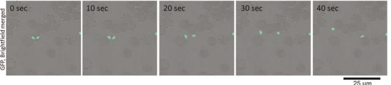
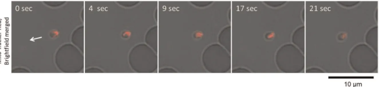
From observations of the GFP-expressingB. bovispopulations, we could confirm the gliding motility of the extra-erythrocytic merozoites in vitro. To characterize the mode of gliding motility, cultured IRBCs were applied to glass slides and egress of the merozoites from the IRBC and their subsequent motility were recorded by confocal laser microscopy. By analyzing the video recordings, straight or meandering motility, but not retreating movement, was observed (Fig. 1 and Video S1). The apical end of the merozoites was at the front position in the direction of movement. To characterize the gliding mode ofB. bovismerozoites, parasite mitochondria were stained with MitoTracker Red. The red fluorescence-stained organelles in the cells enabled us to track the body rotation around the long axis. The merozoites rotated in a counterclockwise direction with respect to the direction of travel (Fig. 2 and Video S2). The motility observed in the B. bovis
merozoites was similar to helical gliding, but other modes of gliding such as circular gliding or twirling were not observed.
B. bovisMerozoites form Trails During Gliding
To confirm that the movement ofB. bovismerozoites is due to gliding, the presence of gliding trails was investigated by an indirect immunofluorescence antibody test using anti-merozoite surface antigen 1 (BbMSA-1) antibody. IRBCs were placed on poly-L-lysine-coated glass slides and the merozoites that egressed from RBCs were allowed to glide on the slides. The slides were stained with the antibody. As a result, B. bovis merozoites were observed to deposit trails that formed straight or winding patterns in accordance with observations from time-lapse video microscopy (Fig. 3).
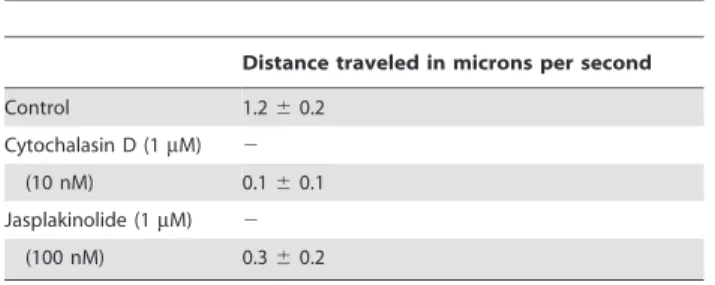
merizer (jasplakinolide) on merozoite motility was analyzed. Motility ofB. bovismerozoites was completely blocked by 1mM cytochalasin D or 1mM jasplakinolide, indicating that the motility was driven by an actomyosin motor. The IRBCs incubated with 1mM of cytochalasin D showed unsuccessful egress of the merozoites and even rupture of the IRBC membrane was obstructed, suggesting that the actomyosin dependent process could also be involved in parasite egress from the IRBCs. A 10 nM cytochalasin D treatment decreased the merozoites’ motility and the velocity was calculated to be 0.1mm/sec, while the velocity under 100 nM jasplakinolide treatment was calculated at 0.3mm/ sec. A related study withT. gondiitachyzoites showed that a high dose of jasplakinolide inhibited gliding motility, while under some conditions the compound madeT. gondiitachyzoites more active in gliding [14]. AlthoughB. bovismerozoites were also treated with jasplakinolide at 100mM, only an inhibition of gliding activity was observed at this concentration and not an increase in activity.
RBC Attachment and Invasion
To observe the invasion of merozoites into new RBCs we recorded a sequence of merozoite motility after the rupture of the IRBC. To enable monitoring of merozoite invasion, cell membranes of bovine RBCs were stained with the red fluorescent dye, PKH26. We observed numerous instances of parasites having successfully invaded fresh RBCs (Fig. 4 and Video S3). Some merozoites attached to several RBCs during their gliding migration before final invasion into a RBC. The ring-shaped red fluorescence around the green merozoite indicated its successful invasion into the new RBC, and it also suggested formation of the PV, which was observed just after the merozoite invasion. It should be noted that when merozoites detached from the RBCs without invasion, the merozoite-RBC interaction was strong enough that pulling of the RBC membrane by the departing merozoite could be observed (Video S4).
Formation and Breakdown of the Parasitophorous Vacuole (PV)
Next, we monitored the fate of the PV, which was observed as a red fluorescent membrane around the parasite. From the video
Figure 1. Time-lapse video microscopy of aB. bovismerozoite engaged in gliding.In vitrocultured IRBCs were placed on a glass slide and their motility documented with confocal laser microscopy. The time elapsed between each frame is indicated in seconds. Gliding of theB. bovis
images, a strongly fluorescent dot appeared beside the parasite and, simultaneously, the ring-shaped red fluorescence disappeared (Fig. 5 and Video S5). The dot fluorescence appeared within 10 min after the invasion of the merozoite into the RBC (the Video S5 shows it appearing approximately 300 sec after invasion). This result suggested that the PV was broken down within several minutes of merozoite invasion into the RBC.
Discussion
To our knowledge this is the first time thatB. bovismerozoites have been demonstrated as displaying gliding behavior in vitro. Gliding motility of apicomplexan parasites was recorded in time-lapse video images by early work inEimeriasporozoites [15], and detailed observation of such locomotion has been conducted mainly in sporozoites of Plasmodium spp. and tachyzoites of
Toxoplasma gondii(former reports and our findings are summarized in Table 2) [2,4,5,16,17,18]. Although bothPlasmodiumandBabesia
merozoites parasitize host RBCs,Plasmodiummerozoites observed
in vitrodo not move across a substrate at all, until contacting the RBC surface [19]. From the analysis of the gliding motility, it seems that Babesia merozoites can glide in any direction. The merozoites of B. bovis rotated their body around the long axis, which was similar to the helical gliding mode of motility defined in
Toxoplasma gondii tachyzoites, for which the force is generated by the actomyosin motor using myosin A proteins anchored on the inner membrane complex, a specialized membranous structure beneath the parasite’s plasma membrane [4]. The rotating direction was counterclockwise, which was also similar to T.
gondii. InT. gondii, it is proposed that closely arranged particle on the inner membrane complex may be connected to the subpellicular microtubule and that the subpellicular microtubule was necessary for the gliding motility [14,20]. InBabesiaparasite, the existence of subpellicular microtubule was also reported inB. major [21] and freeze-fracture electron microscopy of the intraerythrocytic B. ovata merozoites have shown helically ar-ranged intramembranous particles on the inner membrane (S. K., unpublished data), suggesting that there might be helical arrangements of the submembrane cytoskeleton in Babesia
merozoites, and that the direction of this arrangement is consistent with the counterclockwise rotation ofB. bovismerozoites. On the other hand, we did not observe other modes of gliding. In contrast to the crescent shape of Toxoplasma tachyzoites and Plasmodium
sporozoites,B. bovis merozoites have a pyriform shape. It seems that this pyriform shape providesB. bovismerozoites with flexibility in the direction of movement, and therefore the other two forms of motility (circular gliding, upright twirling) observed inToxoplasma
tachyzoites might not be necessary. The cell membrane staining of bovine RBCs facilitated confirmation of the successful invasion of merozoites into the RBCs, which was observed by z-stack analysis from confocal laser microscopy. The recorded images revealed that gliding motility led to invasion RBCs byB. bovismerozoites by a different manner to the well confirmed process inPlasmodium
merozoites [22]. In our study, after the attachment of B. bovis
merozoites, clear reorientation was not observed. Although further detailed analysis is necessary, the video images suggest that the
B. bovis merozoite might act like a screw as it enters the RBC rather than following the serial invasion process observed for
Figure 2. Helical motility ofB. bovismerozoites.In vitrocultured IRBCs were stained with a fluorescent mitochondrial probe (MitoTracker Red) and placed on a glass slide for confocal laser microscopy analysis. The shift in red fluorescence on the merozoites (top to bottom) indicates helical motility of the merozoites. The arrow indicates the direction of merozoite movement.
doi:10.1371/journal.pone.0035227.g002
Plasmodium merozoites. The force generated by the actomyosin motor during gliding motility is transmitted to the outside of the merozoite through interaction of the motor with a transmembrane protein that serves as an adhesion molecule to the substrate. Recent studies inPlasmodiumhave highlighted the essential role of the thrombospondin related anonymous (or adhesive) protein (TRAP) family in gliding and cell invasion of the parasites [23,24]. InBabesia spp., several TRAP family genes have been identified [25]. However, the role of the TRAP-family in gliding motility of
Babesiamerozoites is still unclear.
The experiments with the membrane-stained RBCs enabled us to analyze the PV formation inB. bovis. Since neither the IRBCs nor the merozoites were stained with PKH26 in Fig. 5, the appearance of the ring-shaped red fluorescence around the merozoite indicated that the PV originated from the cell membrane of newly infected RBC. Time-lapse video images revealed that a strongly fluorescent dot appeared beside the merozoite within 10 min after the invasion, and that the ring-shaped fluorescence disappeared. This observation supports the idea that the B. bovis merozoites rapidly escape or are released from the PV. The video images indicate the timing of the event, whereas the molecular mechanism behind this phenomenon is still unclear. Studies of the blood stage of Plasmodium parasites have revealed that the PV membrane is retained until late schizogony [26,27]. This finding suggests that the PV ofB. bovismay not be formed in the same way or sealed as securely as in Plasmodium. Alternatively, because the PV formation is coupled with the merozoite invasion, the difference in the invasion process by these two parasites may result the difference in the PV formation: in contrast to a relatively slow serial invasion process ofPlasmodium
merozoites, quick vivid invasion ofBabesiamerozoites might be too
of PV breakdown. The escape or release of the merozoite from the PV is a unique point in the lifecycle of Babesiaparasites, and it seems to be a critical process for the parasite to establish the intraerythrocytic growth stage. Comparative genome analysis betweenBabesiaand Plasmodiumin terms of PV formation might be an interesting subject to be addressed in future studies.
Materials and Methods
Ethics Statement
Bovine RBCs were collected every two weeks from healthy animals. This study was performed in strict accordance with the recommendations in the Guide for the Care and Use of Laboratory Animals of the Obihiro University of Agriculture and Veterinary Medicine. The protocol was approved by the Committee on the Ethics of Animal Experiments of the Obihiro University of Agriculture and Veterinary Medicine (Permit number 23–26).
Parasite Culture
TheBabesia bovisTexas strain was maintained in purified bovine RBCs with GIT medium (WAKO, Osaka, Japan) by a micro-aerophilic stationary-phase culture system [28]. The parasites were cultured in 1 ml culture medium containing 10% bovine RBCs in 24-well culture plates (Corning, NY, USA). Culture medium was replaced every day and the level of parasitemia was monitored daily by staining thin blood smears with Giemsa solution.
GFP-fluorescentB. bovispopulations were established previously [13]. Briefly, the GFP-expressing plasmid was constructed. Then, the DHFR expression cassette was cloned into the plasmid with the GFP expression cassette. The plasmid constructs were introduced into the Texas strain ofB. bovis by transfection with a NucleofectorH device (Amaxa Biosystems, Cologne, Germany). The transfected parasite population was selected with 5–10 nM of WR99210 and the parasite population with GFP expression was cloned using a limiting dilution. The fluorescent parasite was maintained for more than 7 months under WR99210 drug pressure.
2
(100 nM) 0.360.2
Estimates of rates of motility were based on ten documented individual parasites from two independent experiments. (2) indicates no motility
observed with at least three experiments. doi:10.1371/journal.pone.0035227.t001
Figure 4. Time-lapse video microscopy ofB. bovismerozoite egress from a RBC, gliding to and invasion of a new RBC.The arrow head shows merozoite invasion into a new RBC. The red ring-shaped fluorescence around the merozoite indicates the parasitophorous vacuole. The time elapsed between each frame is indicated in seconds.
Video Microscopy
In vitrocultured IRBCs were applied to glass slides for time-lapse analysis. Time-lapse video microscopy was conducted using a TCS-SP5 confocal laser scanning microscope (Leica Micro-systems, Wetzlar, Germany). Confocal fluorescence images and transmitted images were recorded digitally at approximately one frame per second (0.8–1.5 frame/sec). Time-lapse images were recorded continuously for up to 30 min at room temperature (RT) and merozoite egress from the IRBC and their gliding were monitored. Frames used to create the time lapse series were taken from avi movies and processed using AviUtl software.
Immunofluorescence Detection of Trails
Staining of surface protein in trails was performed by indirect immunofluorescence using the antiserum againstB. bovismerozoite surface antigen 1 (BbMSA-1) [29]. Glass slides were coated in 10mg/ ml poly-L-lysine in PBS for 30 min at RT and rinsed in phosphate buffer saline (PBS). IRBCs were resuspended in GIT medium, added onto the slides, and incubated at 37uC for 30 min. Slides were briefly washed by PBS, dried and fixed with 50% acetone-50% methanol for 5 min at -20uC. Anti-BbMSA-1 mouse antiserum was used at 1:50 and Alexa-Fluor 488 conjugated goat anti-mouse IgG (Molecular probes, OR, USA) was used at 1:200. The slides were incubated with each antibody for 45 min at RT, and then observed with a TCS-SP5 confocal laser scanning microscope.
Gliding Inhibition Assays
For the gliding inhibition assays, cytochalasin D (Wako Pure Chemical, Osaka, Japan) and jasplakinolide (Enzo life sciences, NY, USA) was dissolved in DMSO at 1 mM and stored at230uC. IRBCs were resuspended in GIT medium, and treated with 10 nM or 1mM cytochalasin D, 100 nM or 1mM of jasplakinolide, or 0.1% DMSO for 30 min, respectively. Parasite tracks were traced from the pictures onto transparent sheets and gliding velocity was analyzed for each merozoite. Velocities were calculated over periods of five seconds and the highest speed was taken as the gliding speed. A total of 10 merozoites from two independent experiments were analyzed to obtain mean6SD values.
Mitochondrial Staining ofB. bovis
To stain the mitochondria ofB. bovismerozoites, MitoTracker Red CM-H2XRos (Molecular Probes, OR, USA) was used. The fluorescent probe was diluted to 200 nM in GIT medium, and mixed with an equal amount of IRBCs. The mixture was incubated for 30 min at 37uC and washed with GIT medium 3 times. Parasites with mitochondrial staining were used for analysis on mode of gliding.
Figure 5. Time-lapse video microscopy of the formation and breakdown of the parasitophorous vacuole.To enable monitoring the PV, PKH26 stained fresh RBC was added to the unstained IRBCs. PKH26 fluorescence can be observed around the merozoite just after invasion into the new RBC (arrow). Subsequently, red dot-shaped fluorescence appears (arrow head), and the ring-shaped fluorescence becomes weaker. The time elapsed between each frame is indicated in seconds.
doi:10.1371/journal.pone.0035227.g005
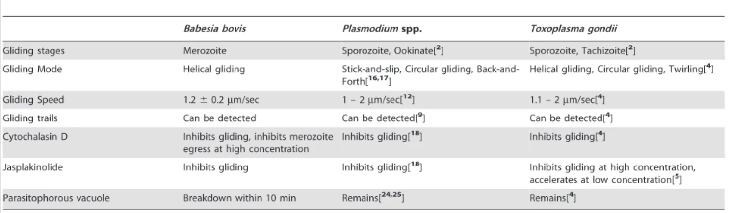
Table 2.Comparison of gliding motility among apicomplexan parasites.
Babesia bovis Plasmodiumspp. Toxoplasma gondii
Gliding stages Merozoite Sporozoite, Ookinate[2] Sporozoite, Tachizoite[2]
Gliding Mode Helical gliding Stick-and-slip, Circular gliding,
Back-and-Forth[16,17] Helical gliding, Circular gliding, Twirling[ 4]
Gliding Speed 1.260.2mm/sec 1 – 2mm/sec[12] 1.1 – 2mm/sec[4]
Gliding trails Can be detected Can be detected[9] Can be detected[4]
Cytochalasin D Inhibits gliding, inhibits merozoite egress at high concentration
Inhibits gliding[18] Inhibits gliding[4]
Jasplakinolide Inhibits gliding Inhibits gliding[18] Inhibits gliding at high concentration, accelerates at low concentration[5]
Parasitophorous vacuole Breakdown within 10 min Remains[24,25] Remains[4]
Supporting Information
Video S1 Time-lapse video microscopy of a B. bovis merozoite engaged in gliding.Video S1 is shown at 6.76real time.
(AVI)
Video S2 Helical motility ofB. bovismerozoites.Video
S2 is shown at 2.86real time. (AVI)
Video S3 Time-lapse video microscopy of B. bovis merozoite egress from a RBC, gliding to and invasion of a new RBC.Video S3 is shown at 15.46real time.
Acknowledgments
We are grateful to Dr. M. Yuda of Mie University for providing plasmids. We are also grateful to Dr. S. Fukumoto of our research center for comments regarding plasmid construction and transfection.
Author Contributions
Conceived and designed the experiments: MA SiK OK. Performed the experiments: MA. Analyzed the data: MA SiK OK YG KY NY SK NI. Contributed reagents/materials/analysis tools: SiK NI OK NY. Wrote the paper: MA SiK OK.
References
1. King CA (1988) Cell motility of sporozoan protozoa. Parasitol Today 4: 315–319.
2. Sibley LD (2004) Intracellular parasite invasion strategies. Science 304: 248–253.
3. Vanderberg JP (1974) Studies on the motility of Plasmodium sporozoites. J Protozool 21: 527–537.
4. Ha˚kansson S, Morisaki H, Heuser J, Sibley LD (1999) Time-lapse video microscopy of gliding motility inToxoplasma gondiireveals a novel, biphasic mechanism of cell locomotion. Mol Biol Cell 10: 3539–3547.
5. Wetzel DM, Schmidt J, Kuhlenschmidt MS, Dubey JP, Sibley LD (2005) Gliding motility leads to active cellular invasion by Cryptosporidium parvum sporozoites. Infect Immun 73: 5379–5387.
6. Russell DG, Sinden RE (1981) The role of the cytoskeleton in the motility of coccidian sporozoites. J Cell Sci 50: 345–359.
7. Chauvin A, Moreau E, Bonnet S, Plantard O, Malandrin (2009) Babesia and its hosts: adaptation to long-lasting interactions as a way to achieve efficient transmission. Vet Res 40: 37.
8. Gohil S, Kats LM, Sturm A, Cooke BM (2010) Recent insights into alteration of red blood cells byBabesia bovis: moovin’ forward. Trends Parasitol 26: 591–599. 9. Stewart MJ, Vanderberg JP (1988) Malaria sporozoites leave behind trails of
circumsporozoite protein during gliding motility. J Protozool 35: 389–393. 10. Sibley LD, Haˆkansson S, Carruthers VB (1998) Gliding motility: an efficient
mechanism for cell penetration. Curr Biol 8: R12–R14.
11. Amino R, Me´nard R, Frischknecht F (2005)In vivoimaging of malaria parasites– recent advances and future directions. Curr Opin Microbiol 8: 407–414. 12. Frischknecht F, Baldacci P, Martin B, Zimmer C, Thiberge S, et al. (2004)
Imaging movement of malaria parasites during transmission by Anopheles mosquitoes. Cell Microbiol 6: 687–694.
13. Asada M, Tanaka M, Goto Y, Yokoyama N, Inoue N, et al. (2012) Stable expression of green fluorescent protein and targeted disruption of thioredoxin peroxidase-1 gene inBabesia boviswith the WR99210/dhfr selection system. Mol Biochem Parasitol 181: 162–170.
14. Wetzel DM, Ha˚kansson S, Hu K, Roos D, Sibley LD (2003) Actin filament polymerization regulates gliding motility by apicomplexan parasites. Mol Biol Cell 14: 396–406.
15. Doran DJ, John TL, Rinaldi R (1962) Encystation and locomotion ofEimeria acervulmasporozoites (motion picture). J Parasitol 48 (Suppl.). 32 p.
16. Mu¨nter S, Sabass B, Selhuber-Unkel C, Kudryashev M, Hegge S, et al. (2009) Plasmodium sporozoite motility is modulated by the turnover of discrete adhesion sites. Cell Host Microbe 6: 551–562.
17. Gilson PR (2009) Malaria parasites do the stick-and-slip shuffle. Cell Host Microbe 6: 499–501.
18. Siden-Kiamos I, Pinder JC, Louis C (2006) Involvement of actin and myosins in Plasmodium bergheiookinete motility. Mol Biochem Parasitol 150: 308–317. 19. Pinder J, Fowler R, Bannister L, Dluzewski A, Mitchell GH (2000) Motile
systems in malaria merozoites: how is the red blood cell invaded? Parasitol Today 16: 240–245.
20. de Souza W (2007) Macro, micro and nano domains in the membrane of parasitic protozoa. Parasitol Int 56: 161–170.
21. Morzaria SP, Bland P, Brocklesby DW (1978) Ultrastructure ofBabesia major vermicules from the tickHaemaphysalis punctataas demonstrated by negative staining. Z Parasitenkd 55: 119–125.
22. Gratzer WB, Dluzewski AR (1993) The red blood cell and malaria parasite invasion. Semin Hematol 30: 232–247.
23. Sultan AA, Thathy V, Frevert U, Robson KJ, Crisanti A, et al. (1997) TRAP is necessary for gliding motility and infectivity of plasmodium sporozoites. Cell 90: 511–522.
24. Huynh MH, Carruthers VB (2006) Toxoplasma MIC2 is a major determinant of invasion and virulence. PLoS Pathog 2: e84.
25. Zhou J, Fukumoto S, Jia H, Yokoyama N, Zhang G, et al. (2006) Characterization of theBabesia gibsoniP18 as a homologue of thrombospondin related adhesive protein. Mol Biochem Parasitol 148: 190–198.
26. Wickham ME, Culvenor JG, Cowman AF (2003) Selective inhibition of a two-step egress of malaria parasites from the host erythrocyte. J Biol Chem 278: 37658–37663.
27. Langreth SG, Jensen JB, Reese RT, Trager W (1978) Fine structure of human malariain vitro. J Protozool 25: 443–452.
28. Bork S, Okamura M, Matsuo T, Kumar S, Yokoyama N, et al. (2005) Host serum modifies the drug susceptibility ofBabesia bovis in vitro. Parasitology 130(Pt 5): 489–492.