This study is an interesting selection of esophageal dynamic images and respective activity/time curves to demonstrate motility ranging from normal to the opposite extreme (advanced-stage achalasia). The technique employed was: 4-hour fast, with restriction of smoking, alcohol and caffeine products; anterior 0.5-second imaging during 2 minutes, covering the region from the mouth to the gastric fundus, followed by a planar 20-second image from the same region (normal transit time: < 10 seconds). The collection is based on a twenty-year experience employing a systematization including several parameters, which is able to discrimi-nate patients with normal total esophageal transit time independently of symptoms.
Keywords: Esophageal scintigraphy; Transit time; Motility; Achalasia; Parameters.
Motilidade esofagiana: ensaio iconográfico sobre cintilografia dinâmica do esôfago.
Este trabalho é uma seleção interessante de imagens dinâmicas do esôfago e de curvas de atividade/tempo, variando da motilidade normal até o extremo oposto, a acalásia em estado avançado. A técnica é a usual: quatro horas de jejum, com restrição de fumo, álcool e cafeína; incidência anterior; imagens de 0,5 segundo durante dois minutos, região da boca ao fundo gástrico, seguida de imagem plana de 20 segundos da mesma região (tempo de trânsito normal: < 10 segundos). A coletânea é baseada em vinte anos de experiência empregando uma sistematização com vários parâmetros de análise que permite discriminar pacientes com tempo total de trânsito normal.
Unitermos: Cintilografia esofagiana; Tempo de trânsito; Motilidade; Acalásia; Parâmetros.
Abstract
Resumo
* Study developed in the Department of Radiology – Nuclear Medicine at Faculty of Medicine, Universidade Federal do Rio de Janeiro (UFRJ), Rio de Janeiro, RJ, Brazil.
1. PhD, Associate Professor, Department of Radiology – Nu-clear Medicine at Faculty of Medicine, Universidade Federal do Rio de Janeiro (UFRJ), Rio de Janeiro, RJ, Brazil.
Mailing address: Profa. Dra. Maria Expósito Penas. Rua Alfredo Corrêa, 150, ap. 204, Ilha do Governador. Rio de Janeiro, RJ, Brazil, 21920-225. E-mail: [email protected]
Received March 8, 2007. Accepted after revision May 8, 2007.
INTRODUCTION
The present essay is aimed at showing
an interesting selection of images and
graphics demonstrating esophageal
motil-ity in a spectrum ranging from normal to
the opposite extreme that is an
advanced-stage achalasia. Besides studies utilizing
room-temperature water for radiotracer
dilution, further studies (Bernstein tests)
were performed with volunteers (with free
and informed consent) utilizing mild
hy-drochloric acid (at 0.1 N and 0.05 N
con-centration) to reproduce symptoms of
gas-troesophageal reflux. All of the studies
were approached by a standard
systemati-zation including the following parameters
(for normal values [n]): total esophageal
transit time (TETT) (n: < 10.0 s), curve
pat-tern (CP) (n: coordinated), residual
activi-ty (RA) (n: < 10%), time for initial entry
into stomach (TIES) (n: < 6.0 s), stomach
entry form (SEF) (n: abrupt), retrograde
movements (RM) (n: absent), curve
varia-tion factor (CVF) (n: < 0.1), transit time in
the proximal, middle and distal esophageal
thirds (PTT, MTT, DTT) (respectively n: <
3.0 s, < 6.0 s and < 10.0 s) and a plain
ra-diographic image of the esophagus (PRI)
at the end of the dynamic scintigraphy (n:
absent or mild residue)
(1–3).
REFERENCES
1. Russell COH, Hill LD, Holmes ER III, Hull DA, Gannon R, Pope CE II. Radionuclide transit: a sensitive screening test for esophageal dysfunc-tion. Gastroenterology 1981;80:887–892. 2. Blackwell JN, Hannan WJ, Adam RD, Heading
RC. Radionuclide transit studies in the detection of oesophageal dysmotility. Gut 1983;24:421–426. 3. Penas ME, Orlando MMC, Koch HA. Dynamic esophageal scintigraphy parameters to analyze in single liquid bolus swallow. Alasbimn J 2006; 8(33).
Penas ME
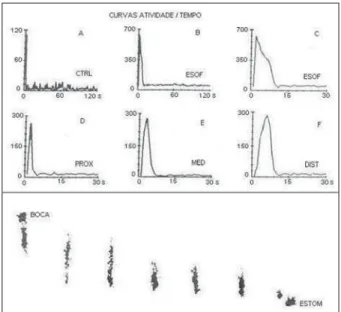
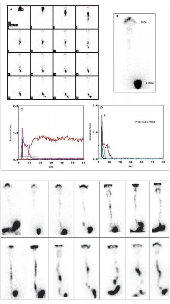
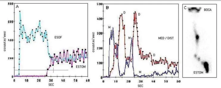
Figure 5. Female, 51-year-old patient with dysphagia and diagnosis of scleroderma, without diabetes. Manometry demonstrating adynamia of the esophageal body. A: Dynamic images showing a tortuous route. B: PRI demonstrating diffuse residual activity in the proximal and middle esophagus. C: Control curve demonstrating a single swallowing. D: Curve demonstrating overlapping of esophageal thirds, where the bolus reflux from the distal to the middle esophagus is clear, likewise in the dynamic images. E: Esophagus/stomach curve demonstrating stepped entry into stomach. F: Curve corresponding to condensed images clearly demonstrates the temporary radioactive bolus retention in the middle esophagus. Significant esophageal motor dysfunction.
Penas ME
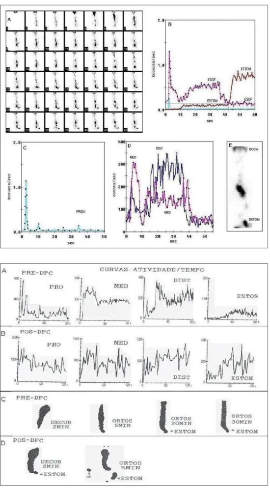
Figure 7. Female, 48-year-old patients with dysphagia and chest pain. Endos-copy suggesting achalasia and positive Bernstein test.. A: Pre-pneumatic dila-tation (PD) phase of the cardia – curves of the proximal, middle and distal esoph-ageal thirds and stomach, all of them with an abnormal aspect. B: The same sequence of curves in the post-PD phase. C: Esophageal images at the end of the dynamic study where an increase in the caliber of the proximal and middle esophagus is observed, without a clear identification of the distal esophagus. The following three images acquired in orthostatic position demonstrate the dis-tal esophagus and a subtle image of the stomach corresponding to the radioac-tive bolus with 10 ml of water that re-mained in the stomach for more than 30 minutes. D: Post-PD phase. Image ac-quired at the end of the dynamic study with a subtle contour of the stomach, a little more evident in the orthostatic po-sition, but still with an intense radiotracer presence in the esophagus. Study typi-cally demonstrating achalasia in the pre-PD phase, evidencing the low efficacy of the PD, in spite of the improvement of the symptoms reported by the patient. This study corroborates the sensitivity of the method.