Correspondence to: Miloš BJELOVIĆ
Hospital for Digestive Surgery First Surgical Hospital Clinical Center of Serbia Dr. Koste Todorovića 6 11000 Belgrade Serbia
SUMMARY
Introduction At the Department of Minimally Invasive Upper Digestive Surgery of the Hospital for Diges-tive Surgery in Belgrade, hybrid minimally invasive esophagectomy (hMIE) has been a standard of care for patients with resectable esophageal cancer since 2009. As a next and final step in the change manage-ment, from January 2015 we utilized total minimally invasive esophagectomy (tMIE) as a standard of care. Objective The aim of the study was to report initial experiences in hMIE (laparoscopic approach) for cancer and analyze surgical technique, major morbidity and 30-day mortality.
Methods A retrospective cohort study included 44 patients who underwent elective hMIE for esophageal cancer at the Department for Minimally Invasive Upper Digestive Surgery, Hospital for Digestive Surgery, Clinical Center of Serbia in Belgrade from April 2009 to December 2014.
Results There were 16 (36%) middle thoracic esophagus tumors and 28 (64%) tumors of distal thoracic esophagus. Mean duration of the operation was 319 minutes (approximately five hours and 20 minutes). The average blood loss was 173.6 ml. A total of 12 (27%) of patients had postoperative complications and mean intensive care unit stay was 2.8 days. Mean hospital stay after surgery was 16 days. The average number of harvested lymph nodes during surgery was 31.9. The overall 30-day mortality rate within 30 days after surgery was 2%.
Conclusion As long as MIE is an oncological equivalent to open esophagectomy (OE), better relation between cost savings and potentially increased effectiveness will make MIE the preferred approach in high-volume esophageal centers that are experienced in minimally invasive procedures.
Keywords: esophageal cancer; surgery; minimally invasive esophagectomy; laparoscopy; outcome
Minimally Invasive Esophagectomy for Cancer:
Single Center Experience after 44 Consecutive Cases
Miloš Bjelović1,2, Tamara Babič2, Dragan Gunjić2, Milan Veselinović2, Bratislav Špica2 1University of Belgrade, School of Medicine, Belgrade, Serbia;
2Department for Minimally Invasive Upper Digestive Surgery, Hospital for Digestive Surgery –
First Surgical Hospital, Clinical Center of Serbia, Belgrade, Serbia
INTRODUCTION
Each year 462,000 people are diagnosed with esophageal cancer worldwide and 386,000 people die from it [1, 2]. Although the patients’ satisfaction with surgery is lower comparing with other treatment options, probably due to its aggressiveness, surgical treatment remains the primary treatment for localized resect-able esophageal cancer. Open transthoracic esophagectomy (OE) as a radical procedure has been the procedure of choice in the treat-ment of resectable esophageal cancer. It is an extensive and traumatic procedure with mor-bidity rate ranging between 30% and 50%. [3]. Transhiatal esophagectomy has been designed to decrease operative trauma in comparison to the transthoracic approach, but it is palliative in origin because it is not feasible to perform lymphadenectomy of the middle and upper me-diastinum. Minimally invasive esophagectomy (MIE) results in a significant decrease of surgi-cal trauma and could provide a proper visual-ization and exposure of the posterior mediasti-num and adequate lymph node dissection [4-7]. In the majority of cases MIE is performed as a total MIE (tMIE) or hybrid MIE (hMIE) [8]. In change management from OE to tMIE, hMIE (laparoscopic or thoracoscopic procedure) is an intermediate but also a very large step. In
1992 Sir Alfred Cuschieri et al. [7] published a paper about thoracoscopic mobilization of the esophagus. The first large series (48 patients) using MIE with transhiatal approach was pub-lished by the Brazilian surgeon DePaula et al. [9]. To our knowledge, the largest personal se-ries of MIE is the sese-ries of Luketich et al. [10] with more than 1,000 operated patients.
Hybrid MIE has been routinely performed by the team of the Department for Minimally Invasive Upper Digestive Surgery, Hospital for Digestive Surgery, Clinical Center of Serbia, since 2009 [11]. As a next and final step in the change management, from 2015 we utilized the total MIE as a standard of care.
OBJECTIVE
In this article, we report initial experience after 44 consecutive cases of hMIE (laparoscopic ap-proach) for cancer and analyze surgical tech-nique, major morbidity and 30-day mortality.
METHODS
Invasive Upper Digestive Surgery, Hospital for Digestive Surgery, Clinical Center of Serbia in Belgrade from April 2009 to December 2014. The standard preoperative work-up included symptoms evaluation, barium swallow radi-ography, upper flexible endoscopy with biopsy, computed tomography of thorax and abdomen and broncho-pulmo-nary evaluation (flexible bronchoscopy and pulmobroncho-pulmo-nary function tests).
All patients underwent antibiotic and deep veins thrombosis prophylaxis. The standard surgical technique for hMIE is described in detail in further text. Control bar-ium test through a nasogastric tube (NG) was performed on the second postoperative day, followed by NG tube extraction in cases of normal pylorus transit. All patients received pulmonary physiotherapy and early mobilization. Control barium radiography was routinely performed on the seventh postoperative day followed by the clear liquid diet. Data regarding demographic characteristic, preopera-tive work-up evaluation, intraoperapreopera-tive data (duration of operation, intraoperative blood loss, etc.) and postopera-tive course details were all analyzed. In addition, postop-erative complications were analyzed separately and graded according to the Dindo–Clavien classification [12].
After hospital discharge, the first check-up was per-formed a month after surgery and then periodically ac-cording to the European Society for Medical Oncology [13]. The standard postoperative annual check-up in-cluded symptoms evaluation, control barium radiography, computed tomography of the thorax and abdomen and upper flexible endoscopy.
Surgical technique
Laparoscopic part
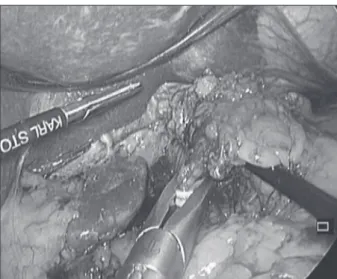
Positions of the patient and trocars were adopted from Luketich et al. [4]. The laparoscopic part commenced with the exploration of the liver, peritoneal surfaces and integrity of the epiploic arcade. Afterwards, the transection of the gastrocolic ligament is performed, keeping in mind that the aim of that part is to preserve the integrity of the arcade as it will be the only blood vessel left to supply the gastric tube. The left gastroepiploic vessels are isolated, clipped and cut. After that, shot gastric vessels should be sealed and cut all the way from the left epiploic vessels to the angle of Hiss. Then, the transection of the gastrocolic ligament is continued backward to meet the pylorus. Pyloromyotomy or pyloroplasty was performed routinely in the majority of cases. However, it seems that this step is not necessary in all patients, especially after narrow gastric tube forma-tion. Subsequently, left gastric vessels are isolated, clipped and transected. In case of squamous cell carcinoma, the left gastric vessels are ligated at the origin and lifted along with lymph nodes around the left gastric artery (Figure 1). In case of adenocarcinoma, dissection along the hepatic and splenic artery is routinely performed. Dissection of the lower mediastinum should be a part of the laparo-scopic procedure. It is easier to do it laparolaparo-scopically than via thoracotomy or thoracoscopy (Figure 2). After sealing and cutting of the gastric arcade on the lesser curvature of the stomach we proceed with gastric tube formation. On average three to four articulating endoscopic linear staplers are used in creating the gastric tube (Figure 3).
Figure 3. Gastric tubulization with endoscopic staplers
Figure 2. Laparoscopic dissection of the lymph nodes in the lower mediastinum
Open thoracic part
The patient is repositioned in the left lateral decubitus, and open thoracotomy is performed. In case of distal thoracic cancer, a typical two-field standard lymph node dissection is performed. When middle thoracic carcinoma is present, a two-field total lymph node dissection should be performed. After subtotal esophagectomy and gastric pull-up, mechan-ic esophagogastrmechan-ic anastomosis is performed in the upper mediastinum and wrapped with the part of the omentum preserved along the greater curvature of the stomach. An NG tube is placed under the direct control and the gastric stump is closed with a linear stapler. The stomach is fixed in place with a couple of stitches to the surrounding pleura, and the thorax is closed after an insertion of a chest tube.
RESULTS
The outcomes of 44 consecutive hMIEs (laparoscopic ap-proach) for esophageal cancer from April 2009 to December 2014 were included. Male patients were more prevalent (84% male and 16% female patients); mean age was 61.4 years. The average Karnofski and ASA scores were 79.8 and 2.3, respectively. The average body mass index was 22.8 kg/m2. There were 16 (36%) middle thoracic esophagus tumors and 28 (64%) tumors of the distal thoracic esophagus. Ana-lyzing tumor histology, there were 23 (52%) squamous cell carcinoma, 21 (48%) adenocarcinoma. Mean duration of operation was 319 minutes (approximately five hours and 20 minutes). In 42 patients (96%) pyloromyotomy or py-loroplasty was performed. All patients underwent stapled intrathoracic esophagogastric anastomosis. Additional surgical procedures included atypical lung resections in two patients (5%). The average blood loss was 173.6 mL. Conversion rate was zero. Mean hospital stay was 16 days. The average number of harvested lymph nodes was 31.9. These data are summarized in Table 1.
The total of 12 patients (27%) had postoperative com-plications, while the mean intensive care unit (ICU) stay was 2.8 days. Postoperative complications mainly included major pulmonary complications, such as: atelectasis in one patient (2%), ARDS in one patient (2%) and pneumonia in six patients (14%). Other significant postoperative com-plications included anastomotic leakage, herniation of the transverse colon into the thorax, hemothorax needed operation, wound infection with dehiscence and bile leak in the case of severe adhesions after previous surgery in one patient each.
In addition, all postoperative complications were ana-lyzed according to the Dindo–Clavien classification (Table 2). Overall, the 30-day mortality rate was 2%.
DISCUSSION
In the literature, under the term of MIE many different procedures can be found (Table 3), and all of these proce-dures could be performed in the left lateral decubitus or
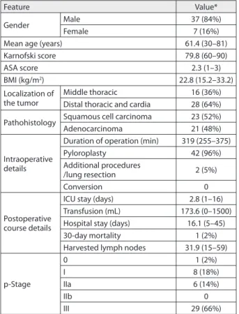
Table 1. Demographic characteristics, preoperative, intraoperative and postoperative course details
Feature Value*
Gender Male 37 (84%)
Female 7 (16%)
Mean age (years) 61.4 (30–81)
Karnofski score 79.8 (60–90)
ASA score 2.3 (1–3)
BMI (kg/m2) 22.8 (15.2–33.2)
Localization of the tumor
Middle thoracic 16 (36%) Distal thoracic and cardia 28 (64%)
Pathohistology Squamous cell carcinoma 23 (52%) Adenocarcinoma 21 (48%)
Intraoperative details
Duration of operation (min) 319 (255–375)
Pyloroplasty 42 (96%)
Additional procedures
/lung resection 2 (5%)
Conversion 0
Postoperative course details
ICU stay (days) 2.8 (1–16) Transfusion (mL) 173.6 (0–1500) Hospital stay (days) 16.1 (5–45) 30-day mortality 1 (2%) Harvested lymph nodes 31.9 (15–59)
p-Stage
0 1 (2%)
I 8 (18%)
IIa 6 (14%)
IIb 0
III 29 (66%)
* The values are presented as the number of patients (with percentage), or as mean value (with range) of the feature.
ASA – classifi cation and peri-operative risk according to the American Society of Anesthesiologists; BMI – body mass index; ICU – intensive care unit; p-Stage – pathologic stage according to UICC Classifi cation (Union for International Cancer Control)
Table 2. Postoperative complications
Feature Number
Total number of postoperative complications 13
Major pulmonary complications
Atelectasis 1 (2%)
Pneumonia 6 (14%)
Respiratory insufficiency 0
ARDS 1 (2%)
Anastomotic leakage 1 (2%)
Thoracic complications without anastomotic leakage* 2 (5%)
Other** 2 (5%)
Number of patients with complications 12 (27%)
Dindo–Clavien classification
II 7 (16%)
III 4 (9%)
IV 0
V 1 (2%)
* Thoracic complications without anastomotic leakage were hiatal hernia and hemothorax needing operation.
** Other complications were wound infection and dehiscence and bile leak in the case of severe adhesions after previous surgery.
ARDS – acute respiratory distress syndrome; Dindo–Clavien classifi cation – classifi cation of the postoperative course
Table 3. Variations of minimally invasive esophagectomy (MIE) [8] Total MIE – thoracoscopic and laparoscopic esophagectomy Hybrid MIE – either thoracoscopic or laparoscopic approach Laparoscopic-assisted transhiatal esophagectomy
prone position. In addition, the procedures could differ in respect to the extent of lymph node dissection and the position of anastomosis [8]. tMIE refers to a combined thoracoscopic and laparoscopic approach. hMIE is esopha-gectomy using either the thoracoscopic or laparoscopic approach. Although transhiatal esophagectomy can be performed using laparoscopy alone, the transcervical me-diastinoscopic approach is added in some institutions to ensure mediastinal dissection. Robot-assisted MIE using the DaVinci system has only been introduced in a limited number of institutions [14, 15]. According to the system-atic review of MIE cases published in the English language up to June 2012, tMIE is the most common procedure (58%), followed by hMIE (29%). hMIE with the thoraco-scopic approach has been performed in 17% of patients and hMIE with laparoscopic approach in 12% [16].
With so many different procedures it is difficult to com-pare postoperative outcomes of MIE with more or less standard OE. A possible solution of the problem is to do a analysis. To our knowledge, there are three meta-analyses comparing MIE with OE published by Biere et al. [17], Sgourakis et al. [18] and Nagpal et al. [19]. In addi-tion, there are at least two well-designed randomized con-trolled trials (RCTs) comparing outcomes after MIE and OE. One is the TIME trial conducted in the Netherlands by Biere et al. [20]. The trial was designed to compare tMIE and OE with 115 patients enrolled and with a primary end-point of 14-day pulmonary infection. The second trial is the MIRO trial conducted in France by Briez et al. [21]. The trial was designed to compare OE and hMIE (laparoscop-ic approach) in 280 patients enrolled and with a primary endpoint of major postoperative pulmonary complications. When duration of MIE is discussed, most agree that the du-ration of MIE is longer than that of OE. However, Nguyen et al. [22] have reported that MIE does not last significantly longer than OE if performed by experienced surgeons. In our series, the average duration of the operation was 319 minutes, which is comparable to results published by other authors [5, 23]. Gaining experience with MIE may reduce the operating time as it significantly depends on surgical experience. The average intraoperative blood loss in our series was 173.6 mL. Biere et al. [20] in their series reported average blood loss of 200 mL which is similar to our series. Also, Biere et al. [20] concluded that there was statistically significant difference in the intraoperative blood loss be-tween MIE and OE, favoring MIE. Similar results were obtained in all three meta-analyses [17, 18, 19].
The overall morbidity in our series was 27%, which is similar to results published by Luketich et al. [4] and sig-nificantly lower compared to OE series [21]. While Biere et al. [17] and Sgourakis et al. [18] found no significant difference in the pulmonary complications, in the meta-analysis performed by Nagpal et al. [19] a significant im-provement of pulmonary morbidity rates was observed in four studies, whereas no significant difference in the frequency of pulmonary complication was reported in 10 studies. The rates of major pulmonary complications in
our series do not differ significantly from those published by Briez et al. [21] with an exception of respiratory insuffi-ciency rates, probably due to the larger number of patients in their series. In the RCTs, both Biere et al. [20] and Briez et al. [21] found significantly lower prevalence of pulmo-nary complications and concluded that lower prevalence of pulmonary complications provided evidence for the short-term benefits of MIE compared with OE for patients with resectable esophageal cancer.
Length of hospital stay and stay in the ICU were reduced in tMIE and thoracoscopic-assisted groups when compared to open surgery group, suggesting early recovery in the case of MIE [17]. Our experience has demonstrated that MIE could be performed safely, with overall 30-days mortal-ity of 2%, mean ICU stay of 2.8 days and average hospital stay of 16 days, which correlates with results published by Yamamoto et al. [24] and Smithers et al. [25]. Oncologic result of different procedures could be compared in respect to the number of harvested lymph nodes and survival rates. In our series, average number of harvested lymph nodes was 31.9, which is similar to results published by Taguchi et al. [26] and even higher compared to the best OE series. Other authors have found that oncologic outcomes of MIE are not inferior to those of OE [17, 25, 27, 28]. On the contrary, the number of retrieved lymph nodes is higher in MIE than in OE. Magnifying the view of the surgical field might contribute to the increase in retrieved nodes.
In terms of overall survival, many authors believe that there is no difference in three-year and five-year survival rates between minimally invasive and open surgical ap-proach [25].
In the past decade MIE has been increasingly per-formed to treat resectable esophageal cancer. In the UK, there has been a steady increase in the uptake of MIE, with 24.7% of esophageal cancer resections in 2009 being per-formed using a hybrid or completely minimally invasive approach [29]. This fact expresses attitude in favor of the acceptance and distribution of minimally invasive pro-cedures for esophageal carcinoma treatment worldwide. Finally, Lee et al. [30] reported that MIE is cost-effective compared to OE in the management of patients with re-sectable esophageal cancer.
CONCLUSION
1. Brown L M, Devesa SS, Chow WH. Incidence of adenocarcinoma of the esophagus among white Americans by sex, stage, and age. J Nati Cancer Inst. 2008; 100(16):1184-7. [DOI: 10.1093/jnci/djn211] [PMID: 18695138]
2. Vizcain o AP, Moreno V, Lambert R, Parkin DM. Time trends incidence of both major histologic types of esophageal carcinomas in selected countries, 1973-1995. Int J Cancer. 2002; 99(6):860-8. [DOI: 10.1002/ijc.10427] [PMID: 12115489]
3. Finks J F, Osborne NH, Birkmeyer JD. Trends in hospital volume and operative mortality for high-risk surgery. N Eng J Med. 2011; 364(22):2128-37. [DOI: 10.1056/NEJMsa1010705] [PMID: 21631325] 4. Luketic h JD, Alvelo-Rivera M, Buenaventura PO, Christie NA,
McCaughan JS, Litle VR, et al. Minimally invasive esophagectomy: outcomes in 222 patients. Ann Surg. 2003; 238(4):486-95. [DOI: 10.1097/01.sla.0000089858.40725.68] [PMID: 14530720] 5. Ngu yen NT, Roberts P, Follette DM, Rivers R, Wolfe BM.
Thoracoscopic and laparoscopic esophagectomy for benign and malignant disease: lessons learned from 46 consecutive procedures. J Am Coll Surg. 2003; 197(6):902-13.
[DOI: 10.1016/j.jamcollsurg.2003.07.005] [PMID: 14644277] 6. Mehran RJ. Minimally invasive surgical treatment of esophageal
carcinoma. Gastrointest Cancer Res. 2008; 2(6):283-6. [PMID: 19259276]
7. Cuschie ri A, Shimi S, Banting S. Endoscopic oesophagectomy through a right thoracoscopic approach. J R Coll Surg Edinb. 1992; 37(1):7-11. [PMID: 1573620]
8. Watanab e M, Baba Y, Nagai Y, Baba H. Minimally invasive esophagectomy for esophageal cancer: an updated review. Surg Today. 2013; 43(3):237-44. [DOI: 10.1007/s00595-012-0300-z] [PMID: 22926551]
9. DePaula AL, Hashiba K, Bafutto M, Machado CA. Laparoscopic reoperations after failed and complicated antireflux operations. Surg Endosc. 1995; 9(6):681-6. [DOI: 10.1007/BF00187939] [PMID: 7482163]
10. Luketic h JD, Pennathur A, Awais O, Levy RM, Keeley S, Shende M, et al. Outcomes after minimally invasive esophagectomy: review of over 1000 patients. Ann Surg. 2012. 256(1):95-103.
[DOI: 10.1097/SLA.0b013e3182590603] [PMID: 22668811] 11. Bjelovi c MN, Neagoe RM. Minimally invasive esophagectomy for
cancer – short up-to-date. Jurnalul de Chirurgie. 2014; 10(3):189-91. [DOI: 10.7438/1584-9341-10-3-1]
12. Dindo D , Demartines N, Clavien PA. Classification of surgical complications: a new proposal with evaluation in a cohort of 6336 patients and results of a survey. Ann Surg. 2004; 240(2):205-13. [DOI: 10.1097/01.sla.0000133083.54934.ae] [PMID: 15273542] 13. Stahl M , Mariette C, Haustermans K, Cervantes A, Arnold D, Group
EGW. Oesophageal cancer: ESMO Clinical Practice Guidelines for diagnosis, treatment and follow-up. Ann Oncol. 2013; 24 Suppl 6:vi51-6. [DOI: 10.1093/annonc/mdt342] [PMID: 24078662] 14. Dunn DH , Johnson EM, Morphew JA, Dilworth HP, Krueger JL,
Banerji N. Robot-assisted transhiatal esophagectomy: a 3-year single-center experience. Dis Esophagus. 2013; 26(2):159-66. [DOI: 10.1111/j.1442-2050.2012.01325.x] [PMID: 22394116] 15. Galvani CA, Gorodner MV, Moser F, Jacobsen G, Chretien C, Espat NJ, et al. Robotically assisted laparoscopic transhiatal esophagectomy. Surg Endosc. 2008; 22(1):188-95. [DOI: 10.1007/s00464-007-9441-3] [PMID: 17939004]
16. Hanna G B, Arya S, Markar SR. Variation in the standard of minimally invasive esophagectomy for cancer – systematic review. Semin Thorac Cardiovasc Surg. 2012; 24(3):176-87.
[DOI: 10.1053/j.semtcvs.2012.10.004]
17. Biere SS, Cuesta MA, van der Peet DL. Minimally invasive versus open esophagectomy for cancer: a systematic review and meta-analysis. Minerva Chir. 2009; 64(2):121-33.
[DOI: 10.1016/j.ejcts.2008.09.024] [PMID: 19365313]
18. Sgourakis G, Gock el I, Radtke A, Musholt TJ, Timm S, Rink A, et al. Minimally invasive versus open esophagectomy: meta-analysis of outcomes. Dig Dis Sci. 2010; 55(11):3031-40.
[DOI: 10.1007/s10620-010-1153-1] [PMID: 20186484] 19. Nagpal K, Ahmed K , Vats A, Yakoub D, James D, Ashrafian H, et
al. Is minimally invasive surgery beneficial in the management of esophageal cancer? A meta-analysis. Surg Endosc. 2010; 24(7):1621-9. [DOI: 10.1007/s00464-009-0822-7] [PMID: 20108155] 20. Biere SS, van Ber ge Henegouwen MI, Maas KW, Bonavina L, Rosman
C, Garcia JR, et al. Minimally invasive versus open oesophagectomy for patients with oesophageal cancer: a multicenter, open-label, randomised controlled trial. Lancet. 2012; 379(9829):1887-92. [DOI: 10.1016/S0140-6736(12)60516-9] [PMID: 22552194] 21. Briez N, Piessen G, Torres F, Lebuffe G, Triboulet JP, Mariette
C. Effects of hybrid minimally invasive oesophagectomy on major postoperative pulmonary complications. Br J Surg. 2012; 99(11):1547-53. [DOI: 10.1002/bjs.8931] [PMID: 23027071] 22. Nguyen NT, Gelfan d D, Stevens CM, Chalifoux S, Chang K, Nguyen P,
et al. Current status of minimally invasive esophagectomy. Minerva Chir. 2004; 59(5):437-46. [PMID: 15494671]
23. Santillan AA, Far ma JM, Meredith KL, Shah NR, Kelley ST. Minimally invasive surgery for esophageal cancer. J Natl Compr Canc Netw. 2008; 6(9):879-84. [PMID: 18926097]
24. Yamamoto S, Kawah ara K, Maekawa T, Shiraishi T, Shirakusa T. Minimally invasive esophagectomy for stage I and II esophageal cancer. Ann Thorac Surg. 2005; 80(6):2070-5.
[DOI: 10.1016/j.athoracsur.2005.06.004] [PMID: 16305846] 25. Smithers BM, Gotl ey DC, Martin I, Thomas JM. Comparison of the
outcomes between open and minimally invasive esophagectomy. Ann Surg. 2007; 245(2):232-40.
[DOI: 10.1097/01.sla.0000225093.58071.c6] [PMID: 17245176] 26. Taguchi S, Osugi H, Higashino M, Tokuhara T, Takada N, Takemura
M, et al. Comparison of three-field esophagectomy for esophageal cancer incorporating open or thoracoscopic thoracotomy. Surg Endosc. 2003; 17(9):1445-50. [DOI: 10.1007/s00464-002-9232-9] [PMID: 12811660]
27. Noshiro H, Iwasaki H, Kobayashi K, Uchiyama A, Miyasaka Y, Masatsugu T, et al. Lymphadenectomy along the left recurrent laryngeal nerve by a minimally invasive esophagectomy in the prone position for thoracic esophageal cancer. Surg Endosc. 2010; 24(12):2965-73. [DOI: 10.1007/s00464-010-1072-4]
[PMID: 20495981]
28. Kent MS, Schucher t M, Fernando H, Luketich JD. Minimally invasive esophagectomy: state of the art. Dis Esophagus. 2006; 19(3):137-45. [DOI: 10.1111/j.1442-2050.2006.00555.x] [PMID: 16722989] 29. Mamidanna R, Bott le A, Aylin P, Faiz O, Hanna GB.
Short-term outcomes following open versus minimally invasive esophagectomy for cancer in England: a population-based national study. Ann Surg. 2012; 255(2):197-203.
[DOI: 10.1097/SLA.0b013e31823e39fa] [PMID: 22173202] 30. Lee L, Sudarshan M, Li C, Latimer E, Fried GM, Mulder DS, et al.
Cost-effectiveness of minimally invasive versus open esophagectomy for esophageal cancer. Ann Surg Oncol. 2013; 20(12):3732-9.
[DOI: 10.1245/s10434-013-3103-6] [PMID: 23838923]
Увод У Оде ље њу за ми ни мал но ин ва зив ну хи рур ги ју гор-њег ди ге стив ног трак та Кли ни ке за ди ге стив ну хи рур ги ју Кли нич ког цен тра Ср би је у Бе о гра ду хи брид на ми ни мал но ин ва зив на езо фа гек то ми ја (МИЕ) је стан дард на те ра пиј ска оп ци ја од 2009. го ди не. Као за вр шни ко рак у упра вља њу про ме ном, од ја ну а ра 2015. го ди не уве ден је кон цепт то-тал не МИЕ.
Циљ ра да Циљ ра да је био да се ана ли зи ра ис ку ство у ле-че њу кар ци но ма јед ња ка хи брид ном МИЕ из пер спек ти ве ана ли зе хи рур шке тех ни ке, те мор би ди те та и мор та ли те та бо ле сни ка.
Ме то де ра да Ре тро спек тив ном ко хорт ном сту ди јом об у хва-ће на су 44 бо ле сни ка ко ја су под врг ну та елек тив ној хи брид-ној МИЕ ра ди ле че ња кар ци но ма јед ња ка из ме ђу апри ла 2009. и де цем бра 2014. го ди не у Оде ље њу за ми ни мал но ин ва зив ну хи рур ги ју гор њег ди ге стив ног трак та Кли ни ке за ди ге стив ну хи рур ги ју Кли нич ког цен тра Ср би је у Бе о гра ду.
Ре зул та ти Од укуп ног бро ја бо ле сни ка, код 16 (36%) је утвр-ђен ту мор сред њег то ра кал ног јед ња ка, а код 28 (64%) ту-мор дис тал ног то ра кал ног јед ња ка. Опе ра ци ја је у про се ку тра ја ла 319 ми ну та (око пет са ти и два де сет ми ну та). Про се-чан гу би так кр ви је био 173,6 ми ли ли та ра. По сто пе ра ци о не ком пли ка ци је су за бе ле же не код 12 бо ле сни ка (27%), а про-сеч но вре ме про ве де но у је ди ни ци ин тен зив не не ге би ло је 2,8 да на. По сле опе ра ци је бо ле сни ци су у бол ни ци оста ја ли у про се ку 16 да на. То ком опе ра ци је укло ње но је про сеч но 31,9 лим фних жле зда. Укуп на сто па смрт ност то ком 30 да на од опе ра ци је би ла је 2%.
За кљу чак Док год је МИЕ он ко ло шки екви ва лент на отво-ре ној езо фа гек то ми ји, бо љи од нос тро шко ва и ефе ка та МИЕ чи ни је про це ду ром из бо ра у цен три ма са ис ку ством у ле че њу бо ле сти јед ња ка и цен три ма ко ји има ју зна чај но ис ку ство у ми ни мал но ин ва зив ној хи рур ги ји.
Кључ не ре чи: кар ци ном јед ња ка; хи рур ги ја; ми ни мал но ин ва зив на езо фа гек то ми ја; ла па ро ско пи ја; ис ход
Минимално
инвазивна
езофагектомија
у
лечењу
карцинома
једњака
:
искуство
након
44
операције
Милош Бјеловић1,2, Тамара Бабич2, Драган Гуњић2, Милан Веселиновић2, Братислав Шпица2
1Универзитет у Београду, Медицински факултет, Београд, Србија;
2Одељење за минимално инвазивну хирургију горњег дигестивног тракта, Клиника за дигестивну хирургију – Прва хируршка клиника,
Клинички центар Србије, Београд, Србија