Engineering a light-controlled F
1
ATPase
using structure-based protein design
Daniel Hoersch
Experimental Molecular Biophysics, Department of Physics, Freie Universita¨t Berlin, Berlin, Germany
ABSTRACT
The F1sub-complex of ATP synthase is a biological nanomotor that converts the free energy of ATP hydrolysis into mechanical work with an astonishing efficiency of up to 100% (Kinosita et al., 2000). To probe the principal mechanics of the machine, I re-engineered the active site ofE.coliF1ATPase with a structure-based protein design approach: by incorporation of a site-specific, photoswitchable crosslinker, whose end-to-end distance can be modulated by illumination
with light of two different wavelengths, a dynamic constraint was imposed on the inter-atomic distances of theaandbsubunits. Crosslinking reduced the ATP
hydrolysis activity of four designs tested in vitro and in one case created a synthetic ATPase whose activity can be reversibly modulated by subsequent illumination with near UV and blue light. The work is a first step into the direction of the long-term goal to design nanoscaled machines based on biological parts that can be precisely controlled by light.
Subjects Biochemistry, Bioengineering, Biophysics, Synthetic Biology
Keywords Azobenzene, Protein design, Light control, Molecular machine
INTRODUCTION
ATP-driven protein machines are fundamental to life. They perform extraordinarily complex and diverse biological functions: DNA replication/transcription (helicases, DNA/RNA polymerases), intracellular trafficking (myosin, kinesin, dynein), ATP production (ATP synthase), protein folding/unfolding (chaperonins, HSP90, proteasome) or maintenance of ion gradients (V-ATPase) just to name a few. However, despite the considerable amount of work invested in characterizing the static and dynamic structural features of these big protein complexes, the understanding of their detailed molecular mechanism has been limited in large by the complexity of the allosteric coupling, which converts the chemical energy of ATP hydrolysis into large-scale conformational changes.
To tackle this problem, predictive engineering is a promising way to rigorously test and improve mechanistic models of complex systems. An example of such an approach is the successful reprogramming of the homo-oligomeric group II chaperonin Mm-cpn to use light instead of ATP hydrolysis to open and close around an internal cavity by artificially constraining its conformational space (Hoersch & Kortemme, 2016;Hoersch et al., 2013). This was realized this by site-specific crosslinking of neighbouring subunits of the protein complex with the thiol-reactive molecular spacer azobenzene-dimaleimide (ABDM,Fig. 1B), that reversibly switches inter-atomic distances upon illumination with
Submitted30 April 2016
Accepted4 July 2016
Published28 July 2016
Corresponding author
Daniel Hoersch,
daniel.hoersch@fu-berlin.de
Academic editor
Kerstin Blank
Additional Information and Declarations can be found on page 8
DOI10.7717/peerj.2286
Copyright
2016 Hoersch
Distributed under
two different wavelengths of light, due to the reversible trans-cisphotoisomerization of the azobenzene group. Controlling the activity of biological and bioactive molecules with the high spatial and temporal resolution of light has a rich history, dating back to the 1970s with the introduction of caged compounds (Beharry & Woolley, 2011;Kaplan, Forbush & Hoffman, 1978;Mayer & Heckel, 2006;Szymanski et al., 2013). The crosslinking of proteins with azobenzene bearing compounds has been harnessed previously to control the secondary structure of peptides and proteins (Kumita et al., 2003;Kumita, Smart & Woolley, 2000;Zhang et al., 2010), to modulate the accessibility of a ligand to ion channels and receptors (Banghart et al., 2004;Numano et al., 2009;Volgraf et al., 2006), to modulate the activity of an enzyme (Schierling et al., 2010), to control the dimerization properties of catherin (Ritterson et al., 2013), and to regulate an ATP-driven protein translocation system (Bonardi et al., 2010).
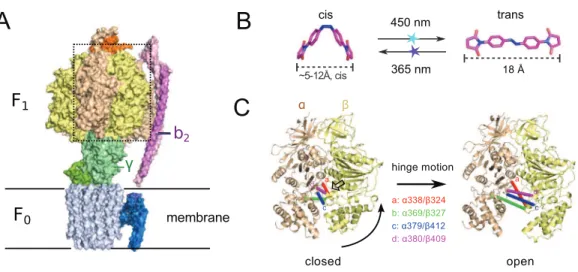
Another interesting target system for predictive re-engineering using azobenzene based crosslinker is F0F1ATP synthase, a complex, physiologically important and highly efficient biological machine (Junge & Nelson, 2015;Okuno, Iino & Noji, 2011;Walker, 2013). The protein complex consists of two fully reversible rotary motor units F0and F1, which are coupled by a rotor (g) and a stator unit (b2) (Fig. 1A). Dependent on the conditions the system either synthesizes ATP using a trans-membrane proton gradient or pumps protons when hydrolyzing ATP. In both cases the energy is transmitted between the F0 and F1units via the torque of the rotatinggunit. Under ATP synthesis conditions this rotation drives the active site, which is located at the interface of thea/bsubunits of
the F1unit (Fig. 1A) to undergo a conformational cycle that supplies the energy necessary for ATP synthesis. This structural change mainly consists of a hinge-bending motion of theb-subunit that opens up the active site for product release (Fig. 1C). F0F1can be disassembled in vitro resulting in a F1sub-complex that rotates thegsubunit upon ATP hydrolysis (Duncan et al., 1995;Noji et al., 1997;Sabbert, Engelbrecht & Junge, 1996) and synthesizes ATP if thegunit is rotated using an external force (torque) (Itoh et al., 2004;Rondelez et al., 2005).
To dissect the mechanical coupling of the ATP binding pocket of F1ATPase it might be interesting to apply an engineering strategy similar to the one applied to Mm-cpn: By picking crosslinking sites in-between thea andbsubunits ofE.coliF1, that are
supposed to undergo a distance change during the hinge bending motion that matches the distance change of ABDM during thecis/transphotoisomerization (Fig. 1B), it
should be possible to perturb the motor function of F1by artificially constraining the mobility/flexibility of the machinery of the motor in a light-dependent fashion.
Here I report a first successful step into this direction: The design and manufacturing of four double cysteine mutants ofE.coliF1ATPase for ABDM crosslinking. Incubation with ABDM leads to a formation of covalently linkeda/bdimers within the F1complex
and a significant decrease of the ATP hydrolysis activity for all tested mutants. In the case of the ABDM coupled mutantaA380C/bV409C, the ATP hydrolysis activity can
MATERIALS AND METHODS
Structure-based design
To design attachment sites for ABDM, the crystal structure ofE.coliF1, ATPase with a resolution of 3.26 A˚ was used (PDB ID: 3OAA). The expected distances between sulphur atoms for every possible pair of cysteines mutations in neighbouringa/bunits
(harbouring the active site) as well as the expected solvent accessible surface area of the sulphur were calculated using the software PyMOL (Schroedinger, LCC). The data set was then screened for residue pairs with an expected sulphur distance of 5–14 A˚ in the closed and 16.5–20.5 A˚ in the open state and a minimum expected solvent accessible surface area for the sulphur atoms of 20 A˚2(25% of the maximum solvent accessible surface area of the sulphur atom in a deprotonated cysteine residue), leading to 39 residue pairs satisfying the matching criteria. This list was visually inspected for candidates with a large distance change between the closed and open state and for which there is enough unoccupied space in between the attachment sites to accommodate ABDM. Four candidate sequence positions for crosslinking were chosen for experimental testing:a338/ b324,a369/b327,a379/b412 anda380/b409.
Plasmids
pKH4 a plasmid containing the operon of a functionalE.coliF0F1ATPase synthase in which all native cysteines are replaced with alanines and a 6xHis-tag is attached to the N-terminus of thebsubunit was a gift from W. Junge and S. Engelbrecht (Kuo, Ketchum & Nakamoto, 1998;Noji et al., 1999). All cysteine double mutants were produced by site-directed mutagenesis with the Quickchange method (Aligent Genomics). The sequences of the primers used for site-directed mutagenesis are listed in theSupplemental Material.
Figure 1 (A) Crystal structure of ATP synthase fromParacoccus denitrificans(Morales-Rios et al., 2015).(B) Chemical structure of the crosslinker ABDM in the two isomerization statescisandtrans
The incorporation of the cysteine mutations was confirmed by plasmid sequencing (Microsynth AG, Balgach, Switzerland).
Protein purification
TheE.coliF1ATPase double cysteine mutants were expressed inE.coliBL21-CodonPlus-RP cells (Aligent Technologies) grown in LB medium and purified via affinity
chromatography using a Ni-NTA agarose resin (Macherey-Nagel, Du¨ren, Germany) as described previously (Greene & Frasch, 2003). The presence of thea,bandgsubunits in
the purified samples indicates the correct folding and assembly of the F1complex.
ABDM crosslinking and photoswitching
ABDM was purchased from BIOZOL (BIOZOL Diagnostica Vertrieb GmbH, Eching, Germany). ABDM was dissolved in DMF to a concentration of 1.2 mM and stored at-20C. ABDM was added to a 0.3 mg/ml solution of the F1mutants in buffer A (20 mM HEPES pH 7.4, 100 mM KCl, 5 mM MgCl2, 5% glycerol) at a ratio of 1ml ABDM solution per 50ml protein solution. The crosslinking reaction was incubated for at least 2 h at room temperature and then quenched by addition of 1 mM DTT. To shift the azobenzene isomer equilibrium, samples were illuminated for 10 s with either a blue 3 W LED (447 nm, LUXEON Rebel) to accumulate thetransstate or a 3 W UV LED (365 nm, LED Engin) to accumulate thecisstate. Longer illumination times did not change thetrans/cisisomer equilibrium (Fig. S2). The crosslinking ratio of the samples was determined via analysis on a 8% sodium dodecyl sulphate (SDS)-PAGE gel by calculating the intensities of thea/bdimer band relative to theaandbmonomer bands
with the ImageJ software package (Schneider, Rasband & Eliceiri, 2012).
UV-VIS spectroscopy
The absorption spectra shown inFigs. S1andS2were recorded with a Shimadzu UV-2450 UV-VIS spectrometer. The samples were prepared by washing and concentrating ABDM crosslinked F1aA380C/bV409C in buffer A using an Amicon-ultra 0.5 ml centrifugal filter with 50 kDa pore size (Millipore, Billerica, MA, USA).
ATPase assay
RESULTS AND DISCUSSION
Structure-based design of F1 double cysteine mutants
In a computational screen of the crystal structure of the asymmetricE.coliF1ATPase unit (PDB ID: 3OAA (Cingolani & Duncan, 2011)) four promising crosslinking sites in between theaandbsubunits were identified, whose distances for the closed and open ATP
binding cleft match the end-to-end distance of ABDM in thecisandtransisomerization states (seeFig. 1BandTable 1). Cysteines for crosslinking were introduced by site-directed mutagenesis into a plasmid with a functional cysteine-free ATP synthase operon.
ABDM crosslinks the double cysteine mutants with varying efficiency
After expression and purification the F1units the double cysteine mutants were
incubated with ABDM in thetransisomerization state (trans-ABDM). The formation of covalently crosslinkeda/bdimers was monitored on a SDS PAGE-gel (Fig. 2). From the
intensities of the monomer and dimer bands it is possible to estimate the crosslinking ratio of the samples defined as the number crosslinks divided by the number of possible crosslinking sites (Hoersch et al., 2013). The crosslinking ratios for the different mutants are 0.2 for ABDM-aS338C/bQ324C and ABDM-aN369C/bS327C, 0.3 for ABDM-aG379C/bG412C and 0.4 for ABDM-aA380C/bV409C. These numbers are below one, the
value for full crosslinking; note however that thea3/b3barrel of the F1sub-complex
is asymmetric in the presence of thegunit and only one of the three active sites is in the open conformation (Abrahams et al., 1994;Cingolani & Duncan, 2011). This limits the maximal achievable crosslinking ratio.
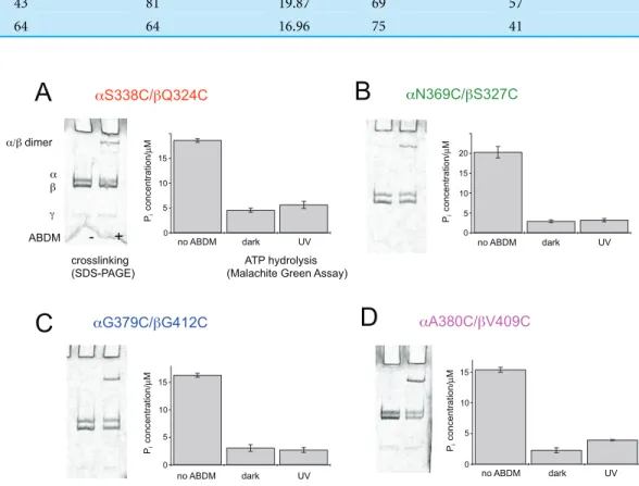
ABDM crosslinking reduces the ATP hydrolysis activity of F1
For the same set of samples the ATPase activity was determined using the colorimetric Malachite Green assay. The assay senses the presence of inorganic phosphate in the sample after incubation with ATP via complex formation with malachite green molybdate. The complex absorbs at 620 nm and its concentration can be determined using a conventional UV-VIS spectrometer.Figure 2shows that for all double cysteine mutants ABDM-crosslinking leads to a decrease of the ATP hydrolysis activity of the sample. The ATPase activity decreases for the different ABDM-incubated mutants to a fraction between 0.14 and 0.24. This behavior is in line with the design strategy as the ABDM crosslinker is supposed to constrain the flexibility of the active site by stabilizing either the open (trans-ABDM) or closed (cis-ABDM) conformation of the nucleotide binding pocket. Note that the ATPase activity of F1is highly cooperative therefore controlling the conformation of oneabdimer might be sufficient to control the ATPase activity of the
whole F1complex (Boyer, 1997;Milgrom & Cross, 2005).
samples (ABDM-aS338C/bQ324C, ABDM-aN369C/bS327C and ABDM-aG379C/ bG412C) UV light illumination did not result in significant change of the ATP hydrolysis activity (Figs. 2A–2C). For ABDM-aA380C/bV409C however, UV light induced an
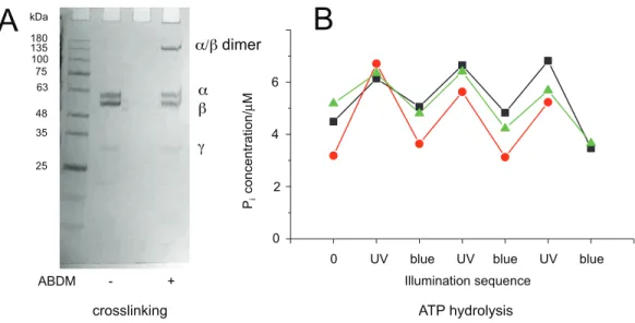
increase in ATP hydrolysis activity (Fig. 2D). To test if this light-dependent change in ATPase activity is reversible, the same assay was performed on three independently crosslinkedaA380C/bV409C samples that were exposed to alternating illumination at
365 and 450 nm (Note that 450 nm illumination induces acis/transphotoisomerization
ABDM).Figure 3shows that for all 3 samples illumination leads to a reversible
modulation of the ATP hydrolysis activity. UV light inducedtrans/cisisomerization of
0 5 10 15 0 5 10 15 0 5 10 15 20 25 0 5 10 15 20
S338C/ Q324C N369C/ S327C
G379C/ G412C A380C/ V409C
crosslinking ATP hydrolysis (SDS-PAGE) (Malachite Green Assay)
ABDM - +
/ dimer
A
B
C
D
no ABDM dark UV
15 10 5 0 P i concentration/ µ M
no ABDM dark UV
15 10 5 0 P i concentration/ µ M
no ABDM dark UV
15 10 5 0 P i concentration/ µ M
no ABDM dark UV
15 10 5 0 P i concentration/ µ M 20
Figure 2 ABDM crosslinking and ATPase activity of the F1double cysteine mutants.(A)aS338C/
bQ324C, (B) aN369C/bS327C, (C) aG379C/bG412C and (D) aA380C/bV409C. (A, C) Depict SDS-PAGE gels of the mutants before and after incubation with ABDM. The appearance of a shifted high molecular weight band indicates the formation of crosslinks between theaandbsubunit. (B, D) Show the F1catalyzed increase of the Piconcentration of inorganic phosphate (Pi) after ATP incubation of the
sample for the F1double cysteine mutant (no ABDM), thetrans-ABDM crosslinked F1mutant (dark)
and the ABDM crosslinked F1mutant after UV light illumination (UV). Shown are the mean values and
standard error of three independent experiments.
Table 1 Results of a computational screen for ABDM crosslinking sites.Resaand Resbare the amino acid sequence positions chosen for cysteine mutation in theaand b subunit of F1. Dist () refers to the expected distance of the sulphur atoms of the engineered cysteines in neighbouring subunits (seeFig. 1B). SASASG() is the expected surface accessible area for the deprotonated SG atom of the corresponding cysteine.
“cl” and “op” denote the open and closed ATP binding pocket, respectively.
PDB ID Resa Resb Dist (cl) (A˚ ) SASASG(a, cl) (A˚2) SASASG(b, cl) (A˚2) Dist (op) (A˚ ) SASASG(a, op) (A˚2) SASASG(b, op) (A˚2)
3OAA 338 324 7.40 63 75 19.73 71 65
369 327 8.66 27 43 20.41 27 67
379 412 6.11 43 81 19.87 69 57
ABDM increases the activity and blue light inducedcis/transisomerization decreases the
activity over several illumination cycles. UV-VIS absorption spectroscopy confirms fully reversible trans/cisphotoswitching of ABDM in ABDM-aA380C/bV409C under the
applied illumination conditions (Fig. S1) (Umeki et al., 2004).
It is possible to explain this behavior with the different molecular properties of the two isomerization states of ABDM:trans-azobenzene is predicted to be more rigid thancis-azobenzene (Fig. 1B). For the calculated distance distributions of the azobenzene isomerization states, see Beharry et al., 2012;Schafer et al., 2007;Zhang et al., 2009. In a consequencetrans-ABDM likely imposes a stronger distance constraint on the conformation of the active site of F1thancis-ABDM. As the active site of F1has to switch constantly between the open and closed conformation for ATP binding, hydrolysis and product release,trans-ABDM might therefore perturb the conformational cycle more efficiently than cis-ABDM. This behavior is exactly what is observed in the experiment.
Previously crosslinking via engineered disulfide bridges has been used to constrain the rotation of thegunit in respect to thea3/b3 barrel of the F1unit and to determine
interaction between subunits of the F0unit of ATP synthase (DeLeon-Rangel et al., 2013;
Duncan et al., 1995;Sielaff et al., 2008). However, to my knowledge the strategy of
crosslinking with azobenzene bearing compounds has not been applied to F0F1ATP synthase so far.
CONCLUSIONS
This work shows for the first time that it is possible to manipulate the molecular machinery ofE.coliF1ATPase reversibly in a light-dependent fashion using the
crosslinking ATP hydrolysis
ABDM - +
/ dimer
Illumination sequence
75
48
25 135 kDa
63
35 100 180
0 UV blue UV blue UV blue 4
2
0 6
P
i
concentration/
µ
M
A
B
Figure 3 Light-dependent ATPase activity of ABDM-crosslinked F1aA380C/bV409C.(A) SDS-PAGE
gels of the mutant before and after incubation with ABDM. The appearance of a shifted high molecular weight band indicates the formation of crosslinks between theaandbsubunit (crosslinking ratio: 0.3). (B) F1catalyzed increase of the Piconcentration after ATP incubation for atrans-ABDM crosslinked F1
photoswitchable molecular spacer ABDM. It is an initial step in the direction of re-engineering protein-based molecular motors using the azobenzene-based spacer to constrain and manipulate the position of moving parts of the machinery. In future experiments I want to test if the azobenzene spacer is able to actively drive the conformational cycle of the active site of F1ATPase using the energy of the photo-isomerizing azobenzene. This will help to elucidate the design principles and dynamic properties of biological motors and might in the long run inspire the bottom-up design of synthetic nano-scaled machines based on biological parts.
ACKNOWLEDGEMENTS
I thank Wolfgang Junge and Siggi Engelbrecht for the gift of pKH4.
ADDITIONAL INFORMATION AND DECLARATIONS
Funding
This research was supported by the DRS POINT fellowship program of Freie Universita¨t Berlin. The funders had no role in study design, data collection and analysis, decision to publish, or preparation of the manuscript..
Competing Interests
The author declares that they have no competing interests.
Author Contributions
Daniel Hoersch conceived and designed the experiments, performed the experiments, analyzed the data, contributed reagents/materials/analysis tools, wrote the paper, prepared figures and/or tables, reviewed drafts of the paper.
Data Deposition
The following information was supplied regarding data availability: The raw data has been supplied asSupplemental Dataset Files.
Supplemental Information
Supplemental information for this article can be found online athttp://dx.doi.org/
10.7717/peerj.2286#supplemental-information.
REFERENCES
Abrahams JP, Leslie AGW, Lutter R, Walker JE. 1994.Structure at 2.8 Aˆ resolution of F1-ATPase from bovine heart mitochondria.Nature370(6491):621–628
DOI 10.1038/370621a0.
Banghart M, Borges K, Isacoff E, Trauner D, Kramer RH. 2004.Light-activated ion channels for remote control of neuronal firing.Nature Neuroscience7(12):1381–1386
DOI 10.1038/nn1356.
photoswitchable distance constraints on the structure of a globular protein.Biochemistry 51(32):6421–6431DOI 10.1021/bi300685a.
Beharry AA, Woolley GA. 2011.Azobenzene photoswitches for biomolecules.Chemical Society Reviews40(8):4422–4437DOI 10.1039/C1CS15023E.
Bonardi F, London G, Nouwen N, Feringa BL, Driessen AJM. 2010.Light-induced control of protein translocation by the SecYEG complex.Angewandte Chemie International Edition 49(40):7234–7238DOI 10.1002/anie.201002243.
Boyer PD. 1997.The ATP synthase–a splendid molecular machine.Annual Review of Biochemistry 66(1):717–749DOI 10.1146/annurev.biochem.66.1.717.
Cingolani G, Duncan TM. 2011.Structure of the ATP synthase catalytic complex (F1) from
Escherichia coliin an autoinhibited conformation.Nature Structural & Molecular Biology 18(6):701–707DOI 10.1038/nsmb.2058.
DeLeon-Rangel J, Ishmukhametov RR, Jiang W, Fillingame RH, Vik SB. 2013.Interactions between subunitsaandbin the rotary ATP synthase as determined by cross-linking.FEBS Letters587(7):892–897DOI 10.1016/j.febslet.2013.02.012.
Duncan TM, Bulygin VV, Zhou Y, Hutcheon ML, Cross RL. 1995.Rotation of subunits during catalysis by Escherichia coli F1-ATPase.Proceedings of the National
Academy of Sciences of the United States of America92(24):10964–10968
DOI 10.1073/pnas.92.24.10964.
Greene MD, Frasch WD. 2003.Interactions amonggR268,gQ269, and thebsubunit catch
loop ofEscherichia coliF1-ATPase are important for catalytic activity.Journal of Biological
Chemistry278(51):51594–51598DOI 10.1074/jbc.M309948200.
Hanwell MD, Curtis DE, Lonie DC, Vandermeersch T, Zurek E, Hutchison GR. 2012.Avogadro: an advanced semantic chemical editor, visualization, and analysis platform.Journal of Cheminformatics4(1):17DOI 10.1186/1758-2946-4-17.
Hoersch D, Kortemme T. 2016.A model for the molecular mechanism of an engineered light-driven protein machine.Structure24(4):576–584DOI 10.1016/j.str.2016.02.015.
Hoersch D, Roh S-H, Chiu W, Kortemme T. 2013.Reprogramming an ATP-driven protein machine into a light-gated nanocage.Nature Nanotechnology8(12):928–932
DOI 10.1038/nnano.2013.242.
Itoh H, Takahashi A, Adachi K, Noji H, Yasuda R, Yoshida M, Kinosita K Jr. 2004.
Mechanically driven ATP synthesis by F1-ATPase.Nature427(6973):465–468
DOI 10.1038/nature02212.
Junge W, Nelson N. 2015.ATP synthase.Annual Review of Biochemistry84(1):631–657
DOI 10.1146/annurev-biochem-060614-034124.
Kaplan JH, Forbush B III, Hoffman JF. 1978.Rapid photolytic release of adenosine 5′ -triphosphate from a protected analogue: utilization by the Na:K pump of human red blood cell ghosts.Biochemistry17(10):1929–1935DOI 10.1021/bi00603a020.
Kinosita K Jr, Yasuda R, Noji H, Adachi K. 2000.A rotary molecular motor that can work at near 100% efficiency.Philosophical Transactions of the Royal Society B: Biological Sciences
355(1396):473–489DOI 10.1098/rstb.2000.0589.
Kumita JR, Flint DG, Woolley GA, Smart OS. 2003.Achieving photo-control of protein conformation and activity: producing a photo-controlled leucine zipper.Faraday Discussions 122:89–103DOI 10.1039/B200897A.
Kumita JR, Smart OS, Woolley GA. 2000.Photo-control of helix content in a short peptide. Proceedings of the National Academy of Sciences of the United States of America97(8):3803–3808
Kuo PH, Ketchum CJ, Nakamoto RK. 1998.Stability and functionality of cysteine-less FOF1
ATP synthase fromEscherichia coli.FEBS Letters426(2):217–220
DOI 10.1016/S0014-5793(98)00337-8.
Mayer G, Heckel A. 2006.Biologically active molecules with a “light switch”.Angewandte Chemie International Edition45(30):4900–4921DOI 10.1002/anie.200600387.
Milgrom YM, Cross RL. 2005.Rapid hydrolysis of ATP by mitochondrial F1-ATPase
correlates with the filling of the second of three catalytic sites.Proceedings of the National Academy of Sciences of the United States of America102(39):13831–13836
DOI 10.1073/pnas.0507139102.
Morales-Rios E, Montgomery MG, Leslie AGW, Walker JE. 2015.Structure of ATP synthase from Paracoccus denitrificansdetermined by X-ray crystallography at 4.0 A˚ resolution.Proceedings of the National Academy of Sciences of the United States of America112(43):13231–13236
DOI 10.1073/pnas.1517542112.
Noji H, Ha¨sler K, Junge W, Kinosita K Jr, Yoshida M, Engelbrecht S. 1999.Rotation of Escherichia coliF1-ATPase.Biochemical and Biophysical Research Communications
260(3):597–599DOI 10.1006/bbrc.1999.0885.
Noji H, Yasuda R, Yoshida M, Kinosita K Jr. 1997.Direct observation of the rotation of F1-ATPase.Nature386(6622):299–302DOI 10.1038/386299a0.
Numano R, Szobota S, Lau AY, Gorostiza P, Volgraf M, Roux B, Trauner D, Isacoff EY. 2009.
Nanosculpting reversed wavelength sensitivity into a photoswitchable iGluR.Proceedings of the National Academy of Sciences of the United States of America106(16):6814–6819
DOI 10.1073/pnas.0811899106.
Okuno D, Iino R, Noji H. 2011.Rotation and structure ofFOF1-ATP synthase.Journal of Biochemistry149(6):655–664DOI 10.1093/jb/mvr049.
Ritterson RS, Kuchenbecker KM, Michalik M, Kortemme T. 2013.Design of a photoswitchable cadherin.Journal of the American Chemical Society135(34):12516–12519
DOI 10.1021/ja404992r.
Rondelez Y, Tresset G, Nakashima T, Kato-Yamada Y, Fujita H, Takeuchi S, Noji H. 2005.Highly coupled ATP synthesis by F1-ATPase single molecules.Nature433(7027):773–777
DOI 10.1038/nature03277.
Sabbert D, Engelbrecht S, Junge W. 1996.Intersubunit rotation in active F-ATPase.Nature 381(6583):623–625DOI 10.1038/381623a0.
Schafer LV, Muller EM, Gaub HE, Grubmuller H. 2007.Elastic properties of photoswitchable azobenzene polymers from molecular dynamics simulations.Angewandte Chemie International Edition46(13):2232–2237DOI 10.1002/anie.200604595.
Schierling B, Noel A-J, Wende W, Hien LT, Volkov E, Kubareva E, Oretskaya T, Kokkinidis M, Ro¨mpp A, Spengler B, Pingoud A. 2010.Controlling the enzymatic activity of a restriction enzyme by light.Proceedings of the National Academy of Sciences of the United States of America107(4):1361–1366
DOI 10.1073/pnas.0909444107.
Schneider CA, Rasband WS, Eliceiri KW. 2012.NIH Image to ImageJ: 25 years of image analysis. Nature Methods9(7):671–675DOI 10.1038/nmeth.2089.
Sielaff H, Rennekamp H, Wachter A, Xie H, Hilbers F, Feldbauer K, Dunn SD, Engelbrecht S, Junge W. 2008.Domain compliance and elastic power transmission in rotary FOF1-ATPase. Proceedings of the National Academy of Sciences of the United States of America105(46):
Szymanski W, Beierle JM, Kistemaker HAV, Velema WA, Feringa BL. 2013. Reversible
photocontrol of biological systems by the incorporation of molecular photoswitches.Chemical Reviews113(8):6114–6178DOI 10.1021/cr300179f.
Umeki N, Yoshizawa T, Sugimoto Y, Mitsui T, Wakabayashi K, Maruta S. 2004.Incorporation of an azobenzene derivative into the energy transducing site of skeletal muscle myosin results in photo-induced conformational changes.Journal of Biochemistry136(6):839–846
DOI 10.1093/jb/mvh194.
Volgraf M, Gorostiza P, Numano R, Kramer RH, Isacoff EY, Trauner D. 2006.Allosteric control of an ionotropic glutamate receptor with an optical switch.Nature Chemical Biology2(1):47–52
DOI 10.1038/nchembio756.
Walker JE. 2013.The ATP synthase: the understood, the uncertain and the unknown.Biochemical Society Transactions41(1):1–16DOI 10.1042/BST20110773.
Zhang F, Timm KA, Arndt KM, Woolley GA. 2010.Photocontrol of coiled-coil proteins in living cells.Angewandte Chemie International Edition49(23):3943–3946
DOI 10.1002/anie.201000909.
Zhang F, Zarrine-Afsar A, Al-Abdul-Wahid MS, Prosser RS, Davidson AR, Woolley GA. 2009.