Correspondence to: Neda STEFANOVIĆ
Resavska 88/33, 11000 Belgrade Serbia
SUMMARY
Introduction Dentofacial deformity, a deviation from normal facial proportions and dental relationships, is corrected by jaw repositioning in all three spatial planes, which changes the position and tension of the surrounding tissues, bones and muscles. These changes may also affect the dimensions of the pharyngeal airways (PA).
Objective The aim of this study was to evaluate and compare three-dimensional PA changes in patients treated by a combination mandibular set-back/maxillary advancement versus patients that had bimaxil-lary advancement with genioplasty.
Methods The sample consisted of 7 patients treated by combined mandibular set-back/maxillary ad-vancement and 7 patients treated with bimaxillary adad-vancement surgery. Nasopharyngeal (NP) volume, oropharyngeal (OP) volume and the area of maximum constriction (AMC) in the OP were measured on CBCT scans (2 mA/120 kV/12’’ FOV) taken before (T1) and 3 months after surgery (T2). Paired samples t-test was used for analyzing statistical significance of changes (p≤0.05).
Results OP volume and AMC increase after bimaxillary advancement was statistically significant, while for the mandibular set-back group the increase was non-significant. NP volume was not reduced in any of the two groups. No significant differences in PA dimensions were found between groups at neither T1 nor T2 time points.
Conclusion Results suggest that the combination of mandibular set-back/maxillary advancement did not reduce airway dimensions, while bimaxillary advancement surgery led to a statistically significant increase in the OP dimensions.
Keywords: cone beam CT; bimaxillary orthognathic surgery; pharyngeal airway
Pharyngeal Airway Changes after Bimaxillary
Orthognathic Surgery – Preliminary Results
Neda Lj. Stefanović1, Branislav Glišić1, Predrag V. Nikolić1, Jovana Juloski1, Juan Martin Palomo2 1University of Belgrade, Faculty of Dental Medicine, Department of Orthodontics, Belgrade, Serbia; 2Case Western Reserve University, School of Dental Medicine, Department of Orthodontics and
Craniofacial Imaging Center, Cleveland, Ohio, USA
INTRODUCTION
Dentofacial deformity is defined as a handicap-ping deviation from normal facial proportions and dental relationships. Treatment of such deformity is complex and involves orthodon-tists, maxillofacial surgeons and other dental specialists. Aesthetic and functional problems are corrected by jaw repositioning in all three spatial planes [1]. Skeletal movements change the position and tension of the surrounding soft tissues, tongue, soft palate, hyoid bone and muscles, which are directly or indirectly connected to the upper and/or lower jaw. These changes may also affect the dimensions of the oral and nasal cavities, as well as the pharyngeal airway space (PAS) [2, 3]. The most commonly preformed bimaxillary orthognathic surgeries are mandibular set-back combined with max-illary advancement and maxillo-mandibular advancement.
Mandibular set-back combined with maxil-lary advancement is a procedure used to treat class III malocclusions. It has been shown that class III correction by mandibular set-back only can cause a reduction in pharyngeal air-way dimensions, therefore additional maxillary advancement is suggested in order to prevent potential breathing problems [4, 5].
Maxillo-mandibular advancement (MMA) combined with genioplasty was first described as a procedure for treating the obstructive sleep apnea (OSA) syndrome [6]. It is performed by means of the Le Fort I and bilateral sagittal split (BSS) osteotomies, after which both jaws are moved anteriorly. This leads to anterior repo-sitioning of the soft palate, tongue and pha-ryngeal tissues.
OBJECTIVE
The aim of this study was to analyze and compare three-dimensional (3D) pharyngeal airway changes in surgical patients treated by mandibular set-back and maxillary advance-ment and patients that had bimaxillary ad-vancement with genioplasty.
METHODS
268
Stefanović N. LJ. et al. Pharyngeal Airway Changes after Bimaxillary Orthognathic Surgery – Preliminary Results
Group A consisted of 7 patients treated by combined mandibular set-back/maxillary advancement, and group B consisted of 7 patients treated by maxillo-mandibular advancement (MMA) with genioplasty. Groups were matched for age and gender.
All patients were treated with standard edgewise appli-ances and orthognathic surgery. CBCT scans were taken before (T1) and 3 months after surgery (T2) using a custom Hitachi CB MercuRay scanner (Hitachi Medical Systems America Inc., Twinsburg, OH). The scanner settings were adjusted in order to fully comply with the ALARA (As Low As Reasonably Achievable) standards, while main-taining acceptable diagnostic image quality [7, 8]. Images were taken at 2 mA, 120 kV, and a 12-inch field of view (F Mode) setting, with the scanning time of 9.6 seconds. Image data for each patient consisted of 512 slices, with isometric voxels sized 0.377 mm. Image resolution was 1024×1024 pixels and 12 bits per pixel (4096 grayscale). Patients were scanned in the sitting position with head in the natural head posture and teeth in maximum intercus-pation. Scanning was performed at the end of the exhala-tion period when the patient was not swallowing. The im-ages were taken during the regular diagnostic procedures of obtaining orthodontic records. Patients have signed the informed consent form that allows the use of their records for research and publication purposes. The research was also approved by the Human Research Ethics Committee of the University of Belgrade Faculty of Dental Medicine (resolution number 36/20 from December 14, 2009).
DICOM (Digital Imaging and Communication in Medicine) images were analyzed using the InVivo Den-tal Software (Anatomage Inc., San Jose, CA, USA). Image orientation was performed in the Section view according to the axial, coronal and sagittal slices (Figure 1). Foramen incisivum served as a reference point for determining the midsagittal plane on the axial slice. On the sagittal slice palatal plane was oriented so that it coincided with the True Horizontal Plane and on the coronal slice Infraor-bitale points were aligned. Images were further worked on in the Volume Render view where orientation was transmitted automatically. Grayscale view images with maximum intensity reconstruction were moved upward or downward with the Patient Orientation tool when needed, so that the palatal plane coincided with the central hori-zontal line of the grid. Slice view and the Volume Render view were then matched.
Positive airway creation and volume calculation was also performed in the Volume Render view. Grayscale im-ages were put in top orientation, with volume rendering reconstruction, and were then inversed. Opacity was de-creased in order to visualize internal structures. Sculpting tool was used to cut away unnecessary parts (Figure 2A) and the partly sculpted images were then oriented to Right Lateral view where sculpting was continued (Figure 2B). Images were then reoriented back to Top view and maxil-lary sinuses were removed (Figure 2C). After obtaining the desired airway, opacity was increased, brightness and contrast were readjusted and a solid airway was created for calculating the final airway volume (Figure 2D).
Nasal passages (NP)
Inferior border of the NP was defined using the horizon-tal line through the palahorizon-tal plane (Figure 3). The superior border was determined in the Section view by moving the axial reference plane on the sagittal slice until reaching the axial slice on which the nasal septum first fuses with the posterior wall of the pharynx (Figure 3). Distance meas-uring tool was used to measure the distance between the superior and inferior borders.
The 3D Volume Clipping Tool in the Volume Render view was used to cut the airway along the axial plane. Clip-ping plane was moved when needed to concur with the inferior NP border by scrolling the mouse wheel. Distance Measuring Tool was used to measure the distance between the borders obtained earlier, and using the Clipping Tool the part above the superior border was eliminated. Maxil-lary sinuses were cut away in Top view orientation, and the definite NP volume was obtained.
Oropharyngeal airways (OP)
Inferior NP border (palatal plane) was used as the supe-rior OP border (Figure 3) and the horizontal line through the most anteroinferior point of the second cervical ver-tebrae as the inferior OP border (Figure 3). The distance between OP borders was measured in the same way as the NP borders.
The NP airway volume was flipped to the side under-neath the palatal plane using the Flip option of the 3D Vol-ume Clipping Tool. The distance between the OP borders was transferred to the airway volume and the part below the inferior border was cut with the Sculpting Tool. OP volume was measured using the Volume Measuring Tool.
All volumes were calculated using automatic segmenta-tion, i.e. the Volumetric Measuring Tool, which calculates and displays the desired volume measurement in cubic millimeters (mm³) and cubic centimeters (cc).
Area of maximum constriction in the OP
The area of maximum constriction (AMC) in the OP was measured on the axial slices in the Sectional view by means of the Area Measuring Tool. The maximum constriction slice was identified by moving the axial reference plane on the sagittal slice while observing the airway area on the corresponding axial slice.
Cephalometric analysis
The methodology used has been previously applied with success [3, 9]. All measuring has been performed and repeated for reliability testing by a single operator (NLjS) trained by an expert (JMP).
Statistical analysis
Data processing and descriptive statistics (means, stand-ard deviations and ranges for pretreatment (T1) and post-treatment (T2) records) was done using the Micro-soft Office Excel 2010 package (MicroMicro-soft Corporation, Redmond, WA). SPSS software package (version 12, SPSS Inc., Chicago, IL) was used for further statistical analysis. Intraoperator reliability for each measurement was deter-mined using the intraclass correlation coefficient (ICC). The Kolmogorov–Smirnov test revealed the normality of distribution for all data, therefore parametric tests were used. Statistical significance of changes between T1 and T2 was analyzed using paired samples t-test, with the level of significance set at p<0.05.
Figure 1. Image orientation according to the axial, sagittal and coronal slices
Figure 2. Image orientation, views and reconstruction during positive airway creation
270
RESULTS
The intraclass correlation coefficient for all measured pa-rameters showed high reliability and reproducibility of mea-surements (r>0.95).
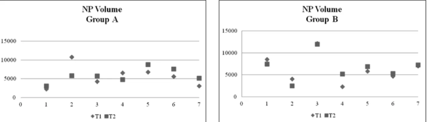
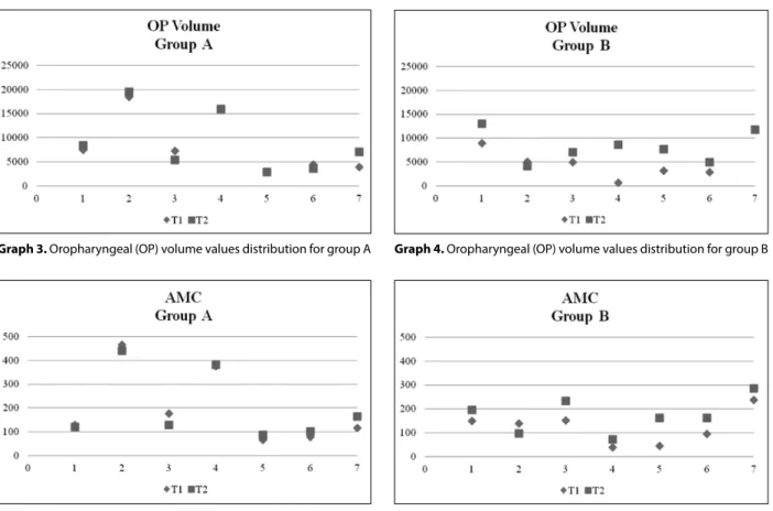
Mean ages and cephalometric measurements at T1 and T2 for both groups are presented in Table 1, while Table 2 contains pharyngeal airway measurements. Postoperative OP and NP volumes, as well as the AMC, increased in both groups. OP volume and AMC increase after bimaxillary advancement (group B) was statistically significant (Table 2). No significant differences were found between groups at T1 and T2 (Table 3).
Distribution of NP volume values before and after sur-gery is presented in Graph 1 for group A and in Graph 2 for group B. Distribution of OP volume values before and after surgery is presented in Graph 3 for group A and in
Graph 4 for group B. Distribution of AMC values before and after surgery is shown in Graph 5 for group A and in Graph 6 for group B.
DISCUSSION
Jaw repositioning by orthognathic surgery changes the position and tension of the surrounding structures, there-fore affecting the dimensions of the pharyngeal airway space. The quantity of PAS dimension changes depends on the intensity and direction of skeletal movement [2]. This study was designed to assess PAS changes in patients treated by a combination of orthodontic treatment and bimaxillary orthognathic surgery. Using the information from the DICOM images provided by a single CBCT scan, we were able to analyze the PAS of our patients easily and
Table 1. Average age and sagittal parameters for groups A and B
Parameter Age (years) SNA SNB ANB A-Nperp B-Nperp
T1 T1 T2 T1 T2 T1 T2 T1 T2 T1 T2
Group A (n=7) 18.18±1.2 82.36±4.37 85.56±3.86 83.11±2.49 81.01±2.43 -0.74±4.14 4.49±3.23 -0.33±5.24 2.94±3.88 0.20±4.26 -3.37±4.06 Group B (n=7) 19.75±3.79 79.94±3.9 83.99±4.64 77.19±5.95 80.16±4.52 2.76±2.72 3.86±0.8 -2.77±4.32 -2.21±10.79 -6.30±7.67 -5.07±7.57
SNA – sagittal position of the maxilla relative to the cranial base; SNB – sagittal position of the mandible relative to the cranial base; ANB – intermaxillary sagittal relation
Table 2. Descriptive statistics and comparison of pharyngeal airway measurements at T1 and T2 for groups A and B
Parameter T1 T2 p Δ (T2–T1)
Mean SD Min Max Mean SD Min Max Mean±SD
Group A (n=7)
NP volume (mm3) 5,590.43 2,835.66 2,238 10,737 5,827.14 1,844.55 3,082 8,722 0.821 236.71±2,652.08
OP volume (mm3) 8,620.71 6,156.43 2,890 18,463 8,962.14 6,367.22 2,870 19,528 0.593 341.43±1,600.51
AMC (mm2) 200.42 156.42 65.11 464.76 202.96 144.74 86.81 439.85 0.843 2.54±32.48
Group B (n=7)
NP volume (mm3) 6,342.29 3,262.56 2,280 12,167 6,642.71 2,907.42 2,482 11,982 0.609 2,993.83±1,471.54
OP volume (mm3) 5,344.29 3,806.64 680 11,775 8,166.43 3,292.97 4,076 12,996 0.047* 2,822.14±300.43
AMC (mm2) 121.43 69.91 37.54 237.28 174.64 73.83 71.23 284.55 0.041* 53.21±54.13 * p<0.05
NP – nasal passage; OP – oropharyngeal; AMC – area of maximal constriction in the OP; SD – standard deviation; Min – minimum value; Max – maximum value
Table 3. Mean differences for pharyngeal airway measurements between groups A and B at T1 and T2
Parameter T1 T2 Δ
Group A (n=7) Group B (n=7) p Group A (n=7) Group B (n=7) p Group A (n=7) Group B (n=7) p NP volume (mm3) 5,590.43±2835.66 6,342.29±3,262.56 0.654 5,827.14±1844.55 6,642.71±2907.42 0.543 236.71±2652.08 2,993.83±1471.54 0.957
OP volume (mm3) 8,620.71±6156.43 5,344.29±3,806.64 0.254 8,962.14±6367.22 8,166.43±3292.97 0.774 341.43±1600.51 2,822.14±300.43 0.077
AMC (mm2) 200.42±156.42 121.43±69.91 0.246 202.96±144.74 174.64±73.83 0.653 2.54±32.48 53.21±54.13 0.055 NP – nasal passage; OP – oropharyngeal; AMC – area of maximal constriction in the OP
Stefanović N. LJ. et al. Pharyngeal Airway Changes after Bimaxillary Orthognathic Surgery – Preliminary Results
in detail [10]. NP and OP volumes and the AMC were calculated for all patients at T1 and T2.
Patients from our sample treated by mandibular set-back/maxillary advancement (group A) showed a non-sig-nificant increase in the NP and OP volumes and the AMC. Using lateral cephalograms Chen et al. [4] also reported a non-significant change in PAS dimensions after bimaxil-lary Class III correction, and a decrease after mandibular set-back only. Because of such results Chen et al. [4], as well as Degerliyurt et al. [5] suggest bimaxillary surgical Class III correction whenever possible in order to prevent PAS narrowing that could lead to the development of the obstructive sleep apnea (OSA) syndrome. This is further supported by the findings of Jakobsone et al. [11] on lateral cephalograms, who state that NP volume increases signifi-cantly in the long-term after bimaxillary Class III correc-tion. However, some other authors who also used lateral cephalograms came to opposing conclusions – Turnbull and Battagel [12] and Foltán et al. [13] found a statisti-cally significant decrease. On the other hand Degerliyurt et al. [5] used CT scans and noted a significant decrease after monomaxillary and non-significant decrease after bimaxillary Class III correction.
Group B of our sample, treated by maxillo-mandibular advancement (MMA) combined with genioplasty, showed a significant increase in the OP volume and the AMC (Fig-ure 4), while the NP volume increase lacked statistical sig-nificance. These results are in line with those of Hernán-dez-Alfaro et al. [14] who, using CBCT scans, found a significant increase of airway volume in patients treated
by MMA. Group B could also be compared to the samples from studies of OSA patients that claim a 75–100% success rate in treating OSA syndrome by MMA [15-19]. Li et al. [17] (using cephalograms and fiberoptic nasopharyngos-copy), Fairburn et al. [15] (using conventional CT scans), and Ronchi et al. [20] (using cephalograms, CT scans and polysomnography) reported a significant increase in PAS dimensions, a decrease in PAS collapsibility, as well as the elimination of OSA symptoms after MMA.
Orthodontists and maxillofacial surgeons alike are frequently faced with the potential link between PAS di-mensions and the sleep-induced breathing disturbances nowadays [21]. The obstructive sleep apnoea syndrome Figure 4. Area of maximum constriction in the pharynx before and after maxillo-mandibular advancement with genioplasty
Graph 3. Oropharyngeal (OP) volume values distribution for group A Graph 4. Oropharyngeal (OP) volume values distribution for group B
Graph 5. Area of maximum constriction (AMC) values distribution for group A
272
Stefanović N. LJ. et al. Pharyngeal Airway Changes after Bimaxillary Orthognathic Surgery – Preliminary Results
1. Proffit WR, White RP, Sarver DM. Contemporary Treatment of Dentofacial Deformity. St. Louis, MO: Mosby; 2003.
2. Lye KW. Effect of orthognathic surgery on the posterior airway space (PAS). Ann Acad Med Singapore. 2008; 37(8):677-82. 3. Stefanović N. The Use of Cone Beam Computerized Tomography in
Airway Analysis. Belgrade: Andrejević Endowment; 2013. 4. Chen F, Terada K, Hua Y, Saito I. Effects of bimaxillary surgery and
mandibular setback surgery on pharyngeal airway measurements in patients with Class III skeletal deformities. Am J Orthod Dentofacial Orthop. 2007; 131(3):372-7.
5. Degerliyurt K, Ueki K, Hashiba Y, Marukawa K, Nakagawa K, Yamamoto E. A comparative CT evaluation of pharyngeal airway changes in class III patients receiving bimaxillary surgery or mandibular setback surgery. Oral Surg Oral Med Oral Pathol Oral Radiol Endod. 2008; 105(4):495-502.
6. Waite PD, Wooten V, Lachner J, Guyette RF. Maxillomandibular advancement surgery in 23 patients with obstructive sleep apnea syndrome. J Oral Maxillofac Surg. 1989; 47(12):1256-62. 7. Kwong JC, Palomo JM, Landers MA, Figueroa A, Hans MG.
Image quality produced by different cone-beam computed tomography settings. Am J Orthod Dentofacial Orthop. 2008; 133(2):317-27.
8. Palomo JM, Rao PS, Hans MG. Influence of CBCT exposure conditions on radiation dose. Oral Surg Oral Med Oral Pathol Oral Radiol Endod. 2008; 105(6):773-82.
9. Stefanovic N, El H, Chenin DL, Glisic B, Palomo JM. Three-dimensional pharyngeal airway changes in orthodontic patients
treated with and without extractions. Orthod Craniofac Res. 2013; 16(2):87-96.
10. Aboudara C, Nielsen I, Huang JC, Maki K, Miller AJ, Hatcher D. Comparison of airway space with conventional lateral headfilms and 3-dimensional reconstruction from cone-beam computed tomography. Am J Orthod Dentofacial Orthop. 2009; 135(4):468-79.
11. Jakobsone G, Stenvik A, Espeland L. The effect of maxillary advancement and impaction on the upper airway after bimaxillary surgery to correct Class III malocclusion. Am J Orthod Dentofacial Orthop. 2011; 139(4 Suppl):e369-76. 12. Turnbull NR, Battagel JM. The effects of orthognathic surgery on
pharyngeal airway dimensions and quality of sleep. J Orthod. 2000; 27(3):235-47.
13. Foltán R, Hoffmannová J, Donev F, Vlk M, Sedý J, Kufa R, et al. The impact of Le Fort I advancement and bilateral sagittal split osteotomy setback on ventilation during sleep. Int J Oral Maxillofac Surg. 2009; 38(10):1036-40.
14. Hernández-Alfaro F, Guijarro-Martínez R, Mareque-Bueno J. Effect of mono- and bimaxillary advancement on pharyngeal airway volume: cone-beam computed tomography evaluation. J Oral Maxillofac Surg. 2011; 69(11):e395-400.
15. Fairburn SC, Waite PD, Vilos G, Harding SM, Bernreuter W, Cure J, et al. Three-dimensional changes in upper airways of patients with obstructive sleep apnea following maxillomandibular advancement. J Oral Maxillofac Surg. 2007; 65(1):6-12. REFERENCES
(OSAS) is a medical condition with a growing incidence in the contemporary population [22]. Several authors [23, 24] hypothesized on the connection between the size of the mandibular region and the occurrence of OSA symp-toms, but no correlation was found. However, Zucconi et al. [25] reported on a significant decrease in sagittal man-dibular dimensions in habitual snorers. One study done on lateral cephalograms and study models focused on the connection between maxillary morphological features and the occurrence of the OSA symptoms in 8–10-year-old mouth-breathers reported that these children had nar-row maxillas, insufficient apical base lengths and reduced palatal plane to cranial base angles [26]. El and Palomo [27, 28] found significantly smaller NP volumes in Class II compared to Class I and Class III subjects, and signifi-cantly smaller OP volumes in Class II compared to Class III subjects. Moreover, Kim et al. [29] reported mean total airway volumes to be smaller in retrognathic compared to normal sagittal skeletal relation subjects. Due to this con-troversy, PAS dimension assessment is slowly becoming an essential part of the diagnostic and treatment planning processes in orthodontics and orthognathic surgery.
The reason methodology is mentioned next to the ref-erences comes from the study published by Park et al. [30] in which they examined PAS dimensions after mandibular set-back using lateral cephalograms and CT scans. They reported a difference in PAS changes depending on the diagnostic tool, namely a decrease on lateral cephalograms and a non-significant change in PAS volume and axial cross-sections on CT scans. This indicates that attention should be paid to methodology when interpreting results of different studies. Moreover, in studies involving orthog-nathic surgery, one should also consider different types of bony fixation, time of obtaining postoperative images and
preoperative airway dimensions, as well as other factors which may influence PAS dimension changes and give a false picture of what actually happened.
Analyzing PAS using CBCT scans is becoming more popular, but even with recent technical advancements, the role of these techniques as clinical tools is limited. We are still lacking normative 3D values for PAS structures and functions, as well as standardized protocols for obtaining these images. Software reliability [31] and operators’ train-ing and experience also need to be considered.
CONCLUSION
Results of this study suggest that bimaxillary surgery had a positive effect on PA dimensions, with no statistically significant differences in the pharyngeal airway values at either T1 or T2 when comparing patients treated by man-dibular set-back/maxillary advancement to those treated with bimaxillary advancement and genioplasty. Mandibu-lar set-back combined with maxilMandibu-lary advancement did not cause a reduction in either the NP and OP volume or the AMC, while bimaxillary advancement resulted in sig-nificant OP volume and AMC increase. The T1 to T2 dif-ference for AMC and OP volume was significantly larger in the bimaxillary advancement group, due to mandibu-lar advancement. Further investigation on mandibu-larger samples and post-retention records is suggested in order to better determine the significance and quantity of PAS changes.
NOTE
КРАТАК САДРЖАЈ
Увод Ден то фа ци јал ни де фор ми те ти пред ста вља ју од сту-па ње у од но су на нор мал не про пор ци је лица и ден тал не од но се. Ле че се ре по зи ци о ни ра њем ви ли ца у све три рав-ни про сто ра, што ме ња по ло жај и на пе тост окол рав-них ме ких тки ва, ко сти ју и ми ши ћа. Ове про ме не мо гу да ути чу на ве-ли чи ну фа рин ге ал них ва зду шних пу те ва.
Циљ ра да Циљ сту ди је је био да се про це не и упо ре де тро-ди мен зи о нал не про ме не фа рин ге ал них ва зду шних пу те ва код осо ба ле че них ре тро по зи ци о ни ра њем ман ди бу ле уз по ме ра ње мак си ле уна пред у од но су на оне ле че не по ме-ра њем обе ви ли це уна пред уз ге ни о пла сти ку.
Ме то де ра да Ис пи та ни ке је чи ни ло се дам па ци је на та ле-че них ком би на ци јом ре тро по зи ци о ни ра ња ман ди бу ле и ан те ри ор ног по зи ци о ни ра ња мак си ле и се дам па ци је на та ле че них би мак си лар ним ан те ри ор ним по зи ци о ни ра њем. За пре ми не на зо фа ринк са, оро фа ринк са и по вр ши на нај-у жег де ла оро фа ринк са ме ре ни снај-у на CBCT сним ци ма (2
mA/120 kV/12'' FOV) на пра вља ним пре операције (Т1) и три
ме се ца на кон хи рур шке ко рек ци је (Т2). Сту ден тов t-тест за упа ре не узор ке ко ри шћен је за ана ли зу ста ти стич ке зна чај-но сти про ме на (p≤0,05).
Ре зул та ти За пре ми на оро фа ринк са и по вр ши на нај у жег де ла оро фа ринк са по ве ћа ле су се у обе гру пе, и то ста ти-стич ки зна чај но код ис пи та ни ка ле че них би мак си лар ним ан те ри ор ним по зи ци о ни ра њем, а ста ти стич ки бе зна чај но код ис пи та ни ка ле че них ком би на ци јом ре тро по зи ци о ни-ра ња ман ди бу ле и ан те ри ор ног по зи ци о ни ни-ра ња мак си ле. Ни у јед ној гру пи ни је до шло до сма ње ња за пре ми не на зо-фа ринк са. Ни пре ни по сле те ра пи је ни су уоче не зна чај не раз ли ке у ве ли чи ни ва зду шних пу те ва из ме ђу гру па. За кљу чак Ре зул та ти ука зу ју на то да ре тро по зи ци о ни ра-ње ман ди бу ле уз ан те ри ор но по зи ци о ни ра ра-ње мак си ле ни је сма њи ло ди мен зи је ва зду шних пу те ва, док је би мак си лар но ан те ри ор но по зи ци о ни ра ње до ве ло до ста ти стич ки зна чај-ног по ве ћа ња ве ли чи не оро фа ринк са.
Кључ не ре чи: CBCT; би мак си лар на ор тог нат ска хи рур ги ја; фа рин ге ал ни ва зду шни пу те ви
Промене фарингеалних ваздушних путева након бимаксиларне ортогнатске
хирургије – прелиминарни резултати
Неда Љ. Стефановић1, Бранислав Глишић1, Предраг В. Николић1, Јована Јулоски1, Хуан Мартин Паломо2
1Универзитет у Београду, Стоматолошки факултет, Клиника за ортопедију вилица, Београд, Србија;
2Универзитет Case Western Reserve, Стоматолошки факултет, Клиника за ортодонцију и краниофацијални имиџинг центар,
Кливленд, Охајо, САД
Примљен • Received: 23/06/2014 Прихваћен • Accepted: 24/09/2014
16. Jones R, Badlani J, Jones C. Maxillary, mandibular and chin advancement surgery for the treatment of obstructive sleep apnoea. Aust Dent J. 2010; 55(3):314-21.
17. Li KK, Guilleminault C, Riley RW, Powell NB. Obstructive sleep apnea and maxillomandibular advancement: an assessment of airway changes using radiographic and nasopharyngoscopic examinations. J Oral Maxillofac Surg. 2002; 60(5):526-31. 18. Okushi T, Tonogi M, Arisaka T, Kobayashi S, Tsukamoto Y,
Morishita H, et al. Effect of maxillomandibular advancement on morphology of velopharyngeal space. J Oral Maxillofac Surg. 2011; 69(3):877-84.
19. Schendel S, Powell N, Jacobson R. Maxillary, mandibular, and chin advancement: treatment planning based on airway anatomy in obstructive sleep apnea. J Oral Maxillofac Surg. 2011; 69(3):663-76.
20. Ronchi P, Novelli G, Colombo L, Valsecchi S, Oldani A, Zucconi M, et al. Effectiveness of maxillo-mandibular advancement in obstructive sleep apnea patients with and without skeletal anomalies. Int J Oral Maxillofac Surg. 2010; 39(6):541-7. 21. Abu Allhaija ES, Al-Khateeb SN. Uvulo-glosso-pharyngeal
dimensions in different anteroposterior skeletal patterns. Angle Orthod. 2005; 75(6):1012-8.
22. Madani M, Madani F. The pandemic of obesity and its relationship to sleep apnea. Atlas Oral Maxillofac Surg Clin North Am. 2007; 15(2):81-8.
23. Sakakibara H, Tong M, Matsushita K, Hirata M, Konishi Y, Suetsugu S. Cephalometric abnormalities in non-obese and obese patients with obstructive sleep apnoea. Eur Respir J. 1999; 13(2):403-10.
24. Hou HM, Hägg U, Sam K, Rabie ABM, Wong RWK, Lam B, et al. Dentofacial characteristics of Chinese obstructive sleep apnea patients in relation to obesity and severity. Angle Orthod. 2006; 76(6):962-9.
25. Zucconi M, Ferini-Strambi L, Palazzi S, Curci C, Cucchi E, Smirne S. Craniofacial cephalometric evaluation in habitual snorers with and without obstructive sleep apnea. Otolaryngol – Head Neck Surg. 1993; 109(6):1007-13.
26. Vučinić P, Vukić-Ćulafić B, Ivić S. Early detection possibilities of obstructive sleep apnoea syndrome. Srp Arh Celok Lek. 2012; 140(3-4):159-63.
27. El H, Palomo JM. Airway volume for different dentofacial skeletal patterns. Am J Orthod Dentofacial Orthop. 2011; 139(6):e511-21.
28. El H, Palomo JM. An airway study of different maxillary and mandibular sagittal positions. Eur J Orthod. 2013; 35(2):262-70. 29. Kim YJ, Hong JS, Hwang YI, Park YH. Three-dimensional analysis
of pharyngeal airway in preadolescent children with different anteroposterior skeletal patterns. Am J Orthod Dentofacial Orthop. 2010; 137(3):306.e1-11; discussion 306-7.
30. Park JW, Kim NK, Kim JW, Kim MJ, Chang YI. Volumetric, planar, and linear analyses of pharyngeal airway change on computed tomography and cephalometry after mandibular setback surgery. Am J Orthod Dentofacial Orthop. 2010; 138(3):292-9. 31. Ghoneima A, Kula K. Accuracy and reliability of cone-beam