Evaluation of upper airways after bimaxillary orthognathic
surgery in patients with skeletal Class III pattern using cone-beam
computed tomography
Marília Spínola Azevêdo1, Andre Wilson Machado2, Inêssa da Silva Barbosa1, Lucas Senhorinho Esteves3, Vanessa Álvares Castro Rocha4, Marcos Alan Vieira Bittencourt5
Introduction: It has been suggested that mandibular setback surgery, combined or not with maxillary advancement as a treatment alternative for patients with mandibular prognathism, can induce changes in upper airway space (UAS). There-fore, this study aimed to assess the response of the upper airway in the oropharynx region of patients with Class III skeletal pattern that underwent bimaxillary orthognathic surgery (maxillary advancement and mandibular setback) combined with mentoplasty. Material and Methods: The sample comprised 26 cone-beam computed tomography (CBCT) scans of 13 patients. The examination was taken before and after surgery. UAS volume, sagittal area, length and minimal axial area with its width, depth and location, were measured with the aid of Dolphin ImagingTM software version 11.5
Pre-mium. Data were statistically treated by applying Shapiro-Wilk test and Student’s paired t-test, considering as statistically significant the results of which p-value was lower than 0.05. Results: No statistically significant differences were found in any measurements evaluated. Conclusions: No significant changes were observed in the oropharynx after bimaxillary orthognathic surgery and mentoplasty.
Keywords:Airway remodeling. Orthognathic surgery. Obstructive sleep apnea. Cone-beam computed tomography.
1 Specialist in Orthodontics, Universidade Federal da Bahia (UFBA), Salvador,
Bahia, Brazil.
2 Adjunct professor, Universidade Federal da Bahia (UFBA), Department of
Orthodontics, Salvador, Bahia, Brazil.
3 Master in OMFS, Unigranrio, Rio de Janeiro, Rio de Janeiro, Brazil and
Fellowship in OMFS, Oklahoma University, Norman, Oklahoma, USA.
4 Residency in OMFS, Universidade de São Paulo (USP), Bauru, SP, Brazil and
Northwestern University, Evanston, Illinois, USA.
5 Associate professor, Universidade Federal da Bahia (UFBA), Department
of Orthodontics, Salvador, Bahia, Brazil. Director, Brazilian Board of Orthodontics and Dentofacial Orthopedics (BBO).
DOI: http://dx.doi.org/10.1590/2177-6709.21.1.034-041.oar
How to cite this article: Azevêdo MS, Machado AW, Barbosa IS, Esteves LS, Rocha VAC, Bittencourt MAV. Evaluation of upper airways after bimaxillary orthognathic surgery in patients with skeletal Class III pattern using cone-beam computed tomography. Dental Press J Orthod. 2016 Jan-Feb;21(1):34-41. DOI: http://dx.doi.org/10.1590/2177-6709.21.1.034-041.oar
Submitted: November 11, 2014 - Revised and accepted: August 04, 2015 Contact address: Marília Spínola Azevêdo – E-mail: [email protected]
» The authors report no commercial, proprietary or financial interest in the products or companies described in this article.
» Patients displayed in this article previously approved the use of their facial and in-traoral photographs.
Introdução: tem sido sugerido que a cirurgia de recuo mandibular — que é uma das alternativas de tratamento para pacientes com prognatismo mandibular —, associada ou não ao avanço maxilar, pode promover alterações no espaço das vias aéreas superiores. Por esse motivo, o presente estudo teve como objetivo avaliar o comportamento das vias aéreas superiores, na região da orofaringe, em pacientes com padrão esquelético de classe III submetidos à cirurgia ortognática bimaxilar (avanço maxilar e recuo mandibular) e mentoplastia. Métodos: a amostra foi composta por 26 tomografias computadorizadas de feixe cônico de 13 pacientes, tomadas antes e após o procedimento cirúrgico. Usando o programa Dolphin Imaging® versão 11.5 Premium, foram mensurados o volume do espaço orofaringeano; sua área (em um corte
sagital); a mínima secção transversal dessa região, com sua largura, profundidade e localização; além de seu comprimento. Os dados obtidos foram tratados estatisticamente, tendo-se utilizado os testes Shapiro-Wilk e T-student pareado, con-siderando-se significativos os resultados com p < 0,05. Resultados: não foram encontradas diferenças estatisticamente significativas em qualquer uma das medidas avaliadas. Conclusão: não foram observadas modificações significativas na orofaringe após a realização da cirurgia ortognática bimaxilar e mentoplastia.
INTRODUCTION
Class III skeletal pattern is characterized by dis-harmony in the anteroposterior direction, with max-illary deiciency, mandibular excess or both. In cases in which orthodontic compensation is not possible, orthodontic-surgical treatment is a therapeutic alterna-tive, usually mandibular setback combined or not with maxillary advancement.1
Ater setback, mandibular spatial position is shit-ed to a region closer to the posterior pharyngeal wall. This new relation to the composing structures of the upper airways may compromise air space and predis-pose the individual to developing obstructive sleep apnea-hypopnea (OSAH), also leading to alteration of other structures, such as hyoid bone and palatal sot tissue, thus narrowing posterior air space (PAS) to a greater extent.2
OSAH is a disorder characterized by recurrent epi-sodes of partial or complete obstruction of upper airway during sleep.3 Patients with this condition show a higher
risk of developing cardiovascular diseases, including hy-pertension, angina, heart attack and stroke.4,5 Mandibular
advancement surgery, combined or not with maxillary advancement aiming at increasing upper airways space, is among a wide range of treatment alternatives proposed in the literature.6,7 On the other hand, some studies have
shown narrowing of PAS ater mandibular setback sur-gery, but on a lesser extend in cases in which setback is associated with maxillary advancement. Advancement of the sot palate and velopharyngeal muscles caused by Le Fort I osteotomy and anterior maxillary replacement may explain the reduced constriction efect caused by man-dibular setback surgery.8,9,10
According to Lee et al,11 bimaxillary orthognathic
sur-gery for maxillary advancement and mandibular setback does not afect total airway volume. The results disclosed by the authors increased volume in the upper region and decreased volume in the lower region. Thus, there was compensation of the total volume, which, according to the authors, would not have occurred if only posterior repositioning of the mandible had been performed.
Another aspect to be taken into consideration is the combined rotational movement of the occlusal plane. Studies show that when counterclockwise rotation movement is performed during maxillo-mandibular advancement, an increase in PAS occurs.12,13 However,
when the same occlusal plane rotation is performed
with maxillary advancement and mandibular setback, there is narrowing of airspace.12
There has been a growing interest in upper airway space evaluation due to reports of post-surgery symp-toms that appear when isolated mandibular setback is performed, such as snoring and narrowing of PAS, which are associated with OSAH.8 However, most upper
air-way evaluations derive from measurements taken on bidimensional radiographic exams.14,15,16 Therefore, in
order to achieve higher reliability, it is mandatory to con-duct studies using tridimensional analysis carried out by means of cone-beam computed tomography.
MATERIAL AND METHODS
This research was approved by Universidade Fede-ral da Bahia, School of Dentistry, Institutional Review Board. It should be noted that all patients signed a free and informed consent form authorizing the use of their examinations, performed exclusively for therapeutic purposes, in this research.
All CT scans were obtained with the aid of i-CATTM
system (Imaging Sciences International, Hatield, PA, USA), with acquisition protocol set at 120 Kvp; 36.9 mA; 0.4 mm voxel; FOV 22 cm and rotation time of 40 seconds. CT scans were performed with the pa-tient seated, Frankfort horizontal plane parallel to the ground, and in maximum and habitual intercuspation. All patients were instructed to keep their tongues at rest during examination.
Digital Imaging and Communications in Medicine (DICOM) iles were imported and a tridimensional reconstruction of maxillary structures was performed by Dolphin ImagingTM sotware version 11.5
Pre-mium (Dolphin Imaging & Management Solutions, Chatsworth, California, USA). CT scans were obtained before surgery (T0) and between the range of four to six months ater surgery (T1).
Before measurements, the digital position of the head was standardized according to the axial, coronal and sagittal planes. In lateral view, right Orbital and Po-rion points were located and positioned, so as to coin-cide with Frankfort horizontal plane. In frontal view, the points of the right and let frontozygomatic sutures were marked and the median line of the sotware placed exactly on the median line of the patient.
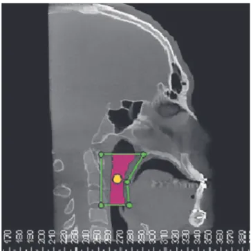
Ater digital head positioning, the sotware sinus/air-way tool, by means of which the sotware provides the
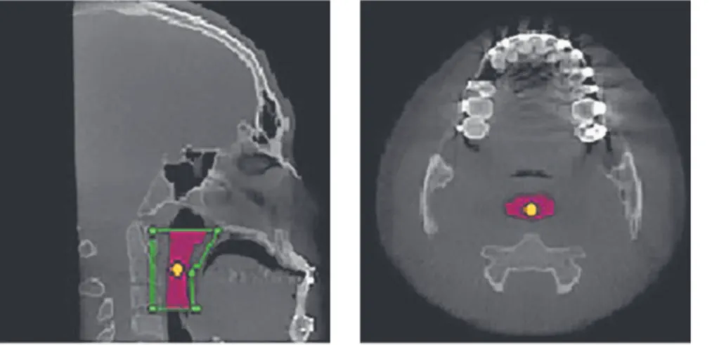
user with the image of a median sagittal section, was used. This image was used to determine the space of the oro-pharynx, while lines passing respectively by the Posterior Nasal Spine point and the apex of the epiglottis parallel to the Frankfort horizontal plane demarcated the upper and lower limits. The anterior and posterior limits were estab-lished so that a prism was formed with the upper and lower lines, thereby encompassing the entire oropharynx. The main marker was placed within the delimited space by ill-ing the whole area (Fig 1). The other multiplanar images (axial and coronal) were checked, so as to verify that the main marker encompassed the whole area (Fig 2).
Thereater, using speciic Dolphin tools, the follow-ing measurements were taken:
1. Sagittal area (SA) recorded in mm2.
2. Airway volume (Vol) recorded in mm3.
3. Minimum axial area (MAA) recorded in mm2.
For this measurement, the lower limit was displaced 5 mm above the apex of the epiglottis.
4. MAA depth (MAAD) recorded in mm. It is the distance between the posterior and anterior walls of the oropharynx in the MAA region performed on the sagit-tal midline.
5. MAA width (MAAW) also recorded in mm. It is the distance between the lateral walls of the oropharynx in the MAA region on the coronal plane.
6. MAA location (Loc) recorded in mm. To deter-mine this location, the distance between the Sella point and the MAA was measured on the sagittal section.
7. Airway length (C) recorded in mm. It is the dis-tance between the upper and lower limits of the oro-pharynx on the sagittal section.
Sagittal and coronal sections used to implement these measures were automatically provided by the sot-ware in the greatest constriction area. Representations of all measures can be seen in Figure 3.
Prior to measuring, ive CT scans were randomly selected in order to calibrate the examiner. All measure-ments were carried out in two stages with an interval of two weeks in between them, under the same conditions. For all variables, random error was calculated according to Dahlberg’s formula (S²=Σd²/2n) in order to verify intraex-aminer agreement. Analysis of measurement reproducibil-ity was performed by testing intraclass correlation, both with a conidence level of 95%. A high reliability rate was found, since all coeicients were greater than or equal to 0.84 for both time points. Thereater, measurements were
Figure 2 - Area determined by the main marker in three multiplanar sections (sagittal, axial and coronal).
Figure 3 - Representations of all measures: 1) sagittal area (SA), 2) airway volume (Vol), 3) minimum axial area (MAA), 4) MAA depth (MAAD), 5) MAA width (MAAW), 6) MAA location (Loc), 7) airway length (L). 1) SA
4) MAAD
7) L
2) Vol
5) MAAW
3) MAA
taken on all CT scans obtained before surgery (T0) and ater surgery (T1). Data were statistically treated. Shapiro-Wilk test was applied and results showed that the values had non-normal distribution. Thereater, Student’s paired t-test was used. All results with p-value lower than 0.05 were considered statistically signiicant.
RESULTS
As previously reported, sample was composed by CT scans of patients aged between 17 and 40 years old, with a mean age of 26 years and standard deviation of seven years, as shown in Table 1.
It is noteworthy that, in comparing the preopera-tive cephalometric tracings with the postoperapreopera-tive trac-ings, it was found that patients who took part in this study experienced a mean maxillary advancement of 3.35 mm and a mean mandibular setback of 3.92 mm, which produced a mean sagittal change of 7.27 mm in the maxillo-mandibular relationship (Table 2).
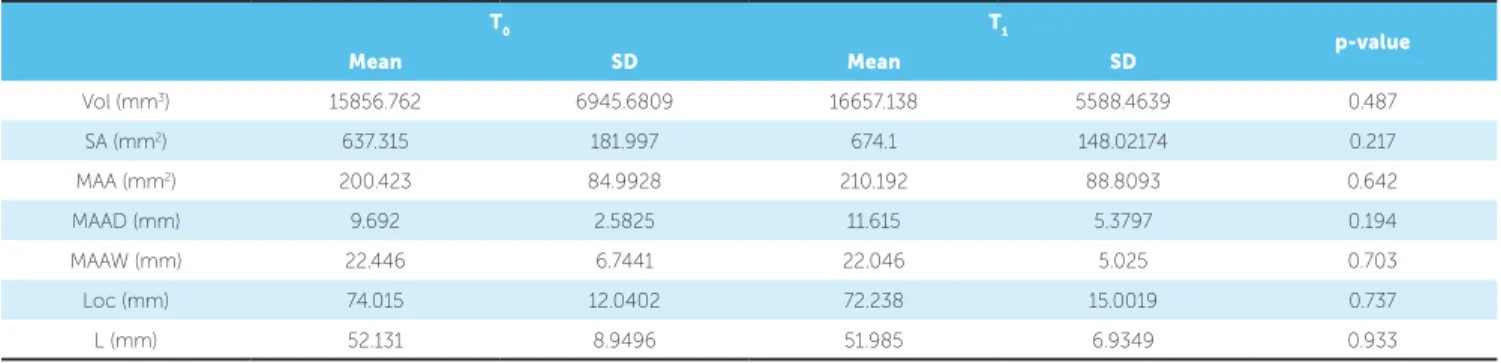
Table 3 shows the mean and standard deviation of each variable measured before (T0) and ater (T1) sur-gery. As it can be seen, there was no statistically signii-cant diference in any of them (p > 0.05).
Age
Mean 26
Standard deviation 7
Maximal value (Vmax) 40
Minimal value (Vmin) 17
Table 1 - Mean, standard deviation, maximal value and minimal value of patients’ ages.
Table 3 - Mean, standard deviation (SD) and p-value for each measurement, pre (T0) and post (T1) operative.
Table 2 - Mean, standard deviation, maximal value and minimal value of maxillary advancement and mandibular setback performed on patients.
Max. advancement Mand. setback
Mean 3.35 3.92
Standard deviation 1.97 2.39
Maximal value (Vmax) 8 6
Minimal value (Vmin) 1 0.5
T0 T1
p-value
Mean SD Mean SD
Vol (mm3) 15856.762 6945.6809 16657.138 5588.4639 0.487
SA (mm2) 637.315 181.997 674.1 148.02174 0.217
MAA (mm2) 200.423 84.9928 210.192 88.8093 0.642
MAAD (mm) 9.692 2.5825 11.615 5.3797 0.194
MAAW (mm) 22.446 6.7441 22.046 5.025 0.703
Loc (mm) 74.015 12.0402 72.238 15.0019 0.737
L (mm) 52.131 8.9496 51.985 6.9349 0.933
DISCUSSION
Anteroposterior skeletal disharmony in Class III skel-etal pattern patients may be surgically treated with max-illary advancement, mandibular setback or a combina-tion of both. The type of surgery to be performed will depend on the location and the amount of discrepancy, also considering facial aesthetics.1 Many times,
maxil-lary advancement is chosen based upon potential impair-ment of upper airways. However, in cases of larger skel-etal discrepancies or those in which there is greater aes-thetic impairment by mandibular inluence, it becomes
unavoidable to perform mandibular setback. In the pres-ent study, mean total discrepancy was 7.27 mm, which justiied the need for bimaxillary surgery. It is important to highlight that one patient required turbinectomy and septoplasty, since there was a 5-mm upper repositioning of the maxilla, and interference of nasal turbines would hinder inal bone position, which could lead to septum deviation and functional alteration.
For proper airway evaluation, linear, surface and volumetric measurements are required. In the present study, all these measurements were taken on CT scans
which provide more accurate anatomical information and are more suitable for evaluation of upper airway tests than conventional radiography.17
The literature on the efects of orthognathic surgery on airway space is controversial. In this study, it was observed that surgery induced a slight increase in up-per airway volume, although no statistically signiicant diference was found (p = 0.487), which corroborates the study by Jakobsone et al18 who used CT scans of
ten patients and also reported a slight increase, albeit not statistically signiicant. On the other hand, Park et al20 found no diference in the total volume of airways,
although they did ind a decrease in the oropharyngeal region. In other study evaluating women, no signiicant changes in volume of the pharyngeal airway were found.
By separately evaluating the upper and lower por-tions of the airways, it was found that maxillary ad-vancement enlarged the upper region whereas mandib-ular setback reduced the lower region, in what appears to be a compensatory process.11 This fact may justify
why there was no signiicant alteration in any of the measurements taken in this study, in the sense that there may have been some compensation in the values found, since all patients had undergone maxillary advancement and mandibular setback surgery.
On the other hand, in a study evaluating a sample of nine individuals who had undergone bimaxillary surgery, a decrease in the volume of the pharynx was noted.10 Their sample, however, difered from the
pres-ent study sample, since patipres-ents did not undergo ad-vancement mentoplasty. It is known that this procedure can by itself cause expansion of upper airways and has been proposed in the literature as a treatment for mild to moderate OSAH.21 Given that, in the present sample,
all patients underwent mentoplasty, thus, it is believed that this might have inluenced the results. Contradict-ing this information, in a study comparContradict-ing patients who had undergone mentoplasty with patients who had not, no statistically signiicant diference was found between them in terms of pharyngeal airway volume.22
Polysomnography and cephalometric radiographs of pre- and postoperative patients without OSAH who had undergone mandibular setback were evaluated, and it was concluded that some narrowing of the upper airway had occurred, but with no evidence that these patients had acquired sleep disorders. According to the authors, the airways of individuals with a skeletal Class III pattern
display wider dimensions than individuals whose values are normal; ater surgery, however, these values remain within normality range.23
Another study showed the inluence of the type of movement performed during bimaxillary surgery, rotational or not, which afects airway behavior. Ac-cording to the authors, if counterclockwise rotation is performed, there will be an increase in the size of air-space. This should therefore be, whenever possible, the movement of choice.13 In this study, this aspect was not
taken into account, as the number of patients who had undergone bimaxillary surgery without rotation was too small, which made it diicult to compare the groups.
Another aspect evaluated in this investigation was the area of upper airway in a sagittal section. It was observed that there was a slight increase, although not statistically signiicant (p = 0.217), from 637.3 mm2 to
674.1 mm2. This inding is consistent with another
re-search which evaluated cephalometric radiographs of 48 patients divided into three groups, one of which comprised patients who had undergone bimaxillary surgery. The authors found no signiicant diferences in the oropharyngeal and hypopharyngeal regions, which together comprised the same area used in this work, i.e., from the Posterior Nasal Spine to the apex of the epiglottis.24
This study measured the area, depth, width and loca-tion of the major airway narrowing region, also known as minimum axial area (MAA). This assessment is para-mount, since airway collapse occurs due to resistance to airlow.25 It was reported that average cross-section
is much smaller in apneic patients compared to healthy patients.26 Galvin et al27 found an average of 134.2 mm2
in healthy individuals. In this study, ater comparing preoperative versus postoperative CT scans, a mean of 200.4 mm2 was found for the former and 210.2 mm2 for
the latter. These values are well above the range described for patients with OSAH, i.e., approximately 50 mm2 or
less.28 It is noteworthy that none of the patients assessed in
As it can be seen in Table 3, the minimal axial area had its size and depth slightly increased, while its width decreased, and the MAA was displaced upwards. How-ever, these measures were not statistically signiicant. Additionally, it can be observed that a slight increase in depth was responsible for a slight increase in MAA, revealing that even ater mandibular setback, the region of greatest constriction showed a slight gain in the an-teroposterior direction. This was probably due to the advancement of the maxilla and chin and counterclock-wise rotation of the maxilla. Furthermore, slight up-ward displacement was not expected, since it was as-sumed that mandibular setback would decrease the up-per airway space, which in turn would shit the area of greatest constriction down to a lower region.
In a similar study, no statistically signiicant dif-ference was found in the area, depth and width of the cross-sections studied.10 In another study, two groups
were evaluated; one had undergone mandibular setback while the other one underwent bimaxillary surgery. The authors observed a decrease in the cross-sectional area of both groups, but found no statistically signiicant diference in the latter.9
According to Segal et al,29 patients diagnosed with
OSAH exhibit a correlation between airway length and OSAH severity to such an extent that, due to air resistance, the greater the length of the airway, the greater the respiratory disturbance index. For this reason, it is important to evaluate this measurement. In the present study, this measurement remained
virtually unchanged, showing a slight, not statistically significant (p = 0.933) decrease. Other study, how-ever, found a significant decrease in the length of the pharynx after bimaxillary surgery.10
Many studies show that mandibular setback can change the dimensions of upper airways and predispose one to OSAH.2,8,30 Other studies show that, in
compar-ing mandibular setback surgery with bimaxillary orthog-nathic surgery, a greater decrease occurs in the airways if only mandibular setback surgery is performed.9,10,19
Thus, in view of the results found in this investigation, one can conclude that when treating patients with skel-etal Class III pattern, bimaxillary orthognathic surgery (maxillary advancement and mandibular setback) does not lead to signiicative dimensional alterations in the oropharynx region.
CONCLUSIONS
In view of the results achieved in this study, it can be concluded that there are no signiicant changes in the upper airway of patients with skeletal Class III pattern ater bimaxillary orthognathic surgery and mentoplasty.
Author contributions
1. Boeck EM, Lunardi N, Pinto AS, Pizzol KEDC, Boeck RJN. Occurrence of skeletal malocclusions in Brazilian patients with dentofacial deformities. Braz Dent J. 2011 May;22(4):340-45.
2. Demetriades N, Chang DJ, Laskarides C, Papageorge M. Efects of mandibular retropositioning, with or without maxillary advancement, on the oro-naso-pharyngeal airway and development of sleep-related breathing disorders. J Oral Maxillofac Surg. 2010 Oct;68(10):2431-6.
3. Masood A, Phillips B. Sleep apnea. Curr Opin Pulm Med. 2000 Nov;6(6):479-84. 4. Harding SM. Complications and consequences of obstructive sleep apnea. Curr
Opin Pulm Med. 2000 Nov;6(6):485-9.
5. Phillips BG, Somers VK. Sleep disordered breathing and risk factors for cardiovascular disease. Curr Opin Pulm Med. 2002 Nov;8(6):516-20. 6. Riley RW, Powell NB, Guilleminault C. Inferior mandibular osteotomy and hyoid
myotomy suspension for obstructive sleep apnea: a review of 55 patients. J Oral Maxillofac Surg. 1989 Feb;47(2):159-64.
7. Sibel AEL, Hakan EL, Palomo JM, Baur DA. A 3-dimensional airway analysis of an obstructive sleep apnea surgical correction with cone beam computed tomography. J Oral Maxillofac Surg. 2011 Sep;69(9):2424-36.
8. Chen F, Terada K, Hua Y, Saito I. Efects of bimaxillary surgery and mandibular setback surgery on pharyngeal airway measurements in patients with Class III skeletal deformities. Am J Orthod Dentofacial Orthop. 2007 Mar;131(3):372-7. 9. Degerliyurt K, Ueki K, Hashiba Y, Marukawa K, Nakagawa K, Yamamoto E.
A comparative CT evaluation of pharyngeal airway changes in Class III patients receiving bimaxillary surgery or mandibular setback surgery. Oral Surg Oral Med Oral Pathol Oral Radiol Endod. 2008 Apr;105(4):495-502.
10. Hong JS, Park YH, Kim YJ, Hong SM, Oh KM. Three-dimensional changes in pharyngeal airway in skeletal class III patients undergoing orthognathic surgery. J Oral Maxillofac Surg. 2011 Nov;69(11):e401-8.
11. Lee Y, Chun YS, Kang N, Kim M. Volumetric changes in the upper airway after bimaxillary surgery for skeletal Class III malocclusions: a case series study using 3-dimensional cone-beam computed tomography. J Oral Maxillofac Surg. 2012 Dec;70(12):2867-75.
12. Mehra P, Downie M, Pita MC, Wolford LM. Pharyngeal airway space changes after counterclockwise rotation of the maxillomandibular complex. Am J Orthod Dentofacial Orthop. 2001 Aug;120(2):154-9.
13. Zinser MJ, Zachow S, Sailer HF. Bimaxillary ‘rotation advancement’ procedures in patients with obstructive sleep apnea: a 3-dimensional airway analysis of morphological changes. Int J Oral Maxillofac Surg. 2013 May;42(5):569-78. 14. Kawakami M, Yamamoto K, Fujimoto M, Ohgi K, Inoue M, Kirita T. Changes in
tongue and hyoid positions, and posterior airway space following mandibular setback surgery. J Craniomaxillofac Surg. 2005 Apr;33(2):107-10.
15. Pereira-Filho VA, Castro-Silva LM, de Moraes M, Gabrielli MF, Campos JA, Juergens P Cephalometric evaluation of pharyngeal airway space changes in Class III patients undergoing orthognathic surgery. J Oral Maxillofac Surg. 2011 Nov;69(11):e409-15.
16. Ribeiro CO, Bittencourt MAV, Brandão RA. Avaliação dos efeitos da cirurgia ortognática de recuo mandibular isolado e combinado no tamanho da orofaringe. Ortho Sci Orthod Sci Pract. 2011 Fev;3(13):414-7.
REFERENCES
17. Lenza MG, Lenza MM, Dalstra M, Melsen B, Cattaneo PM. An analysis of diferent approaches to the assessment of upper airway morphology: a CBCT study. Orthod Craniofac Res. 2010 May;13(2):96-105.
18. Jakobsone G, Neimane L, Krumina G. Two- and three-dimensional evaluation of the upper airway after bimaxillary correction of Class III malocclusion. Oral Surg Oral Med Oral Pathol Oral Radiol Endod. 2010 Aug;110(2):234-42.
19. Park SB, Kim YI, Son WS, Hwang DS, Cho BH. Cone-beam computed tomography evaluation of short- and long-term airway change and stability after orthognathic surgery in patients with Class III skeletal deformities: bimaxillary surgery and mandibular setback surgery. Int J Oral Maxillofac Surg. 2012 Jan;41(1):87-93.
20. Panou E, Motro M, Ateş M, Acar A, Erverdi N. Dimensional changes of maxillary sinuses and pharyngeal airway in Class III patients undergoing bimaxillary orthognathic surgery. Angle Orthod. 2013 Sep;83(5):824-31.
21. Foltán R, Hofmannová J, Pretl M, Donev F, Vlk M. Genioglossus advancement and hyoid myotomy in treating obstructive sleep apnoea syndrome: a follow-up study. J Craniomaxillofac Surg. 2007 Jun-Jul;35(4-5):246-51.
22. Kim MA, Kim BR, Youn JK, Kim YJ, Park YH. Head posture and pharyngeal airway volume changes after bimaxillary surgery for mandibular prognathism. J Craniomaxillofac Surg. 2014 Jul;42(5):531-5.
23. Hochban W, Schürmann R, Brandenburg U, Conradt R. Mandibular setback for surgical correction of mandibular hyperplasia — does it provoke sleep-related breathing disorders? Int J Oral Maxillofac Surg. 1996 Oct;25(5):333-8. 24. Aydemir H, Memikoğlu U, Karasu H. Pharyngeal airway space, hyoid bone
position and head posture after orthognathic surgery in Class III patients. Angle Orthod. 2012 Nov;82(6):993-1000.
25. Abramson Z, Susarla SM, Lawler M, Bouchard C, Troulis M, Kaban LB. Three-dimensional computed tomographic airway analysis of patients with obstructive sleep apnea treated by maxillomandibular advancement. J Oral Maxillofac Surg. 2011 Mar;69(3):677-86.
26. Lan Z, Itoi A, Takashima M, Oda M, Tomoda K. Diference of pharyngeal morphology and mechanical property between OSAHS patients and normal subjects. Auris Nasus Larynx. 2006 Dec;33(4):433-9.
27. Galvin JR, Rooholamini SA, Stanford W. Obstructive sleep apnea: diagnosis with ultrafast CT. Radiology. 1989 Jun;171(3):775-8.
28. Avrahami E, Englender M. Relation between CT axial cross-sectional area of the oropharynx and obstructive sleep apnea syndrome in adults. AJNR Am J Neuroradiol. 1995 Jan;16(1):135-40.
29. Segal Y, Malhotra A, Pillar G. Upper airway length may be associated with the severity of obstructive sleep apnea syndrome. Sleep Breath. 2008 Nov;12(4):311-6.