INTRODUCTION
Root perforations allow the root canal system to communicate with the periradicular tissues and may cause the loss of the tooth due to secondary periodontal involvement. The ideal repair material must show good sealing ability in addition to physical, chemical and biological properties. The properties of an ideal repair material have been demonstrated by several studies since Lee et al. (1) presented mineral trioxide aggregate (MTA). Currently, MTA is considered the material of choice for the repair of root perforations.
MTA is composed by tricalcium silicate, trical-cium oxide, silicate oxide and other mineral oxides that are responsible for its physical and chemical properties. An alkaline pH and calcium release probably stimulate tissue mineralization (2). The major difference between gray and white MTA (WMTA) is the concentration of iron oxide (FeO). The amount of FeO is extremely re-duced in the WMTA (3), while the biological properties
of both types of cement are comparable (4-5).
A new experimental epoxy resin and calcium hydroxide based cement (MBPc) has been developed at the Department of Endodontics of Bauru Dental School, University of São Paulo, Brazil as another option to repair root perforations. The ability of the epoxy resin to seal and the biocompatibility of calcium hydroxide have been proven in vitro (6-8) and have shown good results in vivo (9).
Generally, cytotoxicity is the irst test used to
investigate biocompatibility. The evaluation of new ma-terials using in vitro methods must irst be done to avoid unnecessary animal sacriice. The agar overlay method
with neutral red dye allows great sensitivity using the material as it is used clinically, while protecting the cell monolayer against physical damage (10).
The aim of this study was to evaluate the toxicity of gray MTA, white MTA and the experimental MBPc by comparing their effect on L929 cells, using the agar overlay method with neutral red dye.
Correspondence: Dra. Rosana B. Miranda, Subdivisão de Ensino e Pesquisa, Odontoclínica de Aeronáutica Santos Dumont, Praça Marechal Âncora, 77, 20021-200 Rio de Janeiro, RJ, Brasil. Tel: +55-21-2101-6083 ramal 217. e-mail: [email protected]
L929 Cell Response to Root Perforation Repair
Cements: an
In Vitro
Cytotoxicity Assay
Rosana Belchior MIRANDA1
Sandra Rivera FIDEL2
Maria Aparecida Affonso BOLLER3
1Santos Dumont Brazilian Air Force Dental Clinic, Rio de Janeiro, RJ, Brazil 2Dental School, State University of Rio de Janeiro, Rio de Janeiro, RJ, Brazil 3National Institute for Quality Control in Health/ Oswaldo Cruz Foundation,
Ministry of Health, Rio de Janeiro, RJ, Brazil
This study compared the cytotoxicity of an experimental epoxy-resin and calcium hydroxide-based cement (MBPc), gray mineral tri-oxide aggregate (MTA) and white mineral tritri-oxide aggregate (WMTA) using the agar overlay method with neutral red dye. L929 cells
were seeded into 6-well culture plates where 48-h set test materials were placed on the agar overlay, in triplicate. Telon and natural rubber served as negative and positive controls. After an incubation period of 24 h at 37ºC in a humidiied atmosphere of 5% CO2 in air, a discolored area around the samples and the positive controls could be observed and measured per quadrant. The mean values were compared and converted into grades to classify the results according to the table of cytotoxicity grades according to the Standard Operating Procedures (SOP) of the Oswaldo Cruz Foundation, Brazil. The nonviable cell areas and the morphological changes in the cells were observed with an inverted microscope. The results showed grade 1 (slight) for the two types of MTA (p>0.05) and grade 2 (mild) for the MBPc (p<0.001). All samples met the requirements of the test as none of the cultures showed reactivity higher than grade 2.
MATERIAL AND METHODS
Test Materials
Gray ProRoot MTA (Dentsply/Tulsa Dental, Tul-sa, OK, USA) and Angelus WMTA (Angelus Soluções Odontológicas, Londrina, PR, Brazil) were mixed in a 1:1 ratio of powder: liquid, and the MBPc experimental cement was mixed in a 3:1 ratio of base paste (0.2 g): catalyze paste (0.6 g), according to the manufacturers’ instructions. The mixtures were then immediately placed into a silicone mold to prepare uniform pellets (6 x 6 x 1 mm3). The cements were allowed to set for 48 h.
Cell Culture
L929 cells, derived from mouse ibroblasts
(ATCC, Rockville, MD, USA), were stored frozen Eagle’s minimum essential medium (MEM) with Earle’s salts (Sigma Aldrich Corp., St. Louis, MO, USA),
supplemented with 20% v/v fetal bovine serum (FBS;
Gibco, Grand Island, NY, USA), streptomycin sulfate (100 mg/mL, Sigma Aldrich Corp.), anphotericyn B (100 IU/mL, Sigma Aldrich Corp.), L-glutamine (2 mM,
Sigma Aldrich Corp.) and 10% v/v glycerol into liquid
nitrogen at -70ºC. For use in this experiment, the cells were transferred to 75 cm2 lasks (Costar, Corning, NY,
USA) in Dulbecco’s MEM supplemented with 5% FBS
and antibiotics and were subcultured.
Agar Overlay Method
The study was performed according to the Stan-dard Operating Procedures (SOP) of the Oswaldo Cruz Foundation (FIOCRUZ, Rio de Janeiro, RJ, Brazil) (11). Cells were seeded in 6-well plates (Costar) at a density of 2 x10⁵ cells/mL. Each well, which had an
inner diameter of 35 mm, received 4 mL of the prepared suspension. The plates were incubated for 48 h at 37ºC
in an atmosphere of 5% CO2 in air and 100% humidity,
to allow adhesion and cell monolayer conluence. The
medium was removed from the wells and replaced with
agar 1.8% (Bacto; Difco Laboratories Inc., Detroit, MI, USA) added neutral red dye 0.01%, staining the viable
cells while allowing diffusion of leachable chemicals from cements. Samples in triplicate, positive (natural
rubber) and negative (Telon) controls on the agar over -lay featured the 6-well plates. One well in each plate
received the agar only to serve as a cell control. After an incubation period of 24 h at 37ºC in a
humidiied atmosphere of 5% CO2 in air, discolored areas
could be seen around specimens related to their toxicity. A milimetric paper was placed underneath the plates before pictures were taken with a digital camera (Nikon
Coolpix 4300; Kinon, Tokyo, Japan). Then, the areas of
nonviable cell were measured per quadrant using Image Tool image-analysis software (The University of Texas Health Science Center at San Antonio, Texas, USA)
adjusting the image magniication tool that allowed
improved view of focused areas. The milimetric paper allowed calibrating the digital measurement tool. The method evaluated the biological reactions quantitatively, comparing the extension (in mm) of nonviable cell areas.
The results were classiied according to a 5-point
cytotoxicity grading system related to the agar overlay method, according to which grade 2 or lower means sat-isfactory samples: 0 = absent cytotoxicity (absence of a
zone of lysis underneath the sample; 1= slight (zones of cell lysis underneath the sample only); 2= mild (zones of cell lysis ≤5 mm from the sample); 3=Moderate (zones of cell lysis >5 mm and ≤10 mm from the sample); 4=
severe (zones of cell lysis >10 mm, but not involving the entire well). In addition, the wells were occasionally examined with an inverted microscope (Nikon Eclipse
TS 100; Nikon) to observe cell changes.
Statistical Analysis
Parametric analyses of data were performed by
one-way ANOVA and Tukey’s test at 5% signiicance
level using GraphPad Prism statistical software package (GraphPad Software Inc., San Diego, CA, USA).
RESULTS
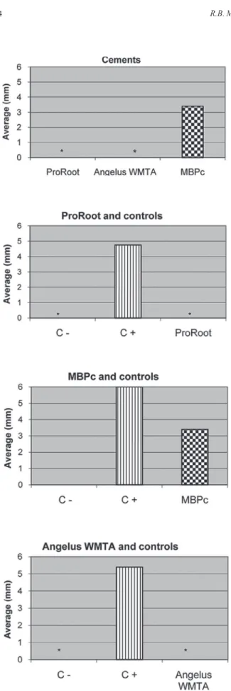
There were statistically signiicant differences
(p<0.05) among the mean zones of cell lysis comparing the cements to each other and comparing the cements to the controls from the respective plates (Fig. 1).
When examining morphological changes in the cells with the inverted microscope, ProRoot MTA showed viable cells around the pellets. Dead cells could be observed underneath the material after removal from the agar layer.
A ×100 magniication of an Angelus WMTA well
clearly showed 4 different moments in the cell morphol-ogy around the cement: cell lysis, rounded cells loosened from the substrate, some morphological alterations, and viable cells with homogeneous cytoplasm in a monolayer (Fig. 2). MBPc revealed the largest zones of cell lysis and morphological cell changes among the 3 cements. Because of this only one section of all wells examined in the study exhibited the different zones within the same
iled of view (Fig. 3).
Figure 1. Comparison of the mean zones of cell lysis among cements and among the cements and their respective controls.
DISCUSSION
Studies with established cell lines are used be-cause of the reproducibility of the results, besides they multiply rapidly with an unlimited life span (12). It
is interesting to evaluate the reaction of ibroblasts to
the materials because those cells are involved with the healing process. The established L929 cell line used in this study is commonly employed in biocompatibility studies (10,13-15).
Using the agar overlay method with vital red dye, Torabinejad et al. (13) recorded a zone of lysis around the samples of fresh and set gray MTA, without any differ-ence between them. Saidon et al. (14) and Haglung et al. (15) found denatured proteins and dead cells underneath fresh MTA samples, morphological changes around them and viable cells in most of the area of the well. The present study obtained similar results for the 48-h set MTA, in agreement with Koulaouzidou et al. (16) who observed little cytotoxicity of 48-h set gray MTA. Although gray and white MTA cements are slightly cytotoxic and have exhibited similar effects on the cells in the present study, it was interesting to note that during the microscopic analysis the cell behavior in the presence of ProRoot MTA was the most favorable.
Accordingly, Pérez et al. (17) and Camilleri et al. (18) showed less cell growth in the presence of white MTA,
but they conirmed its biocompatibility.
Epoxy resin has a favorable property of adhesion to dental structure and calcium hydroxide is known to be biocompatible (19). Cohen et al. (10) demonstrated
severe cytotoxicity of resin-based cements in the irst
48 h after preparation, period in that the most toxic sub-stances are released from resin-based cement, according to the same authors. The present investigation revealed grade 2 (mild) for 48-h set MBPc, probably because the cement had been left to set for 48 h before it has been used, which reduced its toxicity in a considerable way
(20). Even though our morphological indings showed that ibroblasts appeared rounded, unstained and prob -ably in the process of apoptosis in the greatest part of the discolored area as an effect of MBPc, only close to the cement the cells presented vacuolated cytoplasms or lysis, differently from positive controls that determined the entire discolored area with dead cells.
ProRoot MTA and Angelus WMTA were slightly cytotoxic. Although the experimental MBPc was the most cytotoxic cement among the 3 tested, the sample was considered satisfactory. The changes in cell mor-phology occurred in the following sequence of intensity:
gray ProRoot MTA < Angelus WMTA < MBPc, con-sidering that the changes associated with the positive
controls were signiicantly more intense than those
observed these materials.
A further biochemical evaluation would be inter-esting to count viable cells after contact with the tested materials. The experimental MBPc cement must be subjected to other tests to allow an appropriate clinical indication of its biocompatibility. This material may become a good alternative to repair root perforations.
RESUMO
O objetivo deste estudo foi comparar a citotoxicidade de um cimento experimental à base de resina epóxica e hidróxido de cálcio (MBPc), do agregado trióxido mineral (MTA) cinza e do MTA branco, utilizando o ensaio de difusão em agar com o co-rante vermelho neutro. Células L929 foram semeadas em placas de 6 poços e sobre elas a camada de agar, onde foram colocados
os materiais endurecidos por 48 h, em triplicata, além de telon
como controle negativo e látex como controle positivo. Após 24
h em estufa umidiicada a 37ºC com 5% CO2, um halo claro se formou ao redor das amostras e dos controles positivos. As medi-das foram tomamedi-das, por quadrante, e as médias foram comparamedi-das
e convertidas em graus para qualiicar os resultados, de acordo
com a tabela de grau de citotoxicidade do POP/FIOCRUZ. As zonas de inibição e as alterações da morfologia celular foram avaliadas sob microscópio invertido. Os resultados revelaram grau 1 (leve) para os dois tipos de MTA (p>0,05) e grau 2 (branda) para o MBPc (p<0,001). Todas as amostras foram consideradas satisfatórias, pois nenhuma cultura exposta aos cimentos revelou toxicidade superior ao grau 2.
ACKNOWLEDGEMENTS
This study was supported in part by the National Institute of Health Quality (INCQS)/ Oswaldo Cruz Foundation (FIOCRUZ, RJ, Brazil). The authors thank Dr. Ivaldo Gomes de Moraes for supply of MBPc.
REFERENCES
1. Lee SJ, Monsef M, Torabinejad M. Sealing ability of a mineral trioxide aggregate for repair of lateral root perforations. J Endod
1993;19:541-544.
2. Oliveira MG, Xavier CB, Demarco FF, Pinheiro ALB, Costa AT, Pozza DH.Braz Dent J 2007;18:3-7.
3. Asgary S, Parirokh M, Eghbal MJ, Brink F. Chemical differen-ces between white and gray mineral trioxide aggregate. J Endod
2005;31:101-103.
4. Faraco Júnior IM, Holland R. Histomorphological response of dogs`dental pulp capped with white mineral trioxide aggregate.
Braz Dent J 2004;15:104-108.
5. Al-Rabeah E, Perinpanayagam H, Macfarland D. Human alveolar bone cells interact with ProRoot and tooth-colored MTA. J Endod
2006;32:872-875.
6. Silva Neto UX, Moraes IG. Sealing capacity produced by some materials when utilized under furcation perforations of extract
human molars. J Appl Oral Sci 2003;11:27-33.
7. Desai S, Chandler N. Calcium hydroxide-based root canal sealers:
a review. J Endod 2009;35:475-480.
8. Orosco FA, Bramante CM, Garcia RB, Bernardineli NB, Moraes IG. Sealing ability of gray MTA Angelus™, CPM™ and MBPc used as apical plugs. J Appl Oral Sci 2008;16:50-54.
9. Cintra LTA, Moraes IG, Bernabé PFE, Gomes-Filho JE, Bramante CM, Garcia Rb, et al.. Evaluation of the tissue response to MTA and MBPc: microscopic analysis of implants in alveolar bone of
rats. J Endod 2006;32:556-559.
10. Cohen BI, Pagnillo BS, Musikant BL, Deutsch AS. An in vitro
study of the cytotoxicity of two root canal sealers. J Endod
2000;26:228-229.
11. Standard Operating Procedures (SOP)/Quality Brochure. Rio de Janeiro: INCQS/FIOCRUZ. (65.3330.010). In vitro cytotoxicity assay – Agar diffusion method (In Portuguese). 2000. 17 p. 12. Koh ET, McDonald F, Pitt Ford TR, Torabinejad M. Cell response
to mineral trioxide aggregate. J Endod 1998;24:543-547.
13. Torabinejad M, Hong CU, Pitt Ford TR, Kettering JD. Cytotoxicity
of four root end illing materials. J Endod 1995;21:489-492.
14. Saidon J, He J, Zhu WQ, Safavi K, Spångberg LZW. Cell and tis-sue reactions to mineral trioxide aggregate and Portland cement.
Oral Surg Oral Med Oral Pathol Oral Radiol Endod
2003;95:483-489.
15. Haglung R, Safavi KE, Zhu Q. Effects of root-end illing materials on ibroblasts and macrophages in vitro. Oral Surg Oral Med Oral
Pathol Oral Radiol Endod 2003;95:739-745.
16. Koulaouzidou EA, Papazisis KT, Economides NA, Panagiotis B, Kortisaris AH. Antiproliferative effect of mineral trioxide ag-gregate, zinc oxide-eugenol cement, and glass-ionomer cement
against three ibroblastic cell lines. J Endod 2005;31:44-46.
17. Pérez AL, Spears R, Gutmann JL, Opperman LA. Osteoblasts and MG-63 osteosarcoma cells behave differently when in contact with
ProRoot™ MTA and white MTA. Int Endod J 2003;36:564-570.
18. Camilleri J, Montesin FE, Papaioannu S, McDonald F, Pitt Ford TR. Biocompatibility of two commercial forms of mineral trioxide
aggregate. Int Endod J 2004;37:699-704.
19. Schwarze T, Fiedler I, Leyhausen G, Geurtsen W. The cell
compa-tibility of ive endodontic sealers during the setting period. J Endod 2002;28:784-786.
20. Huang FM, Tai KW, Chou MY, Chang YC. Cytotoxicity of resin-, zinc oxide-eugenol-, and calcium hydroxide-based root canal sealers on human periodontal ligament cells and permanent V79
cells. Int Endod J 2002;35:153-158.