C A S E R E P O R T UDC: 618.31-089 DOI: 10.2298/VSP150723160K
Unruptured retroperitoneal pregnancy implanted in the left broad
ligament: A case report
Nerupturirana retroperitonealna trudno
ć
a u levom širokom ligamentu
Ranko M. Kutlešić*, Bojan Lukić†, Marija S. Kutlešić‡, Jasmina Popović†, Milan Stefanović†, Predrag Vukomanović†, Goran Lilić*
*Department of Gynecology and Obstetrics, ‡Center for Anesthesia, Clinical Center of Niš, Niš, Serbia; †Faculty of Medicine, University of Niš, Niš, Serbia
Abstract
Introduction. Retroperitoneal ectopic pregnancy is extremely rare, but potentially fatal condition due to possible massive hemorrhage, representing a great challenge to clinicians. Case report. We presented early retroperitoneal pregnancy in a pa-tient with previous caesarean section, diagnosed at the sixth gestational week, located in the left broad ligament, primary treated by laparoscopy, which had to be converted to laparo-tomy due to massive intraoperative bleeding from the implan-tation site. Conclusion. High index of suspicion, combined with carefully interpreted clinical and ultrasound findings are crucial for the timely diagnosis of retroperitoneal pregnancy, before the occurrence of severe bleeding. The rising, even pla-teau of serum β-human chorionic gonadotropin (β-HCG) lev-els without identification of uterine or ectopic (tubal) preg-nancy should cause suspicion on ectopic pregpreg-nancy in unusual location.
Key words:
pregnancy, ectopic; retroperitoneal space; laparoscopy; intraoperative complications; gynecologic surgical procedures.
Apstrakt
Uvod. Retroperitonealna ektopična trudnoća je krajnje retko, ali
moguće fatalno stanje zbog masivne hemoragije i predstavlja ve-liki izazov za kliničara. Prikaz bolesnika. Prikazali smo ranu retroperitonealnu trudnoću kod pacijentkinje sa carskim rezom u anamnezi, dijagnostikovane u šestoj nedelji gestacije, lo-kalizovanu u ligamentum latumu sa leve strane, primarno lečenu laparoskopski, pri čemu je načinjena konverzija u laparotomiju zbog masivnog intraoperativnog krvarenja sa mesta implantacije.
Zaključak. Da bi se na vreme postavila dijagnoza retroperitone-alne trudnoće, pre pojave obilnog krvarenja, neophodno je da se ima na umu ova mogućnost i pažljivo interpretira klinički i ultra-zvučni nalaz. Nivoi β-humanog horionskog gonadotropina (β-HCG) u serumu koji rastu ili održavaju plato, a da nije identifi-kovana trudnoća u uterusu ili vanmaterična u jajovodu, treba da navedu na pomisao da se radi o ektopičnoj trudnoći neuobičaje-ne lokalizacije.
Ključne reči:
trudnoća, ektopična; retroperitonealni prostor; laparoskopija; intraoperativne komplikacije; hirurgija, ginekološka, procedure.
Introduction
Retroperitoneal pregnancy is a very rare form of ectopic pregnancy. It could be the result of primary retroperitoneal implantation with enigmatic pathogenesis or secondary following tubal rupture in the broad ligament. In modern lite-rature there are less than 25 well-documented cases of primary retroperitoneal pregnancy 1, 2.
We presented a primary retroperitoneal pregnancy im-planted in the left broad ligament.
Case report
A 21-year-old gravida 1, para 1, was admitted into our clinic due to a 6-week history of amenorrhoea, lower
abdo-minal pain and vaginal bleeding. Ectopic gravidity was sus-pected. Before 18 months the patient had term caesarean delivery performed due to breech presentation, oligoamnion and dystocia followed by an uneventful postoperative course. The women was otherwise healthy, had no history of pelvic inflammatory disease and use of intrauterine devices. Her menarche occurred at 12 and menstrual cycles were regular.
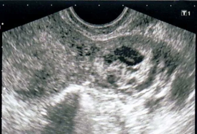
Fig. 1 – Ultrasound image of early retroperitoneal pregnancy in the left broad ligament, just behind the
uterine corpus. The uterine cavity is empty.
Fig. 2 – Ultrasound image of early retroperitoneal pregnancy in the left broad ligament: a round cystic mass, filled with a heterogeneous content and the hypoechogenic structure inside, like a gestational sac without fetal pole.
Fig. 3 – Retroperitoneal pregnancy in the left broad ligament: color Doppler examination revealed reach vascularisation with the typical “ring of fire”, low
resis-tance blood flow around the described mass.
Fig. 4 – Laparoscopic finding of retroperitoneal pregnancy in the left broad ligament at the 6th gestational week: a) round retroperitoneal mass with the intact overlying
peri-toneum; b) the left Fallopian tube is superiorly, macro-scopically normal in size and shape; c) the uterus is slightly
enlarged; d) the part of the left ovary, without any patho-logical findings; e) the pouch of Douglas is empty.
and a tender palpable mass about 4 cm in diameter, on the left adnexal region. Transvaginal ultrasound examination (Toshiba Nemio XG, 6 MHz) showed the empty uterus with 5 mm endometrial strip (Figure 1). A round cystic mass, 4 × 3 × 2 cm in diameter, filled with heterogeneous content and hypoechogenic structure inside, like gestational sac without fetal pole, 1 cm in diameter, was seen just behind the uterine corpus, on the left side (Figure 2). Color Doppler examination revealed reach vascularisation, with the typical “ring of fire”, low resistance blood flow around the described mass (Figure 3). Both ovaries appeared sonographically normal with corpus luteum on the left ovary. There was no intraperitoneal fluid in the pouch of Douglas. Her laboratory results were as follows: white blood cells (WBC) 12.8 ×10⁹/L, red blood cells (RBC) 4.48 ×10¹²/L, Hb 123 g/L, hematocrit (Ht) 37.5, platelets (PLT) 368 ×109/L. Serum electrolytes, coagulation profile and liver function tests were all within physiological limits. At the day of admission her serum β-human chorionic gonadotropin (β-HCG) level (Ab-bott test; Architect-Total-β-HCG) was 28.643 mU/mL and
the next day quantitative β-HCG level decreased to 27,000 mU/mL. Ectopic gravidity was suspected and diagnostic laparoscopy was performed after the written informed con-sent was obtained.
Surgery was conducted under general anesthesia, indu-ced by means of propofol as induction agent, fentanyl as an analgesic and rocuronium as a muscle relaxant. Anesthesia was maintained with 1–1.5% end-tidal sevoflurane in 50% : 50% O2/N2O mixture at 6 L/min flow. The lungs were
ventilated to maintain end-tidal carbon dioxide concentration 30–35 mmHg.
fin-dings. The corpus luteum was located on the left ovary. The-re was no bleeding from the fimbria bilaterally. The perito-neum was opened above the described mass using the ultra-cision, and evacuation of ovulary tissue was started, but suddenly significant bleeding appeared and urgent laparotomy was immediately performed. There was about 500 mL of fresh blood in the abdomen. The bleeding was controlled by two finger digital compression of the left broad ligament and the remaining ovulary tissue was removed. There were no macroscopic signs of communication or fistu-la between the described mass and the uterine cavity or the left Fallopian tube. Hemostasis was completed with hemosta-tic sutures and the abdomen was closed with drainage placed in the pouch of Douglas. Postoperative course was unevent-ful. Serum β-HCG levels decreased to 750 mU/mL two days after the surgery and became negative after seven days. The histopathology report confirmed ectopic gravidity (Figure 5).
Fig. 5 – Histopathology report confirmed ectopic gravidity: chorionic villi (haematoxylin-eosin, ×100).
Discussion
The incidence of ectopic pregnancy is 0.25–1% of all pregnancies. More than 95% of all ectopic pregnancies are tu-bal pregnancies. The incidences of extratutu-bal ectopic pregnan-cies are as follows: abdominal in 1.3%, ovarian and cervical in less than 1% of all ectopic pregnancies 3. Retroperitoneal ecto-pic pregnancy, as a subcategory of abdominal pregnancy, is exceptionally rare. The true incidence of retroperitoneal pregnancy is unknown mainly due to false recognizing of ab-dominal pregnancies with trophoblast invasion as retroperito-neal 1. The first report of retroperitoneal pregnancy was the ca-se of the broad ligament ectopic pregnancy described almost two hundred years ago by Loschge 4. Our own review of the literature (Medline data base, through electronic searches without language restriction) showed a total of 65 reported cases of retroperitoneal pregnancies during the last 57 years, with 26 well documented cases during the last 15 years (Tab-le 1). The largest series of the broad ligament retroperitoneal pregnancies (62 cases) was reported by Champion and Tessi-tore 6 with the incidence of one in 183,900 pregnancies.
The sites of retroperitoneal ectopic implantation include the broad ligament, obturator fossa, areas around large retrope-ritoneal blood vessels, even the upper retroperetrope-ritoneal space – attached to the head of pancreas and major blood vessels 7–10. There have been also reports on broad ligament twin preg-nancaes 11, 12 and heterotopic pregnancies involving the broad ligament and the uterus 13 or broad ligament and interstitial pregnancy 14. Occurrence of partial hydatiform molla was al-so reported in intraligamentous pregnancy 15.
The most often among retroperitoneal pregnancies is the one located in the broad ligament or intraligamentous pregnancy. The original anatomical relationships for diagno-sing broad ligament ectopic pregnancy are: location of the uterus medially, the pelvic side walls laterally, the pelvic floor inferiorly and the Fallopian tube superiorly 6. Recently, original criterions are fulfilled with the statement that overlying peritoneum should be intact in order to confirm the diagnosis of true retroperitoneal implantation 1. Our reported case fulfills all the mentioned criteria.
Retroperitoneal ectopic implantation could appear after spontaneous conception 1, 4, 8, 11, 16–26, intrauterine inseminati-on 27 or after in vitro fertilization/pre-embrio transfer (IVF/ET) 2, 7, 28 (Table 1). Intrauterine device (IUD) in situ
was found in 8% of abdominal pregnancies, so it is specula-ted that IUD could be a factor contributing to the develop-ment of abdominal pregnancy 29.
Retroperitoneal pregnancy could be the result of primary retroperitoneal implantation or secondary following tubal rupture or trophoblast invasion in the broad ligament. There is also the possibility of primary interstitial and secondary retroperitoneal pregnancy 20.
The pathogenesis of primary retroperitoneal pregnancy is quite obscure. It seems that in the majority of cases, primary retroperitoneal implantation could appear after ute-rine or tubal surgery that could develop a communication in the retroperitoneal space resulting with the passage of fertili-zed ovum after spontaneous conception or after IVF/ET (Ta-ble 1). The fistulous tract could be developed after termal injury during laparoscopic salpyngectomy 1, after classical salpyngectomy or salpingoophorectomy or due to inapprop-riate healing of the uterine wall after cesarean section 16, 30. Spontaneous migration of the embryo from the uterus to the retroperitonel space through these communications could re-sult in retroperitoneal pregnancy.
There is also the possibility of false passage during em-brio transfer and placement of embryos into the retroperito-neal space in cases of retroperitoretroperito-neal pregnancies after IVF/ET 7, 28.
In the case we reported here, lymphatic tissue was not found around the trophobalast, even after careful histopathological examination, so we speculate that the development of the de-scribed left broad intraligamentous pregnancy could be explained by spontaneous migration of the embryo from the uterus to the retroperitoneal space through the microscopic fistulous tract caused by inappropriate healing of the uterine wall after the previous caesarean section. Still, we could not exclude with the certainty the possibility of embryo migrati-on via lymphatic vessels, taking into account the localization of described retroperitoneal pregnancy.
The preoperative diagnosis of retroperitoneal pregnancy represents the challenge for clinicians. In fact, in the most cases, the diagnosis is made during surgery.
Maternal morbidity and mortality associated with ab-dominal, especially retroperitoneal, pregnancies could be re-duced by early diagnosis. Transvaginal ultrasound exami-nation is the main tool in the diagnostic of an early abdomi-nal (and retroperitoneal) pregnancy. The proposed criteria are: the absence of an intrauterine gestational sac; the absen-ce of tubal dilatation or complex adnexal mass; a gestational sac surrounded by loops of the bowel and separated from the uterus; and a wide mobility of the gestational sac 33, 34. In fact, sonographic appearance of an early retroperitoneal pregnancy depends on its location. Usually it is fixed deep within the pelvis and not mobile as pregnancy in the non-communicating horn of the unicornuate uterus (cornual pregnancy) 35. The absence of communication between ges-tational sac and endometrial cavity differentiates the retrope-ritoneal broad ligament pregnancy from the pregnancy in non-communicating horn of the unicornuate uterus (cornual pregnancy) and interstitial ectopic pregnancy 35, which was also the truth in the case we reported here. The absence of myometrial layer around this retroperitoneal broad ligament pregnancy differentiates it from interstitial pregnancy. If early retroperitoneal pregnancy is located outside the pelvis, transvaginal ultrasound examination is helpless, and other diagnostic tools, as magnetic resonance imaging (MRI) and other imaging techniques, must be applied.
The suspicion is crucial for the timely diagnosis of ret-roperitoneal ectopic pregnancy. Rising β-HCG levels, or pla-teau, without identification of uterine or ectopic (tubal) pregnancy should cause suspicion on ectopic pregnancy in unusual location. In case we reported here, the diagnosis of ectopic pregnancy was made when the patient was still hemodinamically stable, so we opted for laparoscopic treat-ment during which the definitive diagnosis of left broad li-gament pregnancy was confirmed.
The treatment of retroperitoneal pregnancy also repre-sents a great challenge for clinicians. The most of retroperi-toneal pregnancies are diagnosed and removed during the early stages of gravidity, but there are reports on broad liga-ment pregnancies with viable term fetuses 19, 20, 30, 36–41, even
post term 42.
The great majority of such cases are discovered on surgery for caesarean section.
In spite of many reports on abdominal pregnancies with viable fetuses advanced to term, the risk for the mother is
still very high, especially in cases of retroperitoneal pregnan-cies with the close proximity to large vessels. Immediate surgery is indicated for abdominal pregnancies prior to 23 to 24 weeks because of the high incidence of maternal morbidity and a poor prognosis for the fetus 43.
Fetal anomalies or deformities (facial and cranial asymmetry, joint deformities, CNS anomalies) are associated problem in such pregnancies 41. However, there is a reported case of successful secondary retroperitoneal pregnancy in which the diagnosis had been suspected during the 18th week, discarded due to lack of symptoms and advanced to term with normal course 20.
The main concern with retroperitoneal pregnancy is as-sociated with possible fatal bleeding due to the proximity of large blood vessels. This is also the possibility during the surgery after the attempt to remove the ectopic pregnancy. In the most of the reported cases laparotomy was the treatment. Nowadays, it seems that most unruptured early nontubal pre-gnancies could be managed laparoscopically. Laparoscopic treatment of ectopic pregnancy is minimally invasive proce-dure associated with lower cost, shorter hospital stay and fas-ter recovery. However, the minority of reported cases of ret-roperitoneal pregnancies are treated laparoscopically. Laparoscopy is suitable for hemodynamically stable patients. Laparoscopic surgery has limitations as unnatural hand-eye coordination and impossibility to palpate the organs, especially retroperitoneal, but with improved skills that sho-uld not be a problem.
Hemorrhagy during surgery is the most serious compli-cation. Laparoscopically, it could be controlled by instillati-on of vasopressin 1 or with bipolar electrodes, monopolar scissors and laparoscopic bowel grasper applied across the corneal edge of the uterus 5, or with temporary occlusion of the right hypogastric artery by removable vessel clips to di-minish the risk of bleeding complications 10. There is also the possibility to apply stitches and close the implantation site inside the broad ligament to achieve hemostasis 44. In spite of that, there is still the risk of massive intraoperative hemorr-hage, which was also happened in the case of our patient, so laparoscopy had to be converted to laparotomy. It seems lo-gical that the extent of intraoperative bleeding depends on the viability and vascularisation of retroperitoneal pregnancy. In the case of our patient, color Doppler ultraso-und examination revealed reach vascularisation and the level of serum β-HCG before the surgery was high for ectopic pregnancy at 6th gestational week (over 20 000 mU/mL), both suggesting the vitality of pregnancy which could explain massive intraoperative hemorrhage. Histopathologi-cal examination excluded gestational trophoblast disease in the reported case, already extremely rare in ectopic pregnancy 45, with the prevalence of 0.16 : 1000 deliveries 46.
Conclusion
Retroperitoneal ectopic pregnancy is rare, but potentially fatal condition due to possible massive hemorr-hage, representing a great challenge to clinicians. The early diagnosis and appropriate surgery are conditio sine qua non
for succesfull treatment.
High index of suspicion, combined with carefully in-terpreted clinical and ultrasound findings are crucial for the timely diagnosis, before the occurrence of severe ble-eding.
The rising even plateau of β-HCG levels without iden-tification of uterine or ectopic (tubal) pregnancy should ca-use suspicion on ectopic pregnancy in unusual location.
R E F E R E N C E S
1. Protopapas A, Akrivos N, Athanasiou S, Chatzipapas I, Domal A, Loutradis D. Ultrasound-assisted intraoperative localization and laparoscopic management of a previously missed unruptured retroperitoneal ectopic pregnancy. Gynecol Surg 2014; 11(3): 207−11.
2. Reid F, Steel M. An exceptionally rare ectopic pregnancy. BJOG 2003; 110(2): 222−3.
3. Bouyer J. Sites of ectopic pregnancy: A 10 year population-based study of 1800 cases. Hum Reprod 2002; 17(12): 3224−30.
4. Loschge. Arch F Med Erfahr 1818; 2: 218.
5. Siow A, Chern B, Soong Y. Successful laparoscopic treatment of an abdominal pregnancy in the broad ligament. Singapore Med J 2004; 45(2): 88−9.
6. Champion PK, Tessitore NJ. Intraligamentary pregnancy: a survey of all published cases of over 7 calendar months, with the dis-cussion of an additional case. Am J Obstet Gynecol 1938; 36(5): 281−93.
7. Dmowski WP, Rana N, Ding J, Wu WT. Retroperitoneal sub-pancreatic ectopic pregnancy following in vitrofertilization in a patient with previous bilateral salpingectomy: How did it get there. J Assist Reprod Genet 2002; 19: 90−3.
8. Lin J, Liu Q, Ju Y, Guan Q, Wu Y, Zheng N. Primary obturator foramen pregnancy: a case report and review of literature. Chin Med J 2008; 121(14): 1328−30.
9. You JY, Lee YR, Oak SA, Park JH, Lee EH. A case of retrop-eritoneal ectopic pregnancy of obturator fossa. Korean J Ob-stet Gynecol 2011; 54(12): 830−35.
10.Persson J, Reynisson P, Måsbäck A, Epstein E, Saldeen P. Histopa-thology indicates lymphatic spread of a pelvic retroperitoneal ectopic pregnancy removed by robot-assisted laparoscopy with temporary occlusion of the blood supply. Acta Obstet Gyne-col Scand 2010; 89(6): 835−9.
11.Phupong V, Tekasakal P, Kankaew K. Broad ligament twin preg-nancy: A case Report. J Reprod Med 2001; 46: 144−6. 12.Deshpande N. Case Report: Broad ligament twin pregnancy
fol-lowing in-vitro fertilization. Hum Reprod 1999; 14(3): 852−4. 13.Atalla RK, Murphy PC, Balachandar C. Combined intrauterine
and broad ligament ectopic pregnancy. J Obstet Gynaecol 1997; 17(2): 203.
14.Olsen ME. Bilateral twin ectopic gestation with intraligamen-tous and interstitial components. A case report. J Reprod Med 1994; 39(2): 118−20.
15.Cordero DR, Adra A, Yasin S, O’Sullivan MJ. Intraligamentary Pregnancy. Obstet Gynecol Surv 1994; 49(3): 206−9.
16.Rama C, Lepakshi G, Raju SN. Broad ligament ectopic preg-nancy. J Clin Sci Res 2015; 4: 45−8.
17.Sagili H, Rani R. Preoperative Methotrexate in Broad-Ligament Pregnancy: Is There a Role. J Gynecol Surg 2013; 29(2): 102−4.
18.Yoon SH, Lee JY, Yoo MW, Park SW, Sohn IS, Bae JM. A case of retroperitoneal ectopic pregnancy. Korean J Obstet Gynecol 2012; 55(1): 59−63.
19.Seckin B, Turkcapar FA, Tarhan I, Yalcin HR. Advanced in-traligamentary pregnancy resulting in a live birth. J Obstet Gy-naecol 2011; 31(3): 260−1.
20.Milićević S, Jovanović D, Vilendecić Z, Ljubić A, Bozanović T, Niketić L. Full term interstitial retroperitoneal pregnancy with delivery of a healthy infant. J Obstet Gynaecol Res 2010; 36(4): 869−71.
21.Okorie CO. Retroperitoneal ectopic pregnancy: is there any place for non-surgical treatment with methotrexate. J Obstet Gynaecol Res 2010; 36(5): 1133−6.
22.Abdul M, Tabari A, Kabiru D, Hamidu N. Broad ligament preg-nancy: A report of two cases. Ann Afr Med 2008; 7(2): 86. 23.Chang Y, Ko P, Yen C. Retroperitoneal abdominal pregnancy at
left paracolic sulcus. J Minim Invasive Gynecol 2008; 15(6): 660−1.
24.Holzhacker S, Elito JJ, Santana RM, Hisaba W. Advanced in-traligamentary abdominal pregnancy: Case report. Rev Assoc Med Bras 2008; 54(5): 387−9. (Portuguese)
25.Cormio G, Ceci O, Loverro G, Bettocchi S. Spontaneous left broad ligament pregnancy after ipsilateral salpingo-oophorectomy. J Minim Invasive Gynecol 2006; 13(2): 84−5.
26.Lee JW, Sohn KM, Jung HS. Retroperitoneal ectopic pregnancy. AJR Am J Roentgenol 2005; 184(5): 1600−1.
27.Martínez-Varea A, Hidalgo-Mora J, Payá V, Morcillo I, Martín E, Pellicer A. Retroperitoneal ectopic pregnancy after intrauterine insemination. Fertil Steril 2011; 95(7): 2433.e1−3.
28.Apantaku O, Rana P, Inglis T. Broad ligament ectopic pregnancy following in-vitro fertilisation in a patient with previous bilat-eral salpingectomy. J Obstet Gynaecol 2006; 26(5): 474. 29.Poole A, Haas D, Magann EF. Early abdominal ectopic
preg-nancies: a systematic review of the literature. Gynecol Obstet Invest 2012; 74(4): 249−60.
30.Rudra S, Gupta S, Taneja BK, Gupta R. Full term broad ligament pregnancy through a Cesarean scar. Obstet Gynecol Sci 2013; 56(6): 404−7.
31.Hall JS, Harris M, Levy RC, Walrond ER. Retroperitoneal ec-topic pregnancy. J Obstet Gynaecol Br Commonw 1973; 80(1): 92−4
32.Liang C, Li X, Zhao B, Du Y, Xu S. Demonstration of the route of embryo migration in retroperitoneal ectopic preg-nancy using contrast-enhanced computed tomography. J Ob-stet Gynaecol Res 2014; 40(3): 849−52.
33.Gerli S, Rossetti D, Baiocchi G, Clerici G, Unfer V, Di Renzo GC. Early ultrasonographic diagnosis and laparoscopic treatment of abdominal pregnancy. Eur J Obstet Gynecol Reprod Biol 2004; 113(1): 103−5.
34.Jurkovic D, Mavrelos D. Catch me if you scan: ultrasound diag-nosis of ectopic pregnancy. Ultrasound Obstet Gynecol 2007; 30: 1−7.
35.Mavrelos D, Sawyer E, Helmy S, Holland TK, Ben-Nagi J, Jurkovic D. Ultrasound diagnosis of ectopic pregnancy in the non-communicating horn of a unicornuate uterus (cornual preg-nancy). Ultrasound Obstet Gynecol 2007; 30(5): 765−70. 36.Deneke F. Advanced abdominal pregnancy in an Ethiopian
37.Vierhout ME, Wallenburg HC. Intraligamentary pregnancy re-sulting in a live infant. Am J Obstet Gynecol 1985; 152(7 Pt 1): 878−9.
38.Engelhardt W, Elser H. Ultrasound diagnoses and cardioto-cography in case of full-time carried intraligamentous gravidity (author's transl). Geburtshilfe Frauenheilkd 1976; 36(11): 965−9. (German)
39.Pollmann H. A full-term, retroperitoneally developed extra-uterine pregnancy. Zentralbl Gynakol 1961; 83: 1545−52. (German)
40.Mittal S, Chhabra P, Khanna R. Advanced abdominal intraliga-mentary pregnancy with live birth. Int J Gynaecol Obstet 1994; 46(3): 327−8.
41.Singh U, Singh N, Sankhwar P. Full term viable broad ligament pregnancy surgically managed with favorable feto-maternal outcome. J Obstet Gynaecol India 2012; 62(1): 23−4.
42.Rakotomahenina H, Andrianampy HA, Ramamonjinirina P, Solo-fomalala GD, Brun JL. Post-term pregnancy in the broad liga-ment. Gynecol Obstet Fertil 2014; 42(7−8): 537−9. (French) 43.Paternoster DM, Santarossa C. Primary abdominal pregnancy. A
case report. Minerva Ginecol 1999; 51(6): 251−3.
44.Cheung CS, Cheung VY. Broad ligament ectopic pregnancy. CRSLS e2014.00102.
45.Hassadia A, Kew FM, Tidy JA, Wells M, Hancock BW. Ectopic gestational trophoblastic disease: A case series review. J Re-prod Med 2012; 57(7−8): 297−300.
46.Cortés-Charry R, Figueira LM, García-Barriola V, Gomez C, Garcia I, Santiago C. Gestational trophoblastic disease in ectopic preg-nancy: A case series. J Reprod Med 2006; 51(10): 760−3. 47.Jiang W, Lv S, Sun L, Singer G, Xu C, Lu X. Diagnosis and
treatment of retroperitoneal ectopic pregnancy: review of the literature. Gynecol Obstet Invest 2014; 77(4): 205−10. 48.Salomón AJ, Ortiz HA, Medrano GM, Rivera ES. Ectopic
in-traligamentary pregnancy. Ginecol Obstet Mex M 2013; 81(4): 211−4. (Spanish)
49.Bae SU, Kim CN, Kim KH, Hwang IT, Choi YJ, Lee MK, et al. Laparoscopic treatment of early retroperitoneal abdominal pregnancy implanted on inferior vena cava. Surg Laparosc En-dosc Percutan Tech 2009; 19: e156−8.
50.Phupong V, Lertkhachonsuk R, Triratanachat S, Sueblinvong T. Pregnancy in the broad ligament. Arch Gynecol Obstet 2003; 268(3): 233−5.