Instituto do Coração do Hospital das Clínicas - FMUSP
Mailing address: Walkiria Samuel Avila – InCor – Av. Dr. Enéas C. Aguiar, 44 – 05403-000 – São Paulo, SP – Brazil - E-mail: [email protected]
Objective - To assess pregnancy outcome in women with peripartum cardiomyopathy and to compare it with idiopathic cardiomyopathy.
Methods - Twenty-six pregnant women, aged 28.4±6.1 years, with dilated cardiomyopathy were followed. Eigh-teen patients had peripartum cardiomyopathy [11 with per-sistent left ventricular systolic dysfunction (EF=45.2±2) and 7 with recovered ventricular function (EF=62.3±3.6)]. The 8 remaining patients had idiopathic cardiomyo-pathy (EF= 43.5±4.1). During the prenatal period, limited physical activity and a low-sodium diet were recommended, and hospitalization was recommended when complications occurred.
Results - Of the 26 patients, 11 (42.3%) had a normal delivery; 9(35.5%) had cardiac complications, 6 (22.2%) had obstetric complications. Two patients (7.7%) died. Two preterm pregnancies occurred, with 26 health new-borns (2 sets of twins). Two miscarriages took place. The cardiac complication rate during pregnancy was lower (p<0.009) in the peripartum cardiomyopathy group without ventricular dysfunction and greater (p=0.01) in the idiopathic group when compared with the peripartum group with ventricular dysfunction. Changes in left ventricu-lar ejection fraction were not observed (p<0.05) in the post-partum period, when compared with that during preg-nancy in the 3 groups.
Conclusion - Pregnancy in patients with dilated car-diomyopathy is associated with maternal morbidity. Left ventricular function is a prognostic factor and must be the most parameter when counseling patients with peripartum cardiomyopathy about a new pregnancy.
Key words: peripartum cardiomyopathy, pregnancy, ma-ternal complication, fetal complication
Arq Bras Cardiol, volume 79 (nº 5), 489-93, 2002
Walkiria Samuel Avila, Maria Elisa Carneiro de Carvalho, Cleide K. Tschaen, Eduardo Giusti Rossi, Max Grinberg, Charles Mady, José Antonio Franchini Ramires
São Paulo, SP - Brazil
Pregnancy and Peripartum Cardiomyopathy.
A Comparative and Prospective Study
Peripartum cardiomyopathy is a rare disease of unknown cause that affects women of reproductive age. Its incidence is related to the peripartum period. Hypotheses of its cause are focused on the physiologic relationship bet-ween pregnancy and the postpartum period and infective genetic disorders and hormonal and metabolic changes 1-3.
The criteria for diagnosis of peripartum cardiomyopa-thy include heart failure in the last month of pregnancy or within the first 5 postpartum months, in the absence of a terminable cause of cardiac failure or the absence of de-monstrable preexisting heart disease 4,and systolic
dys-function confirmed by a lower ejection fraction (EF) or the left ventricular fractional shortening, or both of these, shown by echocardiographic measurement 5.
Studies 6-8 about the natural cause of peripartum
cardiomyopathy estimate that more than half of these patients experience a regression in ventricular dysfunction, while about 25% evolve to death within 3 months due to heart failure, arrhythmias, or thromboembolism and the remaining patients develop dilated cardiomyopathy.
No consensus, therefore, exists regarding recommen-dations for future pregnancies in women who have peripar-tum cardiomyopathy. The persistence of ventricular dys-function is associated with a high risk of complications and maternal death. On the other hand, the recovery of ventricu-lar function does not assure a good prognosis of the next pregnancy, in addition to the hypotheses about the recur-rence of the disease, decreased EF, and heart failure in the peripartum period 9,10.
We undertook a study to evaluate the clinical and obstetric evolvement of pregnancy in women with a pre-vious diagnosis of peripartum cardiomyopathy, to evaluate the factors associated with its prognosis, and to compare it with the evolvement of patients with idiopathic dilated car-diomyopathy.
Methods
evaluation at the Instituto do Coração (InCor) (Heart Institute). Eighteen presented with peripartum cardiomyo-pathy, and 8 presented with idiopathic dilated cardiomyopa-thy. Of the 18 patients with peripartum cardiomyopathy, 11 had persistent left ventricular systolic dysfunction (EF = 45.2±2%), whereas 7 patients recovered ventricular function (EF = 62.3±3.6%), prior to pregnancy in the study. Mean EF in the idiopathic dilated cardiomyopathy group was 43.5±4.1%. Diagnosis of cardiomyopathy and classifi-cation in peripartum cardiomyopathy and idiopathic dilated cardiomyopathy followed the criteria adopted by the World Health Organization Committee, defined in 1980 and modi-fied in 1996 11. We performed either clinical,
echocardiogra-phic, or hystologic examinations, or all of these.
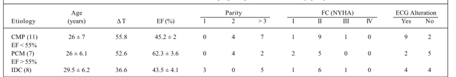
The mean age of patients with peripartum cardiomyo-pathy and ventricular dysfunction was 26±7 years. Of those patients without ventricular dysfunction, it was 26±6,1. The mean age for those patients with idiopathic dilated cardiomyopathy was 29.5±6.2 (tab. I). All patients were in functional class I/II (NYHA) at the beginning of pregnancy, and all had a history of heart failure and decreased EF on echocardiography, except for 2 patients, 1 with peripartum cardiomyopathy and the other with idiopathic dilated cardiomyopathy.
Twenty patients had electrocardiographic alterations (right branch conduction disturbance, supraventricular and ventricular extrasystoles, left ventricular hypertrophy) that were equally distributed between the groups with ventricu-lar dysfunction (tab. I).
In patients with peripartum cardiomyopathy, the elap-sed time between the onset of the disease and the studied pregnancy was, on average, 52.6 months for those patients that recovered ventricular function and 55.8 months for the remaining patients.
Prenatal care included periodic appointments with a cardiologist and an obstetrician, a control ECG, and echo-cardiograms during pregnancy and within 3 months after the delivery. We recommended that patients refrain from physical exercise and begin a diet low in sodium after the second trimester of pregnancy, and we also recommended hospitalization when cardiac and obstetric complications occurred. Medications previously prescribed, such as digi-talis, diuretics, and antiarrhythmic drugs, were continued, except for angiotensin-converting enzyme inhibitors and
anticoagulants, respectively, replaced by hydralazine and heparin, according to the Cardiopathy and Pregnancy Con-sensus of the Brazilian Society of Cardiology 12.
Eight patients became pregnant while taking captopril, which was suspended at the first routine prenatal evalua-tion, which occurred in the majority of case during the first trimester of pregnancy. Four patients continued taking a-miodarone during pregnancy, which was considered ne-cessary for the control of arrhythmias.
An analysis of the occurrence of clinical events (heart failure, thromboembolism, cardiac arrhythmia, and death), and of ECG, and EF changes before and after parturition was performed by calculating minimum, maximum, and median values, and standard deviation, for quantitative va-riables. Tables of absolute and relative frequencies were used for the qualitative variables. The comparative analysis was performed on 3 subgroups: peripartum thy with ventricular dysfunction, peripartum cardiomyopa-thy without ventricular dysfunction, and idiopathic dilated cardiomyopathy. Pearson c2 tests were used to analyze functional class, electrocardiographic alterations, use of me-dication and maternal-fetal complications. Fisher’s exact test and the Student t test were used for age, ejection fraction, and fetal and maternal death variables. A probabi-lity value lower than 0.05 was considered significant.
Results
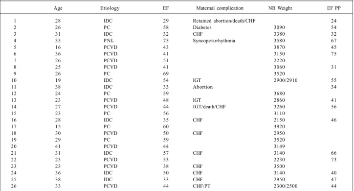
Clinical and obstetrical evolvement is shown in table II. Of the 26 patients, 11 (42.3%) had a normal delivery; 9 (35.5%) had cardiac complications, and 6 (22.2%) had obs-tetric complications. Of the cardiac complications, 8 resulted from heart failure, 2 from sustained ventricular tachycardia, and 1 from thromboembolism. Two maternal deaths occurred: one due to a retained abortion, infection, and heart failure in a patient with idiopathic dilated cardiomyopathy and another because of heart failure in a patient with peripartum cardiomyopathy with ventricular dysfunction. Obstetric complications were 2 miscarriages, 1 case of gestational diabetes, and 3 cases of urinary infec-tion. The 26 healthy newborns included twins, without mal-formations related to maternal cardiomyopathy or to the type of therapeutic regimen adopted.
During pregnancy, 7 patients did not take cardiovas-cular drugs, whereas 21 patients needed medication, such
Table I - Characteristics of the 26 pregnant patients with cardiomyopathy
Age Parity FC (NYHA) ECG Alteration
Etiology (years) ∆ T EF (%) 1 2 > 3 I II III IV Yes No
CMP (11) 26 ± 7 55.8 45.2 ± 2 0 4 7 1 9 1 0 9 2
EF < 55%
PCM (7) 26 ± 6.1 52.6 62.3 ± 3.6 0 4 2 2 5 0 0 2 5
EF > 55%
IDC (8) 29.5 ± 6.2 36.6 43.5 ± 4.1 3 0 5 1 6 1 0 4 4
as digitalis, hydralazine, heparin or amiodarone, or both, for prevention or treatment of complications.
A comparative analysis between the 3 groups, regar-ding age, parity, and functional class in the beginning of pregnancy were not different (p<0.05). Of the cases of peri-partum cardiomyopathy with ventricular dysfunction and idiopathic dilated cardiomyopathy, no differences existed regarding age (p=0.06), functional class at the beginning of pregnancy (p=0.37), electrocardiogram alterations (p=0.38), EF (p=0.06), use of captopril (p=1.0), use of amiodarone (p=0.59), and fetal death (p=0.59) (tab. II).
A cardiac complication rate during gestation was lower (p<0.009) in the peripartum cardiomyopathy without ven-tricular dysfunction subgroup and higher in the idiopathic dilated cardiomyopathy subgroup, when compared with that in the peripartum cardiomyopathy with ventricular dysfunction subgroup (p=0.01). A comparative analysis of 17 records within the first 3 months after delivery showed that no significant changes occurred between the electrocardiogram (p>0.05) and EF (p>0.05) after delivery when compared with that in the pregnancy records in the 3 groups (tab. III).
Discussion
Diversity in the clinical features of peripartum cardio-myopathy causes difficulties in making recommendations about future pregnancies, when the factors of its prognosis are still not clearly established.
This prospective study examined the evolvement of pregnancy in patients who had experienced peripartum cardiomyopathy in a previous pregnancy, with or without re-covery of ventricular function, and compared this with the evolvement of patients with idiopathic dilated cardiomyo-pathy, observing similarities in age, functional capacity, and electrocardiographic and EF alterations in the echocardiogram. Results showed that clinical evolvement of pregnancy in patients with peripartum cardiomyopathy who recover ventricular function was favorable, and patients did not
ex-Table II - Clinical and obstetric evolvement of 26 patients with cardiomyopathy
Age Etiology EF Maternal complication NB Weight EF PP
1 28 IDC 29 Retained abortion/death/CHF 24
2 26 PC 58 Diabetes 3090 54
3 31 IDC 32 CHF 3380 32
4 35 PNL 75 Syncope/arrhythmia 3580 67
5 16 PCVD 43 3870 45
6 36 PCVD 41 3150 75
7 26 PCVD 51 2220
8 25 PCVD 41 3060 31
9 26 PC 69 3520
10 19 IDC 54 IGT 2900/2910 55
11 38 IDC 33 Abortion 34
12 24 PC 59 3680
13 23 PCVD 48 IGT 2860 41
14 27 PCVD 44 IGT/death/CHF 3260 56
15 23 PC 56 3110
16 28 IDC 35 CHF 2150 46
17 15 PC 60 3920
18 30 PCVD 50 CHF 2950
19 29 PC 59 3520
20 41 PCVD 44 3149
21 31 IDC 57 CHF 3140 66
22 23 PCVD 53 2230 73
23 23 PCVD 38 CHF 3500
24 36 IDC 50 CHF 3140 40
25 38 IDC 33 CHF 2950 47
26 33 PCVD 44 CHF/PT 2300/2500 44
EF- left ventricle ejection fraction during pregnancy; EF PP- postpartum left ventricle ejection fraction; IDC- idiopathic dilated cardiomyopathy; PC- peripartum cardiomyopathy without ventricular dysfunction; PCVD- peripartum cardiomyopathy with ventricular dysfunction; IGT- infection of genitourinary tractus; CHF-congestive heart failure; PT- pulmonary of tromboembolism.
Table III - Comparative results between the 3 groups of cardiomyopathy
Peripartum cardiomyopathy Idiopathic p value Groups With Without Cardiomyopathy
dysfunction dysfunction
Age (years) 26 ± 7 26 ± 6.1 29.5 ± 6 NS EF before 45.2 ± 2* 62.3 ± 3.6 43.5 ± 4.1* NS* pregnancy (%)
Cardiac 3 (27.3%)** 1 (14.2%) 5 (62.5%)** p = 0.01** complication
Maternal death 1 ( 9.1%) - 1 (14.2%) NS EF 47.1 ± 2.1*** 65.4 ± 4.3*** 42.4 ± 4.5*** NS*** postpregnancy (%)
perience complications regarding cardiopathy. Additio-nally, EF was maintained at the same level as that observed within the first 3 months after delivery.
These data are in accordance with those from the studies performed by Albanesi Filho et al 13 and Sutton et al 14
who showed in a prospective analyses good evolvement, absence of mortality, or recurrence of peripartum cardio-myopathy in 11 pregnancies of women who had recovered ventricular function after a diagnosis of peripartum cardio-myopathy.
On the other hand, our results are not in accordance with those of Elkayam et al 9 who found a reduction of 8% in
EF in the postpartum period in women with peripartum car-diomyopathy, considered as having recovered from ventri-cular dysfunction, and who began pregnancy with mean EF of 56±7%. In addition, we cannot agree with the authors’ assumption that the recurrence of peripartum cardiomyopa-thy following pregnancy was based on a reduction in EF after delivery. It is possible that bias in the selection of the retrospective cohort, in the criteria of the diagnosis of the di-sease, and in ventricular dysfunction are reasons why the results of the studies do not correlate.
Lampert et al 15 demonstrated that 7 patients with
peri-partum cardiomyopathy, who regained normal resting left ventricular size and performance have decreased contractile reserve revealed by the use of dobutamine. These data and controversies reported in the literature emphasize the need to revise current criteria of ventricular function recovery in peripartum cardiomyopathy. Perhaps current parameters based on conventional echocardiographic analysis are not enough to ensure a diagnosis of normal ventricular func-tion in women with a previous history of peripartum cardio-myopathy.
On the other hand, a mean clinical follow-up of 8.6 years of 42 women with peripartum cardiomyopathy showed that the 75% of patients that recovered ventricular function did not have any kind of limitation in functional capacity and had a good quality of life 16.
Another important aspect of the natural history of pe-ripartum cardiomyopathy is the expectation of a better prog-nosis because of advances in the therapeutics of heart fai-lure in recent decades. Indeed, the 50% rate of ventricular function recovery reported in the 1970s 3 is below the 75%
reported in recent publications 16. Additionally, mortality
rates among patients that remained with dilated cardiomyo-pathy also decreased from 85% to 30% because of the benefits of cardiac transplantation 17 and to the strategies of
drug therapy for this group of patients 18.
Thus, greater life expectancy in short-term and long-term follow-up and the good evolvement of a subsequent pregnancy 13-15 do not justify ruling out pregnancy in
women who experienced peripartum cardiomyopathy in a previous pregnancy, but who recovered left ventricular function. Prospective studies, with a greater cohort and assessment of ventricular function are necessary to define the correct counseling for these women.
On the other hand, our study showed that the cardiac complication rate in women with peripartum cardiomyopa-thy with persistent ventricular dysfunction, including 1 ma-ternal death, highlight the high risk in this group of patients. However, no signs of worsening of ventricular dysfunction were estimated through postpartum EF or signs of recur-rence of the disease even in the 2 sets of twins, a recognized factor of predisposition to peripartum cardiomyopathy 3.
Hemodynamic overload after the first trimester of preg-nancy, demonstrated by a 10% increase in final left ventricu-lar diastolic volume and a 45% increase in cardiac output 19
worsens left ventricular systolic function, which usually re-verses after delivery.
However, we believe that this circulatory response is ventricular dysfunction-dependent and not cause-depen-dent. Therefore, the remaining question is whether or not peripartum cardiomyopathy with ventricular dysfunction persistence has a natural history similar to that of other cau-ses of dilated cardiomyopathy, including pregnancy 20.
In this regard, our results corroborate those of pre-vious studies that had similar patterns regarding obstetric and cardiologic evolvement between peripartum cardio-myopathy and idiopathic dilated cardiocardio-myopathy. It is worth mentioning that among clinical, echocardiographic, and hemodynamic variables previously shown 20,21,
hysto-logic findings of myocarditis in endomyocardial biopsies was controversial 22,23, and in some series it was
significan-tly higher in peripartum cardiomyopathy 24.
An additional factor of maternal fetal morbidity is the suitability of therapeutics during pregnancy in patients with dilated cardiomyopathy. Sodium and physical activity restrictions in association with drugs like digoxin and furo-semide, which are not contraindicated in this period, help control heart failure during pregnancy. However, use of an-giotensin-converting enzyme inhibitors is associated with side effects, such as oligohydramnios, delay in intrauterine development, prematurity, fetal renal failure, bone malforma-tion, and neonatal death 25. In our study, these effects were
not identified in 8 patients who became pregnant while taking captopril; however, this medication was replaced in the first trimester, as soon as pregnancy was confirmed.
Hydralazine, with or without nitrates, as an alternative to angiotensin- converting enzyme inhibitors was based on the results of the V-HeFT-II study 26 that demonstrated
en-hancement in functional capacity of oxygen consumption, and of EF in a 12-month period, equivalent to the peripartum period. The experience with hydralazine in the treatment of hypertensive disease due to pregnancy shows that no obs-tetric and fetal contraindication exists regarding its use, during any phase of pregnancy 27.
Ventricular arrhythmias are usually complex and are re-lated to death in patients with dire-lated cardiomyopathy, the-refore, requiring effective control with antiarrhythmic drugs like amiodarone. However, this medication may be toxic for mother and baby, leading to hypothyroidism and retarded growth, with the risk of perinatal death 28.In our study, we
to 200mg/day) in 4 patients that already used it, because we considered it essential for controlling complex ventricular arrhythmias, and we did not observe side effects in the baby. Systemic or pulmonary thromboembolism is another frequent complication, described in more than half of the cases of peripartum cardiomyopathy with ventricular dys-function. During pregnancy and the postpartum period, hy-percoagulability, including activation of coagulation factors, increase in plasma fibrinogen, and platelet adhe-sion, increases the risk of thrombosis, which is aggravated by the need for prolonged rest due to congestive heart fai-lure. Because of this, patients with dilated cardiomyopathy with ventricular dysfunction must be anticoagulated with heparin at prophylactic doses, because this medication does not cross the placenta.
In summary, peripartum cardiomyopathy has been re-ferred to as a distinct entity from other dilated cardiomyopa-thies because of its relation to the peripartum period and the peculiarities of its natural history. Evolvement of pregnancy in patients with peripartum cardiomyopathy with
ven-tricular dysfunction was not different when compared with that of the idiopathic dilated cardiomyopathy group. However, patients who recovered ventricular function did not experience complications, which is similar to the expe-rience of women considered to be healthy. The present study reveals the limitations inherent in a small cohort and the conventional methods of ventricular function assess-ments. However, because it is a rare disease, this prospec-tive analysis may add to the data about a very controversial subject.
Our results enabled us to conclude that left ventricular function is the determinant factor in pregnancy following the diagnosis of peripartum cardiomyopathy. Based on our understanding, discouragement of a new pregnancy must be reserved for patients with peripartum cardiomyopathy who have ventricular dysfunction.
Acknowledgments
To Dr. Maeve de Barros Correia for the valuable suggestions given in the review of the text.
1. Pearson GD, Veille JC, Rahimtoola S, et al. Peripartum cardiomyopathy. JAMA-Brazil 2000; 283: 1183-8.
2. Veille J, Zaccaro D. Peripartum cardiomyopathy: summary of international survey on peripartum cardiomyopathy. Am J Obstet Gynecol 1999; 181: 315-9. 3. Homans DC. Peripartum cardiomyopathy. N Eng J Med 1985; 312: 1432-6. 4. Demakis JG, Rahimtoola SH. Peripartum cardiomyopathy. Circulation 1971; 44:
964-8.
5. Lampert MB, Lange RM. Peripartum cardiomyopathy. Am Heart J 1995; 130: 860-70. 6. Demakis JG, Rahimtoola SH, Sutton GC, et al. Natural course of peripartum
car-diomyopathy. Circulation 1971; 44: 1053-61.
7. Veille J. Peripartum cardiomyopathies: a review. Am J Obstet Gynecol 1984; 148: 805-17.
8. Souza JL, Carvalho FC, Nastari L, Mady CC. Left ventricular function after new pregnancy in patients with peripartum cardiomyopathy. J Card Fail 2001; 7: 30-5. 9. Elkayam U, Tummala PP, Rao K, et al. Maternal and fetal outcomes of subsequent pregnancies in women with peripartum cardiomyopathy. N Engl J Med 2001; 344: 1567-71.
10. Ceci O, Berardesca C, Caradonna F, et al. Recurrent peripartum cardiomyopathy. Eur J Obstet Gynecol and Reprod Biol 1998; 76: 29-30.
11. Richardson P, McKenna W, Bristow MR, et al. Report of the 1995 World Health Or-ganization/International Society an Federation of Cardiology Task Force on the definition and classification of cardiomyopathies. Circulation 1996; 93: 841-2. 12. Consenso do Departamento de Cardiopatia e Gravidez da Sociedade Brasileira
de Cardiologia. Arq Bras Cardiol, 1999; 72(supl III): 8.
13. Albanesi Fo FM, Silva TT. O comportamento das gestações subseqüentes na
cardiomiopatia periparto. Arq Bras Cardiol 1999; 73: 47-52.
14. Sutton MSJ, Cole P, Plappert M, et al. Effects of subsequent pregnancy on left ventricular function in peripartum cardiomyopathy. Am Heart J 1991; 121; 1776-8. 15. Lampert MB, Weinert L, Hisbbard J, et al. Contractile reserve in patients with peripartum cardiomyopathy and recovered left ventricular function. Am J Obstet Gynecol 1997; 176: 189-95.
References
16. Felker GM, Jaeger CJ, Klodas E, et al. Myocarditis and long-term survival in peripartum cardiomyopathy. Am Heart J 2000; 140: 785-91.
17. Carvalho AG, Almeida D, Cohen M, et al. Successful pregnancy, delivery and puerperium in a heart transplant patient with previous peripartum cardiomyopa-thy. Eur Heart J 1992; 13: 1589-91.
18. The SOLVD Investigators. SOLVD treatment study. Am J Cardiol 1990; 66: 315-22. 19. Robson SC, Hunter S, Boys RJ, Dunlop W. Serial study of factors influencing changes in cardiac output during human pregnancy. Am J Physiol 1989; 226: H1060-5.
20. Felker GM, Hu W, Hare JM, et al. The spectrum of dilated cardiomyopathy: the Johns Hopkins Experience with 1,278 patients. Medicine 1999; 78: 270-83. 21. Chan L, Hill D. Echocardiography for peripartum cardiomyopathy. Am J Em Med
1999; 17: 578-80.
22. Rizeq MN, Rickenbacher PR, Fowler MB, et al. Incidence of myocarditis in peri-partum cardiomyopathy. Am J Cardiol 1994; 74: 474-7.
23. Mady C, Barretto ACP, Belotti G, et al. Biópsia endomiocárdica em pacientes por-tadoras de miocardiopatia periparto. Arq Bras Cardiol 1986; 47: 403-5. 24. O’Connell JB, Constanzo O, Nordin MR, et al. Peripartum cardiomyopathy:
Cli-nical, hemodynamic, histologic and prognostic characteristics. J Am Coll Car-diol 1986; 8: 52-6.
25. Pryde PG, Thorpe SS, Lamont CA. Angiotensin-converting enzyme inhibitor fetopathy. J Am Soc Nephrol 1993; 3: 1575-82.
26. Chon JN, Johnson G, Zeische S, et al. A comparison of enalapril with hydralazi-ne-isosorbide dinitrate in the treatment of chronic congestive heart failure. N Engl J Med 1991; 325: 303-10.
27. Paterson-Brown S, Robson SC, Redfern N, Walkinshaw SA, Swiet M. Hydrala-zine boluses for the treatment of severe hypertension in preeclampsia. Br J Obstet Gynaecol 1994; 101: 409-15.