Submitted 23 September 2015 Accepted 18 November 2015 Published10 December 2015
Corresponding author Stevens K. Rehen, [email protected]
Academic editor Prasanna Krishnamurthy
Additional Information and Declarations can be found on page 14
DOI10.7717/peerj.1486
Copyright 2015 Lages et al.
Distributed under
Creative Commons CC-BY 4.0
OPEN ACCESS
Low oxygen alters mitochondrial
function and response to oxidative stress
in human neural progenitor cells
Yury M. Lages1, Juliana M. Nascimento2, Gabriela A. Lemos3, Antonio Galina3, Leda R. Castilho4and Stevens K. Rehen1,2
1Institute of Biomedical Sciences, Federal University of Rio de Janeiro, Rio de Janeiro, RJ, Brazil 2IDOR, D’Or Institute for Research and Education, Rio de Janeiro, RJ, Brazil
3Institute of Medical Biochemistry Leopoldo De Meis, Federal University of Rio de Janeiro,
Rio de Janeiro, RJ, Brazil
4COPPE, Chemical Engineering Program, Federal University of Rio de Janeiro, Rio de Janeiro, RJ,
Brazil
ABSTRACT
Oxygen concentration should be carefully regulated in all living tissues, beginning at the early embryonic stages. Unbalances in oxygen regulation can lead to cell death and disease. However, to date, few studies have investigated the consequences of variations in oxygen levels for fetal-like cells. Therefore, in the present work, human neural progenitor cells (NPCs) derived from pluripotent stem cells grown in 3% oxygen (v/v) were compared with NPCs cultured in 21% (v/v) oxygen. Low oxygen concentrations altered the mitochondrial content and oxidative functions of the cells, which led to improved ATP production, while reducing generation of reactive oxygen species (ROS). NPCs cultured in both conditions showed no differences in proliferation and glucose metabolism. Furthermore, antioxidant enzymatic activity was not altered in NPCs cultured in 3% oxygen under normal conditions, however, when exposed to external agents known to induce oxidative stress, greater susceptibility to DNA damage was observed. Our findings indicate that the management of oxygen levels should be considered forin vitromodels of neuronal development and drug screening.
Subjects Biochemistry, Cell Biology, Developmental Biology, Neuroscience, Toxicology
Keywords Low oxygen, Cell metabolism, Reactive oxygen species, DNA damage, Mitochondria, Human neural progenitor cells
INTRODUCTION
By describing events that alter mitochondrial metabolism and facilitate tumor formation,
Warburg (1956)shed light on the importance of oxygen concentrations for cellular health. Accordingly, matching thein vitroconditions of a given cell type to the respectivein vivooxygen concentration has become a relevant issue that accompanies the growing number of applications of human pluripotent stem cells, which are particularly relevant for modeling fetal and/or neurological disorders.
progression of brain disorders, including Parkinson’s disease, Alzheimer’s disease, and schizophrenia (Paulsen et al., 2013;Yan, Wang & Zhu, 2013). Impairment of mitochondrial function or the redox state may be especially problematic for highly metabolically demanding neurons. Mismanagement of these processes is massively problematic, negatively impacting energy metabolism, neurochemical signaling and/or synaptic plasticity, and emergent cognitive processes of these functions (Cheng, Hou & Mattson, 2010;Janc & Muller, 2014;Tait & Green, 2012).
Despite the well-recognized relationship between oxidative metabolism and the onset of neural disorders (Paulsen et al., 2013;Yan, Wang & Zhu, 2013;Carreau et al., 2011), few studies have focused on analyzing changes occurring at atmospheric oxygen concentrations (i.e., 21% O2(v/v); typical levels in cell culture, normoxia), compared with
physiological levels (3% O2(v/v)). Studies carried out on murine neural progenitor cells
(NPCs) have considerable differences in proliferation, death, and differentiation (Bae et al., 2012;Chen et al., 2007;Rosafio & Pellerin, 2014;Ross et al., 2012;Stacpoole et al., 2011;
Studer et al., 2000). These studies have shown unexpected deviations in cell fate, including altered relative proportions of neuronal and glial populations (Chen et al., 2007;Stacpoole et al., 2011;Studer et al., 2000).
Studies that specifically address the impacts of oxygen levels on the metabolic behavior of NPCs are still rare. Recent reports have described increased dispersion of mitochondria as well as modifications in mitochondrial efficiency and reactive oxygen species (ROS) production of rat neurons grown under 1–5% O2 (Tiede et al., 2011). In addition,
Tiede and colleagues (2011) have reported increased cell death in physiological oxygen concentrations (physioxia (Rosafio & Pellerin, 2014)) when NPCs are exposed to viral infection proteins; however, their study did not elucidate the cause of the alterations. Therefore, the aim of this study was to compare NPCs grown in physioxia and normoxia (3% and 21% (v/v) O2, respectively) in terms of growth kinetics, glycolytic metabolism,
mitochondrial content, mitochondrial membrane potential(ΔΨM), oxygen uptake, ATP production, ROS production, and antioxidant enzymatic activity.
METHODS
Differentiation of human embryonic stem cells into NPCs
To generate NPCs, human embryonic stem cells BR-1 (Fraga et al., 2011) (kindly provided by Prof. Lygia Pereira, S˜ao Paulo University - USP) were grown on polystyrene plates (TPP, Switzerland) covered with Matrigel (BD Biosciences, Franklin Lakes, NJ, USA) in StemPro medium containing 8 ng/mL basic fibroblast growth factor and 0.1 mM
β-mercaptoethanol (all from Thermo Fischer Scientific, Waltham, MA, USA). After prop-agation, the cells were differentiated as neural cells using inhibitors of bone morphogenetic protein (Noggin; R&D Systems, USA), and transforming growth factor-beta (SB431542; Tocris Bioscience, Bristol, UK) (Chambers et al., 2009). At this moment, cells showed morphology and expression of markers consistent to those of neural progenitor cellsFigs.
Cultivation of NPCs in a physiological environment
NPCs were grown in an environment containing 3% oxygen (physioxia) in an oxygen control chamber (ProOx model C21; BioSpherix, Parish, NY, USA). This equipment was kept at 37◦C and 5% CO
2, and 3% pO2was established by a N2-controlled injection and
monitored by an external probe (Mettler Toledo, Colombus, OH, USA).
Growth kinetics
Cellular growth under different oxygen conditions was evaluated for 18 days (3 passages of 6 days). A total of 6×10⁵NPCs/mL were plated into 24-well tissue culture plates. Each day, cells from two wells of each condition were detached with Accutase (Millipore, Darmstadt, Germany) and counted in a Neubauer chamber.
Glucose and lactate measurements
Glucose and lactate concentrations were determined using a YSI-2700 biochemistry analyzer (Yellow Springs Instruments, Yellow Springs, OH, USA). This measurement is based on quantification of hydrogen peroxide generated upon reaction of these organic molecules catalyzed by glucose or lactate oxidases immobilized on membranes.
Immunostaining assays
NPCs were seeded in 96-multiwell µClear dishes (Greiner, Austria) covered with
2.5µg/mL laminin (Sigma-Aldrich, USA). After 6 days, these cells were fixed with 4%
paraformaldehyde (Sigma-Aldrich, USA) in phosphate-buffered saline for 30 min. Then, the cells were treated with 0.5% Triton X-100 (Sigma-Aldrich, St. Louis, MO, USA), blocked with 5% bovine serum albumin (Sigma-Aldrich, St. Louis, MO, USA), incubated with the following primary antibodies: rabbit anti-human-histone H2A (H2A.X) (1:100; Cell Signaling, USA), mouse anti-Nestin (1:100; Chemicon, Temecula, CA, USA), mouse anti-PSA-NCAM (1:100; Millipore, Germany) and rabbit anti-Tbr2 (1:100; Millipore, Darmstadt, Germany). Subsequently, samples were incubated with the following secondary antibodies: goat anti-rabbit AlexaFluor 488 IgG (1:400; Thermo Fischer Scientific, Waltham, MA, USA) and goat anti-mouse Alexa Fluor 594 IgG (1:400; Thermo Fischer Scientific, Waltham, MA, USA). Nuclei were stained with 0.5µg/mL
4′-6-diamino-2-phenylindole (DAPI).
Regions of interest were visualized and identified, and the immunofluorescence emission of the cells was quantified using an Operetta high content analysis system and Harmony software (PerkinElmer, Waltham, MA, USA). In these experiments, three technical replicates of each biological replicate (N) were performed.
Mitochondrial content andΔΨM quantification assays
Measurement of the mitochondrial mass of NPCs was performed using 0.3 µM
Mitotracker DeepRed FM (Thermo Fischer Scientific, Waltham, MA, USA), a dye that integrates into active mitochondria (568-nm excitation and 675-nm emission). TheΔΨM
was estimated by cationic staining with 1.6µM JC-1 (Thermo Fischer Scientific, Waltham,
MA, USA) (488-nm excitation). This dye exists as a monomer at low concentrations, with fluorescence emission at 525 nm (shown here in green). As it accumulates in the mito-chondria, which is membrane potential-dependent, the dye forms aggregates that exhibit a maximum emission at 590 nm (shown here in yellow). The ratio of aggregate to monomer concentration can be used as a measurement ofΔΨM(Reers, Smith & Chen, 1991).
MitoTracker and JC-1 dyes, diluted in Dulbecco’s modified Eagle’s medium/F12 (Thermo Fischer Scientific, Waltham, MA, USA), were applied to NPCs for 40 min at 37◦C. Fluorescence emission readings were performed in a controlled 5% CO
2and 37◦C
environment. Hoechst 33342 (1µM, Thermo Fischer Scientific, Waltham, MA, USA) was
used for nuclear staining.
Thirty-three fields per well were captured randomly. An average of 825 cells were analyzed per well.
ROS measurement assay
Quantification of superoxides was performed using 10µM dihydroethidium (DHE;
Thermo Fischer Scientific, Waltham, MA, USA). This dye, when oxidized in the cytosol, intercalates with DNA and emits fluorescence at 605 nm. DHE was applied similarly to MitoTracker and JC-1. As a positive control, NPCs were induced to produce ROS by a 40 min pretreatment with 3.6µM antimycin A, a mitochondrial complex III inhibitor that
stabilizes semi-quinone radicals and favors the escape of electrons to oxygen, thus forming superoxide anions.
Twenty-five fields per well were captured randomly. An average of 6,800 nuclei were analyzed per well.
Oxygen consumption measurement
Oxygen consumption was measured by high-resolution respirometry using an Oroboros O2k Oxygraph at 37◦C. DataLab software (Oroboros Instruments, Innsbruck, Austria)
was used for data acquisition and analysis. NPCs were enzymatically detached from the plate, diluted in culture medium, and seeded to the Oroboros at a concentration of 1×10⁶cells/mL. The routine oxygen consumption of cells, measured before the addition of modulators of mitochondrial function, was determined after stabilization of the steady state of oxygen consumption for 10–15 min. Subsequently, ATP synthesis was inhibited with 2µg/mL oligomycin. Oxygen consumption related to oxidative phosphorylation
coupled to ATP synthesis was determined by the difference between routine respiration and oligomycin-insensitive respiration.
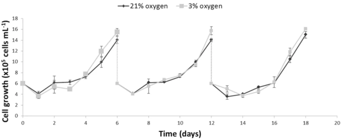
Figure 1 Comparison of the growth profiles of NPCs cultured either in physioxia (gray line) or normoxia (black line) over an 18-day period, including passaging on days 6 and 12.
electron transport system. Finally, the nonoxidative phosphorylation oxygen flux was determined by blocking the electron transporting system with 1µg/mL antimycin A. The
residual oxygen flux represents oxidases in the cell sample.
Antioxidant enzymatic activity assay
The activities of the antioxidant enzymes superoxide dismutase (SOD) and glutathione peroxidase (GPx) were measured using commercial kits (cat. 19160 and CGP1, respec-tively; Sigma-Aldrich, St. Louis, MO, USA). Briefly, the SOD colorimetric assay (440 nm) determines the presence of superoxide radicals in a tetrazolium-coupled reaction. The GPx assay measures NADPH depletion (340 nm). The SOD and GPx enzymatic activities were calculated according to the kit instructions.
To induce ROS production and a possible increase in antioxidant enzymatic activity, the cells were treated with antimycin A, as described previously. Antimycin A showed no effect on apoptosis during the time of treatment (Fig. S1).
Statistical analysis
The unpairedttest was used to compare average differences between two groups. In the case of multiple variable comparisons, one-way analysis of variance was used with the Bonferroni post-test. The null hypothesis of equality between averages was refuted if
p<0.05 (*),p<0.01 (**), orp<0.001 (***). Means and standard errors of the mean were plotted.
RESULTS
Growth and glycolytic metabolism of human NPCs are not altered in physioxia
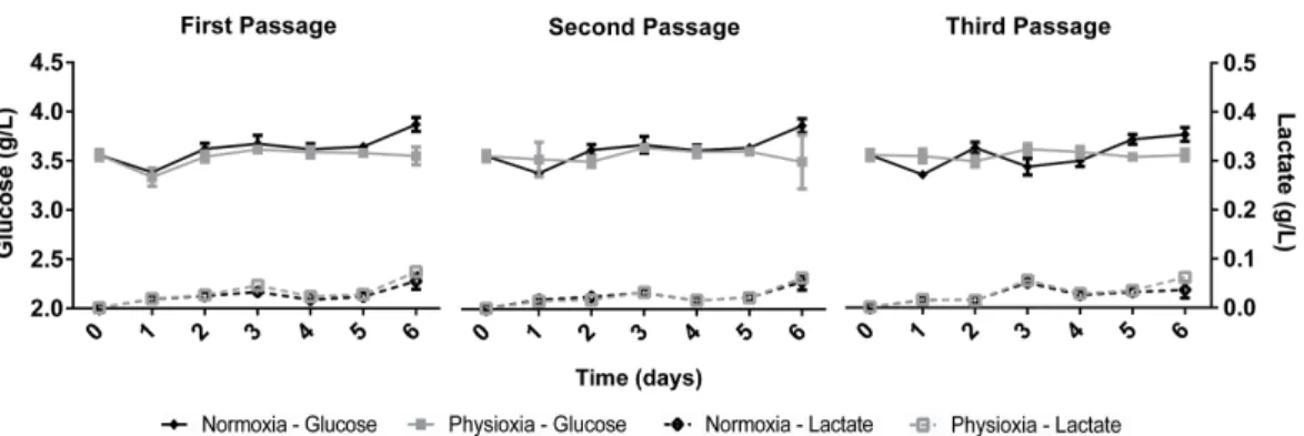
Figure 2 Glucose uptake and lactate concentration in the supernatant of NPCs grown in physioxia (gray lines) and normoxia (black lines) throughout the first, second, and third passages.
Mitochondrial content andΔΨM are altered in NPCs grown in
physioxia
Mitochondrial labeling was performed using the MitoTracker probe, which is internalized by active mitochondria. Fluorescence intensity measurements indicated less mitochondrial content in physioxia-grown NPCs (Figs. 3Aand3B). As a first approach to evaluate mitochondrial function, we quantifiedΔΨMas an indicator of the proton motive force. An increased difference ofΔΨMcan be directly related to a strict commitment to ATP
formation by FoF1ATP-synthase and/or to decreased proton leakage to the mitochondrial
matrix (Jastroch et al., 2010). The data showed increasedΔΨMin physioxia-grown NPCs
(Figs. 3Cand3D).
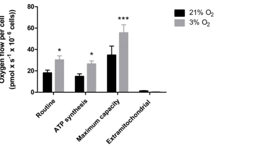
Physioxia-grown NPCs feature an increased mitochondrial respiration capacity
Our data suggest that changes in the mitochondrial physiology are induced by O2
availability (Fig. 3). A possible outcome of improvedΔΨMduring mitochondrial function
could be a more efficient oxygen consumption directed to ATP production. To verify the full potential of the mitochondria to destine environmental oxygen to oxidative respiration efficiently, we tested the O2consumption in NPCs grown previously either in normoxia or
physioxia in conditions in which plenty of O2was available, i.e., atmospheric
concentra-tions, by high resolution respirometry. Our data show higher oxygen uptake rates for NPCs previously cultured at 3% O2(v/v). The oxygen flux coupled to ATP production in NPCs
cultured at 3% O2(v/v) was approximately 70% higher than that of cells cultured at 20%
O2(v/v) (Fig. 4). Additionally, an increased maximum respiratory capacity of
physioxia-grown NPCs was observed, compared to those cultured in normoxia. This observation corroborates our previous results, showing that the NPCs did not suffer from restricted mitochondrial function due to lower oxygen availability. Instead, the physioxia conditions increased the potential of oxidative phosphorylation in NPCs cultured at 3% O2(v/v).
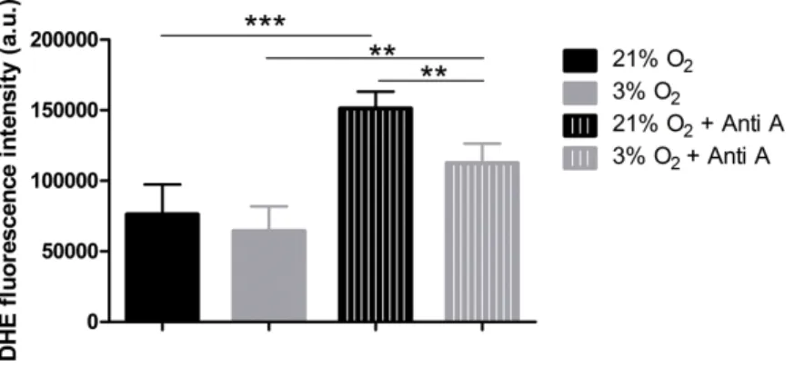
NPCs grown in physioxia produce less ROS
Figure 3 Evaluation of mitochondrial content by MitoTracker (A and B), in red; and mitochondrial membrane potential by JC-1 (C and D), in green and yellow.Nuclei are stained in blue by DAPI.p<0.05 (*),p<0.01 (**). Scale bar: 50µm.
Figure 5 ROS quantification with the probe DHE.p<0.01 (**).
from the respiratory chain, we measured ROS production. We used the dye DHE, a permeable probe oxidized in the cytoplasm, which further intercalates DNA and emits fluorescence. As DHE fluorescence was measured in NPCs in their native oxygen environments (i.e., normoxia and physioxia), we observed that, under routine conditions, ROS production was equivalent in both situations (Fig. 5). However, after antimycin A addition, physioxia-grown cells showed lower ROS levels from mitochondrial complex III induced by semi-quinone radical stabilization, compared to normoxia conditions (Fig. 5). These results suggest that NPCs grown under physiological oxygen are more resistant to redox imbalance. Moreover, this finding also suggests that their antioxidant defense system may be more efficient to scavenge the mitochondrial ROS generated.
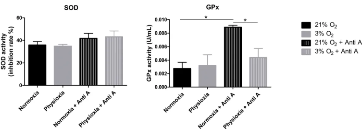
GPx activity is lower in NPCs grown in physioxia
To assess whether the antioxidant defense system of physioxia-grown NPCs is more active under routine conditions, we measured the activities of SOD and GPx in these cells. SOD is responsible for catalyzing the dismutation of superoxide anions in hydrogen peroxide and water. Then, GPx oxidizes intracellular glutathione, reducing peroxide to alcohol and water.
Our data show that NPCs grown either in normoxia or physioxia have equivalent enzymatic activities; however, when treated with antimycin A, the NPCs cultured under lower oxygen concentrations showed a less-pronounced increase of GPx activity in response to the ROS increase, while the SOD activity level was sustained (Fig. 6). This finding indicates that, unlike what was hypothesized previously, NPCs grown in physioxia and treated with antimycin A do not show decreased ROS production due to greater antioxidant activity in the pathway analyzed herein. Instead, GPx, one of the core enzymes of the cellular antioxidant machinery, depicts a decreased response to the ROS increase in these NPCs.
Figure 6 Enzymatic activity of the antioxidant enzymes superoxide dismutase (SOD) and glutathione peroxidase (GPx).Activity quantification under routine conditions and after antimycin A treatment, which stimulates maximum ROS production.p<0.05 (*).
Physioxia-grown NPCs suffer increased DNA damage when exposed to hydrogen peroxide
Although physioxia-grown NPCs produced lower levels of ROS when treated with antimycin A (Fig. 5), our data show that these cells also had a decreased activity of GPx
(Fig. 6), one of the main enzymes responsible for peroxide detoxification. Therefore,
we investigated whether NPCs cultivated under low oxygen concentrations would be more susceptible to stress. To this end, we quantified DNA damage, an indication of oxidative stress (Konyalioglu et al., 2013), in NPCs grown under routine conditions and after hydrogen peroxide treatment.
To quantify the impact of these treatments on DNA damage, the antibody against H2A.X was used to measure altered DNA. H2A.X is phosphorylated when DNA strands break, which signals for DNA repair and cell cycle arrest. Staining of H2A.X appears within the nucleus and can be monitored by both the overall fluorescence intensity and the number of visibly detectable aggregated structures.
Under our routine conditions, we observed that NPCs grown in normoxia or physioxia had equivalent levels of DNA damage (Fig. 7). However, after exposure to hydrogen peroxide, cells cultured under lower oxygen concentrations showed increased DNA damage, which was observed by increased fluorescence intensity both in the whole nucleus and in H2A.X spots (Fig. 7C). Moreover, the population of nuclei containing more than 20 spots decreased; while the whole stained nuclei population, which depicted so many spots that they became indistinguishable from one another, was increased in physioxia-grown NPCs (Fig. 7D).
Figure 7 DNA damage as evidenced by H2A.X (green), either after hydrogen peroxide treatment (A) or under routine conditions (B).Total nuclei are stained with DAPI (blue). Whole nucleus fluorescence is shown in both oxygen concentrations, after hydrogen peroxide treatment (C) and under routine conditions (D). The fluorescence of the whole nucleus and of the H2A.X spots was analyzed in the four categories.p<0.01 (**),p<0.001 (***). Scale bar: 25µm.
DISCUSSION
Evidence of increased susceptibility to exogenous stress agents in physioxia-grown NPCs highlights the importance of culturing human neural cells under low oxygen conditions to better evaluate the effect of drugs, especially the ones known to trigger oxidative stress.
We observed that NPCs in physioxia had reduced mitochondrial content, without changes in morphology (Fig. S2A), which might indicate stress (Giedt et al., 2012). Other studies have described lower mitochondrial DNA mass in human carcinoma, which is known to have a decreased oxygen gradient from the border to the center (Chiba et al., 2013). Moreover, the NPCs showed no alterations in fusion or fission of the mitochondria, which are linked to the dynamics of these organelles and influence their morphology and amount (Chan, 2012).
compensation by increased glycolysis and an unaltered growth profile of physioxia-grown NPCs do not indicate reduced mitochondrial activity in these cells. Thus, we evaluated the hypothesis that, instead, there may be improved function of these organelles to maintain ATP production rates necessary for cellular metabolism, even under low O2
concentrations.
Our data reveal that the mitochondria increased theΔΨMin physioxia-grown NPCs, indicating a higher activity and mitochondrial efficiency in ATP synthesis. As previous reports have demonstrated that mitochondrial membrane potential increases as oxygen availability is lowered below certain levels (Tiede et al., 2011), we believe our system is beneath this threshold. To confirm our hypothesis that increasedΔΨM could directly affect mitochondrial efficiency and commitment to oxidative respiration in physioxia, we investigated the parameters related to ATP production by measuring the oxygen consumption rates in an atmospheric environment, where oxygen is abundant.
Our high-resolution respirometry results corroborate that, in physioxia, oxygen consumption used for ATP production is increased. Moreover, using this technique, we concluded that a greaterΔΨMcould allow cells to maintain ATP production by oxidative
phosphorylation with fewer mitochondria. Thus, we hypothesized that this outcome may reflect an adaptive mechanism conferring a more efficient use of scarce oxygen.
Notably, even with the increased potential respiratory capacity, ROS production was not altered in physioxia compared to normoxia. Notwithstanding, when NPCs were exposed to antimycin A, stimulating an enhanced ROS production by mitochondria (Chen et al., 2003), we observed a greater amount of ROS in normoxia-grown NPCs. In this oxygen environment, oxygen availability as a source of ROS production is much higher. Thus, when electron leakage from the respiratory chain is stimulated by antimycin A and the oxygen concentrations increase, superior ROS formation is expected. On the other hand, in physioxia, conditions in which the free oxygen concentration is reduced, the ROS production is decreased even when NPCs are treated with antimycin A, corroborating our hypothesis of greater mitochondrial commitment to ATP production.
Keeping in mind the core importance of the antioxidant enzymes SOD and GPx in the detoxification process of superoxide anions generated by mitochondria in neural cells (even overcoming catalase activity (Mitozo et al., 2011)), we evaluated whether the decreased production of these ROS was a consequence not only of increasedΔΨMbut also of improved enzymatic activity.
The SOD activity measurements revealed that this enzyme function was not altered in physioxia, neither under routine conditions nor after antimycin A treatment (Fig. 6). As superoxide anion dismutation in hydrogen peroxide can also occur spontaneously, ROS production can increase after antimycin A treatment even if there are no alterations in SOD activity (Drose & Brandt, 2008).
currently exists, earlier studies correlating reduced oxygen concentrations to GPx activity indicate decreased activity of this and other enzymes involved in ROS detoxification, such as peroxiredoxin 3 (Becker et al., 2014;Duranton et al., 2012;Hidalgo et al., 2014;Xi et al., 2014). Indeed, minor increases of ROS production in response to antimycin A treatment were not enough to generate greater GPx activation in physioxia-grown NPCs.
The importance of GPx peroxide detoxification was investigated once more, measuring the susceptibility of these cells to hydrogen peroxide insults by quantifying the amount of DNA damage, a marker of oxidative stress. Increased DNA damage caused by peroxide, shown previously in other models (Konyalioglu et al., 2013;Mitozo et al., 2011), was herein reproduced by NPCs in normoxia. These cells showed a greater H2A.X fluorescence intensity and more spots in the nuclei, and the amount of marked cells rose from 25% to 100% after treatment.
Comparing NPCs grown in normoxia vs. physioxia, all parameters indicated a similar amount of DNA damage in routine conditions (without insult). However, after hydrogen peroxide insult, physioxia-grown NPCs showed nuclei with increased H2A.X fluorescence and more spots, indicating increased DNA damage as compared to normoxia-grown NPCs.
These data indicate a greater susceptibility of human NPCs grown in physiological oxy-gen conditions to insults caused by ROS. This evidence is of great relevance, since models to study disease related to oxidative stress are usually performed under atmospheric oxygen environments, and possibly may not represent actual processes and responses.
Recent studies, using human NPCs differentiated from donors with brain disorders, have shown alterations in mitochondrial membrane potential and oxidative stress, in addition to increased DNA damage, when compared to control NPCs generated from healthy donors; these data indicate phenotype reversibility when classical drugs are used (Brennand et al., 2014;Paulsen et al., 2013). Just as these studies, many others have been performed under atmospheric oxygen concentrations; thus, the results could be more reliable if the cells had been grown at the same oxygen concentrations foundin vivo.
ACKNOWLEDGEMENTS
This work is part of the MSc thesis of YL. We thank Michelle Kormann for the support during cell cultivation.
ADDITIONAL INFORMATION AND DECLARATIONS
Funding
This study was funded by the following Brazilian funding agencies: CNPq, FAPERJ, CAPES, FINEP, and BNDES. The funders had no role in study design, data collection and analysis, decision to publish, or preparation of the manuscript.
Grant Disclosures
The following grant information was disclosed by the authors: CNPq.
FAPERJ. CAPES. FINEP. BNDES.
Competing Interests
The authors declare there are no competing interests.
Author Contributions
• Yury M. Lages performed the experiments, analyzed the data, wrote the paper, prepared figures and/or tables, reviewed drafts of the paper.
• Juliana M. Nascimento conceived and designed the experiments, performed the experiments, analyzed the data, prepared figures and/or tables, reviewed drafts of the paper.
• Gabriela A. Lemos performed the experiments, analyzed the data.
• Antonio Galina, Leda R. Castilho and Stevens K. Rehen conceived and designed the experiments, contributed reagents/materials/analysis tools, wrote the paper, reviewed drafts of the paper.
Data Availability
The following information was supplied regarding data availability: The raw data is provided in theSupplemental Information.
Supplemental Information
Supplemental information for this article can be found online athttp://dx.doi.org/
REFERENCES
Bae D, Mondragon-Teran P, Hernandez D, Ruban L, Mason C, Bhattacharya SS, Veraitch FS. 2012.Hypoxia enhances the generation of retinal progenitor cells from human induced pluripotent and embryonic stem cells. Stem Cells and Development 21:1344–1355
DOI 10.1089/scd.2011.0225.
Becker NP, Martitz J, Renko K, Stoedter M, Hybsier S, Cramer T, Schomburg L. 2014.
Hypoxia reduces and redirects selenoprotein biosynthesis.Metallomics 6:1079–1086
DOI 10.1039/C4MT00004H.
Brennand K, Savas JN, Kim Y, Tran N, Simone A, Hashimoto-Torii K, Beaumont KG, Kim HJ, Topol A, Ladran I, Abdelrahim M, Matikainen-Ankney B, Chao SH, Mrksich M, Rakic P, Fang G, Zhang B, Yates 3rd JR, Gage FH. 2014.Phenotypic differences in hiPSC NPCs derived from patients with schizophrenia.Molecular Psychiatry20:361–368DOI 10.1038/mp.2014.22.
Carreau A, El Hafny-Rahbi B, Matejuk A, Grillon C, Kieda C. 2011.Why is the partial oxygen pressure of human tissues a crucial parameter? Small molecules and hypoxia.Journal of Cellular and Molecular Medicine15:1239–1253DOI 10.1111/j.1582-4934.2011.01258.x.
Chambers SM, Fasano CA, Papapetrou EP, Tomishima M, Sadelain M, Studer L. 2009.Highly efficient neural conversion of human ES and iPS cells by dual inhibition of SMAD signaling.
Nature Biotechnology27:275–280DOI 10.1038/nbt.1529.
Chan DC. 2012.Fusion and fission: interlinked processes critical for mitochondrial health.Annual Review of Genetics46:265–287DOI 10.1146/annurev-genet-110410-132529.
Chen HL, Pistollato F, Hoeppner DJ, Ni HT, McKay RD, Panchision DM. 2007.Oxygen tension regulates survival and fate of mouse central nervous system precursors at multiple levels.Stem Cells25:2291–2301DOI 10.1634/stemcells.2006-0609.
Chen Q, Vazquez EJ, Moghaddas S, Hoppel CL, Lesnefsky EJ. 2003.Production of reactive oxygen species by mitochondria: central role of complex III.Journal of Biological Chemistry
278:36027–36031DOI 10.1074/jbc.M304854200.
Cheng A, Hou Y, Mattson MP. 2010.Mitochondria and neuroplasticity.ASN Neuro2:e00045
DOI 10.1042/AN20100019.
Chiba M, Yokoyama C, Okada M, Hisatomi H. 2013.Mitochondrial DNA reduced by hypoxic conditions in three-dimensional (3D) spheroid cell cultures.Tumour Biology
35(12):12689–12693DOI 10.1007/s13277-014-2593-6.
Clanton TL, Hogan MC, Gladden LB. 2013.Regulation of cellular gas exchange, oxygen sensing, and metabolic control.Comprehensive Physiology3:1135–1190DOI 10.1002/cphy.c120030.
Drose S, Brandt U. 2008.The mechanism of mitochondrial superoxide production by the cytochrome bc1 complex.Journal of Biological Chemistry283:21649–21654
DOI 10.1074/jbc.M803236200.
Duranton C, Rubera I, Cougnon M, Melis N, Chargui A, Mograbi B, Tauc M. 2012.CFTR is involved in the fine tuning of intracellular redox status: physiological implications in cystic fibrosis.American Journal of Pathology181:1367–1377DOI 10.1016/j.ajpath.2012.06.017.
Eliasson P, Jonsson JI. 2010.The hematopoietic stem cell niche: low in oxygen but a nice place to be.Journal of Cellular Physiology222:17–22DOI 10.1002/jcp.21908.
Erler JT, Cawthorne CJ, Williams KJ, Koritzinsky M, Wouters BG, Wilson C, Miller C, Demonacos C, Stratford IJ, Dive C. 2004.Hypoxia-mediated down-regulation of Bid and Bax in tumors occurs via hypoxia-inducible factor 1-dependent and -independent mechanisms and contributes to drug resistance.Molecular and Cellular Biology24:2875–2889
Forristal CE, Christensen DR, Chinnery FE, Petruzzelli R, Parry KL, Sanchez-Elsner T, Houghton FD. 2013a.Environmental oxygen tension regulates the energy metabolism and self-renewal of human embryonic stem cells.PLoS ONE8:e62507
DOI 10.1371/journal.pone.0062507.
Forristal CE, Winkler IG, Nowlan B, Barbier V, Walkinshaw G, Levesque JP. 2013b.
Pharmacologic stabilization of HIF-1alpha increases hematopoietic stem cell quiescence
in vivo and accelerates blood recovery after severe irradiation. Blood 121:759–769
DOI 10.1182/blood-2012-02-408419.
Fraga AM, Sukoyan M, Rajan P, Braga DP, Iaconelli Jr. A, Franco Jr. JG, Borges Jr. E, Pereira LV. 2011.Establishment of a Brazilian line of human embryonic stem cells in defined medium: implications for cell therapy in an ethnically diverse population.Cell Transplantation
20:431–440DOI 10.3727/096368910X522261.
Giedt RJ, Pfeiffer DR, Matzavinos A, Kao CY, Alevriadou BR. 2012.Mitochondrial dynamics and motility inside living vascular endothelial cells: role of bioenergetics.Annals of Biomedical Engineering40:1903–1916DOI 10.1007/s10439-012-0568-6.
Hidalgo M, Marchant D, Quidu P, Youcef-Ali K, Richalet JP, Beaudry M, Besse S, Launay T. 2014.Oxygen modulates the glutathione peroxidase activity during the L6 Myoblast early differentiation process.Cellular Physiology and Biochemistry33:67–77DOI 10.1159/000356650.
Janc OA, Muller M. 2014.The free radical scavenger Trolox dampens neuronal hyperexcitability, reinstates synaptic plasticity, and improves hypoxia tolerance in a mouse model of Rett syndrome.Frontiers in Cellular Neuroscience8:56DOI 10.3389/fncel.2014.00056.
Jang YY, Sharkis SJ. 2007. A low level of reactive oxygen species selects for primitive hematopoietic stem cells that may reside in the low-oxygenic niche.Blood110:3056–3063
DOI 10.1182/blood-2007-05-087759.
Jastroch M, Divakaruni AS, Mookerjee S, Treberg JR, Brand MD. 2010.Mitochondrial proton and electron leaks.Essays in Biochemistry47:53–67DOI 10.1042/bse0470053.
Konyalioglu S, Armagan G, Yalcin A, Atalayin C, Dagci T. 2013.Effects of resveratrol on hydrogen peroxide-induced oxidative stress in embryonic neural stem cells.Neural Regeneration Research8:485–495DOI 10.3969/j.issn.1673-5374.2013.06.001.
Kubota Y, Takubo K, Suda T. 2008.Bone marrow long label-retaining cells reside in the sinusoidal hypoxic niche.Biochemical and Biophysical Research Communications366:335–339
DOI 10.1016/j.bbrc.2007.11.086.
Li Z. 2014.Editorial: high content screening for lead identification and optimization.Current Chemical Genomics and Translational Medicine8:1–2DOI 10.2174/2213988501408010001.
Li Z, Okamoto K, Hayashi Y, Sheng M. 2004.The importance of dendritic mitochondria in the morphogenesis and plasticity of spines and synapses.Cell119:873–887
DOI 10.1016/j.cell.2004.11.003.
Mitozo PA, De Souza LF, Loch-Neckel G, Flesch S, Maris AF, Figueiredo CP, Dos Santos AR, Farina M, Dafre AL. 2011.A study of the relative importance of the peroxiredoxin-, catalase-, and glutathione-dependent systems in neural peroxide metabolism.Free Radical Biology and Medicine51:69–77DOI 10.1016/j.freeradbiomed.2011.03.017.
Narva E, Pursiheimo JP, Laiho A, Rahkonen N, Emani MR, Viitala M, Laurila K, Sahla R, Lund R, Lahdesmaki H, Jaakkola P, Lahesmaa R. 2013.Continuous hypoxic culturing of human embryonic stem cells enhances SSEA-3 and MYC Levels.PLoS ONE8:e78847
Paulsen S, Da Silveira MS, Galina A, Rehen SK. 2013.Pluripotent stem cells as a model to study oxygen metabolism in neurogenesis and neurodevelopmental disorders.Archives of Biochemistry and Biophysics534:3–10DOI 10.1016/j.abb.2012.10.009.
Pereira SL, Graos M, Rodrigues AS, Anjo SI, Carvalho RA, Oliveira PJ, Arenas E, Ramalho-Santos J. 2013. Inhibition of mitochondrial complex III blocks neuronal differentiation and maintains embryonic stem cell pluripotency.PLoS ONE8:e82095
DOI 10.1371/journal.pone.0082095.
Reers M, Smith TW, Chen LB. 1991. J-aggregate formation of a carbocyanine as a quantitative fluorescent indicator of membrane potential.Biochemistry 30:4480–4486
DOI 10.1021/bi00232a015.
Rosafio K, Pellerin L. 2014.Oxygen tension controls the expression of the monocarboxylate transporter MCT4 in cultured mouse cortical astrocytes via a hypoxia-inducible
factor-1alpha-mediated transcriptional regulation.Glia62:477–490DOI 10.1002/glia.22618.
Rosova I, Dao M, Capoccia B, Link D, Nolta JA. 2008.Hypoxic preconditioning results in increased motility and improved therapeutic potential of human mesenchymal stem cells.
Stem Cells26:2173–2182DOI 10.1634/stemcells.2007-1104.
Ross HH, Sandhu MS, Cheung TF, Fitzpatrick GM, Sher WJ, Tiemeier AJ, Laywell ED, Fuller DD. 2012.In vivointermittent hypoxia elicits enhanced expansion and neuronal differentiation in cultured neural progenitors.Experimental Neurology235:238–245
DOI 10.1016/j.expneurol.2012.01.027.
Song X, Liu X, Chi W, Liu Y, Wei L, Wang X, Yu J. 2006.Hypoxia-induced resistance to cisplatin and doxorubicin in non-small cell lung cancer is inhibited by silencing of HIF-1alpha gene.
Cancer Chemotherapy and Pharmacology58:776–784DOI 10.1007/s00280-006-0224-7.
Stacpoole SR, Bilican B, Webber DJ, Luzhynskaya A, He XL, Compston A, Karadottir R, Franklin RJ, Chandran S. 2011.Derivation of neural precursor cells from human ES cells at 3% O(2) is efficient, enhances survival and presents no barrier to regional specification and functional differentiation.Cell Death and Differentiation18:1016–1023
DOI 10.1038/cdd.2010.171.
Studer L, Csete M, Lee SH, Kabbani N, Walikonis J, Wold B, McKay R. 2000.Enhanced
proliferation, survival, and dopaminergic differentiation of CNS precursors in lowered oxygen.
Journal of Neuroscience20:7377–7383.
Tait SW, Green DR. 2012.Mitochondria and cell signalling.Journal of Cell Science125:807–815
DOI 10.1242/jcs.099234.
Tiede LM, Cook EA, Morsey B, Fox HS. 2011.Oxygen matters: tissue culture oxygen levels affect mitochondrial function and structure as well as responses to HIV viroproteins.Cell Death Disease2:e246DOI 10.1038/cddis.2011.128.
Warburg O. 1956.On the origin of cancer cells.Science123:309–314
DOI 10.1126/science.123.3191.309.
Xi H, Gao YH, Han DY, Li QY, Feng LJ, Zhang W, Ji G, Xiao JC, Zhang HZ, Wei Q. 2014.
Hypoxia inducible factor-1alpha suppresses Peroxiredoxin 3 expression to promote proliferation of CCRCC cells.FEBS Letters588:3390–3394DOI 10.1016/j.febslet.2014.07.030.
Yan MH, Wang X, Zhu X. 2013.Mitochondrial defects and oxidative stress in Alzheimer disease and Parkinson disease.Free Radical Biology and Medicine62:90–101
DOI 10.1016/j.freeradbiomed.2012.11.014.
Zachar V, Prasad SM, Weli SC, Gabrielsen A, Petersen K, Petersen MB, Fink T. 2010.The effect of human embryonic stem cells (hESCs) long-term normoxic and hypoxic cultures on the maintenance of pluripotency.In Vitro Cellular & Developmental Biology-Animal46:276–283