Faculdade de Farmácia
Role of catecholamines and reactive
oxygen species in the mechanism of
oxidative stress-induced heart disease:
studies using freshly isolated
Vera Marisa Freitas Costa
ROLE OF CATECHOLAMINES AND REACTIVE OXYGEN SPECIES
IN THE MECHANISM OF OXIDATIVE STRESS-INDUCED HEART
DISEASE:
IN VITRO
STUDIES USING FRESHLY ISOLATED RAT
CARDIOMYOCYTES
Faculdade de Farmácia
U
niversidade do Porto
ROLE OF CATECHOLAMINES AND REACTIVE OXYGEN
SPECIES IN THE MECHANISM OF OXIDATIVE
STRESS-INDUCED HEART DISEASE:
IN VITRO
STUDIES USING
FRESHLY ISOLATED RAT CARDIOMYOCYTES
Vera Marisa Freitas Costa
Dissertação de candidatura ao grau de Doutor
em Toxicologia submetida à Faculdade de
Farmácia da Universidade do Porto
SUPERVISOR
Professor Doutor Fernando Remião
Professor Associado
da Faculdade de Farmácia
Universidade do Porto, Porto, Portugal
CO-SUPERVISORES
Professor Doutor Rui Albuquerque Carvalho
Professor Auxiliar
da Faculdade de Ciências e Tecnologia
Universidade de Coimbra, Coimbra, Portugal
Professora Doutora Márcia Carvalho
Professora Auxiliar
da Faculdade de Ciências da Saúde
“A miséria do meu ser, Do ser que tenho a viver, Tornou-se uma coisa vista. Sou nesta vida um qualquer Que roda fora da pista.
Ninguém conhece quem sou Nem eu mesmo me conheço E, se me conheço, esqueço, Porque não vivo onde estou. Rodo, e o meu rodar apresso.
É uma carreira invisível, Salvo onde caio e sou visto, Porque cair é sensível Pelo ruído imprevisto... Sou assim.”
Fernando Pessoa
“Não há comparação entre o que se perde por fracassar e o que se perde por
não tentar”
Ao abrigo do nº 2 do artigo 8º do Decreto-Lei nº 388/70, declara-se que fazem
parte integrante desta dissertação os seguintes trabalhos publicados. Para
esses trabalhos, o autor da dissertação contribuiu maioritariamente na
execução das experiências laboratoriais, interpretação de resultados e
preparação dos manuscritos.
PUBLICAÇÕES
Artigos em revistas de circulação internacional com arbitragem científica
incluídos nesta dissertação:
I. Costa, V. M., Silva, R., Ferreira, L. M., Branco, P. S., Carvalho, F., Bastos, M. L., Carvalho, R. A., Carvalho, M. and Remião, F. (2007) Oxidation process of adrenaline in freshly isolated rat cardiomyocytes: formation of adrenochrome, quinoproteins and GSH-adduct. Chem Res Toxicol20, 1183-1191.
II. Costa, V. M., Ferreira, L. M., Branco, P. S., Carvalho, F., Bastos, M. L., Carvalho, R. A., Carvalho, M. and Remião, F. (2009) Cross-functioning between the extraneuronal monoamine transporter and multidrug resistance protein 1 in the uptake of adrenaline and export of 5-(glutathion-S-yl)adrenaline in rat cardiomyocytes. Chem Res Toxicol 22, 129-135.
III. Costa, V. M., Silva, R., Ferreira, R., Amado, F., Carvalho, F., Bastos, M. L., Carvalho, R. A., Carvalho, M. and Remião, F. (2009) Adrenaline in pro-oxidant conditions elicits intracellular survival pathways in isolated rat cardiomyocytes. Toxicology 257, 70-79.
tomá-la apenas como minha…
Ao Professor Doutor Fernando Remião não poderei esquecer que foi quem primeiro encarou esta aposta e que graças a ele esta tese se tornou possível. Espero nunca o ter defraudado nas expectativas, que certamente tinha. Agradeço-lhe, sentidamente, a oportunidade que me deu e o sentido crítico com que sempre me orientou, mas também o facto de me ter permitido descobrir o meu próprio trajecto.
À Professora Doutora Márcia Carvalho reconheço todos os esforços e dedicação que dedicou a este trabalho. Agradeço-lhe todos os bons conselhos e ânimo, mas em especial, todas as horas no laboratório e que me permitiram seguir muito mais confiante.
Ao Professor Doutor Rui de Carvalho agradeço a disponibilidade sempre demonstrada, a rapidez e o sentido crítico com que sempre acompanhou este trabalho. Agradeço também o apoio sempre disponível.
Ao Professor Doutor Félix Carvalho devo inúmeros agradecimentos: pelos pedidos sempre atendidos, pela boa disposição, pela confiança e optimismo constantes, mas igualmente pelas sugestões, a análise científica e o rigor com que sempre avaliou os trabalhos realizados.
À Professora Doutora Maria Lourdes Bastos agradeço a oportunidade de poder trabalhar num laboratório que tudo lhe deve. Agradeço-lhe também a disponibilidade com que sempre me agraciou.
Carneiro pela sua inesgotável paciência e fascinante energia e ao Dr. Luís Vouga pela gentileza com que sempre nos recebeu.
Além de todos acima referidos, a muitos outros devo um sincero agradecimento. Realço a Professora Doutora Luísa Ferreira e Professora Doutora Paula Branco pela paciência insuperáveis e disponibilidade constantes, à Rita Ferreira, ao Rui Vitorino e ao Professor Doutor Francisco Amado pelo trabalho e cooperação; ao João, ao Virgílio, à Inês da Universidade de Coimbra pelo acolhimento e atenção com que sempre me distinguiram… Deixo também aqui um agradecimento especial à Patrícia do Departamento de Química da Universidade de Coimbra pelos conselhos, amizade e infinita generosidade sempre demonstrada: espero poder retribuir.
Devo também sentidas palavras de agradecimento a todos os que, na Faculdade de Farmácia, me acompanharam neste percurso. Agradeço, em primeiro lugar, a todos os colaboradores do Departamento de Toxicologia. Aos meus colegas, Carla, Ricardo, Helena Pontes, Luciana, Miguel e ao Görkem, agradeço a boa disposição e alegria que caracterizou o departamento. A todos os que lá passaram deixaram a sua marca e que não esqueço agora, deixo um obrigada: o Marco, a Filipa, a Cecília, a Rita, a Paulinha, o Daniel, a Susana (IBMC)... Agradeço, em especial, à Renata, por todo o tempo que me dispensou e toda ajuda que nunca me negou, e à Maria João pela amizade e cooperação. Reconheço e agradeço igualmente toda a solicitude e atenção que a Professora Doutora Helena Carmo me proporcionou durante o meu Doutoramento. Agradeço ainda aos restantes colaboradores do Departamento, sem as quais, este não seria o mesmo: à Engenheira Maria Elisa, pelas palavras e atitudes que sempre me fizeram prosseguir; à Doutora Paula Guedes pelo companheirismo e simpatia; à D. Júlia pela personalidade e boa disposição inexcedíveis; à D. Graziela pela assistência e à Conceição pelo trabalho árduo que, em tanto, nos facilita o nosso, mas também pela paciência inigualável. Muito obrigada a todos!
Doutora Eduarda pela solicitude e, em especial à Marlene, pela generosidade e atenção. Deixo um agradecimento especial ao Laboratório de Análises Clínicas da FFUP, ao Professor Doutor Franklim por nos permitir utilizar as suas instalações, à Dra. Laura e à Bárbara pela disponibilidade e, finalmente, à Ana pela simpatia e paciência. À Clara, ao Bruno Fonseca, ao Bruno Sarmento e à Sofia Magalhães agradeço pela troca de experiências e amizade. Um obrigado à D. Casimira pela disponibilidade. Finalmente, agradeço à Ana Margarida, que pelo seu trabalho e dedicação sempre tornou o nosso possível, mas principalmente, agradeço pela amizade e sinceridade.
As próximas palavras, deixo-as aos outros amigos, não “cientistas” que enriquecem a minha vida. Nunca esqueci ninguém, acreditem e quero apenas deixar um obrigada! À minha inigualável Martinha tenho que agradecer a simples presença na minha vida: apesar dos 500 km que nos separam estás sempre presente; à Ágata pela amizade, mas também por me deixar partilhar a sua vida e família; à Ivone, espero não te ter faltado ou desiludido nunca, e à minha querida Rute, pelo optimismo e generosidade que comovem qualquer um; à Andreia e ao Victor, à Rita Maia… obrigada.
Mais importante que tudo o resto, tenho que agradecer à minha família sem a qual não estaria aqui. Aos meus pais agradeço, profunda e sinceramente, o esforço e sacrifício de tantos anos, para que pudesse ter as oportunidades que nunca tiveram. Agradeço-lhes também pela educação, mas também pelo exemplo constante de trabalho e dedicação que sempre demonstraram. Espero poder corresponder e seguir esse exemplo (um agradecimento especial à minha mãe pela paciência em aturar os meus silêncios ou tristezas).
À Ana e ao Zé, embora não espere que leiam esta tese, deixo aqui nestas páginas um reconhecimento infinito. Sem que alguma vez soubessem, fizeram--me sorrir em momentos muito difíceis e, principalmente, sempre me permitiram manter a perspectiva sobre o que é realmente importante na vida. Adoro-vos!
Apesar da tristeza por já não estares comigo, padrinho, não te esqueço jamais. Os meus padrinhos foram como pais para mim e sou uma afortunada por fazerem parte da minha vida. Daí que este projecto seja também deles.
Ao João, meu amor, tenho tudo a agradecer. Agradeço-te, em nome de todos, o companheirismo no laboratório e todas as novas ideias que trouxeste. Com essa troca de experiências, todos ficamos a ganhar e nunca te recusaste a ajudar ninguém ou regateaste esforços a nenhum de nós. Pessoalmente, agradeço-te a amizade e o amor. Mais que tudo, agradeço pelo constante incentivo e por nunca me deixares desistir. Obrigada por estares comigo nos desesperos e tristezas e por nunca deixares de acreditar em mim, mesmo quando eu duvidei. Fizeste um esforço inigualável que nunca poderei retribuir. Quero que saibas que se este sonho, que começou meu, acabou como nosso…
Fundação para a Ciência e Tecnologia (FCT) pela bolsa de Doutoramento SFRD/BD/17677/2004 sem a qual não seria possível esta tese e pelo financiamento dos projectos POCI/SAU-OBS/55849/2004 e PPCDT/SAU-OBS/55849/2004.
TABLE OF CONTENTS
INDEX... xvii
TABLE OF CONTENTS... xix
INDEX OF FIGURES... xxv
INDEX OF TABLES... xxix
ABBREVIATIONS LIST... xxxi
OUTLINE... 1
-ABSTRACT... 5
-RESUMO... - 11 -
PART I... - 17 -
I. GENERAL INTRODUCTION... - 19 -
I.1. An Overview On The Role Of Catecholamines: Basic Physiology And Pharmacology... - 19 -
I.1.1. Historic introduction and background... - 19 -
I.1.2. Structure and main location of catecholamines... 20
I.1.3. Synthesis of catecholamines... 24
I.1.4. Intracellular storage of catecholamines... 27
I.1.5. Release of catecholamines... 30
I.1.6. Transporters involved in catecholamine reuptake... 31
I.1.7. Catecholamine extraneuronal transporters... 35
I.1.8. Metabolism and action termination... 37
-I.1.8.1. Monoaminoxidase metabolism... 38
-Table of contents_________________________________________________
xx
I.1.8.3. Catecholamine metabolites... 40 -I.1.8.4. The overall metabolism and action termination of catecholamines... - 43 - I.1.8.5. Another pathway to destroy catecholamines... 46 I.1.8.6. Catecholamines and their metabolites in the plasma and
urine ... 49 I.1.9. Adrenergic receptors... 53 -I.1.9.1. Historic introduction and background... 53 -I.1.9.2. Alpha (α)-adrenoceptors... - 54 - I.1.9.3. Beta (β)-adrenoceptors... 56 -I.1.9.4. Dopamine receptors... 58 -I.1.9.5. Location and regulation of adrenoceptors... 59 I.1.10. Summary on the basic physiology and pharmacology of
catecholamines... - 62 - I.2. Heart, Reactive Oxygen Species And Catecholamines: Clinical And Pathological Features... - 63 - I.2.1. Heart and cardiovascular system... - 63 - I.2.1.1. General anatomophysiology of the heart... 63
-I.2.1.1.1. Cardiomyocytes contractile and cytoskeleton characteristics... 68 -I.2.1.2. Action potential and conduction system of the heart 72 -I.2.1.3. General characterization of excitation-contraction (EC) coupling ... 76
-I.2.1.3.1. Control of heart function... 80 -I.2.1.4. The energetic metabolism of the heart... - 82 - I.2.2. Physiological actions and pathological features of
catecholamines in the heart... 86 -I.2.2.1. Characterization of adrenergic receptors in the heart- 86 -
I.2.2.1.1. α-Adrenoceptors subtypes in the heart... 88 -I.2.2.1.2. β-Adrenoceptors in the heart... 90 -I.2.2.2. The catecholamines and the heart: physiological and clinical signals... 95
I.2.2.2.3. Dopamine (DA)... 97 -I.2.2.3. Embryonic heart development and catecholamines
extraneuronal and extra-adrenal formation... - 98 - I.2.2.4. Pathologies and catecholamines... 100
-I.2.2.4.1. Heart failure (HF) and other cardiomyopathies.... 100
-I.2.2.4.2. Stress cardiomyopathy... - 105 -
I.2.2.4.3. Ischemia-reperfusion (I/R) in the heart... 106
I.2.2.4.3.1. Introductory remarks of I/R in the heart... 106 I.2.2.4.3.2. I/R and catecholamines... 110
-I.2.2.4.4. Ageing... 115
-I.2.2.4.5. Stress... 116
-I.2.2.4.6. Pheochromocytoma... 118 -I.2.2.5. Toxic features of catecholamines in the heart... 120
-I.3. The Heart And Oxidative Stress... 124
I.3.1. Reactive oxygen and nitrogen species... 124 -I.3.1.1. Sources and types of oxidant species... 124
-I.3.1.1.1. Different sources of reactive oxygen and nitrogen species ... - 127 - I.3.1.2. Endogenous or exogenous defenses to oxidative stress 132
-I.3.1.2.1. Antioxidant molecules... 133
-I.3.1.2.2. Antioxidant enzymes... 136
-I.3.1.2.3. Heat shock proteins (HSPs)... - 139 -
I.3.1.2.4. Proteins that minimize the availability of the pro-oxidant transition metals iron and copper... 141
-I.3.1.2.5. ATP-binding cassette (ABC) transporters... - 142 -
I.3.2. Pathologies associated with oxidative stress... 143 -I.3.2.1. Heart failure (HF)... 143
-I.3.2.1.1. Some redox sensitive signaling pathways activated in HF ... - 144 - I.3.2.2. Ischemia-reperfusion (I/R)... 146
-I.3.2.2.1. Some redox sensitive signaling pathways activated in I/R ... 150
Table of contents_________________________________________________
xxii
OBJECTIVES... 157
-I.4. OBJECTIVES... 159
-PART II... 161
-II. EXPERIMENTAL SECTION... 163
-II.1. Brief considerations on the experimental model and concentrations used in the studies... 163
II.1.1. The in vitro model... 163
II.1.2. Concentrations and adrenaline’s model of oxidation... 166 -II.2. MANUSCRIPT I... 169 -Oxidation process of adrenaline in freshly isolated rat cardiomyocytes: formation of adrenochrome, quinoproteins, and GSH adduct
II.3. MANUSCRIPT II... 181 -Cross-functioning between the extraneuronal monoamine transporter and multidrug resistance protein 1 in the uptake of adrenaline and export of 5-(glutathion-S-yl)adrenaline in rat cardiomyocytes
II.4. MANUSCRIPT III... 191 -Adrenaline in pro-oxidant conditions elicits intracellular survival pathways in isolated rat cardiomyocytes
PART III... 219
-III. DISCUSSION AND GENERAL CONCLUSIONS... 221
-III.1. Oxidative stress elicited by catecholamines... 221 -III.2. Transport of catecholamines and their metabolites in isolated cardiomyocytes... 232 -III.3. Catecholamine elicited alterations in intracellular signaling.. 235 -III.4. The mitochondria and energetic metabolism as targets for catecholamines and oxidative stress... 242 -III.5. The cell’s fate: a tight regulated equilibrium... 246 -III.6. New insights on the preconditioning phenomenon... 250 -III.7. Final remarks and future directions... 255
-PART IV... 257
-INDEX OF FIGURES
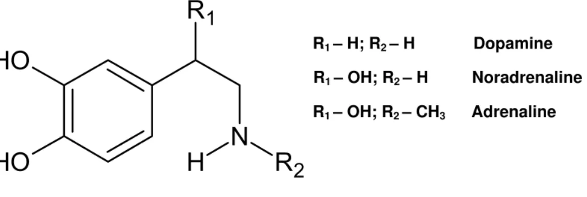
Figure 1 Chemical structure of biogenic catecholamines. ... 21
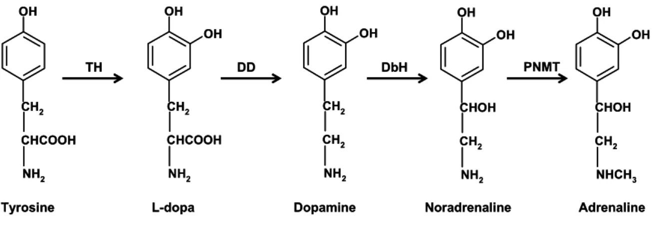
-Figure 2 - Catecholamine biosynthetic pathway. Tyrosine is sequentially 3-hydroxylated to form L-3,4-dihydroxyphenylalanine (L-dopa) by tyrosine hydroxilase (TH) and then decarboxylated to form dopamine (DA) by dopa descarboxilase (DD) in the cytoplasm. Dopamine β-hydroxylase (DbH) β -hydroxylates DA to yield noradrenaline (NA), which is N-methylated by phenylethanolamine N-methyltransferase (PNMT) to form adrenaline (ADR) in adrenergic neurons and other ADR producing cells. ... 24
-Index of Figures__________________________________________________
xxvi
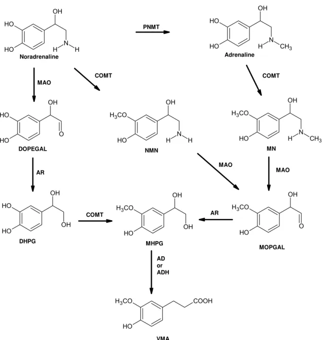
Figure 4 - Representative pathway of adrenaline (ADR) and noradrenaline (NA) metabolism. ADR is synthesized from NA by phenylethanolamine N-methyltransferase (PNMT). NA is metabolized preferably by monoamine oxidase (MAO) to dihydroxyphenylglycoaldehyde (DOPEGAL) that is reduced by aldehyde reductase (AR) to dihydroxyphenylglycol (DHPG). ADR and NA can be metabolized by catechol-O-methyltransferase (COMT) resulting in metanephrine (MN) and normetanephrine (NMN), respectively. Both MN and NMN can be further metabolized by MAO to 3-methoxy-4-hydroxyphenyl-glycoaldehyde (MOPGAL) and by AR to form methoxyhydroxyphenylglycol (MHPG). MHPG is further metabolized and can be transformed to vanillylmandelic acid (VMA) by alcohol dehydrogenase (ADH) or aldehyde dehydrogenase (AD). VMA is the main catecholamine metabolite in urine (adapted from Goldstein 2003)... 41
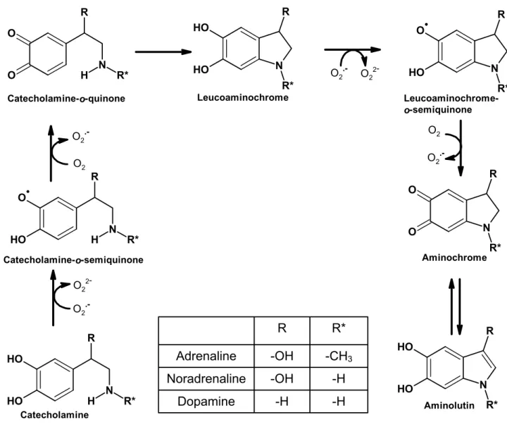
-Figure 5 - Postulated pathway for the oxidation of catecholamines. The oxidation process of catecholamines initially involves their conversion to o-quinones through o-semio-quinones intermediates. The o-semiquinone reduces oxygen forming superoxide anion (O2●–). The o-quinone can undergo an
irreversible 1,4-intramolecular cyclization, forming leucoaminonochrome. The oxidation of the unstable leucoaminonochrome to aminochrome is rapid, but differs according to the different catecholamines. The formation of aminochrome from leucoaminochrome is a reaction where a total of two electrons are removed and leucoaminochrome semiquinone is the intermediary with O2●– as a
by-product. Aminochrome formed in the cells can lead to the formation of aminolutin. ... 47
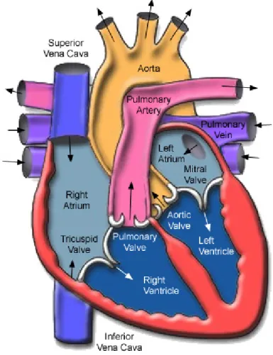
-Figure 6 - The human heart is represented with its four internal chambers: two atria and two ventricles. The right atrium communicates to the right ventricle through the tricuspid valve. The left atrium communicates to the left ventricle through the mitral valve (Adapted from Seeley 2003). ... 64
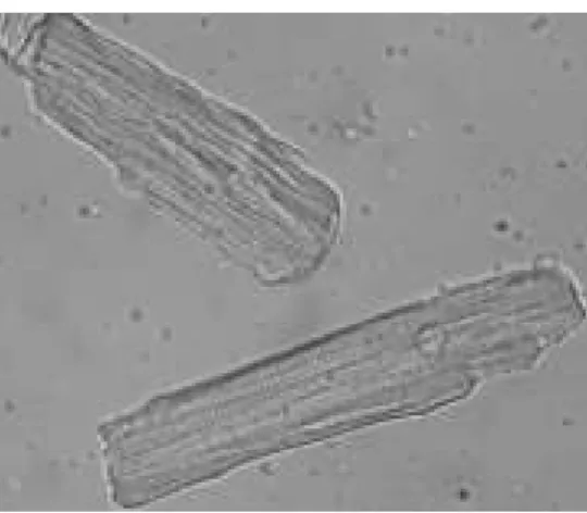
-Figure 8 - Image obtained by optic microscopy of the cardiac muscle cell: the
cardiomyocyte. ... 66
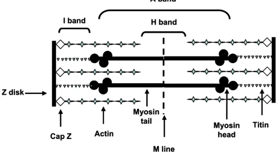
-Figure 9 - Representative figure of the structure of the muscle’s sarcomere. The
sarcomere is divided into several bands according to the main contractile
proteins that form it... 69
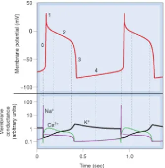
-Figure 10 - The graphic above represents the 5 phases in the membrane
potential during the action potential of a cardiac cell (phase 0, rapid
depolarization; phase 1, partial repolarization; phase 2, the plateau; phase 3,
repolarization; and phase 4, the pacemaker potential). In the graphic below is
presented the different conductivity of the ion channels involved in the
generation, maintenance and termination of the action potential in cardiac
muscle cells (adapted from Rang, 2007). ... 73
-Figure 11 - The structure of the cardiomyocyte illustrating some of the key
elements involving excitation contraction (EC) coupling. The network of the
sarcoplasmatic reticulum (SR) wraps both mitocondria and myofibrils and
contains the ryanodine receptors (RyR). Note the overlapping locations of RyR
and dihydropyridine receptor (DHPR) L-type Ca2+ channels in the transverse
tubules (T tubule). ... 77
-Figure 12 - Postulated pathway for the oxidation of adrenaline (ADR) in
cardiomyocytes and incubation medium (for simplification purposes, not all
intermediates are shown). ADR enters in the cardiomyocytes by the active
extraneuronal monoamine transporter (EMT). The oxidation process of ADR
initially involves its conversion to o-quinone with an o-semiquinone
intermediary. The o-quinone can react glutathione (GSH), to form the
corresponding GSH conjugate [5-(glutathion-S-yl)adrenaline], or react with
protein nucleophilic groups in the cells, forming quinoproteins. The
5-(glutathion-S-yl)adrenaline is transported out of the cell by multidrug resistance
protein 1 (MRP1). Meanwhile, the o-quinone can undergo an irreversible
1,4-intramolecular cyclization, forming leucoadrenochrome. The oxidation to
adrenochrome resulting of the unstable leucoadrenochrome is rapid. The
Index of Figures__________________________________________________
xxviii
removed and adrenochrome semiquinone is the intermediate. Adrenochrome formed in the cells can also suffer conjugation with cellular GSH, leading to its depletion, or it can polymerize into several other compounds like melanins. During ADR oxidation process, the superoxide anion (O2•–) is formed. Through
the Fenton or/and Haber-Weiss reactions hydroxyl (OH•) is formed (intermediaries are not shown for simplification). Furthermore, the reaction between O2•– and nitric oxide (NO●) generates peroxynitrite (ONOO–). Those
reactive species can oxidize GSH to form oxidized glutathione (GSSG). The GSSG is extruded from the cardiomyocyte through MRP1 or converted again to GSH by GSH reductase (GR)... 223
-Figure 13 - Postulated mechanism for the cellular signaling pathways altered by adrenaline (ADR) oxidation in cardiomyocytes in the presence of a superoxide anion (O2●−) generating system: xanthine and xanthine oxidase (XXO). The
oxidation process of ADR involves the formation of many reactive intermediaries and reactive species (RS) in the cardiomyocytes. The metabolization by monoaminoxidase (MAO) can also form reactive oxygen species (ROS) (1). The initial burst of RS and oxidants resulting from ADR oxidation leads to trimerization of the monomeric HSF-1 (2) that translocates to the nucleus (3). Afterwards, RS lead to NF-κB activation, presumably through the action of IκB protein kinases (4) and proteasome activity (5), leading to
-INDEX OF TABLES
ABBREVIATIONS USED IN THE TEXT
ΔΨ Potential gradient
ABC ATP-binding cassette
ACE Angiotensin converting-enzyme
ACh Acetylcholine
AD Aldehyde dehydrogenase
ADH Alcohol dehydrogenase ADR Adrenaline; Epinephrine AIF Apoptosis inducing factor
Apaf-1 Apoptosis protease activating factor-1
AR Aldehyde reductase
ATP Adenosine-5'-triphosphate ATPase Adenosine triphosphatase
AV Atrio-ventricular
Ca2+ Calcium ion
CaMKII Ca2+/calmodulin-dependent protein kinase II cAMP Cyclic adenosine monophosphate
cDNA Complementary DNA
CICR Ca2+-induced Ca2+ release
Cl- Chloride ion
CNS Central nervous system COMT Catechol-O-methyltransferase
CORT Corticosterone
CO2 Carbon dioxide
Abbreviations__________________________________________________________
xxxiv
DA Dopamine
DAG Diacylglycerol
DAT Dopamine transporter DbH Dopamine β-hydroxylase
DD Dopa descarboxilase
DHPG Dihydroxyphenylglycol DHPGALD Dihydroxyphenylglycoaldehyde DHPR Dihydropyridine receptor DNA Deoxyribonucleic acid DOMA 3,4-Dihydroxymandelic acid DOPAC 3,4-Dihydroxyphenylacetic acid DOPEGAL Dihydroxyphenylglycoaldehyde EC Excitation-contraction
EMT Extraneuronal monoamine transporter ERK Extracellular signal-regulated kinases FAD Flavin adenine dinucleotide
FADH2 Reduced flavin adenine dinucleotide FKBP12.6 FK-506 binding proteins; Calstabin
G protein Guanine nucleotide binding regulatory protein
GF120918 9,10-dihydro-5-methoxy-9-oxo-N-[4-[2-(1,2,3,4-tetrahydro
6,7-dimethoxy-2-isoquinolinyl)ethyl]phenyl]-4-acridine
carboxamide
Gi Inhibitory G Protein
GLUT Glucose transporters
GSSG Glutathione disulfide; Oxidized glutathione GTP Guanosine-5'-triphosphate
HF Heart failure
HSE Heat shock element HSF-1 Heat shock factor 1
HSP Heat shock protein HSR Heat shock response HOO● Hydroperoxyl radical H2O2 Peroxide hydrogen
5-HT 5-Hydroxytriptamine; Serotonin HVA Homovanillic acid
ICA Intrinsic cardiac adrenergic IκB Inhibitory kappa B
IP3 Inositol trisphosphate ISO Isoproterenol; Isoprenaline I/R Ischemia and reperfusion JNK c-Jun NH2-terminal kinase
K+ Potassium ion
Km Half-saturation constant; Michaelis constant
KO Knockout
LDH Lactate dehydrogenase L-dopa L-3,4-dihydroxyphenylalanine LOOH Fatty acid hydroperoxide MAO Monoamine oxidase
MAPK Mitogen-activated protein kinase
Abbreviations__________________________________________________________
xxxvi
MDA Malondialdehyde
MG-132 Z-Leu-Leu-Leu-aldehyde
MK-571 (E)-3-[[[3-[2-(7-chloro-2-quinoli-nyl)ethenyl]phenyl]-[[3 dimethylamino)-3-oxopropyl]thio]meth-yl]thio]propanoicacid
Mg2+ Magnesium ion
MHPG 3-Methoxy-4-hydroxy-phenylethylene glycol
MN Metanephrine
MOPGAL 3-Methoxy-4-hydroxyphenyl-glycoaldehyde MnSOD Manganese superoxide dismutase
MRP Multidrug resistance protein mRNA Messenger ribonucleic acid
MTT 3-(4,5-dimethylthiazol-2yl)-2,5-diphenyl tetrazolium bromide
Na+ Sodium ion
NADPH Nicotinamide adenine dinucleotide phosphate NADH Reduced nicotinamide adenine dinucleotide NAD+ Oxidized nicotinamide adenine dinucleotide NE Noradrenaline; Norepinephrine
NET Noradrenaline transporter NF-ĸB Nuclear factor kappa B
-NH2 Amine group
NMN Normetanephrine
NO● Nitric oxide
NPY Neuropeptide Y O2●– Superoxide anion
OCT Organic cation transporters
OH● Hydroxyl radical
-OH Hydroxyl group
ONOO– Peroxynitrite radical
p38 MAPK p38 mitogen-activated protein kinase P-gp P-glycoprotein
PKA Protein kinase A PKC Protein kinase C
PMAT Plasma membrane monoamine transporter
PMNT Phenylethanolamine N-methyltransferase; Noradrenaline N-methyltransferase
RIRR Reactive oxygen species-induced reactive oxygen species- release
RNA Ribonucleic acid
RNS Reactive nitrogen species ROO● Peroxyl radical
ROS Reactive oxygen species
RS Reactive species
RyR Ryanodine receptors RyR2 Type 2 ryanodine receptors
SA Sino-auricular node; sinoatrial node
s.c. Subcutaneous
S-COMT Soluble catechol-O-methyltransferase
Abbreviations__________________________________________________________
xxxviii
-SH Sulfhydryl group
SOD Superoxide dismutase SR Sarcoplasmatic reticulum
SULT1A3 Monoamine-preferring phenolsulfotransferase TBARS Thiobarbituric acid reactive substances TH Tyrosine hydroxilase
TNF-α Tumor Necrosis Factor-α
TT Transverse tubules
TTC 2,3,5-triphenyltetrazolium chloride VF Ventricular fibrillation
VMA Vanillylmandelic acid; Methoxy-4-hydroxymandelic acid VMAT Vesicular monoamine transporter
Vmax Maximum velocity
XXO Xanthine and xanthine oxidase
XD Xanthine dehydrogenase
OUTLINE OF THE DISSERTATION
The present dissertation is divided into four main sections:
• Part I – General Introduction
In this section, a review on the existing literature on cardiotoxic effects of catecholamines is presented, in order to provide a good basis for understanding the objectives and the obtained results of the experimental studies. Furthermore, and to make it clearer, the general introduction is subdivided in two subchapters. The general objectives are included in this part I.
• Part II – Experimental section
In part II, the articles published in the scope of this dissertation are presented.
• Part III – Discussion and Conclusions
In this section an integration of the results obtained in the scope of this dissertation is performed. The discussion of their potential relevance and their connection with existing scientific reports is also addressed here. Moreover, part III includes the main conclusions taken from the works of the present dissertation.
• Part IV – References
ABSTRACT
In cardiovascular pathologic conditions, like cardiac ischemia/reperfusion or heart failure, the sustained elevation of plasma and interstitial catecholamine levels, namely adrenaline, and the generation of reactive oxygen species (ROS) are hallmarks. Despite a century of research, the mechanisms underlying adrenaline-induced cardiotoxicity are still far from being fully elucidated. The present-dissertation aimed to contribute to increase the knowledge on the mechanisms by which adrenaline produces cardiotoxicity and the possible influence of ROS on that cardiotoxicity. Freshly isolated calcium tolerant cardiomyocytes from adult rats were incubated at 37ºC with adrenaline alone (ADR group), in the presence of a ROS generating system [xanthine with xanthine oxidase (XXO group)], or both, (ADR with XXO) during a short period of time (maximum 3 hours).
Abstract_______________________________________________________________
- 8 -
the classical EMT inhibitor, corticosterone, or by GF120918, the levels of intracellular 5-(glutathion-S-yl)adrenaline decreased, thus demonstrating that the intracellular formation of the adduct is dependent on adrenaline´s intracellular concentration.
The experiments also revealed that the adrenaline redox behavior altered the intracellular signaling pathways of cardiomyocytes, either when incubated alone or in the presence of XXO. The pro-oxidant signal elicited by adrenaline promoted the translocation to the nucleus of the transcription factors, Heat Shock Factor-1 (HSF-1) and Nuclear Factor-κB (NF-κB). In addition, the proteasome activity was compromised in the experimental groups where the generation of reactive species occurred hence showing its redox sensitivity. Proteasome activity decrease was prevented by adding the ROS scavenger, tiron. Proteasome inhibition seemed to have elicited an increase in the levels of Heat Shock Protein 70 (HSP70) levels, which occurred after HSF-1 translocation to the nucleus. Notwithstanding the several insults inflicted by the adrenaline redox behavior to cardiomyocytes, namely, GSH depletion, quinoproteins accumulation, proteasome activity inhibition, and reactive species formation, all potentially capable of promoting cell death, no change in the cellular viability was observed. Accordingly, it can be assumed that the protection provided by HSP70 against the oxidative injury may, at least partially, explain the absence of cell death. In fact, the increase in the levels of HSP70 was time-dependent in all treatment groups, followed by a decrease in apoptotic factors, namely caspase 3 activity (in all groups) and cytoplasmatic cytochrome c levels (in the XXO and ADR with XXO groups). Furthermore, the ADR group had a delayed outset in HSP70 expression and no significant differences in mitochondrial or cytoplasmatic cytochrome c content were observed when compared to control. Presumably, the cardiomyocytes exposed to an initial burst of ROS generated by XXO may have developed an early “preconditioning defense”, through proteasome inhibition and HSP70 expression.
energetic metabolism proteins, encompassing ATP synthase alpha chain and several enzymes of the tricarboxilic acid cycle; (iv) the stress response proteins, like HSP; and, (v) several regulatory proteins. The ROS generating system (XXO) had a particular noxious effect to contractile proteins and caused impairment in glucose metabolism, namely with high lactate production. On the other hand, the ADR group showed a remarkable increase in the activity of mitochondrial complexes, known to be accompanied by high “electron leakage”. This high “electron leakage” was accompanied by an increased in the expression and activity of SOD. In the ADR group, the ratio of alanine/lactate and the lactate levels were the lowest, reflecting an increase in cytosolic redox state (NADH/NAD+) due to high glycolytic activity. The increase in the cytosolic redox state led to an active transfer of reduced equivalents from the cytosol towards the mitochondrial matrix and hence high activity of the electron transport chain complexes in the ADR group.
RESUMO
Os níveis plasmáticos e intersticiais das catecolaminas, entre elas a adrenalina, assim como a geração de espécies reactivas de oxigénio, estão aumentados em várias patologias cardíacas, tais como a isquemia/reperfusão e a insuficiência cardíaca. Todavia, após mais de um século de investigação nesta área, os mecanismos envolvidos na cardiotoxicidade induzida pela adrenalina ainda estão longe de estarem plenamente esclarecidos. Desta forma, esta dissertação pretende contribuir para um melhor conhecimento dos mecanismos de cardiotoxicidade desencadeados pela adrenalina e a possível influência da formação de espécies reactivas nessa mesma cardiotoxicidade. Para a prossecução deste objectivo, suspensões de cardiomiócitos tolerantes ao cálcio isolados de rato adulto foram incubadas a 37ºC com adrenalina (grupo ADR), com um sistema gerador de radicais livres de oxigénio [xantina com xantina oxidase (grupo XXO)] ou com ambos (grupo ADR+XXO) durante um curto período de tempo (máximo de 3 horas).
Resumo_______________________________________________________________
- 14 -
durante a incubação com a adrenalina foram exportadas dos cardiomiócitos pela proteína de resistência a múltiplos fármacos 1 (MRP1). Igualmente, o transportador extraneuronal das monoaminas (EMT) mostrou-se funcional nos cardiomiócitos isolados e demonstrou-se que a sua actividade é determinante para a formação intracelular do aducto. Quando o EMT foi inibido pelo seu inibidor clássico, a corticosterona, ou pelo GF120918, os níveis de 5-(glutation -S-il)adrenalina diminuíram, confirmando-se que a formação intracelular do aducto depende da concentração intracelular da adrenalina.
oxigénio através do sistema XXO desenvolveram uma “defesa de precondicionamento” precoce, através da inibição da actividade do proteasoma e da expressão de HSP70.
Por último, a incubação com adrenalina ou XXO, alterou de forma diferente a abundância de várias proteínas e a actividade de alguns complexos enzimáticos. A maioria das alterações proteicas ocorreu em 5 grupos proteicos: (i) do sarcómero e citoesqueleto, especialmente cadeia leve da miosina-2; (ii) das proteínas envolvidas na regulação redox, como a dismutase do superóxido; (iii) das proteínas do metabolismo energético, como a cadeia alfa da ATP sintetase; (iv) das proteínas envolvidas na resposta ao stress, como as HSP; (v) e de várias proteínas reguladoras. O sistema gerador de espécies reactivas de oxigénio (XXO) mostrou ter um efeito particularmente nocivo sobre as proteínas contrácteis e desencadeou alterações no metabolismo da glucose, nomeadamente com elevada formação de lactato. Por outro lado, o grupo ADR mostrou um aumento considerável da actividade dos complexos mitocondriais, reconhecida por ser acompanhada pela elevada “libertação de electrões”. Essa “libertação de electrões” foi acompanhada por um aumento de expressão e actividade da dismutase do superóxido. No grupo ADR, a razão alanina/lactato e os níveis de lactato foram os mais baixos, reflectindo um aumento do estado redox citosólico (NADH/NAD+) devido à elevada actividade glicolítica. O aumento do estado redox citosólico levou a um aumento da transferência activa de equivalentes redutores do citosol para a matriz mitocondrial e daí a elevada actividade dos complexos da cadeia mitocondrial no grupo ADR.
PART I
I. GENERAL INTRODUCTION
I.1. An Overview On The Role Of Catecholamines: Basic
Physiology And Pharmacology
I.1.1. Historic introduction and background
In 1856, Vulpian applied a solution of ferric chloride to slices of the
adrenal glands and noticed that the medulla stained green while the cortex did
not. He also observed that the same reaction occurred in samples of venous
blood leaving the adrenal, but not in arterial blood entering the gland. To
account for these observations, he assumed that the medulla synthesized a
substance that was released to the circulation (Vulpian, 1856). In 1895, Oliver
and Schäfer demonstrated that injection of extracts of adrenal gland caused the
arterial blood pressure to rise, due to the catecholamines there present (Oliver
and Schäfer, 1895). Soon after these discoveries, the role of catecholamines
became the centre of one of the most endurable and passionate scientific
discussions and research, with tremendous breakthroughs that crossed the
twentieth century. Even in recent years, a remarkable amount of information
about catecholamines and related compounds has accumulated partly because
of the important interactions between the endogenous catecholamines and
many of the drugs used in the treatment of hypertension, mental disorders, and
in a variety of other conditions, namely drug addiction.
Under the general heading are noradrenaline (NA) (also known as
norepinephrine), the principal transmitter of sympathetic postganglionic fibers
and of certain tracts of the central nervous system (CNS); dopamine (DA), the
predominant transmitter of the mammalian extrapyramidal system and of
several mesocortical and mesolimbic neuronal pathways; and adrenaline (ADR)
(also known as epinephrine), the major hormone of the adrenal medulla.
Collectively, these three amines are called biogenic catecholamines (Westfall
General Introduction____________________________________________________
- 20 -
learning and memory, sleep-wake cycle regulation, as well as endocrine and visceral functions (Wevers et al., 1999).
Many studies have been performed concerning the toxicity of these molecules, namely cardiotoxicity (Raab et al., 1968; Fluck, 1972; Opie, 1975; Rona, 1985; Todd et al., 1985; Wheatley et al., 1985; Rupp et al., 1994; Lameris et al., 2000; Remião et al., 2001b; Remião et al., 2002; Remião et al., 2004), and neurotoxicity (Fornstedt et al., 1990; Spencer et al., 1995; Spencer et al., 1998; Segura-Aguilar et al., 2001). Furthermore, the toxicity mechanisms of illicit drugs, namely 3,4-methylenedioxymethamphetamine (MDMA or “ecstasy”) (Badon et al., 2002; Carvalho et al., 2004b; Patel et al., 2005; Capela et al., 2006b), methamphetamine (He et al., 1996; Varner et al., 2002; Wijetunga et al., 2003), cocaine (Kloner et al., 1992; Rump et al., 1995) involve and are related to alterations in catecholamine pathways (Newton et al., 2005).
The basic physiological, biochemical, and pharmacological features of catecholamines will be described here, before a more detailed look into the heart and the cardiotoxicity of these transmitters.
I.1.2. Structure and main location of catecholamines
NA, ADR, and DA are known to be catecholamines, as they contain a catechol moiety, an amine side-chain and are all derived from the amino acid tyrosine (Westfall and Westfall, 2006; Baynes and Dominiczak, 2007; Rang et al., 2007). In common with other amino groups containing compounds, such as 5-hydroxytryptamine (5-HT; serotonin), they are known as biogenic amines. In contrast, isoproterenol (ISO) (previously isoprenaline), a synthetic derivative of NA not present in the body, is a catecholamine but not a biogenic amine (Rang et al., 2007).
vasoconstriction in the skin vessels. NA containing neurons are also present in
the CNS, mainly in the brain stem, the most prominent cluster being the locus
ceruleus. Their axons extend in a wide network throughout the cortex and alter
the overall state of alertness (Westfall and Westfall, 2006).
Figure 1 - Chemical structure of biogenic catecholamines.
In both the CNS and peripheral nervous system of mammals, NA is the
principal catecholamine released from noradrenergic neurons (Axelrod and
Kopin, 1969). However, ADR was the first catecholamine isolated (Figure 1). In
1897, Abel and Crawford obtained an extract from the suprarenal gland that
they called “epinephrin” (Abel and Crawford, 1897). The mono-benzoyl
derivative isolated was however less active than the crude extracts, so the
quest for the bioactive compound continued. In 1901, Takamine, an industrial
chemist, isolated the active principle, that he called “adrenalin” (Takamine,
1902). Takamine’s notation was adopted by the Europeans but not by North
American researchers, thus resulting in the different nomenclature still used
today in the opposite sides of the Atlantic Ocean.
ADR, synthesized from NA by norepinephrine N-methyltransferase, best
known as phenylethanolamine N-methyltransferase (PNMT) (Axelrod, 1962) is
released from the adrenal gland as an endocrine hormone by the influence of
acetylcholine (ACh)-containing nerves analogous to the sympathetic
preganglionic nerves (Baynes and Dominiczak, 2007). ADR triggers
coordinated metabolic and physiological processes in response to stress
(Westfall and Westfall, 2006). ADR is more active than NA in the heart and
O
H
O
H
N
R
1
H
R
2
General Introduction____________________________________________________
- 22 -
lungs, causes redirection of blood from the skin to skeletal muscle, and it has important stimulatory effects in the glycogen metabolism at the liver (Westfall and Westfall, 2006; Baynes and Dominiczak, 2007; Rang et al., 2007).
Although generally treated as an endocrine hormone, ADR also acts as a neurotransmitter being released in central and peripheral adrenergic neurons (Loewi, 1936; Jarrott, 1970; Fuller, 1982). Indeed, Loewi demonstrated the presence of “acceleranstoff” released from sympathetic neurons innervating the frog’s heart (Loewi, 1921), which later was found to reflect the synthesis, storage, and release of ADR (Loewi, 1936; Azuma et al., 1965; Norberg and McIsaac, 1967).
Further down in this dissertation, other tissues in addition to the CNS or adrenal medulla, where PNMT is expressed and ADR is produced, will be described in more detail.
DA (3,4-dihydroxyphenylethylamine) is the immediate metabolic precursor of NA and ADR. Although DA originally was regarded only as a precursor of NA, assays on distinct CNS regions revealed that the distributions of DA and NA neurons are markedly different. DA is a central neurotransmitter particularly important in the regulation of movement and with important intrinsic pharmacological properties (Bloom, 2006). In fact, more than half of the CNS catecholamine content is DA, and extremely large amounts are found in the basal ganglia (especially the caudate nucleus), the nucleus accumbens, the olfactory tubercle, the central nucleus of the amygdala, the median eminence, and restricted fields of the frontal cortex (Bloom, 2006). Of these myriad connections, the greatest attention has been given to the long projections between the major DA-containing nuclei located at the substantia nigra and ventral tegmentum and their targets in the striatum, and the limbic zones of the cerebral cortex and in other major limbic regions (but in general not the hippocampus). In addition, two important central dopaminergic systems are functionally located peripheral to the blood-brain barrier, in the chemoceptor trigger zone and in the anterior pituitary. In the periphery, DA is synthesized in epithelial cells of the proximal tubule and is thought to exert local diuretic and natriuretic effects in the kidney (Esler et al., 1990; Bloom, 2006). Likewise, DA receptors are found in the proximal gastrointestinal tract where activation of DA receptors delays gastric emptying. DA receptors are also present in renal, mesenteric, coronary and intracerebral arteries, favoring vasodilatation (Bloom, 2006).
General Introduction____________________________________________________
- 24 - OH CH2 CHCOOH NH2 OH OH CH2 CHCOOH NH2 OH OH CH2 CH2 NH2 OH OH CHOH CH2 NH2 OH OH CHOH CH2 NHCH3 Tyrosine L-dopa Dopamine Noradrenaline Adrenaline
TH DD DbH PNMT
OH CH2 CHCOOH NH2 OH OH CH2 CHCOOH NH2 OH OH CH2 CH2 NH2 OH OH CHOH CH2 NH2 OH OH CHOH CH2 NHCH3 Tyrosine L-dopa Dopamine Noradrenaline Adrenaline
TH DD DbH PNMT
I.1.3. Synthesis of catecholamines
The understanding of the mechanisms of synthesis, storage, release and binding sites of catecholamines derives mostly from studies of sympathetically innervated organs and the adrenal medulla (Westfall and Westfall, 2006).
Figure 2 - Catecholamine biosynthetic pathway. Tyrosine is sequentially 3-hydroxylated
to form L-3,4-dihydroxyphenylalanine (L-dopa) by tyrosine hydroxilase (TH) and then
decarboxylated to form dopamine (DA) by dopa descarboxilase (DD) in the cytoplasm.
Dopamine β-hydroxylase (DbH) β-hydroxylates DA to yield noradrenaline (NA), which is N-methylated by phenylethanolamine N-methyltransferase (PNMT) to form
adrenaline (ADR) in adrenergic neurons and other ADR producing cells.
may also be substrates. For example, 5-HT can be produced from 5-hydroxy-L-tryptophan by DD. DD also converts methyldopa to α-methyldopamine, which, in turn, is converted by DbH to methylnoradrenaline (Westfall and Westfall, 2006).
The hydroxylation of tyrosine by TH is generally regarded as the rate-limiting step in the biosynthesis of catecholamines (Zigmond et al., 1989). This enzyme is activated after stimulation of sympathetic nerves or of adrenal medulla and is tightly regulated. In fact, TH is a substrate for protein kinase A (PKA), protein kinase C (PKC) and calcium/calmodulin-dependent protein kinase II (CaMKII). The kinase-catalyzed phosphorylation may be related to increased hydroxylase activity of TH (Zigmond et al., 1989; Daubner et al., 1992). This is an important acute mechanism for increasing catecholamine synthesis in response to elevated nerve stimulation. A delayed response can also occur, resulting in alteration upon the th gene. The increased production of TH can occur at multiple levels of regulation, including transcription, RNA processing, regulation of RNA stability, translation, and enzyme stability (Kumer and Vrana, 1996). These mechanisms serve to maintain the content of catecholamines in response to increased transmitter release. Likewise, TH is subject to feedback inhibition by catechol compounds, which allosterically modulate the enzyme activity (Kumer and Vrana, 1996).
TH deficiency has been reported in humans and is characterized by generalized rigidity, hypokinesia, and other extrapyramidal symptoms, with low cerebrospinal fluid levels of NA and DA metabolites, homovanillic acid (HVA) and 3-methoxy-4-hydroxy-phenylethylene glycol (MHPG) (Wevers et al., 1999; Carson and Robertson, 2002). The TH knockout (KO) is unviable in mice as they die in the embryonic stage, presumably because catecholamine loss results in altered cardiac function (Zhou et al., 1995). Interestingly, residual levels of DA are present in these embryonic mice, suggesting that tyrosinase may be an alternate way to form catecholamines. The amounts of tyrosinase-derived catecholamines are clearly not sufficient for mice’s survival (Carson and Robertson, 2002).
General Introduction____________________________________________________
- 26 -
(Westfall and Westfall, 2006). In the case of DbH-deficient mice, there is about 90% embryonic mortality (Carson and Robertson, 2002).
In adrenergic neurons, the enzymes that participate in NA formation are synthesized in the cell bodies of neurons and are then transported along the axon to the terminals. In the course of NA synthesis, both the hydroxylation of tyrosine to L-dopa and the decarboxylation of L-dopa to DA take place in the cytoplasm. About half of the DA formed in the cytoplasm is then actively transported into the DbH-containing storage vesicles, where it is converted to NA. The remainder escapes active transport to the vesicles and is deaminated to 3,4-dihydroxyphenylacetic acid (DOPAC) and subsequently O-methylated to HVA (Westfall and Westfall, 2006).
The NA formed in the vesicles leaves these structures and is methylated in the cytoplasm to ADR by PNMT. ADR then re-enters the vesicles, where it is stored and concentrated for subsequent release (Masson et al., 1999; Westfall and Westfall, 2006).
As stated, the ADR producing cells contain the enzyme PNMT. Although the adrenal gland and the brain are the most prominent sites of PNMT expression in the adult mammal, this enzyme has also been detected in the retina (Hadjiconstantinou et al., 1983; Foster et al., 1985; Park et al., 1986), spleen (Pendleton et al., 1978), lungs (Pendleton et al., 1978; Kennedy et al., 1990), and heart (Axelrod, 1962; Pendleton et al., 1978; Culman et al., 1987; Torda et al., 1987; Kennedy and Ziegler, 1991). The distribution of PNMT activity in various regions of the rat, monkey, and human brain was already investigated (Moore and Phillipson, 1975b; Lew et al., 1977). High and intermediate enzyme activity values were found in the same regions of the primate and rat brain, namely in the medulla, nucleus reticularis (C1), locus
ceruleus, and periventricular areas of the hypothalamus (Lew et al., 1977). Intermediate or low activity levels were found in various regions of the primate brain, e.g., basal ganglia, amygdala, septum, and habenula (Lew et al., 1977). The PNMT activity is more widely distributed in the primate brain than in the rat’s brain. The brain’s PNMT has the same substrate specificity and similar kinetic properties as the PTMN in the adrenal medulla (Lew et al., 1977).
1972). Glucocorticoids levels are very important in the expression of PMNT. The glucocorticoids are very relevant in the rate of ADR synthesis, and hence in the size of the ADR store available for release in the brain (Moore and Phillipson, 1975b; Moore and Phillipson, 1975a), adrenal medulla (Kelner and Pollard, 1985; Stachowiak et al., 1988; Wan and Livett, 1989; Ross et al., 1990; Betito et al., 1992; Wong et al., 1992), heart (Kennedy and Ziegler, 1991; Krizanova et al., 2001; Kvetnansky et al., 2004) or lungs (Kennedy et al., 1993). The activities of both TH and DbH are also increased in the adrenal medulla by the secretion of glucocorticoids (Carroll et al., 1991). Thus, any stress that persists sufficiently to evoke an enhanced secretion of corticotrophin mobilizes the appropriate hormones of both the adrenal cortex (predominantly cortisol in humans) and adrenal medulla, where it influences the synthesis of catecholamines.
I.1.4. Intracellular storage of catecholamines
General Introduction____________________________________________________
- 28 -
the circulation, supporting the argument that the process of release following adrenergic nerve stimulation involves exocytosis (Westfall and Westfall, 2006).
The storage process decreases intraneuronal metabolism of transmitters and their leakage outside the cell, thus controlling their concentration gradient across the plasma membrane and preventing the possible toxic effects of neurotransmitters that could occur when the catecholamines cytoplasmic concentration exceeds a critical level (Schuldiner, 1994; Masson et al., 1999). The storage process is mediated by specific vesicular transporters (Schuldiner et al., 1978; Erickson et al., 1992; Schuldiner, 1994; Masson et al., 1999). In mammals, there are two closely related vesicular monoamine transporters (VMAT) isoforms of the monoamine transporters and they are called VMAT-1 and VMAT-2. VMAT-1 is primarily present in endocrine and paracrine cells, and VMAT-2 is the predominant monoamine transporter in the nervous system (Masson et al., 1999). VMAT-2 is expressed mainly in axon terminals in dense core vesicles (Figure 3) (Nirenberg et al., 1997).
Monoamine transporters are relatively promiscuous and transport DA, NA, ADR, and 5-HT, as well as meta-iodobenzylguanidine, which can be used to image chromaffin cell tumors (Schuldiner, 1994), but they differ in their substrate preferences and affinities. Reserpine inhibits monoamine transport into storage vesicles and ultimately leads to depletion of catecholamines in nerve endings (von Euler, 1972; Masson et al., 1999).
The vesicular transport system appears to be driven by pH and potential gradients that are established by an ATP-dependent proton transporter. The ATP-driven H+ pump, on one hand, acidifies the vesicular lumen and, on the other hand, generates a potential gradient (ΔΨ)(Schuldiner et al., 1978; Johnson and Scarpa, 1979). For every molecule of amine taken in, two internal H+ ions are extruded by VMAT-2 in an antiporter process (Masson et al., 1999). The monoamine steady-state concentration gradients are dependent both on ΔΨ and square ΔpH (Njus et al., 1986; Rottenberg, 1986; Johnson, 1988; Schuldiner, 1994; Masson et al., 1999). In addition, the monoamine uptake is modulated by vesicle-associated heterotrimeric guanine nucleotide binding regulatory proteins (G proteins), Gαo2 and Gαq (Ahnert-Hilger et al., 1998; Holtje
mediates the inhibition uptake by G proteins. In fact, the monoamines, with the exception of ADR, induce G protein-mediated inhibition and VMATs have a receptor-like function localized in its first luminal loop that senses the intravesicular concentration of monoamines (Brunk et al., 2006).
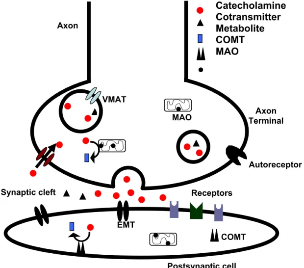
Figure 3 - Storage, release and metabolism of catecholamines in the nerve endings of
catecholaminergic neurons. The action potential reaches the axon terminal of neurons.
The axon terminals have neurotransmitter storage vesicles rich in catecholamines and
cotransmitters. The synthesis of catecholamines occurs in the cytoplasm and the
vesicular monoamine transporter (VMAT) is responsible for the transport of
catecholamines to the storage vesicles, maintaining their low cytosolic concentrations.
When catecholamines are released to the synaptic cleft, they are reuptaken to the
presynaptic terminal by noradrenaline transporter (NET), and taken to extraneuronal
cells by the extraneuronal transporters, namely extraneuronal monoamine transporter Synaptic cleft
Axon Terminal Axon
Postsynaptic cell
▲
▲
▲
▲
EMT
COMT MAO
VMAT
Autoreceptor
Receptors
▲
General Introduction____________________________________________________
- 30 -
(EMT). Catecholamines are metabolized by intracellular enzymes. Monoamine oxidase
(MAO) is located in the mitochondria, mainly in neurons but also in extraneuronal cells,
while catechol-O-methyl transferase (COMT) is located at extraneuronal cells, either in
the soluble or the membrane bound form. Both enzymes are the main responsible for
the metabolism of catecholamines. From the enzymatic process can result a wide
variety of metabolites, due to the complex and dynamic processes involved. To exert
their actions, the catecholamines and other neurotransmitters in the synaptic cleft bind
to different pre and postsynaptic receptors. Such binding results in alterations of the
postsynaptic cell, but also feed-back responses in their own liberation by the
presynaptic neurons, through the autoreceptors.
Several vesicular transport complementary deoxyribonucleic acid (cDNA) have been cloned; these cDNAs reveal open reading frames predictive of proteins with 12 transmembrane domains (Masson et al., 1999). Regulation of the expression of these vesicular transporters may be important in the regulation of synaptic transmission (Varoqui and Erickson, 1997).
I.1.5. Release of catecholamines
Depolarization of the axonal terminal triggers the release of catecholamines to the cleft. The vesicles content, including enzyme, neurotransmitters and hormones, are discharged to the exterior through a process termed exocytosis (Figure 3). Triggered exocytosis is the most common cellular mechanism for secretion of polar molecules and is a chain of complex mechanisms that involve vesicle docking, priming and fusion, and it can be regulated or constitutive. In the synaptic transmission, a synchronized secretion provides cells with a mechanism for precisely timed release of molecules into the extracellular space (Pocock and Richards, 2006). The full sequence of steps by which the nerve impulse affects the release of NA from sympathetic neurons is not fully understood.
translated into an intracellular signal that regulates the fusion rate of the preformed secretory vesicles will fuse with the plasma membrane. In most types of cells, regulated exocytosis is triggered by an increase in the concentration of free calcium ions (Ca2+) within the cytosol (Meir et al., 1999; Pocock and Richards, 2006). The increase in cytosolic Ca2+ is derived from the extracellular medium, through voltage-gated Ca2+ channels which open following depolarization of the plasma membrane, making the Ca2+ able to diffuse into the cell down its electrochemical gradient. Ca2+ is also mobilized from intracellular stores (mainly the endoplasmic reticulum) following activation of G protein-coupled receptor systems. As a result, the intracellular free Ca2+ increases and triggers the fusion of docked secretory vesicles with the plasma membrane. Ca2+-triggered secretion involves interaction of highly conserved molecular scaffolding proteins that ultimately lead to docking of granules (Aunis, 1998; Meir et al., 1999). As fusion proceeds, a pore is formed that connects the extracellular space with the interior of the vesicle and this pore provides a pathway for the contents of the vesicle to diffuse into the extracellular space (Pocock and Richards, 2006). Other aspects of the modulation of catecholamines transmission will be further addressed in a subsequent section that focuses on the adrenoceptors and their role in augmenting or diminishing the synaptic release (section I.1.10).
I.1.6. Transporters involved in catecholamine reuptake
General Introduction____________________________________________________
- 32 -
transporters include those for DA, NA, 5-HT, and a variety of aminoacid transmitters (Masson et al., 1999). These transporters are members of an extended family sharing common structural motifs, particularly the putative 12-transmembrane helices (Bönisch and Brüss, 2006), several similarly configured intracellular and extracellular loops or regions with respective phosphorylation and glycosylation sites, and intracellularly located amino- and carboxy-terminal residues (Eisenhofer, 2001). In mammals, DA, NA and ADR are removed from extracellular spaces by specialized transporters localized in the plasma membrane of neuronal terminals, varicosities, and dendrites (Figure 3). DA is cleared by DA transporter (DAT) whilst NA and ADR by NA transporter (NET) (Trendelenburg, 1988; Apparsundaram et al., 1997; Eisenhofer, 2001).
These plasma membrane transporters appear to have greater substrate specificity than do vesicular transporters and may be viewed as targets (“receptors”) for specific drugs such as cocaine (NET) or fluoxetine (5-HT transporter) (Bönisch and Brüss, 2006). DAT and NET exhibit overlapping, yet distinct, substrate selectivity, translocation efficiency, and antagonist sensitivity (Buck and Amara, 1994; Giros et al., 1994; Pifl et al., 1996; Bönisch and Brüss, 2006). Besides the corresponding neurons, NET is also present in the adrenal medulla, liver, and placenta, whereas DAT is present in the stomach, pancreas, and kidney (Eisenhofer, 2001).
The reuptake process aims to assure constant and high levels of neurotransmitters in the releasing neuron and low concentrations in the cleft (Eisenhofer, 2001; Bönisch and Brüss, 2006). It works as integrated part of the neurotransmitters recycling process, since in addition to their de novo synthesis, the monoamines stores in the terminal portions of the neural fibers are also replenished by their active transport of neurotransmitters back to the terminals. Moreover, the reuptake system contributes to the degradation of catecholamines, since the metabolizing enzymes are located intracellularly (Westfall and Westfall, 2006).
radiolabelled ADR and NA in sympathetically innervated organs, which was dependent on intact sympathetic nerve terminals and was inhibited by cocaine (Axelrod et al., 1959; Axelrod et al., 1961). It was discovered afterwards that the accumulation of catecholamines occurred through NET.
Neurotransmitter transporters, like NET and DAT, are dependent on sodium ion (Na+) and chloride ion (Cl−) concentrations. The Na+-gradient (concentration of Na+ outside is high) across the plasma membrane is the main driving force, dictating the transport direction of the neurotransmitter and co-substrate ions (in these case Na+), which is normally inwards (Masson et al., 1999; Sonders et al., 2005; Bönisch and Brüss, 2006). The negative inside membrane potential created by the potassium ion (K+) gradient also contributes for the above mentioned driving force (Bönisch and Brüss, 2006).
Transport of the naturally occurring substrate NA by the NET is saturable and characterized by a half-saturation constant or Michaelis constant (Km) of about 0.8 μM, while to ADR the Km is 2.8 μM in a maximum velocity (Vmax) two times lower than that for NA transport (Apparsundaram et al., 1997; Bönisch and Brüss, 2006). NET has a high affinity for NA and a somewhat lower affinity for ADR, while the synthetic β-adrenergic agonist ISO is not a substrate for this system (Eisenhofer, 2001; Bönisch and Brüss, 2006). The common structural requirement for uptake by NET is the presence of an ionizable nitrogen not incorporated into the aromatic ring system (Eisenhofer, 2001).
In amphibians, an ADR specific transporter has been identified, with molecular distinct characteristics from those of DAT or NET (Apparsundaram et al., 1997). In mammals, the presence of ADR specific transporter was not yet clarified, however in situ hybridization studies indicate the absence of both DAT and NET messenger ribonucleic acid (mRNA) in ADR-synthesizing neurons in the brainstem of adult rats (Lorang et al., 1994). Thus, if ADR is cleared at these sites by reuptake, these terminals might elaborate a catecholamine transporter distinct from DAT and NET, or ADR acts as an endocrine regulator that does not require rapid reuptake (Lorang et al., 1994; Apparsundaram et al., 1997).
General Introduction____________________________________________________
- 34 -
remove approximately 87% of released NA by NET, while 5% is removed by extraneuronal monoamine transporter (EMT) (see section I.1.7), and the remainder 8% is diffused to the circulation (Eisenhofer, 2001).
The importance of neuronal catecholamine transporters in maintaining vesicular levels of catecholamines is strikingly illustrated in mutant mice lacking either DAT or NET. In mice lacking DAT, brain tissue stores of DA are severely decreased to less than 5% of the levels in wild-type animals (Jones et al., 1998). As a result of the decrease in store size, neuronal release of DA is substantially reduced by 75%. Thus, despite a 300-fold lower rate of extracellular DA clearance, the decrease in exocytotic DA release leads to only a 5-fold increase in the DA extracellular fluid levels. The neuronal reuptake block and the resulting increase in extracellular fluid levels of DA lead to a marked enhancement of extraneuronal DA metabolism and an accompanying 2-fold increase in DA synthesis. The minimal increase in transmitter synthesis occurs not only as a result of the decrease in exocytotic release, but more importantly, reflects the large contribution of transmitter leakage from storage vesicles to turnover and intraneuronal metabolism, and the dependence of this event to the size of vesicular transmitter stores (Floor et al., 1995; Eisenhofer et al., 1998b).